IntracellularCa2 StoresandCa2 InfluxAreBothRequiredfor ... · 2 Neural Plasticity studies have...
11
Hindawi Publishing Corporation Neural Plasticity Volume 2012, Article ID 203536, 10 pages doi:10.1155/2012/203536 Research Article Intracellular Ca 2+ Stores and Ca 2+ Influx Are Both Required for BDNF to Rapidly Increase Quantal Vesicular Transmitter Release Michelle D. Amaral and Lucas Pozzo-Miller Department of Neurobiology, Civitan International Research Center, The University of Alabama at Birmingham, SHEL-1002, 1825 University Boulevard, Birmingham, AL 35294-2182, USA Correspondence should be addressed to Lucas Pozzo-Miller, [email protected] Received 12 April 2012; Accepted 29 May 2012 Academic Editor: Jean-Luc Gaiarsa Copyright © 2012 M. D. Amaral and L. Pozzo-Miller. This is an open access article distributed under the Creative Commons Attribution License, which permits unrestricted use, distribution, and reproduction in any medium, provided the original work is properly cited. Brain-derived neurotrophic factor (BDNF) is well known as a survival factor during brain development as well as a regulator of adult synaptic plasticity. One potential mechanism to initiate BDNF actions is through its modulation of quantal presynaptic transmitter release. In response to local BDNF application to CA1 pyramidal neurons, the frequency of miniature excitatory postsynaptic currents (mEPSC) increased significantly within 30 seconds; mEPSC amplitude and kinetics were unchanged. This effect was mediated via TrkB receptor activation and required both full intracellular Ca 2+ stores as well as extracellular Ca 2+ . Consistent with a role of Ca 2+ -permeable plasma membrane channels of the TRPC family, the inhibitor SKF96365 prevented the BDNF-induced increase in mEPSC frequency. Furthermore, labeling presynaptic terminals with amphipathic styryl dyes and then monitoring their post-BDNF destaining in slice cultures by multiphoton excitation microscopy revealed that the increase in frequency of mEPSCs reflects vesicular fusion events. Indeed, BDNF application to CA3-CA1 synapses in TTX rapidly enhanced FM1-43 or FM2-10 destaining with a time course that paralleled the phase of increased mEPSC frequency. We conclude that BDNF increases mEPSC frequency by boosting vesicular fusion through a presynaptic, Ca 2+ -dependent mechanism involving TrkB receptors, Ca 2+ stores, and TRPC channels. 1. Introduction Brain-derived neurotrophic factor (BDNF) is a member of the neurotrophic factor family and is well known as a survival factor and chemoattractant during development of the central nervous system [1]. In recent years, however, it has been shown that BDNF also plays a significant role mod- ulating synaptic plasticity in the hippocampus [2–6]. These functions include the enhancement of synaptic transmission at excitatory synapses [7] and alterations of dendritic archi- tecture [8], for example, increasing dendritic spine density [9–12]. BDNF exerts its effects both at the presynapse and the postsynapse: at the presynaptic level, BDNF modulates quan- tal synaptic transmission, and at hippocampal CA3-CA1 pyramidal neuron synapses, this is manifested as transient increases in the frequency of miniature synaptic currents. Acute application of BDNF enhances spontaneous and evoked glutamatergic excitatory postsynaptic currents in cultured neurons [13–17]. Long-term treatment of post- natal hippocampal slice cultures with BDNF increases the frequency of miniature excitatory postsynaptic currents (mEPSCs) recorded from CA1 pyramidal neurons, without affecting their amplitude or kinetics [12]. The latter effect of BDNF was specific on a rapidly recyclable pool of vesicles. Indeed, BDNF selectively increased the density of synaptic vesicles docked at the active zone of asymmetric spine synapses, without affecting those in the main vesicle cluster [12]. Furthermore, BDNF selectively enhanced evoked and spontaneous FM1-43 destaining in acute hippocampal slices, but only when presynaptic terminals were dye-loaded with a hyperosmotic shock using sucrose [18], a manipulation that only engages the readily releasable pool of vesicles [19, 20]. Two potential mechanisms mediating the BDNF-induced increase in transmitter release include synapsin-I phospho- rylation [21] and TrkB-initiated PLCγ activation leading to Ca 2+ mobilization from IP3-sensitive stores [22]. Previous
Transcript of IntracellularCa2 StoresandCa2 InfluxAreBothRequiredfor ... · 2 Neural Plasticity studies have...

Hindawi Publishing CorporationNeural PlasticityVolume 2012, Article ID 203536, 10 pagesdoi:10.1155/2012/203536
Research Article
Intracellular Ca2+ Stores and Ca2+ Influx Are Both Required forBDNF to Rapidly Increase Quantal Vesicular Transmitter Release
Michelle D. Amaral and Lucas Pozzo-Miller
Department of Neurobiology, Civitan International Research Center, The University of Alabama at Birmingham, SHEL-1002,1825 University Boulevard, Birmingham, AL 35294-2182, USA
Correspondence should be addressed to Lucas Pozzo-Miller, [email protected]
Received 12 April 2012; Accepted 29 May 2012
Academic Editor: Jean-Luc Gaiarsa
Copyright © 2012 M. D. Amaral and L. Pozzo-Miller. This is an open access article distributed under the Creative CommonsAttribution License, which permits unrestricted use, distribution, and reproduction in any medium, provided the original work isproperly cited.
Brain-derived neurotrophic factor (BDNF) is well known as a survival factor during brain development as well as a regulator ofadult synaptic plasticity. One potential mechanism to initiate BDNF actions is through its modulation of quantal presynaptictransmitter release. In response to local BDNF application to CA1 pyramidal neurons, the frequency of miniature excitatorypostsynaptic currents (mEPSC) increased significantly within 30 seconds; mEPSC amplitude and kinetics were unchanged. Thiseffect was mediated via TrkB receptor activation and required both full intracellular Ca2+ stores as well as extracellular Ca2+.Consistent with a role of Ca2+-permeable plasma membrane channels of the TRPC family, the inhibitor SKF96365 preventedthe BDNF-induced increase in mEPSC frequency. Furthermore, labeling presynaptic terminals with amphipathic styryl dyes andthen monitoring their post-BDNF destaining in slice cultures by multiphoton excitation microscopy revealed that the increase infrequency of mEPSCs reflects vesicular fusion events. Indeed, BDNF application to CA3-CA1 synapses in TTX rapidly enhancedFM1-43 or FM2-10 destaining with a time course that paralleled the phase of increased mEPSC frequency. We conclude thatBDNF increases mEPSC frequency by boosting vesicular fusion through a presynaptic, Ca2+-dependent mechanism involvingTrkB receptors, Ca2+ stores, and TRPC channels.
1. Introduction
Brain-derived neurotrophic factor (BDNF) is a memberof the neurotrophic factor family and is well known as asurvival factor and chemoattractant during development ofthe central nervous system [1]. In recent years, however, ithas been shown that BDNF also plays a significant role mod-ulating synaptic plasticity in the hippocampus [2–6]. Thesefunctions include the enhancement of synaptic transmissionat excitatory synapses [7] and alterations of dendritic archi-tecture [8], for example, increasing dendritic spine density[9–12]. BDNF exerts its effects both at the presynapse and thepostsynapse: at the presynaptic level, BDNF modulates quan-tal synaptic transmission, and at hippocampal CA3-CA1pyramidal neuron synapses, this is manifested as transientincreases in the frequency of miniature synaptic currents.
Acute application of BDNF enhances spontaneous andevoked glutamatergic excitatory postsynaptic currents in
cultured neurons [13–17]. Long-term treatment of post-natal hippocampal slice cultures with BDNF increases thefrequency of miniature excitatory postsynaptic currents(mEPSCs) recorded from CA1 pyramidal neurons, withoutaffecting their amplitude or kinetics [12]. The latter effect ofBDNF was specific on a rapidly recyclable pool of vesicles.Indeed, BDNF selectively increased the density of synapticvesicles docked at the active zone of asymmetric spinesynapses, without affecting those in the main vesicle cluster[12]. Furthermore, BDNF selectively enhanced evoked andspontaneous FM1-43 destaining in acute hippocampal slices,but only when presynaptic terminals were dye-loaded with ahyperosmotic shock using sucrose [18], a manipulation thatonly engages the readily releasable pool of vesicles [19, 20].
Two potential mechanisms mediating the BDNF-inducedincrease in transmitter release include synapsin-I phospho-rylation [21] and TrkB-initiated PLCγ activation leading toCa2+ mobilization from IP3-sensitive stores [22]. Previous

2 Neural Plasticity
studies have suggested that Ca2+ influx following Ca2+ storedepletion may contribute to spontaneous vesicular release[23]. However, studies regarding BDNF-mediated increasesin mEPSC frequency in dissociated cultured neurons haveconcluded that this effect is dependent upon Ca2+ releasefrom intracellular stores only [16]. We herein report thatBDNF activation of TrkB receptors rapidly enhances sponta-neous quantal transmitter release in CA1 pyramidal neuronsof hippocampal slice cultures by Ca2+ mobilization fromintracellular stores, a signal further amplified by Ca2+ entrythrough TRPC plasma membrane channels. Additionally,BDNF causes immediate FM dye destaining in the absence ofaction potentials, directly proving its presynaptic effect andthe vesicular origin of postsynaptic quantal responses. Therate of BDNF-induced destaining was similar for two FMdyes with different membrane departition rates, suggestingthat BDNF increases mEPSC frequency by either full-fusionevents or through fusion pores with limited FM dye permea-bility.
2. Materials and Methods
2.1. Organotypic Slice Culture. All animal procedures strictlyadhered to national and international guidelines for the eth-ical use of research animals. The Institutional Animal Careand Use Committee (IACUC) of The University of Alabamaat Birmingham (UAB) reviews and approves all animal pro-cedures described in the present paper on an annual basis.Briefly, hippocampi were dissected from anesthetized post-natal days 7–11 Sprague-Dawley rats (Harlan, Indianapo-lis, IN, or Charles River Laboratories, Wilmington, MA,USA) and cut transversely into 400 μm thick slices using acustom-made wire slicer fitted with 20 μm thick gold-coatedplatinum wire [24]. Hippocampal slices were individuallyplated on Millicell-CM filter inserts (Millipore, Billerica, MA,USA) and cultured in 36◦C, 5% CO2, 98% relative humidityincubators (Thermo-Forma, Waltham, MA, USA). Sliceswere maintained in culture media (Neurobasal-A plus B27;Invitrogen, Carlsbad, CA), containing 20% equine serumfor the first 4 days in vitro (div). To avoid the confoundingeffects of hormones and growth factors in the serum, its con-centration was gradually reduced over a period of 48 h start-ing at 4 div (24 h each in 10 and then 5% serum). After aperiod of 24 h in serum-free media (Neurobasal-A plus B27),7–10 div slices were used for electrophysiology [12].
2.2. Whole-Cell Intracellular Recordings. Individual 7–10 divslices were transferred to a recording chamber mountedon a fixed-stage upright microscope (Axioskop FS; Zeiss,Oberkochen, Germany) and continuously perfused (2mL/min) with artificial CSF (aCSF) at room temperature (24◦C)containing the following (in mM): 124 NaCl, 2 KCl, 1.24KH2PO4, 1.3 MgSO4, 17.6 NaHCO3, 2.5 CaCl2, 10 glucose,and 29.2 sucrose (310–320 mOsm). aCSF was bubbledwith 95% O2/5% CO2, pH 7.4. Superficial CA1 pyramidalneurons were visualized with a water-immersion 63x objec-tive (0.9 numerical aperture (NA)) using infrared differen-tial interference contrast (IR-DIC) microscopy. Whole-cell
intracellular recordings were performed as previously des-cribed [10, 24].
Human recombinant mature BDNF (supplied by Amgen,Thousand Oaks, CA, or purchased from Promega, Madison,WI, USA) was pressure applied to hippocampal slices fromglass pipettes (∼5 MΩ) using a Picospritzer III (ParkerHannifin, Cleveland, OH, USA) as described [10, 22].
2.3. Loading FM Dyes into Presynaptic Terminals in Slice Cul-tures. Hippocampal slices, maintained in serum-free med-ium for 10 to 12 days, were labeled with either FM1-43 orFM2-10 dyes. The total recycling pool of vesicles was loadedvia endocytosis by incubating slices in 40 mM K+ aCSF con-taining 0.5 μM TTX and either 15 μM FM1-43 or 100 μMFM2-10; the solution had been prewarmed to 37◦C andwas under constant aeration (95% O2/5% CO2) throughoutthe 15 min incubation. Individual slices were then placedin standard aCSF containing 0.5 μM TTX and either 5 μMFM1-43 or 100 μM FM2-10 for 3 minutes, then in dye-free,nominally zero Ca2+ aCSF containing 0.5 μM TTX for a 30-minute washout. Imaging was conducted in standard aCSFcontaining 0.5 μM TTX, and FM dye fluorescence inten-sity was monitored during pressure application of BDNFfrom glass pipettes (∼5 MΩ) using a Picospritzer-III (ParkerHannifin, Cleveland, OH, USA).
2.4. Multiphoton Excitation Microscopy of FM-Labeled Termi-nals in Slice Cultures. FM dye fluorescence images were col-lected with a 60x, 0.9 NA water-immersion objective (LUM-PlanFI/IR2; Olympus, Melville, NY, USA), using a custom-built multiphoton excitation laser scanning microscopeconsisting of an upright BX50WI microscope and a modifiedFV300 scanhead (Olympus). A Ti-sapphire laser pumped bya 12 W diode Ar laser (Chameleon; Coherent, Santa Clara,CA, USA) was tuned to 840 nm center wavelength. Infraredlaser intensity was controlled by an external Pockels cell(Conoptics, Danbury, CT, USA) before entering the scan-head. Infrared-filtered FM dye fluorescence emission wasdetected by a GaASP photomultiplier (PMT) (H7422P-40;Hamamatsu) in nondescanned mode. The lowest intensitynecessary for adequate signal-to-noise ratio was used toavoid photodamage and FM dye bleaching. Average powerin the back focal plane of the objective never exceeded50 mW. IR-filtered FM dye fluorescence was detected by las-er-scanning microscopy and time-lapse image acquisitionswere controlled by FluoView-Tiempo software (Olympus).
Full-frame images (512 × 512 pixels, ∼72 × 72 μm) wereacquired every 1.1 second. All image fields were obtainedfrom within ∼200 μm of the slice surface, typically ∼25–150 μm deep. Ten image frames were acquired before stim-ulation and used to determine baseline fluorescence (Fb).Once stimulation was delivered, FM dye emission intensitywas measured, and changes were expressed as ΔF/Fb. FM dyebleaching was measured by image sequences of similar dura-tion but without stimulation and was less than 5% overthe 30-minute long movies. FM dye fluorescence intensitywas measured from visually identified puncta or clusters ofseveral puncta that could be individually tracked in a singlez-plane (focal) section over time. The fluorescence intensity

Neural Plasticity 3
of each of these regions-of-interest (ROI) was individuallymeasured, normalized, and plotted over time, after back-ground and bleaching subtraction [18, 25–28]. Fluorescencechanges over time in single puncta were analyzed usingImageJ [29]. All data analyses were performed blindly andincluded only those FM puncta, whose fluorescence intensitywas more than two times the SD of the baseline FM dyefluorescence intensity [28]. Identical ROIs with a constantdiameter of 1 μm (seven pixels) were used to select individualpuncta and ensure consistent analysis over time and regionsin a slice. Data from experiments where lateral displacement(x-y) or focal drift in the z-axis of FM dye puncta occurred,were discarded. To avoid imaging nonselective FM staining,only puncta that showed stimulus-dependent destainingwere included in the analyses.
2.5. Statistical Analyses. Data were analyzed using pairedStudent’s t-test for comparing BDNF effects on mEPSC meanfrequency in the same cells, Kolmogorov-Smirnov test forcomparing median mEPSC frequencies, and unpaired Stu-dent’s t-test for comparing FM dye destaining rates (Prism;Graph-Pad Software, San Diego, CA, USA). P < 0.05 wasconsidered significant. Data are presented as mean ± SEM.
3. Results
Long-term (minutes to hours) exposure to BDNF inducesvaried effects on hippocampal neurons, ranging from mod-ulation of synaptic transmission and plasticity to structuralchanges of dendrites, spines, and presynaptic terminals [3, 4,6, 30]. In order to investigate whether short-term exposurehas similar effects, we applied BDNF from picospritzer-con-trolled pipettes placed 100 μm above hippocampal slice cul-tures, a configuration that prevents pressure and mechanicalartifacts [10].
3.1. Acute Exposure to BDNF Rapidly Increases mEPSCFrequency in Hippocampal CA1 Pyramidal Neurons. BDNF-containing pipettes were positioned over CA1 neuron den-drites within stratum radiatum, 200 μm away from their cellbodies, to reproduce the spatiotemporal profile of a para-crine neuropeptide released from dense-core vesicles actingon perisynaptic receptors [31]. Under these conditions, asingle BDNF puff of 30 sec duration increased the meanmEPSC frequency from 3.6 ± 0.6 Hz to 44.8 ± 6.6 Hzmeasured at 70 ± 10 sec following BDNF application (n = 3,P = 0.0262) (Figures 1(a) and 1(b)), which is a ∼14 foldincrease over the baseline levels. The BDNF-induced eleva-tion of mEPSC frequency was evident within seconds, andlasted 176.7 ± 11.6 sec before returning to baseline levels.The median baseline mEPSC frequency was 3.9 Hz, while thepeak median mEPSC frequency following BDNF applicationreached 21.3 Hz. A Kolmogorov-Smirnov test for differencesbetween baseline mEPSC frequency and BDNF-inducedmEPSC frequency yielded a Z value of 14.6 ± 0.7, whichwas highly significant (P < 0.05) (Figure 1(c)). The inwardcurrent observed in the representative example shown inFigure 1(a) is mediated by TRPC3-containing channels andwas fully characterized in Amaral and Pozzo-Miller [10].
The amplitude of individual mEPSCs before and afterBDNF exposure was not statistically different (20.9 ± 0.9 pAversus 22.6 ± 0.5 pA, n = 3, P > 0.05) (Figure 1(c)).Similarly, the kinetics of individual mEPSCs was not affectedby BDNF (rise time: 1.7 ± 0.3 ms versus 2 ± 0.3 ms, n = 3,P > 0.05).
3.2. BDNF Increases mEPSC Frequency by Activation of TrkReceptors. The BDNF-induced increase in mEPSC frequencywas mediated by Trk receptors, as the effect did not occurafter bath application of k-252a, an inhibitor of tyrosinekinases [32] (mean: 2.4 ± 0.6 Hz versus 2.6 ± 0.8 Hz, n =3, P = 0.51; median: 2.6 Hz versus 3.3 Hz; Kolmogorov-Smirnov Z value: 1.9 ± 1.3) (Figure 2(a)). On the otherhand, intracellular application of k-252b (an analog of lowermembrane permeability) reduced but not fully preventedthe BDNF effect on mEPSC frequency (mean: 1.41 ± 0.4 Hzversus 9.6 ± 2.7 Hz, n = 3, P = 0.1250; median: 1.94 Hzversus 3.59 Hz; Kolmogorov-Smirnov Z value: 4.31 ± 3.7),which was a 6.77 fold increase over the baseline (Figure 2(b)).The BDNF response in k-252b-loaded neurons peaked at80 ± 40 sec following BDNF application (not different incontrol conditions) but lasted significantly less (76.67 ±37.86 sec versus 176.7 ± 11.6 sec; n = 3, P = 0.0289). Thepersistence of such response—albeit attenuated—reflects thecontribution of presynaptic Trk receptors (which are notinhibited in k-252b-loaded neurons) to the overall effectmeasured under control conditions. The fact that intracel-lular k-252b application reduced the amplitude and durationof the BDNF effect on mEPSC frequency suggests theintriguing possibility that postsynaptic Trk receptors signalretrogradely to modulate presynaptic transmitter release.Alternatively, k-252b could simply leak out of the postsy-naptic cell to act on nearby presynaptic terminals. Both ofthese pharmacological agents were used at a concentration of200 nM in 0.01% DMSO, which is specific for receptor tyro-sine kinases of the trk gene family [33]. Vehicle controlsrevealed no effects of either extracellular or intracellularDMSO on mEPSC frequency, amplitude, and kinetics.
3.3. BDNF-Induced Elevation in mEPSC Frequency Is Sensitiveto Manipulations of Ca2+ Levels. Since the frequency ofasynchronous synaptic transmission is sensitive to changes inCa2+ and BDNF signaling engages intracellular Ca2+ signal-ing, we next examined whether the BDNF effect on mEPSCfrequency was sensitive to manipulations of Ca2+ levels.Indeed, BDNF failed to increase the mean mEPSC frequencyin the absence of extracellular Ca2+ (2.26 ± 1.2 Hz versus2.94 ± 1.3 Hz, n = 3, P = 0.7523) (Figure 3(a)). The medianbaseline frequency was 0.69 Hz, which briefly reached amedian frequency of 6.41 Hz (Kolmogorov-Smirnov Z value:1.9 ± 1.3), demonstrating that influx of extracellular Ca2+ isrequired for the effect of BDNF on mEPSC frequency.
Next, intracellular Ca2+ stores were depleted by bathapplication of the SERCA pump inhibitor thapsigargin(1 μM) [34]. Under this condition, the magnitude of theBDNF effect on mEPSC frequency (0.87 ± 0.3 Hz versus14.11 ± 3.5 Hz, n = 3) was significantly smaller than thatobserved under control conditions (P < 0.05). The median
4 Neural Plasticity
100 pA
100 pA
30 s
30 ms
BDNF
(a)
0 50 100 150 200 250 300 350 400 ControlaCSF
0
20
40
60
+ BDNF
BDNF
Freq
uen
cy (
Hz)
Freq
uen
cy (
Hz)
50
40
30
20
10
0
Time (s)
(b)
0 20 40 60 80 100
Amplitude (pA)
Amplitude (pA)0 20 40 60 80 100
Amplitude (pA)
Nu
mbe
r of
eve
nts
Nu
mbe
r of
eve
nts
200
100
0
200
100
0
Control aCSF
Control aCSF Control aCSFPost-BDNF Post-BDNF
Cu
mu
lati
ve p
roba
bilit
y
Cu
mu
lati
ve p
roba
bilit
y
0 500 1000 1500 20000 50 100 150Inter event interval (ms)
+ BDNF
0
0.2
0.4
0.6
0.8
1
0
0.2
0.4
0.6
0.8
1
(c)
Figure 1: BDNF rapidly increases mEPSC frequency. (a) Representative trace demonstrating the effect of BDNF on mEPSC frequencyrecorded in CA1 pyramidal cells. BDNF was applied at a concentration of 100 ng/mL from a Picrospritzer-controlled pipette aimed at CA1striatum radiatum in a 14 div slice culture. Insets show representative mEPSCs at higher time resolution. (b) Left: representative runningaverage plot of mEPSC frequency as a function of time (sec); right: mean mEPSC frequency before and after BDNF application. (c) Top:tepresentative probability distributions of mEPSC amplitudes prior to and following application of BDNF; bottom: cumulative probabilitydistributions for amplitude and interevent intervals prior to and following application of BDNF.

Neural Plasticity 5
Baseline + BDNFk-252a (bath)
100 pA
30 s
50
40
30
20
10
0
30
20
10
00 50 100 150 200 250
Time (s)
BDNF
BDNF
Freq
uen
cy (
Hz)
Freq
uen
cy (
Hz)
200 nM k-252a (bath)
(a)
Baselinek-252b
(intracellular)
+ BDNF
100 pA30 s
50
40
30
20
10
0
30
20
10
00 100 200 300 400 500 600
Time (s)
BDNF
BDNF
Freq
uen
cy (
Hz)
Freq
uen
cy (
Hz)
200 nM k-252b (intracellular)
(b)
Figure 2: The increase in mEPSC frequency requires TrkB receptor activation. (a) Top: representative trace showing the BDNF effect ina CA1 pyramidal cell in the presence of the receptor tyrosine kinase inhibitor k-252a (200 nM); left: representative running average plotof mEPSC frequency in the presence of k252a; right: mean frequency of mEPSCs before and after BDNF in the continuous presence ofk252a. (b) Top: representative trace recorded showing the BDNF effect in a CA1 pyramidal cell dialyzed with k252b (200 nM), a membraneimpermeable inhibitor of the Trk receptor family; left: running average plot of mEPSC frequency in a representative k252b-loaded neuron;right: mean frequency of mEPSCs before and after BDNF in k252b-loaded neurons.
mEPSC frequency changed from 1.1 Hz to 3.7 Hz (Kolmo-gorov-Smirnov Z value was 6.6 ± 2.6) (Figure 3(b)). Thetemporal features of this significantly smaller response,which peaked at 102.5 ± 63.1 sec after BDNF application,and lasted 135 ± 90.37 sec, are not significantly differentfrom control conditions. Such significantly smaller BDNFresponse in the presence of thapsigargin may reflect incom-plete store depletion, or the contribution by capacitative Ca2+
entry triggered by store depletion.
Consistent with a presynaptic mechanism, the inclusionof 20 mM BAPTA in the neurons under recording did notaffect the ability of BDNF to increase the mean mEPSC from1.58 ± 0.59 Hz to 20.04 ± 4.73 Hz (n = 5, P = 0.0196)(Figure 3(c)). The median mEPSC frequency increased from0.93 Hz to 8.68 Hz following BDNF application, and aKolmogorov-Smirnov test yielded a Z value of 9.51 ± 1.93.
Taken together, these results suggest that an elevationof intracellular Ca2+ in presynaptic terminals mediated by

6 Neural Plasticity
Baseline + BDNF
100 pA
30 s
50
40
30
20
10
0
30
20
10
00 100 200 300
Time (s)
BDNF
BDNF
Freq
uen
cy (
Hz)
Freq
uen
cy (
Hz)
Nominally zero calcium (no EGTA)
zero Ca2+
(a)
Baseline
thapsigargin+ BDNF
100 pA30 s
50
40
30
20
10
0
30
20
10
00 100 200 300 400
Time (s)
BDNF
BDNF
Freq
uen
cy (
Hz)
Freq
uen
cy (
Hz)
1 μM thapsigargin
(b)
Baselinethapsigargin
+ BDNF
100 pA
30 s50
40
30
20
10
0
30
20
10
00 100 200 300 400
Time (s)
BDNF
BDNFFreq
uen
cy (
Hz)
Freq
uen
cy (
Hz)
20 mM BAPTA (intracellular)
(c)
Figure 3: BDNF requires Ca2+ influx and intracellular Ca2+ mobilization to increase mEPSC frequency. (a) Top: representative trace froma CA1 pyramidal cell in the absence of extracellular Ca2+ prior to and following BDNF application; left: representative running averageplot of mEPSC frequency in the absence of extracellular Ca2+; right: mean frequency of mEPSCs before and after BDNF in the absence ofextracellular Ca2+. (b) Top: representative trace from a CA1 pyramidal cell in the presence of the SERCA pump inhibitor thapsigargin (1 μM)prior to and following BDNF application; left: representative running average plot of mEPSC frequency in the presence of thapsigargin;right: mean frequency of mEPSCs before and after BDNF in the continuous presence of thapsigargin. (c) Top: representative trace froma CA1 pyramidal cell loaded with BAPTA prior to and following BDNF application; left: running average plot of mEPSC frequency in arepresentative BAPTA-loaded neuron, right: mean frequency of mEPSCs before and after BDNF in BAPTA-loaded neurons.

Neural Plasticity 7
both Ca2+ influx and mobilization of intracellular stores isnecessary for BDNF to increase mEPSC frequency.
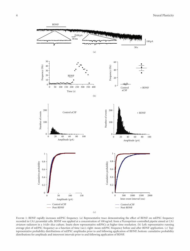
3.4. TRPC Channels Are Necessary for BDNF to IncreasemEPSC Frequency. The requirement of both Ca2+ influxand intracellular Ca2+ stores suggested the involvement ofTRPC channels, which are activated by the PLC cascade andmediated the activation of postsynaptic currents and den-dritic Ca2+ signals by BDNF in CA1 and CA3 pyramidalneurons [10, 22, 35]. Bath application of the TRPC inhibitorSKF-96365 [36] completely prevented the effect of BDNF onmEPSC frequency (3.04± 2.29 Hz versus 2.29± 0.78 Hz; n =3, P = 0.4256) (Figure 4(a)). The median mEPSC frequencychanged from 2.75 Hz to 2.28 Hz after BDNF application(Kolmogorov-Smirnov test Z value 2.3 ± 1.9).
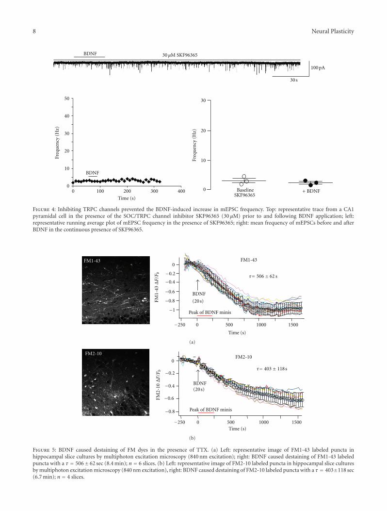
3.5. BDNF Rapidly Evokes FM Dye Destaining in the Presenceof TTX. To directly demonstrate that the elevated mEPSCfrequency evoked by BDNF represents vesicular transmitterrelease, we performed multiphoton imaging of FM dyes inslice cultures. The total pool of recycling synaptic vesicleswas labeled by exposing slices to a depolarizing solution(40 mM K+) containing 5 μM FM1-43 (Figure 5(a)) [18, 26].Following FM1-43 dye washout in a zero Ca2+ solution (toprevent FM1-43 destaining by spontaneous vesicle fusions),slices were imaged by multiphoton excitation microscopy(840 nm excitation) in Ca2+-containing aCSF with TTX toblock Na+-dependent action potentials. BDNF was appliedto apical dendrites in area CA1 using a Picospritzer in thesame manner as for whole-cell recordings of mEPSCs, andFM1-43 fluorescence intensity was followed over time (0.9frames per second, 30 min movies). Figure 5(a) shows arepresentative example of BDNF-induced FM1-43 destainingin the presence of TTX to block spike-dependent transmitterrelease. The rate of the exponential decay of FM1-43 fluo-rescence intensity immediately following BDNF applicationwas 506 ± 62 sec (n = 6 slices). These observations wereconfirmed with FM2-10, a styryl lipophilic dye with lowermembrane-binding affinity (Figure 5(b)). The exponentialdecay rate of FM2-10 intensity evoked by BDNF in TTX was403 ± 118 sec (n = 4 slices), which was not significantly dif-ferent that that observed using FM1-43 (P > 0.05). Theseresults show that BDNF induces FM dye loss by a vesicularfusion mechanism that is independent of the membraneaffinity and molecular size of FM1-43 and FM2-10 dyes. Fur-thermore, these imaging results directly demonstrate thatthe elevated mEPSC frequency caused by BDNF representSvesicular transmitter release from presynaptic terminals.
4. Discussion
The present study demonstrates that the BDNF-inducedincrease in mEPSC frequency recorded in hippocampal CA1pyramidal neurons represents Ca2+-dependent vesicle releasefrom presynaptic terminals. The BDNF-induced increase inmEPSC frequency is prevented by bath application of thetyrosine kinase inhibitor k-252a. Depletion of intracellularCa2+ stores with thapsigargin also inhibited the BDNF-induced mEPSC frequency increase, as did the removal of
extracellular Ca2+. These results demonstrate the criticalrequirement of TrkB-initiated Ca2+-dependent signaling inthe BDNF-induced increase in mEPSC frequency.
One of the signaling cascades activated by Trk receptors,the hydrolysis of PIP2 by PLCγ leading to IP3 formation, haslong been known to cause intracellular Ca2+ mobilizationin neurons [37]. Indeed, BDNF increased Ca2+ levels withinpresynaptic terminals of Xenopus neuromuscular junctionsin culture, leading to a transient enhancement of transmitterrelease that was dependent on extracellular Ca2+ levels andpresynaptic depolarization [38, 39]. Similarly, extracellul-ar Ca2+ was required for BDNF to increase the frequency ofmEPSCs and somatic Ca2+ levels in cultured hippocampalneurons [40]. In addition to well-established voltage- andligand-gated mechanisms of Ca2+ influx, Ca2+ signals medi-ated by depletion of intracellular Ca2+ stores are fundamentalcomponent of neuronal Ca2+ signaling. In non-excitablecells, the depletion of Ca2+ stores activates plasma mem-brane channels often called store-operated channels (SOCs),allowing Ca2+ entry for the replenishment of emptied stores;this process is called capacitative Ca2+ entry [41, 42]. Recentevidence suggests that cultured hippocampal neurons exhibitsuch capacitative Ca2+ entry [23, 43, 44]. Furthermore, werecently demonstrated that BDNF evokes postsynaptic cur-rents and dendritic Ca2+ signals in CA1 and CA3 pyramidalneurons through the activation of TrkB/PLC and mediatedby TRPC3-containing channels [10, 22, 35].
Here, we present evidence consistent with the model thatthe source of Ca2+ that contributes to the BDNF-inducedincrease in mEPSC frequency originates from presynaptic ERstores. The contribution of intracellular ER stores as a sourceof Ca2+ to asynchronous (spontaneous) quantal transmit-ter release has been a highly debated issue. Although somereports demonstrate that store depletion in hippocampalpyramidal neurons leads to Ca2+-induced Ca2+ release(CICR) and subsequent paired-pulse facilitation of excita-tory postsynaptic potentials [23], other reports are to thecontrary. Using thapsigargin, which empties ER stores bypoisoning the SERCA pump, or ryanodine, which blocksryanodine receptors, it has been demonstrated that ER storesand CICR do not affect paired-pulse facilitation of excita-tory postsynaptic potentials [45]. These studies were con-ducted at four different excitatory synapses in the brain:hippocampal CA3-CA1 synapses; mossy fiber synapsesbetween the dentate granule cells and CA3 pyramidal cells;synapses between CA3 neurons; cerebellar parallel fiber toPurkinje cell synapses [45]. Still others have shown thatimmediate mEPSC frequency increases are dependent uponmitochondrial excitatory postsynaptic potentials [46].
Our studies have directly demonstrated that the elevatedfrequency of mEPSCs evoked by brief and local BDNF appli-cation indeed reflects vesicular fusion events from presy-naptic terminals. We also attempted to determine whetherthe vesicular fusions underlying FM1-43 loss from terminalsrepresented kiss-and-run or full-fusion events by comparingthem to FM2-10 loss, which has a slower departition ratefrom lipid membranes [47, 48]. However, the kinetics ofBDNF-induced FM2-10 destaining was not different thanthat of FM1-43. This suggests that BDNF caused dye loss
8 Neural Plasticity
BaselineSKF96365
+ BDNF
100 pA
30 s
50
40
30
20
10
0
30
20
10
00 100 200 300 400
Time (s)
BDNF
BDNF
30 μM SKF96365
Freq
uen
cy (
Hz)
Freq
uen
cy (
Hz)
Figure 4: Inhibiting TRPC channels prevented the BDNF-induced increase in mEPSC frequency. Top: representative trace from a CA1pyramidal cell in the presence of the SOC/TRPC channel inhibitor SKF96365 (30 μM) prior to and following BDNF application; left:representative running average plot of mEPSC frequency in the presence of SKF96365; right: mean frequency of mEPSCs before and afterBDNF in the continuous presence of SKF96365.
FM1-43FM1-43
BDNF
(20 s)
−250 0 500 1000 1500
0
−0.2
−0.4
−0.6
−0.8
−1 Peak of BDNF minis
τ= 506± 62 s
FM1-
43ΔF/F
b
Time (s)
(a)
Peak of BDNF minis
BDNF(20 s)
0
−0.2
−0.4
−0.6
−0.8
−250 0 500 1000 1500
FM2-10FM2-10
τ= 403± 118s
FM2-
10ΔF/F
b
Time (s)
(b)
Figure 5: BDNF caused destaining of FM dyes in the presence of TTX. (a) Left: representative image of FM1-43 labeled puncta inhippocampal slice cultures by multiphoton excitation microscopy (840 nm excitation); right: BDNF caused destaining of FM1-43 labeledpuncta with a τ = 506± 62 sec (8.4 min); n = 6 slices. (b) Left: representative image of FM2-10 labeled puncta in hippocampal slice culturesby multiphoton excitation microscopy (840 nm excitation), right: BDNF caused destaining of FM2-10 labeled puncta with a τ = 403±118 sec(6.7 min); n = 4 slices.

Neural Plasticity 9
either via full-fusion events or through fusion pores witha permeability for styryl dyes that is not affected by themolecular sizes of FM1-43 and FM2-10. Recently, it has beendetermined that the FM family of dyes is not suitable fordetermining whether vesicle release occurs via full-fusion orkiss-and-run events [49].
Taken together, these results demonstrate that BDNFincreases mEPSC frequency by enhancing vesicular fusionthrough a presynaptic Ca2+-dependent mechanism involvingTrkB receptors, Ca2+ stores, and TRPC channels.
Funding
This research is supported by NIH Grants NS40593 and NS-065027. The funders had no role in study design, data col-lection and analysis, decision to publish, or preparation ofthe paper.
References
[1] Y. A. Barde, “Trophic factors and neuronal survival,” Neuron,vol. 2, no. 6, pp. 1525–1534, 1989.
[2] D. C. Lo, “Neurotrophic factors and synaptic plasticity,” Neu-ron, vol. 15, no. 5, pp. 979–981, 1995.
[3] B. Lu, “BDNF and activity-dependent synaptic modulation,”Learning and Memory, vol. 10, no. 2, pp. 86–98, 2003.
[4] M. M. Poo, “Neurotrophins as synaptic modulators,” NatureReviews Neuroscience, vol. 2, no. 1, pp. 24–32, 2001.
[5] H. Thoenen, “Neurotrophins and neuronal plasticity,” Science,vol. 270, no. 5236, pp. 593–598, 1995.
[6] W. J. Tyler, M. Alonso, C. R. Bramham, and L. D. Pozzo-Miller, “From acquisition to consolidation: on the role ofbrain-derived neurotrophic factor signaling in hippocampal-dependent learning,” Learning and Memory, vol. 9, no. 5, pp.224–237, 2002.
[7] A. M. Lohof, N. Y. Ip, and M. Poo, “Potentiation of developingneuromuscular synapses by the neurotrophins NT-3 andBDNF,” Nature, vol. 363, no. 6427, pp. 350–353, 1993.
[8] A. K. McAllister, “Subplate neurons: a missing link amongneurotrophins, activity, and ocular dominance plasticity?”Proceedings of the National Academy of Sciences of the UnitedStates of America, vol. 96, no. 24, pp. 13600–13602, 1999.
[9] M. Alonso, J. H. Medina, and L. Pozzo-Miller, “ERK1/2activation is necessary for BDNF to increase dendritic spinedensity in hippocampal CA1 pyramidal neurons,” Learningand Memory, vol. 11, no. 2, pp. 172–178, 2004.
[10] M. D. Amaral and L. Pozzo-Miller, “TRPC3 channels are nec-essary for brain-derived neurotrophic factor to activate a non-selective cationic current and to induce dendritic spine form-ation,” Journal of Neuroscience, vol. 27, no. 19, pp. 5179–5189,2007.
[11] C. A. Chapleau, M. E. Carlo, J. L. Larimore, and L. Pozzo-Miller, “The actions of BDNF on dendritic spine densityand morphology in organotypic slice cultures depend on thepresence of serum in culture media,” Journal of NeuroscienceMethods, vol. 169, no. 1, pp. 182–190, 2008.
[12] W. J. Tyler and L. D. Pozzo-Miller, “BDNF enhances quantalneurotransmitter release and increases the number of dock-ed vesicles at the active zones of hippocampal excitatory syn-apses,” Journal of Neuroscience, vol. 21, no. 12, pp. 4249–4258,2001.
[13] B. Berninger, A. F. Schinder, and M. M. Poo, “Synaptic relia-bility correlates with reduced susceptibility to synaptic poten-tiation by brain-derived neurotrophic factor,” Learning andMemory, vol. 6, no. 3, pp. 232–242, 1999.
[14] V. Lessmann, K. Gottmann, and R. Heumann, “BDNF andNT-4/5 enhance glutamatergic synaptic transmission in cul-tured hippocampal neurones,” NeuroReport, vol. 6, no. 1, pp.21–25, 1994.
[15] E. S. Levine, C. F. Dreyfus, I. B. Black, and M. R. Plummer,“Brain-derived neurotrophic factor rapidly enhances synaptictransmission in hippocampal neurons via postsynaptic tyro-sine kinase receptors,” Proceedings of the National Academy ofSciences of the United States of America, vol. 92, no. 17, pp.8074–8077, 1995.
[16] Y. X. Li, Y. Zhang, H. A. Lester, E. M. Schuman, and N.Davidson, “Enhancement of neurotransmitter release inducedby brain-derived neurotrophic factor in cultured hippocampalneurons,” Journal of Neuroscience, vol. 18, no. 24, pp. 10231–10240, 1998.
[17] A. F. Schinder, B. Berninger, and M. M. Poo, “Postsynaptictarget specificity of neurotrophin-induced presynaptic poten-tiation,” Neuron, vol. 25, no. 1, pp. 151–163, 2000.
[18] W. J. Tyler, X. L. Zhang, K. Hartman et al., “BDNF increasesrelease probability and the size of a rapidly recycling vesiclepool within rat hippocampal excitatory synapses,” Journal ofPhysiology, vol. 574, no. 3, pp. 787–803, 2006.
[19] C. Rosenmund and C. F. Stevens, “Definition of the readilyreleasable pool of vesicles at hippocampal synapses,” Neuron,vol. 16, no. 6, pp. 1197–1207, 1996.
[20] C. F. Stevens and T. Tsujimoto, “Estimates for the pool size ofreleasable quanta at a single central synapse and for the timerequired to refill the pool,” Proceedings of the National Academyof Sciences of the United States of America, vol. 92, no. 3, pp.846–849, 1995.
[21] J. N. Jovanovic, A. J. Czernik, A. A. Fienberg, P. Greengard,and T. S. Sihra, “Synapsins as mediators of BDNF-enhancedneurotransmitter release,” Nature Neuroscience, vol. 3, no. 4,pp. 323–329, 2000.
[22] M. D. Amaral and L. Pozzo-Miller, “BDNF induces calciumelevations associated with IBDNF, a nonselective cationic cur-rent mediated by TRPC channels,” Journal of Neurophysiology,vol. 98, no. 4, pp. 2476–2482, 2007.
[23] N. J. Emptage, C. A. Reid, and A. Fine, “Calcium stores in hip-pocampal synaptic boutons mediate short-term plasticity,store-operated Ca2+ entry, and spontaneous transmitterrelease,” Neuron, vol. 29, no. 1, pp. 197–208, 2001.
[24] L. D. Pozzo Miller, J. J. Petrozzino, and J. A. Connor, “G pro-tein-coupled receptors mediate a fast excitatory postsynapticcurrent in CA3 pyramidal neurons in hippocampal slices,”Journal of Neuroscience, vol. 15, no. 12, pp. 8320–8330, 1995.
[25] N. Axmacher, J. Winterer, P. K. Stanton, A. Draguhn, andW. Muller, “Two-photon imaging of spontaneous vesicularrelease in acute brain slices and its modulation by presynapticGABAA receptors,” NeuroImage, vol. 22, no. 2, pp. 1014–1021,2004.
[26] S. S. Mathew, L. Pozzo-Miller, and J. J. Hablitz, “Kainatemodulates presynaptic GABA release from two vesicle pools,”Journal of Neuroscience, vol. 28, no. 3, pp. 725–731, 2008.
[27] P. K. Stanton, U. Heinemann, and W. Muller, “FM1-43 imag-ing reveals cGMP-dependent long-term depression of presy-naptic transmitter release.,” The Journal of Neuroscience, vol.21, no. 19, article RC167, 2001.
[28] S. S. Zakharenko, L. Zablow, and S. A. Siegelbaum, “Visual-ization of changes in presynaptic function during long-term

10 Neural Plasticity
synaptic plasticity,” Nature Neuroscience, vol. 4, no. 7, pp. 711–717, 2001.
[29] W. S. Rasband, U. S. National Institutes of Health, Bethesda,Maryland, USA, http://imagej.nih.gov/ij/, 1997–2012.
[30] A. K. McAllister, L. C. Katz, and D. C. Lo, “Neurotrophins andsynaptic plasticity,” Annual Review of Neuroscience, vol. 22, pp.295–318, 1999.
[31] V. Lessmann, K. Gottmann, and M. Malcangio, “Neurotro-phin secretion: current facts and future prospects,” Progress inNeurobiology, vol. 69, no. 5, pp. 341–374, 2003.
[32] P. Tapley, F. Lamballe, and M. Barbacid, “K252a is a selectiveinhibitor of the tyrosine protein kinase activity of the trk fami-ly of oncogenes and neurotrophin receptors,” Oncogene, vol. 7,no. 2, pp. 371–381, 1992.
[33] B. Knusel and F. Hefti, “K-252 compounds: modulators ofneurotrophin signal transduction,” Journal of Neurochemistry,vol. 59, no. 6, pp. 1987–1996, 1992.
[34] O. Thastrup, P. J. Cullen, B. K. Drobak, M. R. Hanley, and A. P.Dawson, “Thapsigargin, a tumor promoter, discharges intra-cellular Ca2+ stores by specific inhibition of the endoplasmicreticulum Ca2+-ATPase,” Proceedings of the National Academyof Sciences of the United States of America, vol. 87, no. 7, pp.2466–2470, 1990.
[35] Y. Li, G. Calfa, T. Inoue, M. D. Amaral, and L. Pozzo-Miller,“Activity-dependent release of endogenous BDNF from mossyfibers evokes a TRPC3 current and Ca2+ elevations in CA3pyramidal neurons,” Journal of Neurophysiology, vol. 103, no.5, pp. 2846–2856, 2010.
[36] J. E. Merritt, W. P. Armstrong, C. D. Benham et al., “SK and F96365, a novel inhibitor of receptor-mediated calcium entry,”Biochemical Journal, vol. 271, no. 2, pp. 515–522, 1990.
[37] M. J. Berridge, P. Lipp, and M. D. Bootman, “The versatilityand universality of calcium signalling,” Nature Reviews Molec-ular Cell Biology, vol. 1, no. 1, pp. 11–21, 2000.
[38] L. Boulanger and M. M. Poo, “Presynaptic depolarization faci-litates neurotrophin-induced synaptic potentiation,” NatureNeuroscience, vol. 2, no. 4, pp. 346–351, 1999.
[39] R. Stoop and M. M. Poo, “Synaptic modulation by neu-rotrophic factors: differential and synergistic effects of brain-derived neurotrophic factor and ciliary neurotrophic factor,”Journal of Neuroscience, vol. 16, no. 10, pp. 3256–3264, 1996.
[40] B. Berninger, D. E. Garcia, N. Inagaki, C. Hahnel, and D.Lindholm, “BDNF and NT-3 induce intracellular Ca2+ eleva-tion in hippocampal neurones,” NeuroReport, vol. 4, no. 12,pp. 1303–1306, 1993.
[41] M. J. Berridge, “Capacitative calcium entry,” Biochemical Jour-nal, vol. 312, pp. 1–11, 1995.
[42] J. W. Putney Jr., “A model for receptor-regulated calciumentry,” Cell Calcium, vol. 7, no. 1, pp. 1–12, 1986.
[43] A. Baba, T. Yasui, S. Fujisawa et al., “Activity-evoked capacita-tive Ca2+ entry: implications in synaptic plasticity,” Journal ofNeuroscience, vol. 23, no. 21, pp. 7737–7741, 2003.
[44] A. Bouron, “Activation of a capacitative Ca2+ entry pathwayby store depletion in cultured hippocampal neurones,” FEBSLetters, vol. 470, no. 3, pp. 269–272, 2000.
[45] A. G. Carter, K. E. Vogt, K. A. Foster, and W. G. Regehr, “Asses-sing the role of calcium-induced calcium release in short-termpresynaptic plasticity at excitatory central synapses,” Journal ofNeuroscience, vol. 22, no. 1, pp. 21–28, 2002.
[46] Y. G. Tang and R. S. Zucker, “Mitochondrial involvement inpost-tetanic potentiation of synaptic transmission,” Neuron,vol. 18, no. 3, pp. 483–491, 1997.
[47] J. L. Pyle, E. T. Kavalali, E. S. Piedras-Renterıa, and R. W. Tsien,“Rapid reuse of readily releasable pool vesicles at hippocampalsynapses,” Neuron, vol. 28, no. 1, pp. 221–231, 2000.
[48] T. A. Ryan, H. Reuter, B. Wendland, F. E. Schweizer, R. W.Tsien, and S. J. Smith, “The kinetics of synaptic vesicle recycl-ing measured at single presynaptic boutons,” Neuron, vol. 11,no. 4, pp. 713–724, 1993.
[49] Q. Zhang, Y. Q. Cao, and R. W. Tsien, “Quantum dots providean optical signal specific to full collapse fusion of synapticvesicles,” Proceedings of the National Academy of Sciences of theUnited States of America, vol. 104, no. 45, pp. 17843–17848,2007.

Submit your manuscripts athttp://www.hindawi.com
Neurology Research International
Hindawi Publishing Corporationhttp://www.hindawi.com Volume 2014
Alzheimer’s DiseaseHindawi Publishing Corporationhttp://www.hindawi.com Volume 2014
International Journal of
ScientificaHindawi Publishing Corporationhttp://www.hindawi.com Volume 2014
Hindawi Publishing Corporationhttp://www.hindawi.com Volume 2014
BioMed Research International
Hindawi Publishing Corporationhttp://www.hindawi.com Volume 2014
Research and TreatmentSchizophrenia
The Scientific World JournalHindawi Publishing Corporation http://www.hindawi.com Volume 2014
Hindawi Publishing Corporationhttp://www.hindawi.com Volume 2014
Neural Plasticity
Hindawi Publishing Corporationhttp://www.hindawi.com Volume 2014
Parkinson’s Disease
Hindawi Publishing Corporationhttp://www.hindawi.com Volume 2014
Research and TreatmentAutism
Sleep DisordersHindawi Publishing Corporationhttp://www.hindawi.com Volume 2014
Hindawi Publishing Corporationhttp://www.hindawi.com Volume 2014
Neuroscience Journal
Epilepsy Research and TreatmentHindawi Publishing Corporationhttp://www.hindawi.com Volume 2014
Hindawi Publishing Corporationhttp://www.hindawi.com Volume 2014
Psychiatry Journal
Hindawi Publishing Corporationhttp://www.hindawi.com Volume 2014
Computational and Mathematical Methods in Medicine
Depression Research and TreatmentHindawi Publishing Corporationhttp://www.hindawi.com Volume 2014
Hindawi Publishing Corporationhttp://www.hindawi.com Volume 2014
Brain ScienceInternational Journal of
StrokeResearch and TreatmentHindawi Publishing Corporationhttp://www.hindawi.com Volume 2014
Neurodegenerative Diseases
Hindawi Publishing Corporationhttp://www.hindawi.com Volume 2014
Journal of
Cardiovascular Psychiatry and NeurologyHindawi Publishing Corporationhttp://www.hindawi.com Volume 2014