
Intra-Vascular Occlusion of the Aorta for Massive Pelvic...
5
Articles © The authors | Journal compilation © J Curr Surg and Elmer Press Inc™ | www.currentsurgery.org This article is distributed under the terms of the Creative Commons Attribution Non-Commercial 4.0 International License, which permits unrestricted non-commercial use, distribution, and reproduction in any medium, provided the original work is properly cited 13 Case Report J Curr Surg. 2018;8(1-2):13-17 Intra-Vascular Occlusion of the Aorta for Massive Pelvic Trauma: A New Application Paul Hanna a, c , Paul Seo b , John Yoon a , Manrique Guerrero a , Hoan Bui a , Robert Madlinger a , Jamshed Zuberi a Abstract Globally, trauma remains the leading cause of morbidity and mor- tality for all age groups with uncontrolled hemorrhage as the most common form of preventable death in the trauma setting. Specifically, non-compressible torso hemorrhage in trauma patients is known to have high mortality rates. Open aortic cross clamping via anterolat- eral thoracotomy has been the standard approach, but the procedure carries a high mortality risk. Resuscitative endovascular balloon oc- clusion of the aorta (REBOA) is a technique that promptly controls hemorrhage, increases cardiac afterload, and increases central aortic pressure. REBOA in different forms has existed since the Korean War in 1950. It involves the insertion of a balloon into the aorta via a fem- oral access approach that is subsequently inflated to provide hemosta- sis. Despite the potential of REBOA to save lives, many non-vascular surgeons and emergency healthcare providers may be reluctant to use the new procedure. This may be due to a lack of knowledge, skill, or equipment required to perform the procedure. We present a case of a patient with multiple pelvic injuries and massive bleeding for which REBOA was utilized safely and correctly. Keywords: Trauma; Resuscitative endovascular balloon occlusion of the aorta; Aortic occlusion; Hemorrhagic shock; Hemorrhage; Pelvic bleeding; Pelvic hemorrhage Introduction Non-compressible torso hemorrhage in trauma patients is known to have high mortality rates. Open aortic cross clamp- ing via anterolateral thoracotomy has been the standard ap- proach, but the procedure carries a high mortality risk [1]. Re- cently a minimally invasive endovascular approach has been developed to occlude the aorta proximally and control torso hemorrhage. Resuscitative endovascular balloon occlusion of the aorta (REBOA) is a technique that promptly controls hemorrhage, increases cardiac afterload, and increases central aortic pres- sure. Studies demonstrate that REBOA surpasses the aortic cross-clamping procedure with higher survival rates and fewer premature deaths [1, 2]. REBOA can be used as a proactive measure when a risk of cardiovascular collapse is identified. Despite the potential of REBOA to save lives, many non- vascular surgeons and emergency healthcare providers may be reluctant to use the new procedure. This may be due to a lack of knowledge, skill, or equipment required to perform the pro- cedure. However, recent studies demonstrate that with proper training, nonsurgical providers can properly place REBOA catheters effectively in austere prehospital settings [3]. REBOA is a novel procedure, with not many documented successful cases for pelvic trauma. In this paper we present a case of a patient with multiple pelvic injuries and massive bleeding for which REBOA was utilized safely. This case is unique in that there is sound coordination between trauma surgery, critical care, vascular surgery, orthopedic surgery and urology in order to effectively manage the patient. Case Report A 23-year-old male with no prior medical history was brought into the emergency department following a 10-foot fall. The patient stated he was climbing a wall, fell backwards, and sub- sequently a large concrete block fell onto his pelvis. He com- plained of pain in the pelvic region and was unable to move his lower extremities secondary to the pain. He denied hitting his head or losing consciousness, and was able to recollect the events. His vitals in the trauma bay were temperature 35.7 °C, pulse 58 beats/min, blood pressure 112/63 mm Hg, respiratory rate 14 breaths/min with saturations at 100% on 3 L of oxygen nasal cannula. Routine labs and an arterial blood gas were per- formed: hemoglobin 11.8 mg/dL, hematocrit 36%, INR 1.1, pH 7.41, lactate 1.4 mg/dL, base deficit -2.1. On initial assessment, the patient’s airway was intact with normal cardiopulmonary, neurological and abdominal exams. On musculoskeletal exam the patient had tenderness with pal- pation of the pelvis. Vascular exam confirmed palpable femo- ral and dorsalis pedis pulses bilaterally. A fast exam was performed which was negative in all four Manuscript submitted May 7, 2018, accepted May 16, 2018 a Saint Joseph’s Regional Medical Center, Paterson, NJ 07503, USA b New York Medical College, Valhalla, NY 10595, USA c Corresponding Author: Paul Hanna, Department of Surgery, Saint Joseph’s Regional Medical Center, 703 Main Street, Paterson, NJ 07503, USA. Email: [email protected] doi: https://doi.org/10.14740/jcs349w
-
Upload
truongminh -
Category
Documents
-
view
215 -
download
0
Transcript of Intra-Vascular Occlusion of the Aorta for Massive Pelvic...
Articles © The authors | Journal compilation © J Curr Surg and Elmer Press Inc™ | www.currentsurgery.orgThis article is distributed under the terms of the Creative Commons Attribution Non-Commercial 4.0 International License, which permits
unrestricted non-commercial use, distribution, and reproduction in any medium, provided the original work is properly cited13
Case Report J Curr Surg. 2018;8(1-2):13-17
Intra-Vascular Occlusion of the Aorta for Massive Pelvic Trauma: A New Application
Paul Hannaa, c, Paul Seob, John Yoona, Manrique Guerreroa, Hoan Buia, Robert Madlingera, Jamshed Zuberia
Abstract
Globally, trauma remains the leading cause of morbidity and mor-tality for all age groups with uncontrolled hemorrhage as the most common form of preventable death in the trauma setting. Specifically, non-compressible torso hemorrhage in trauma patients is known to have high mortality rates. Open aortic cross clamping via anterolat-eral thoracotomy has been the standard approach, but the procedure carries a high mortality risk. Resuscitative endovascular balloon oc-clusion of the aorta (REBOA) is a technique that promptly controls hemorrhage, increases cardiac afterload, and increases central aortic pressure. REBOA in different forms has existed since the Korean War in 1950. It involves the insertion of a balloon into the aorta via a fem-oral access approach that is subsequently inflated to provide hemosta-sis. Despite the potential of REBOA to save lives, many non-vascular surgeons and emergency healthcare providers may be reluctant to use the new procedure. This may be due to a lack of knowledge, skill, or equipment required to perform the procedure. We present a case of a patient with multiple pelvic injuries and massive bleeding for which REBOA was utilized safely and correctly.
Keywords: Trauma; Resuscitative endovascular balloon occlusion of the aorta; Aortic occlusion; Hemorrhagic shock; Hemorrhage; Pelvic bleeding; Pelvic hemorrhage
Introduction
Non-compressible torso hemorrhage in trauma patients is known to have high mortality rates. Open aortic cross clamp-ing via anterolateral thoracotomy has been the standard ap-proach, but the procedure carries a high mortality risk [1]. Re-cently a minimally invasive endovascular approach has been developed to occlude the aorta proximally and control torso
hemorrhage.Resuscitative endovascular balloon occlusion of the aorta
(REBOA) is a technique that promptly controls hemorrhage, increases cardiac afterload, and increases central aortic pres-sure. Studies demonstrate that REBOA surpasses the aortic cross-clamping procedure with higher survival rates and fewer premature deaths [1, 2]. REBOA can be used as a proactive measure when a risk of cardiovascular collapse is identified.
Despite the potential of REBOA to save lives, many non-vascular surgeons and emergency healthcare providers may be reluctant to use the new procedure. This may be due to a lack of knowledge, skill, or equipment required to perform the pro-cedure. However, recent studies demonstrate that with proper training, nonsurgical providers can properly place REBOA catheters effectively in austere prehospital settings [3].
REBOA is a novel procedure, with not many documented successful cases for pelvic trauma. In this paper we present a case of a patient with multiple pelvic injuries and massive bleeding for which REBOA was utilized safely. This case is unique in that there is sound coordination between trauma surgery, critical care, vascular surgery, orthopedic surgery and urology in order to effectively manage the patient.
Case Report
A 23-year-old male with no prior medical history was brought into the emergency department following a 10-foot fall. The patient stated he was climbing a wall, fell backwards, and sub-sequently a large concrete block fell onto his pelvis. He com-plained of pain in the pelvic region and was unable to move his lower extremities secondary to the pain. He denied hitting his head or losing consciousness, and was able to recollect the events.
His vitals in the trauma bay were temperature 35.7 °C, pulse 58 beats/min, blood pressure 112/63 mm Hg, respiratory rate 14 breaths/min with saturations at 100% on 3 L of oxygen nasal cannula. Routine labs and an arterial blood gas were per-formed: hemoglobin 11.8 mg/dL, hematocrit 36%, INR 1.1, pH 7.41, lactate 1.4 mg/dL, base deficit -2.1.
On initial assessment, the patient’s airway was intact with normal cardiopulmonary, neurological and abdominal exams. On musculoskeletal exam the patient had tenderness with pal-pation of the pelvis. Vascular exam confirmed palpable femo-ral and dorsalis pedis pulses bilaterally.
A fast exam was performed which was negative in all four
Manuscript submitted May 7, 2018, accepted May 16, 2018
aSaint Joseph’s Regional Medical Center, Paterson, NJ 07503, USAbNew York Medical College, Valhalla, NY 10595, USAcCorresponding Author: Paul Hanna, Department of Surgery, Saint Joseph’s Regional Medical Center, 703 Main Street, Paterson, NJ 07503, USA. Email: [email protected]
doi: https://doi.org/10.14740/jcs349w
Articles © The authors | Journal compilation © J Curr Surg and Elmer Press Inc™ | www.currentsurgery.org14
REBOA for Massive Pelvic Trauma J Curr Surg. 2018;8(1-2):13-17
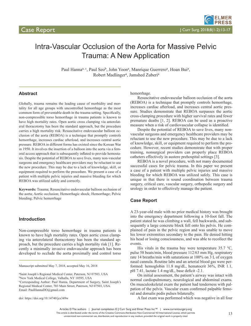
regions. A bedside chest X-ray showed clear lung fields bilat-erally. Pelvic X-ray showed a comminuted, displaced fracture of the right superior and inferior pubic rami as well as non-displaced fractures of the left superior pubic ramus and left acetabulum (Fig. 1).
En route to the CT scan, the patient became hypotensive to 70/50 mm Hg. Two units of packed red blood cells (pRBCs) were immediately transfused with an initial response to 100 - 110 mm Hg systolic BP. However, he became hypotensive again to 80 mm Hg systolic. The patient was then transferred back to the trauma bay with activation of massive transfusion protocol (MTP).
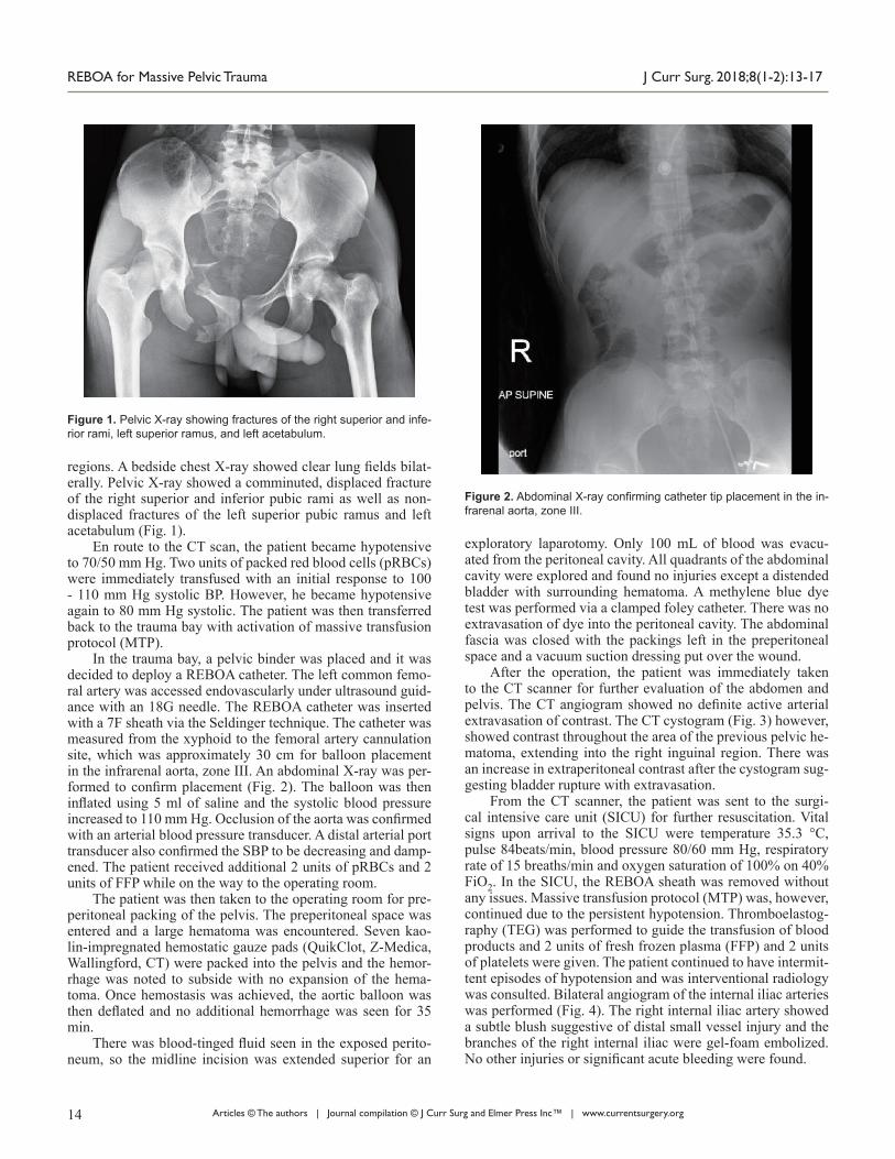
In the trauma bay, a pelvic binder was placed and it was decided to deploy a REBOA catheter. The left common femo-ral artery was accessed endovascularly under ultrasound guid-ance with an 18G needle. The REBOA catheter was inserted with a 7F sheath via the Seldinger technique. The catheter was measured from the xyphoid to the femoral artery cannulation site, which was approximately 30 cm for balloon placement in the infrarenal aorta, zone III. An abdominal X-ray was per-formed to confirm placement (Fig. 2). The balloon was then inflated using 5 ml of saline and the systolic blood pressure increased to 110 mm Hg. Occlusion of the aorta was confirmed with an arterial blood pressure transducer. A distal arterial port transducer also confirmed the SBP to be decreasing and damp-ened. The patient received additional 2 units of pRBCs and 2 units of FFP while on the way to the operating room.
The patient was then taken to the operating room for pre-peritoneal packing of the pelvis. The preperitoneal space was entered and a large hematoma was encountered. Seven kao-lin-impregnated hemostatic gauze pads (QuikClot, Z-Medica, Wallingford, CT) were packed into the pelvis and the hemor-rhage was noted to subside with no expansion of the hema-toma. Once hemostasis was achieved, the aortic balloon was then deflated and no additional hemorrhage was seen for 35 min.
There was blood-tinged fluid seen in the exposed perito-neum, so the midline incision was extended superior for an
exploratory laparotomy. Only 100 mL of blood was evacu-ated from the peritoneal cavity. All quadrants of the abdominal cavity were explored and found no injuries except a distended bladder with surrounding hematoma. A methylene blue dye test was performed via a clamped foley catheter. There was no extravasation of dye into the peritoneal cavity. The abdominal fascia was closed with the packings left in the preperitoneal space and a vacuum suction dressing put over the wound.
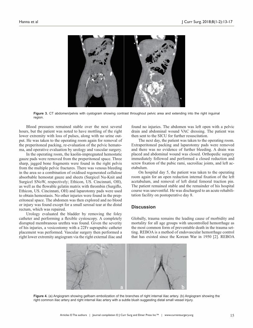
After the operation, the patient was immediately taken to the CT scanner for further evaluation of the abdomen and pelvis. The CT angiogram showed no definite active arterial extravasation of contrast. The CT cystogram (Fig. 3) however, showed contrast throughout the area of the previous pelvic he-matoma, extending into the right inguinal region. There was an increase in extraperitoneal contrast after the cystogram sug-gesting bladder rupture with extravasation.
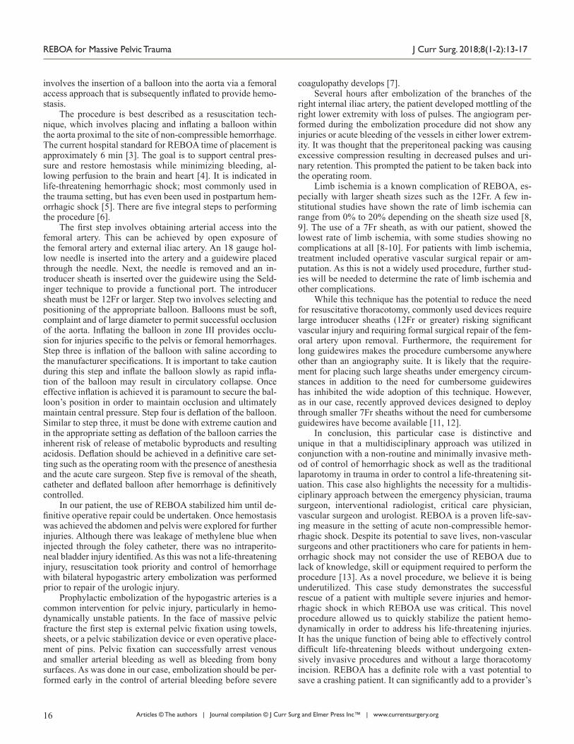
From the CT scanner, the patient was sent to the surgi-cal intensive care unit (SICU) for further resuscitation. Vital signs upon arrival to the SICU were temperature 35.3 °C, pulse 84beats/min, blood pressure 80/60 mm Hg, respiratory rate of 15 breaths/min and oxygen saturation of 100% on 40% FiO2. In the SICU, the REBOA sheath was removed without any issues. Massive transfusion protocol (MTP) was, however, continued due to the persistent hypotension. Thromboelastog-raphy (TEG) was performed to guide the transfusion of blood products and 2 units of fresh frozen plasma (FFP) and 2 units of platelets were given. The patient continued to have intermit-tent episodes of hypotension and was interventional radiology was consulted. Bilateral angiogram of the internal iliac arteries was performed (Fig. 4). The right internal iliac artery showed a subtle blush suggestive of distal small vessel injury and the branches of the right internal iliac were gel-foam embolized. No other injuries or significant acute bleeding were found.
Figure 1. Pelvic X-ray showing fractures of the right superior and infe-rior rami, left superior ramus, and left acetabulum.
Figure 2. Abdominal X-ray confirming catheter tip placement in the in-frarenal aorta, zone III.
Articles © The authors | Journal compilation © J Curr Surg and Elmer Press Inc™ | www.currentsurgery.org 15
Hanna et al J Curr Surg. 2018;8(1-2):13-17
Blood pressures remained stable over the next several hours, but the patient was noted to have mottling of the right lower extremity with loss of pulses, along with no urine out-put. He was taken to the operating room again for removal of the preperitoneal packing, re-evaluation of the pelvic hemato-ma, and operative evaluation by urology and vascular surgery.
In the operating room, the kaolin-impregnated hemostatic gauze pads were removed from the preperitoneal space. Three sharp, jagged bone fragments were found in the right pelvis from the multiple pelvic fractures. There was venous bleeding in the area so a combination of oxidised regenerated cellulose absorbable hemostat gauze and sheets (Surgicel Nu-Knit and Surgicel SNoW, respectively; Ethicon, US. Cincinnati, OH), as well as the flowable gelatin matrix with thrombin (Surgiflo, Ethicon, US. Cincinnati, OH) and laparotomy pads were used to obtain hemostasis. No other injuries were found in the prep-eritoneal space. The abdomen was then explored and no blood or injury was found except for a small serosal tear at the distal rectum, which was repaired.
Urology evaluated the bladder by removing the foley catheter and performing a flexible cystoscopy. A completely disrupted membranous urethra was found. Given the severity of his injuries, a vesicostomy with a 22Fr suprapubic catheter placement was performed. Vascular surgery then performed a right lower extremity angiogram via the right external iliac and
found no injuries. The abdomen was left open with a pelvic drain and abdominal wound VAC dressing. The patient was then sent to the SICU for further resuscitation.
The next day, the patient was taken to the operating room. Extraperitoneal packing and laparotomy pads were removed and there was no evidence of further bleeding. A drain was placed and abdominal wound was closed. Orthopedic surgery immediately followed and performed a closed reduction and screw fixation of the pubic rami, sacroiliac joints, and left ac-etabulum.
On hospital day 5, the patient was taken to the operating room again for an open reduction internal fixation of the left acetabulum, and removal of left distal femoral traction pin. The patient remained stable and the remainder of his hospital course was uneventful. He was discharged to an acute rehabili-tation facility on postoperative day 8.
Discussion
Globally, trauma remains the leading cause of morbidity and mortality for all age groups with uncontrolled hemorrhage as the most common form of preventable death in the trauma set-ting. REBOA is a method of endovascular hemorrhage control that has existed since the Korean War in 1950 [2]. REBOA
Figure 3. CT abdomen/pelvis with cystogram showing contrast throughout pelvic area and extending into the right inguinal region.
Figure 4. (a) Angiogram showing gelfoam embolization of the branches of right internal iliac artery. (b) Angiogram showing the right common iliac artery and right internal iliac artery with a subtle blush suggesting distal small vessel injury.
Articles © The authors | Journal compilation © J Curr Surg and Elmer Press Inc™ | www.currentsurgery.org16
REBOA for Massive Pelvic Trauma J Curr Surg. 2018;8(1-2):13-17
involves the insertion of a balloon into the aorta via a femoral access approach that is subsequently inflated to provide hemo-stasis.
The procedure is best described as a resuscitation tech-nique, which involves placing and inflating a balloon within the aorta proximal to the site of non-compressible hemorrhage. The current hospital standard for REBOA time of placement is approximately 6 min [3]. The goal is to support central pres-sure and restore hemostasis while minimizing bleeding, al-lowing perfusion to the brain and heart [4]. It is indicated in life-threatening hemorrhagic shock; most commonly used in the trauma setting, but has even been used in postpartum hem-orrhagic shock [5]. There are five integral steps to performing the procedure [6].
The first step involves obtaining arterial access into the femoral artery. This can be achieved by open exposure of the femoral artery and external iliac artery. An 18 gauge hol-low needle is inserted into the artery and a guidewire placed through the needle. Next, the needle is removed and an in-troducer sheath is inserted over the guidewire using the Seld-inger technique to provide a functional port. The introducer sheath must be 12Fr or larger. Step two involves selecting and positioning of the appropriate balloon. Balloons must be soft, complaint and of large diameter to permit successful occlusion of the aorta. Inflating the balloon in zone III provides occlu-sion for injuries specific to the pelvis or femoral hemorrhages. Step three is inflation of the balloon with saline according to the manufacturer specifications. It is important to take caution during this step and inflate the balloon slowly as rapid infla-tion of the balloon may result in circulatory collapse. Once effective inflation is achieved it is paramount to secure the bal-loon’s position in order to maintain occlusion and ultimately maintain central pressure. Step four is deflation of the balloon. Similar to step three, it must be done with extreme caution and in the appropriate setting as deflation of the balloon carries the inherent risk of release of metabolic byproducts and resulting acidosis. Deflation should be achieved in a definitive care set-ting such as the operating room with the presence of anesthesia and the acute care surgeon. Step five is removal of the sheath, catheter and deflated balloon after hemorrhage is definitively controlled.
In our patient, the use of REBOA stabilized him until de-finitive operative repair could be undertaken. Once hemostasis was achieved the abdomen and pelvis were explored for further injuries. Although there was leakage of methylene blue when injected through the foley catheter, there was no intraperito-neal bladder injury identified. As this was not a life-threatening injury, resuscitation took priority and control of hemorrhage with bilateral hypogastric artery embolization was performed prior to repair of the urologic injury.
Prophylactic embolization of the hypogastric arteries is a common intervention for pelvic injury, particularly in hemo-dynamically unstable patients. In the face of massive pelvic fracture the first step is external pelvic fixation using towels, sheets, or a pelvic stabilization device or even operative place-ment of pins. Pelvic fixation can successfully arrest venous and smaller arterial bleeding as well as bleeding from bony surfaces. As was done in our case, embolization should be per-formed early in the control of arterial bleeding before severe
coagulopathy develops [7].Several hours after embolization of the branches of the
right internal iliac artery, the patient developed mottling of the right lower extremity with loss of pulses. The angiogram per-formed during the embolization procedure did not show any injuries or acute bleeding of the vessels in either lower extrem-ity. It was thought that the preperitoneal packing was causing excessive compression resulting in decreased pulses and uri-nary retention. This prompted the patient to be taken back into the operating room.
Limb ischemia is a known complication of REBOA, es-pecially with larger sheath sizes such as the 12Fr. A few in-stitutional studies have shown the rate of limb ischemia can range from 0% to 20% depending on the sheath size used [8, 9]. The use of a 7Fr sheath, as with our patient, showed the lowest rate of limb ischemia, with some studies showing no complications at all [8-10]. For patients with limb ischemia, treatment included operative vascular surgical repair or am-putation. As this is not a widely used procedure, further stud-ies will be needed to determine the rate of limb ischemia and other complications.
While this technique has the potential to reduce the need for resuscitative thoracotomy, commonly used devices require large introducer sheaths (12Fr or greater) risking significant vascular injury and requiring formal surgical repair of the fem-oral artery upon removal. Furthermore, the requirement for long guidewires makes the procedure cumbersome anywhere other than an angiography suite. It is likely that the require-ment for placing such large sheaths under emergency circum-stances in addition to the need for cumbersome guidewires has inhibited the wide adoption of this technique. However, as in our case, recently approved devices designed to deploy through smaller 7Fr sheaths without the need for cumbersome guidewires have become available [11, 12].
In conclusion, this particular case is distinctive and unique in that a multidisciplinary approach was utilized in conjunction with a non-routine and minimally invasive meth-od of control of hemorrhagic shock as well as the traditional laparotomy in trauma in order to control a life-threatening sit-uation. This case also highlights the necessity for a multidis-ciplinary approach between the emergency physician, trauma surgeon, interventional radiologist, critical care physician, vascular surgeon and urologist. REBOA is a proven life-sav-ing measure in the setting of acute non-compressible hemor-rhagic shock. Despite its potential to save lives, non-vascular surgeons and other practitioners who care for patients in hem-orrhagic shock may not consider the use of REBOA due to lack of knowledge, skill or equipment required to perform the procedure [13]. As a novel procedure, we believe it is being underutilized. This case study demonstrates the successful rescue of a patient with multiple severe injuries and hemor-rhagic shock in which REBOA use was critical. This novel procedure allowed us to quickly stabilize the patient hemo-dynamically in order to address his life-threatening injuries. It has the unique function of being able to effectively control difficult life-threatening bleeds without undergoing exten-sively invasive procedures and without a large thoracotomy incision. REBOA has a definite role with a vast potential to save a crashing patient. It can significantly add to a provider’s
Articles © The authors | Journal compilation © J Curr Surg and Elmer Press Inc™ | www.currentsurgery.org 17
Hanna et al J Curr Surg. 2018;8(1-2):13-17
armamentarium in the trauma patient.
Conflict of Interest
There is no disclosure or conflict of interest in this project.
References
1. Manzano Nunez R, Naranjo MP, Foianini E, Ferrada P, Rincon E, Garcia-Perdomo HA, Burbano P, et al. A meta-analysis of resuscitative endovascular balloon occlusion of the aorta (REBOA) or open aortic cross-clamping by resuscitative thoracotomy in non-compressible torso hemorrhage patients. World J Emerg Surg. 2017;12:30.
2. Moore LJ, Brenner M, Kozar RA, Pasley J, Wade CE, Baraniuk MS, Scalea T, et al. Implementation of resus-citative endovascular balloon occlusion of the aorta as an alternative to resuscitative thoracotomy for noncom-pressible truncal hemorrhage. J Trauma Acute Care Surg. 2015;79(4):523-530; discussion 530-522.
3. Ross EM, Redman TT. Feasibility and proposed training pathway for austere application of resuscitative balloon occlusion of the aorta. J Spec Oper Med.18(1):37-43.
4. Valkenburg A, Bennett D, Bishop J, Smith G. Resuscita-tive endovascular balloon occlusion of the aorta as a po-tential prehospital procedure for the control of non-com-pressible haemorrhage: A literature review. Australasian Journal of Paramedicine. 2015;12(4).
5. White JM, Cannon JW, Stannard A, Burkhardt GE, Spencer JR, Williams K, Oh JS, et al. Direct vascu-lar control results in less physiologic derangement than proximal aortic clamping in a porcine model of non-
compressible extrathoracic torso hemorrhage. J Trauma. 2011;71(5):1278-1286; discussion 1286-1277.
6. Stensaeth KH, Sovik E, Haig IN, Skomedal E, Jorgensen A. Fluoroscopy-free Resuscitative Endovascular Bal-loon Occlusion of the Aorta (REBOA) for controlling life threatening postpartum hemorrhage. PLoS One. 2017;12(3):e0174520.
7. Stannard A, Eliason JL, Rasmussen TE. Resuscita-tive endovascular balloon occlusion of the aorta (RE-BOA) as an adjunct for hemorrhagic shock. J Trauma. 2011;71(6):1869-1872.
8. Bogert JN, Davis KM, Kopelman TR, Vail SJ, Pieri PG, Matthews MR. Resuscitative endovascular balloon occlu-sion of the aorta with a low profile, wire free device: A game changer? Trauma Case Rep. 2017;7:11-14
9. Taylor JR, 3rd, Harvin JA, Martin C, Holcomb JB, Moore LJ. Vascular complications from resuscitative endovascu-lar balloon occlusion of the aorta: Life over limb? J Trau-ma Acute Care Surg. 2017;83(1 Suppl 1):S120-S123.
10. Saito N, Matsumoto H, Yagi T, Hara Y, Hayashida K, Motomura T, Mashiko K, et al. Evaluation of the safe-ty and feasibility of resuscitative endovascular bal-loon occlusion of the aorta. J Trauma Acute Care Surg. 2015;78(5):897-903; discussion 904.
11. Lopera JE. Embolization in trauma: principles and tech-niques. Semin Intervent Radiol. 2010;27(1):14-28.
12. Belenkiy SM, Batchinsky AI, Rasmussen TE, Cancio LC. Resuscitative endovascular balloon occlusion of the aorta for hemorrhage control: Past, present, and future. J Trau-ma Acute Care Surg. 2015;79(4 Suppl 2):S236-242.
13. Teeter WA, Matsumoto J, Idoguchi K, Kon Y, Orita T, Funabiki T, Brenner ML, et al. Smaller introducer sheaths for REBOA may be associated with fewer complications. J Trauma Acute Care Surg. 2016;81(6):1039-1045.


![Postoperative Management of Vascular Surgery Patients: A ... · benefited from inotropes, where as a hypodynamic empty ventricle needs volume therapy [5]. High intra-abdominal pressure](https://static.fdocuments.net/doc/165x107/5e784bfdb0dc9b23aa36bcda/postoperative-management-of-vascular-surgery-patients-a-benefited-from-inotropes.jpg)


