Intra Ocular Tumours
35
INTRA-OCULAR TUMOURS
-
Upload
nahiyah-isnanda -
Category
Documents
-
view
235 -
download
0
description
intra okular tumor
Transcript of Intra Ocular Tumours
INTRA-OCULAR TUMOURS
Retinoblastoma
Definition: Retinoblastoma is a proliferation of neural cells that have failed to evolve normally. Retinal cell division continues unchecked and results in development of retinal tumour. Retinoblastoma is found in infants and very young children.
Epidemiology
• Disease is rare • Frequently tumour is congenital • Tumour is exclusively seen in infants and very
young children• Fellow eye is affected independently, not by
metastasis in approximately 1/4th cases• In approximately 10% cases a relative may
have suffered from bilateral retinoblastoma
Epidemiology
• Presentation – usually 15 months to 24 months of age, most of the cases presents before the age of 3 years
• Rare after the age of 5 years • 30% are bilateral and 70% are unilateral • 40% are familial and 60% are non-familial
Epidemiology
• Genetic transmission – inheritance is dominant with variable gene penetrance
• Related to chromosomal abnormality with deletion or mutation of q14 band
• Mutation of Rb1 gene has been reported
Pathology
• Growth consists mainly of small round cells with large nuclei resembling the ceels of nuclear layer of retina. Many of the cells stain poorly indicating necrosis.
• Rosette shaped arrangement may be present, resembling rods and cones (Flexner Wintersteiner rosettes)
• In retinoblastoma lesions are usually multiple, with large lesion surrounded by multiple small lesions
Pathology
• Microscopically minute deposits are seen scattered in various parts of globe
• Tumour may grow outwards , separating retina from choroid – Exophytic tumour, condition resembles retinal detachment
• Or it grow inwards towards vitreous cavity – Endophytic tumour seen as polypoid masses , sometimes haemorrhage on the surface
Clinical Features
• SYMPTOMS1. Child is brought to ophthalmologist with history of yellow /white reflex in pupillary area sometimes called leucocoria or amaurotic cat’s eye2. Squint, usually convergent , at times divergent 3. Cataract/ Bulging eye/ large eye (buphthalmos)
Clinical Features
• Signs 1. Leucocoria2. Squint3. Cataract4. Buphthalmos (large eye, raised tension, corneal edema, blue sclera)5. Hypopyon / Proptosis / ocular inflammation 6. Mydriasis/ hyphema failed school vision test, dysmorphic appearance
Stages of Retinoblastoma
1. Quiescent stage (six months to one year)2. Glaucomatous stage (enlargement of globe and
severe pain)3. Stage of extra-ocular extension (globe rupture
usually at limbus with rapid enlargement of fungating growth)
4. Stage of metastasis ( first in preauricular and neighbouring lymph nodes followed by metastasis to cranial and other bones)
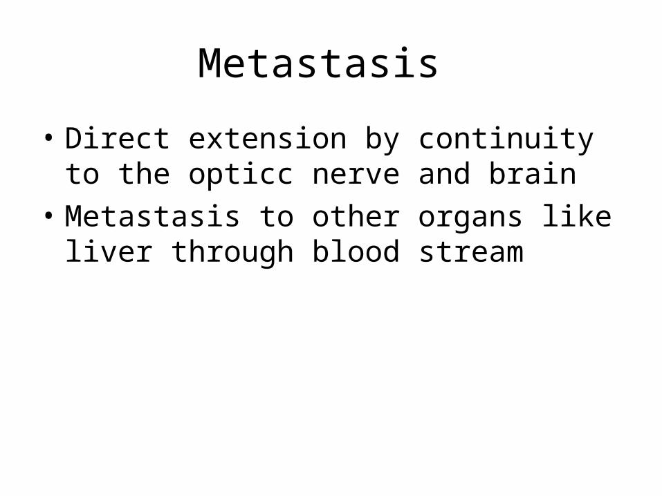
Metastasis
• Direct extension by continuity to the opticc nerve and brain
• Metastasis to other organs like liver through blood stream
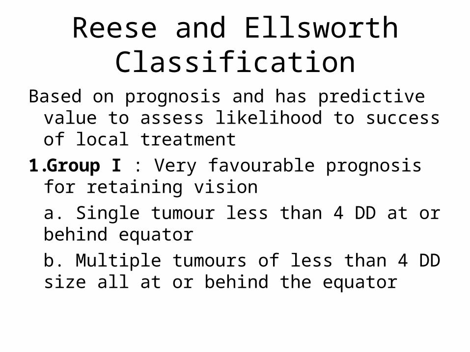
Reese and Ellsworth Classification
Based on prognosis and has predictive value to assess likelihood to success of local treatment
1.Group I : Very favourable prognosis for retaining vision a. Single tumour less than 4 DD at or behind equatorb. Multiple tumours of less than 4 DD size all at or behind the equator
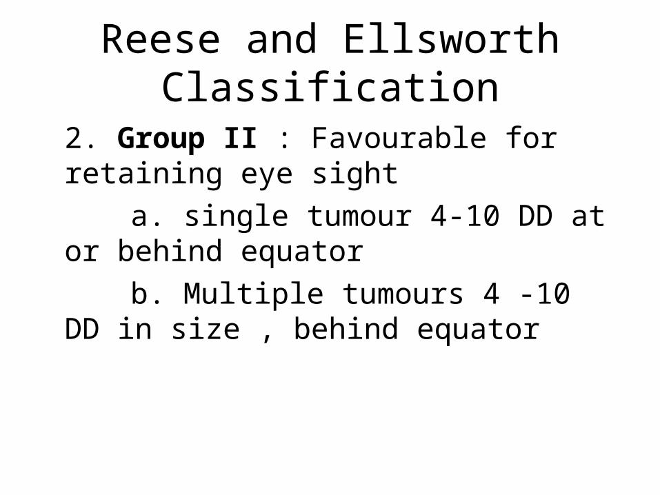
Reese and Ellsworth Classification
2. Group II : Favourable for retaining eye sight a. single tumour 4-10 DD at or behind
equator b. Multiple tumours 4 -10 DD in size ,
behind equator
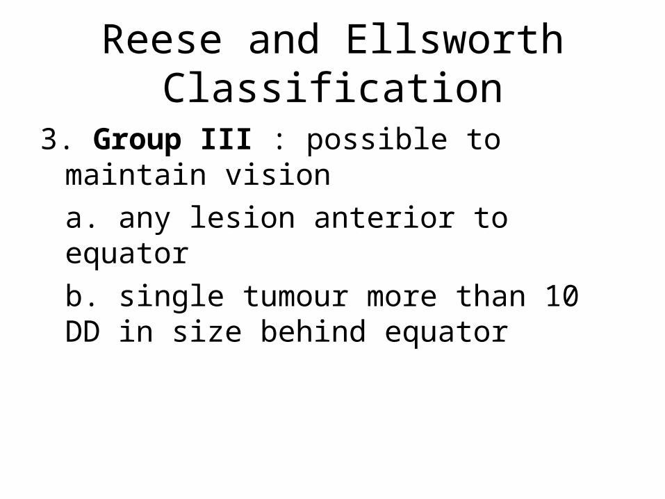
Reese and Ellsworth Classification
3. Group III : possible to maintain vision a. any lesion anterior to equatorb. single tumour more than 10 DD in size behind equator
Reese and Ellsworth Classification
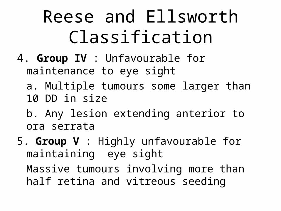
4. Group IV : Unfavourable for maintenance to eye sight a. Multiple tumours some larger than 10 DD in sizeb. Any lesion extending anterior to ora serrata
5. Group V : Highly unfavourable for maintaining eye sight Massive tumours involving more than half retina and vitreous seeding
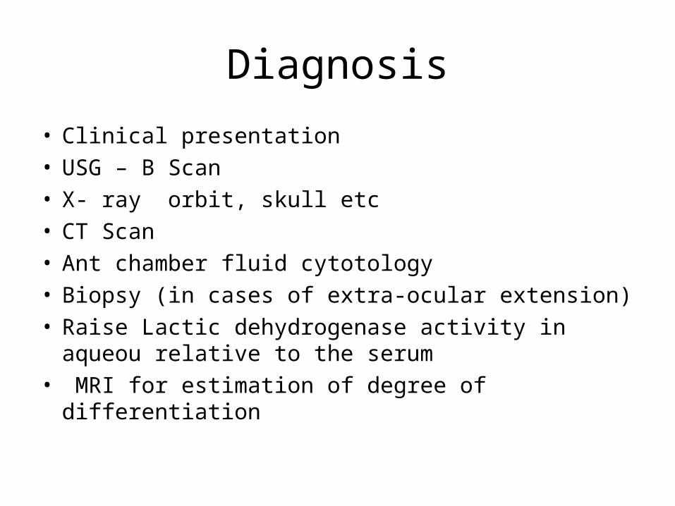
Diagnosis
• Clinical presentation• USG – B Scan• X- ray orbit, skull etc• CT Scan• Ant chamber fluid cytotology• Biopsy (in cases of extra-ocular extension)• Raise Lactic dehydrogenase activity in aqueou
relative to the serum• MRI for estimation of degree of differentiation
Treatment
• Prognosis of retinoblastoma if left untreated , is always bad
• Prognosis is fair if extra-ocular extension is avoided
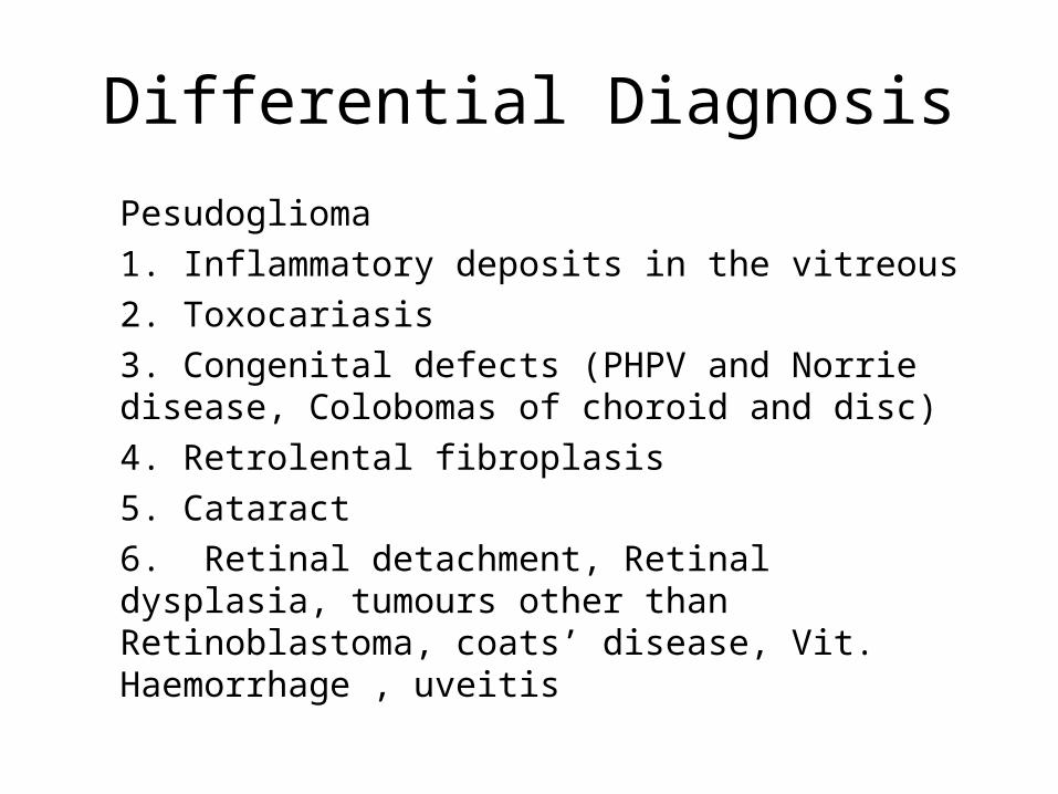
Differential Diagnosis
Pesudoglioma1. Inflammatory deposits in the vitreous2. Toxocariasis3. Congenital defects (PHPV and Norrie disease, Colobomas of choroid and disc) 4. Retrolental fibroplasis5. Cataract6. Retinal detachment, Retinal dysplasia, tumours other than Retinoblastoma, coats’ disease, Vit. Haemorrhage , uveitis
Treatment
1. Small tumourLocal modalities like cryotherapy (for ant lesion), Photocoagulation for posterior lesion, brachytherapy with Co 60 or 125 I and steriotactic radiation Suturing of radioactive cobalt discs – it deliver a dose of 4000 rad to summit of the tumour in one week.
Treatment
PhotocoagulationPlacing a double row of confluent burns around
each tumours with a photocoagulator.Repeat treatment may be required.Cryotherapy- For anteriorly located tumours.
Under direct visualization freezing until ice ball incorporates entire tumour. Tumour is allowed to thaw and refreez- thaw cycle is repeated 3-4 times.
Treatment
2. Enucleation3. Exenteration of the orbit4. External beam radiation 5. Chemoreduction with chemotherapy
(Vincristine, etoposide and carboplatin)
Malignant Melanoma
• Malignant Melanoma is highly malignant tumour arising from the outer layers of choroid. It is commonest intra-ocular tumour.

Clinical Features
• Adults between the age of 40 – 60 years affected.
• Less common amongst African and Asians
Symptoms
• Visual acuity is markedly affected when tumour is located centrally near macula
• In glaucomatous stage patient presents with severe pain in and around eye
• In stage of extra-ocular extension: growth in orbit / fungating mass
• In stage of metastasis: varied presentation depending on organ involved
Signs
• Tumour is primarily single and unilateral • A lens shaped mass raising the Retina above• It stretches the Bruch membrane which
ruptures and then tumour proliferates through the opening and retinal pigment epithelium to form a globular mass in the subretinal space
• Lens becomes opaque as its nutrition suffers
Signs
• Tumour fills the globe then perforate the sclera
• Orbital extension may occur in early stage due to spread along vortex vein or ciliary vessels
• Orbital tissue is infiltrated with tumour cells – presenting as proptosis
• Lymph nodes are not commonly affected
Signs
• Distant metastasis occurs to liver and elsewhere
• Growth is usually pigmented but may be occasionally unpigmented ( pigments are chiefly melanin)
• Surface of tumour may have mottled orange and black appearance
Signs
• Flat malignant melanoma:Choroid is widely infiltrated so that a uniform thickening results with shallow Retinal Detachment
Clinical stages
1. The quiescent stage2. The Glaucomatous stage 3. The stage of extra-ocular extension4. The stage of metastasis
Pathology
• Composed of spindle shaped cells• Cells may also be cylindrical or palisade-like,
arranged in columns or around blood vessels• Most of the tumours are of mixed type
Spindle – ASpindle – BEpitheloid (most malignant)Mixed Contains variable amount of reticulin fibres
Differential Diagnosis
1. Choroidal naevus2. Cavernous haemangioma3. Posterior scleritis
Diagnosis
• Ultrasonography – A and B scan• Radioactive tracers- increased rate of
phosphate uptake and its retaintion for longer time
• Fluorescein Angiography- double circulation with increased fluorescence in the mass (Indocyanine green angiography)
Treatment
• A pigmented lesion with diameter larger than 5 DD (7.5 mm) should be considered a malignant melanoma until proved otherwise
• Goals – eradication of tumour , maintenance of vision and cosmetically acceptable eye
• Tumour less than 10 mm and upto 2 mm thickeness is treated by brachytherapy using Radioactive discs of gold, cobalt 60 or iodine 125
Treatment
For treatment of small tumour • External beam radiation • Cryotherapy• Laser ablation • Transpupillary thermotherapyFor treatment of medium size tumour (10-15 mm in
diameter and 3-5 mm in height) Plaque or external proton beam radiation
Treatment
• Enucleation • Exenteration
PROGNOSISDisease is usually fatal in 5 years if not treated
successfullyIn cases with metastasis – death usually occurs
within a year of detection of metastasis