Intra-Cranial Manifestations of the Neurocutaneous Syndromes · Intra-Cranial Manifestations of the...
17
Pictorial Review Intra-Cranial Manifestations of the Neurocutaneous Syndromes J. HERRON, R. DARRAH, G. QUAGHEBEUR* Department of Radiology, John Radcliffe Hospital, Headley Way, Headington, Oxford, OX3 9DU, *Department of Neuroradiology, Radcliffe Infirmary, Woodstock Road, Oxford, OX2 6HE, U.K. Received: 18 February 1999 Revised: 26 June 1999 Accepted: 9 July 1999 The neurocutaneous syndromes or phakomatoses are a heterogeneous group of congenital disorders primarily involving structures derived from the embryological neuroectoderm. All of the syndromes involve the central nervous system (CNS). Peripheral nerves, skin and other organ systems may also be involved. Twenty to 30 disorders are now classified as neurocutaneous syndromes. This article reviews the intra-cranial imaging features of some of the commonest. Herron, J. et al. (2000). Clinical Radiology 55, 82–98. q 2000 The Royal College of Radiologists Key words: neurocutaneous, central nervous system, imaging features. NEUROFIBROMATOSIS First described over 100 years ago by Von Recklinghausen, it is now clear that there may be as many as eight different clinical subtypes. Types one and two (NF-1 and NF-2) account for over 99% of cases. Neurofibromatosis Type 1 (Von Recklinghausen’s Disease/‘Peripheral’ NF) This disease is characterized by mutliple cutaneous skin lesions, CNS tumours and mesoderm dysplasia. It is autosomal dominant with an incidence of one in 3000 and is due to a mutation of chromosome 17. It has a high penetrance with variable expressivity but about 50% of patients have mutations with no family history. It is not an absolute clinical entity and up to seven forms may exist. CNS findings include both neoplastic lesions and harmartomas [1,2]. The incidence of CNS manifestations in NF-1 is 15–20%, and there is a four-fold increased risk of developing a CNS neoplasm as compared to the general population. Intracranial Manifestations Optic pathway gliomas are the most common brain abnorm- ality in NF-1 and occur in 30% of cases. These tumours appear as either a focal mass on the optic nerve or as a diffuse enlargement of a long segment [2]. T2W MRI may show high signal (thought to be posterior extension) in the optic chiasm, tracts and radiations (Figs 1, 2). These gliomas are most commonly juvenile pilocytic astro- cytomas, but other low and higher-grade gliomas occur. The mesencephalic tectum is the commonest site after the optic pathways, and lesions also occur in the brainstem and cerebrum (Figs 3, 4). Hydrocephalus is not uncommon and may result either from benign acqueductal stenosis or gliomas of the tectum or tegmentum. Characteristic foci of increased signal intensity are present in about 75% of patients with NF-1. These lesions are usually multiple, show no mass effect or contrast enhancement, and elicit no oedema. They are commonest in the pons, cerebellar white matter, internal capsule and splenium (Figs 1, 4). They tend to appear at age 3 years, increasing in size and number till age 10/11 years, and then progressively disappear. They are though to represent harmartomas, abnormal glial cells or dysplastic myelination. Follow-up MRI is recommended to differentiate these lesions from low-grade neoplasms [4]. Similar foci of altered signal are seen in globus pallidus, and although these may show a little mass effect and slight hyperintense signal on T1W MRI they are also presumed to be hamartomas [1,5]. Other abnormalities include cranial nerve tumours (rarely); bone dysplasias such as sphenoid wing hypoplasia with subsequent pulsating exopthalmos (Fig. 5); plexiform neuro- fibromas (Fig. 5); and orbital abnormalities such as bupthalmos (enlargement of the globe due to congenital glaucoma), retinal phakomas and Lisch nodules. Clinical Radiology (2000) 55, 82–98 doi:10.1053/crad.1999.0328, available online at http://www.idealibrary.com on 0009-9260/00/020082+17 $35.00/0 q 2000 The Royal College of Radiologists Correspondence to: Dr J. Herron, Specialist Registrar Radiology, Depart- ment of Radiology, John Radcliffe Hospital, Headley Way, Headington, Oxford OX3 9DU, U.K. Guarantor of study: Dr G. Quaghebeur.
Transcript of Intra-Cranial Manifestations of the Neurocutaneous Syndromes · Intra-Cranial Manifestations of the...
Clinical Radiology(2000)55, 82–98doi:10.1053/crad.1999.0328, available online at http://www.idealibrary.com on
Pictorial Review
Intra-Cranial Manifestations of the NeurocutaneousSyndromes
J. HERRON, R. DARRAH, G. QUAGHEBEUR*
Department of Radiology, John Radcliffe Hospital, Headley Way, Headington, Oxford, OX3 9DU,*Department of Neuroradiology, Radcliffe Infirmary, Woodstock Road, Oxford, OX2 6HE, U.K.
Received: 18 February 1999 Revised: 26 June 1999 Accepted: 9 July 1999
ers
lsocle
ts
0009-9260/00/02
Correspondenment of RadioloOxford OX3 9DU
Guarantor of s
The neurocutaneous syndromes or phakomatoses are a heterogeneous group of congenital disordprimarily involving structures derived from the embryological neuroectoderm. All of the syndromesinvolve the central nervous system (CNS). Peripheral nerves, skin and other organ systems may abe involved. Twenty to 30 disorders are now classified as neurocutaneous syndromes. This artireviews the intra-cranial imaging features of some of the commonest.Herron, J.et al. (2000).ClinicalRadiology55, 82–98. q 2000 The Royal College of Radiologis
Key words: neurocutaneous, central nervous system, imaging features.
sen,rentunt
skinomao awithionsd
oth
%,NS
rm-pearfuse
owptic
tro-Theopticbrumsultthe
nt inallyandellarheyer tilly are
orto[4].
andightd to
ly);with
NEUROFIBROMATOSIS
First described over 100 years ago by Von Recklinghauit is now clear that there may be as many as eight diffeclinical subtypes. Types one and two (NF-1 and NF-2) accofor over 99% of cases.
Neurofibromatosis Type 1 (Von Recklinghausen’sDisease/‘Peripheral’ NF)
This disease is characterized by mutliple cutaneouslesions, CNS tumours and mesoderm dysplasia. It is autosdominant with an incidence of one in 3000 and is due tmutation of chromosome 17. It has a high penetrancevariable expressivity but about 50% of patients have mutatwith no family history. It is not an absolute clinical entity anup to seven forms may exist. CNS findings include bneoplastic lesions and harmartomas [1,2].
The incidence of CNS manifestations in NF-1 is 15–20and there is a four-fold increased risk of developing a Cneoplasm as compared to the general population.
Intracranial Manifestations
Optic pathway gliomas are the most common brain abnoality in NF-1 and occur in 30% of cases. These tumours apas either a focal mass on the optic nerve or as a dif
uro-mostinal
0082+17 $35.00/0
ce to: Dr J. Herron, Specialist Registrar Radiology, Depart-gy, John Radcliffe Hospital, Headley Way, Headington,, U.K.tudy: Dr G. Quaghebeur.
l
enlargement of a long segment [2]. T2W MRI may shhigh signal (thought to be posterior extension) in the ochiasm, tracts and radiations (Figs 1, 2).
These gliomas are most commonly juvenile pilocytic ascytomas, but other low and higher-grade gliomas occur.mesencephalic tectum is the commonest site after thepathways, and lesions also occur in the brainstem and cere(Figs 3, 4). Hydrocephalus is not uncommon and may reeither from benign acqueductal stenosis or gliomas oftectum or tegmentum.
Characteristic foci of increased signal intensity are preseabout 75% of patients with NF-1. These lesions are usumultiple, show no mass effect or contrast enhancement,elicit no oedema. They are commonest in the pons, cerebwhite matter, internal capsule and splenium (Figs 1, 4). Ttend to appear at age 3 years, increasing in size and numbage 10/11 years, and then progressively disappear. Thethough to represent harmartomas, abnormal glial cellsdysplastic myelination. Follow-up MRI is recommendeddifferentiate these lesions from low-grade neoplasmsSimilar foci of altered signal are seen in globus pallidus,although these may show a little mass effect and slhyperintense signal on T1W MRI they are also presumebe hamartomas [1,5].
Other abnormalities include cranial nerve tumours (rarebone dysplasias such as sphenoid wing hypoplasiasubsequent pulsating exopthalmos (Fig. 5); plexiform nefibromas (Fig. 5); and orbital abnormalities such as bupthal(enlargement of the globe due to congenital glaucoma), rephakomas and Lisch nodules.
q 2000 The Royal College of Radiologists

PICTORIAL REVIEW 83
Fig. 1 – A 14-year-old patient with NF-1. (a) MRI (T2-weighted axial with fat saturation) shows bilateral optic nerve gliomas. These are seen as diffuseenlargement of the nerves. These are seen in 10–70% of patients. (b) MRI (T2-weighted axial) shows a bulky chiasm and a lesion in the cerebral pedunclerepresenting a probable hamartoma. (c) MRI (enhanced T1-weighted axial) shows high signal within the chiasmatic tumour. (d) MRI (T2-weighted axial)shows high signal within the basal ganglia and the cerebral peduncles. These are seen in 75% of patients and are thought to represent hamartomas, abnormalglial cells or dysplastic myelination.
(d)
(c)
(a)
(b)

84 CLINICAL RADIOLOGY
Fig. 2 – An 8-year-old patient with NF-1. (a, b) MRI (T2-weighted axial)shows high signal within the optic tracts and radiations due to posteriorextension of an optic chiasm glioma, (c) MRI (enhanced T1-weightedcoronal) shows high signal within the optic radiations, and anintraventricular glioma.
(a)
(b)
lt inmal
rings
the
PICTORIAL REVIEW 85
(c)
Fig. 2 – continued.
Vascular dysplasias due to intimal proliferation can resustenoses and occlusions. Aneurysms and arterio-venousformations (AVM) are also described (Fig. 6).
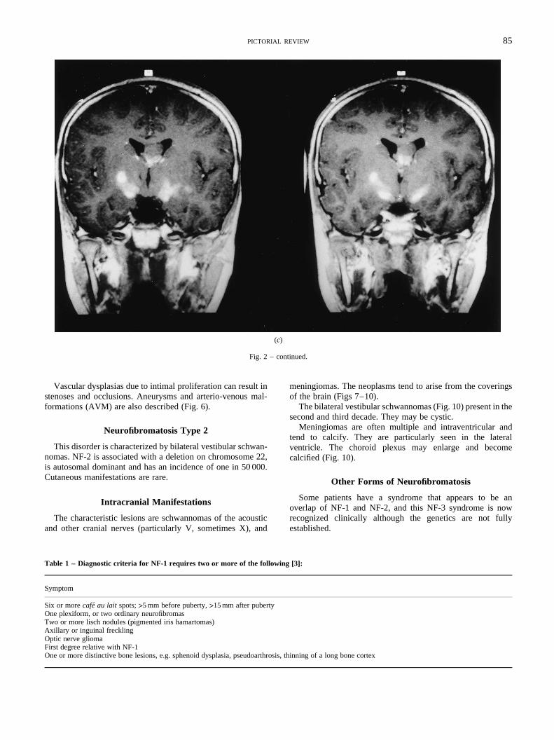
an-22
000.
ndralme
Neurofibromatosis Type 2
This disorder is characterized by bilateral vestibular schwnomas. NF-2 is associated with a deletion on chromosomeis autosomal dominant and has an incidence of one in 50Cutaneous manifestations are rare.
usticand
e anowlly
Intracranial Manifestations
The characteristic lesions are schwannomas of the acoand other cranial nerves (particularly V, sometimes X),
Table 1 – Diagnostic criteria for NF-1 requires two or more of the following
Symptom
Six or morecafeau lait spots;>5 mm before puberty,>15 mm after pubertyOne plexiform, or two ordinary neurofibromasTwo or more lisch nodules (pigmented iris hamartomas)Axillary or inguinal frecklingOptic nerve gliomaFirst degree relative with NF-1One or more distinctive bone lesions, e.g. sphenoid dysplasia, pseudoarth
-
,
meningiomas. The neoplasms tend to arise from the coveof the brain (Figs 7–10).
The bilateral vestibular schwannomas (Fig. 10) present insecond and third decade. They may be cystic.
Meningiomas are often multiple and intraventricular atend to calcify. They are particularly seen in the lateventricle. The choroid plexus may enlarge and becocalcified (Fig. 10).
Other Forms of Neurofibromatosis
Some patients have a syndrome that appears to boverlap of NF-1 and NF-2, and this NF-3 syndrome is nrecognized clinically although the genetics are not fuestablished.
[3]:
rosis, thinning of a long bone cortex

eenith
s ofear
86 CLINICAL RADIOLOGY
Several other forms of neurofibromatosis have also bdescribed by Riccardi, all of which may present wintracranial lesions [6].
e. Itari-000s 9t inumthirdow
t inthan
.
thecallyThetheome
inaltioneal
TUBEROUS SCLEROSIS (BOURNEVILLE’S DISEASE)
Tuberous sclerosis (TS) is a familial multisystem diseasis an autosomal dominant disorder with low penetrance, vable expressivity and a prevalence of one in 34 000–100[1,7]. It has been linked to an abnormality on chromosomeand 16 [7]. The classic triad of symptoms described by Vol1908 of seizures, mental retardation and adenoma sebaceone end of a clinical spectrum and is seen in less than one-of cases [2]. Criteria for the diagnosis of this disorder have nbeen established [1]. Radiological criteria are importanestablishing this diagnosis and may be manifest earliersome clinical features [7].
bersonsrtioncifiedTS
maythisionsthepeaatedhitethe
anon
hey
% othe
icto ththe
MRR,
ses.how
nts.ging12)oney orm atant.
hesecener-mal
ealdialficialus
ondeitemaysedtheesebyig.
rack
13)theof
thenousciallood
ara-theThe
rialing,f the
ofs of
nce
Intracranial Manifestations
TS derives its name from the cortical hamartomas or tuseen in the gyri (most commonly of frontal lobe). These lesiare seen in 95% of cases. Some calcify although the propothat does so has not been established. The number of calcortical lesions increases with age, by age 10 50% ofpatients have calcified cortical tubers. On CT the tubersappear as lucencies in widened gyri in young patients,becomes more difficult to see with age unless the lescalcify. MRI detects these lesions well at all ages, againfeatures change with age (Fig. 11). In neonates, the gyri apenlarged and hyperintense to the surrounding unmyelinwhite matter on T1W images and hypointense to the wmatter on T2W MR images. As the white matter myelinatesappearance alters and the lesions are hypointense on T1Whyperintense on T2W MR, ultimately becoming isointenseT1W images with increasing age. As the lesions calcify tmay again show some T1 hyperintensity [1].
Subependymal nodules or hamartomas are seen in 90cases. The majority calcify and are typically found alongventricular surface of the lateral ventricle in the striothalamgroove between the caudate and the thalamus, just posteriorforamen of Munro. Less often, they are seen elsewhere inventricular system. The imaging characteristics on CT andalter with age, showing progressive calcification on CT. On Mthe nodules alter in signal intensity as myelination progresThey are best seen on T1W MR images. They may senhancement with intravenous GdDTPA (Fig. 11).
Giant cell astrocytomas occur in up to 15% of TS patieThey occur at the foramen of Munro and present as enlarpartly calcified masses with associated hydrocephalus (Fig.Serpentine flow voids due to dilated vessels are seen inthird. Their appearance varies and neither signal intensitcontrast enhancement can differentiate the tumour frosubependymal nodule – the change in size is most imporHistologically these tumours tend to be astrocytomas.
White matter lesions are seen in 90% of cases. Many of trepresent islets of disordered neurones and glial cells. In a restudy, 21% of patients [9] showed linear or curvilinear hypintensities in the white matter, extending from subependy
nodules to cortical tubers. These are thought to be bandhypomyelinated disordered or gliotic cells along the linradial glial-neuronal unit.
Cerebellar white matter lesions occur in 10% of patients
is
r
d
f
e
.-
STURGE WEBER SYNDROME(ENCEPHALOTRIGEMINAL ANGIOMATOSIS)
This is a congenital disorder of vasculature of the face,choroid of the eye, and the leptomeninges. It occurs sporadiaffecting both sexes equally, but familial cases are reported.facial angioma (port wine naevus) can involve all or part offace and is present at birth. It does not change with age. In scases of SWS there is no facial angioma [1]. The origdescription by Schirmer in 1860 of a naevus in the distribuof the trigeminal nerve with an ipsilateral leptomeningabnormality in the occipital region is not characteristic.
The major pathologic abnormality in SWS is a meningangioma in the pia mater, possibly due to persistent primorsinusoidal vascular channels. This leads to a lack of supercortical venous drainage resulting in chronic venohypertension and ischaemia [1,10].
Intracranial Manifestations
The leptomeningeal vascular malformation is best seencontrast-enhanced MRI (Fig. 13). Other MR findings inclulow signal on T2W of the affected gyri and adjacent whmatter. The aetiology of this signal change is unclear andbe due to calcification, accelerated myelination or increadeoxyhaemoglobin in the capillaries and veins. Many ofother radiological findings result from the venous stasis. Thinclude atrophy of the underlying cortex accompaniedcerebral calcification which is seen from 1 year of age (F14). The calcified opposed gyri are seen as parallel tram-tcalcification on plain film.
Enlargement and calcification of the choroid plexus (Fig.on the affected side is well documented, and the size ofchoroid plexus correlates positively with the extentleptomeningeal enhancement [11].
Enlarged vessels can be seen on MRI and CT deep inhemisphere probably due to enlargement of deep vevessels as a result of slow flow or thrombosis in the superfiabnormal venous system, and subsequent shunting of bthrough deep medullary veins.
Cranial asymmetry can occur due to hemiatrophy, or pdoxically there may be a large hemicranium on the side ofatrophy secondary to associated subdural collections.Dyke-Davidoff-Maison syndrome is a constellation of calvachanges which is sometimes seen. It includes skull thickenpneumatization of the paranasal sinuses, ipsilateral shift ofalx and elevation of the petrous ridge and lesser wingsphenoid. This picture may also occur with other causehemicerebral atrophy [1].
tVON HIPPEL-LINDAU SYNDROME
An autosomal dominant disorder with incomplete penetraand a genetic defect at chromosome 3p25-p26 [1].

leceralumd to
ofduleRIle
nt isrelyhave
mo-thesionmas
eouso besisg toroup
ntsach, butow-tro-it isarly.ingellingaticilyayonof
thers isandan[12]
andto
nal
PICTORIAL REVIEW 87
Fig. 3 – A 20-year-old patient with NF-1. (a) MRI (T2-weighted axial) shows a glioma of the septum pellucidum. Although optic nerve gliomas are the mostcommon glioma found in NF-1, gliomas do occur in the brain stem and cerebrum. (b) MRI (enhanced T1-weighted axial) shows avid enhancement of theglioma.
(a)(b)
It is characterized by tumours of the CNS, typically multiphaemangioblastomas and tumours of the abdominal visThe haemangioblastomas occur primarily in the cerebel(65%) brain stem (20%) and spinal cord (15%) [4]. They tenbe sharply demarcated with smooth borders. Two thirdsthese lesions are cystic with a solid component or mural no[13] (Fig. 15). The nodule is isointense to brain on T1W Mand hyperintense on T2W MRI. Flow voids within the nodudue to tumour vessels are common. The cystic componehyperintense to CSF on T2W MRI and the wall of the cyst raenhances. The mural nodule enhances avidly. Solid tumoursintermediate to high signal on T2W MRI and enhance hogenously. Small areas of increased signal on T2W MRI inwhite matter are sometimes seen. The exact nature of these leis unclear, they may represent subclinical haemangioblastoareas of gliosis or dystrophic white matter.
Many other disorders are now classified as neurocutansyndromes, however the vast majority of cases continue tdue to above five conditions. MRI is invaluable in diagnoand shows previously unsuspected lesions. It is helpinincrease our understanding of this complex and diverse gof disorders.
.
s,
The issue of screening or long-term follow-up in patiewith these syndromes is as yet unresolved. In 1988 Ro[14] addressed the use of screening for neurofibromatosisthere is no more recent work. He recommended annual ‘follup’ of these patients, much of which can be clinical or elecphysiological. MRI is now a useful and acceptable tool andthe practice in our department to examine these patients ye
If patients develop new or altered symptomatology, imagis obtained as appropriate. Genetic assessment and counsis vital, and may recommend screening of asymptomrelatives. Griffiths [7] states that diagnosis of TS is readmade on CT imaging although MRI is more sensitive and mbe useful for prognosis. Interval imaging to detect changeSENs and the development of giant cell astrocytoma islimited value, as patient symptomatology determines wheany intervention is needed. Clinical examination of relativeonly minimally less sensitive than radiological screeningfar more cost-effective. In VHL patients, screening onannual basis has been recommended by several groupsand this includes enhanced MRI of brain and spineabdominal CT or ultrasound. The latter is recommendedallow early detection of the potentially life threatening re

88 CLINICAL RADIOLOGY
Fig. 4 – A 12-year-old patient with NF-1. (a) MRI (enhanced T1-weightedsagittal) shows a tectal plate glioma. (b) MRI (enhanced T1-weighted axial)shows a chiasmatic glioma. (c) MRI (T2-weighted axial) shows high signalin the putamen on the right.
(a)
(b)
(c)

PICTORIAL REVIEW 89
Fig. 5 – A 6-year-old patient with NF-1. (a) CT showing sphenoid wingdysplasia. This is associated with pulsating exopthalmos. (b) MRI (T2-weighted axial) shows the plexiform neurofibroma of the right orbit.
(a)
(b)
(a)
(b)
(c)
Fig. 6 – An 18-year-old patient with NF-1. (a) MRI (T2-weighted axial)shows right sided neurogenic oedema due to infarction. (b) MRI (T2-weighted axial) shows the absence of flow void in the right middle cerebralartery. (c) MR angiogram shows absence of signal from the right internalcarotid artery.

efulome
90 CLINICAL RADIOLOGY
Fig. 7 – A 23-year-old patient with NF-2. (a) MRI (T2-weighted axial)shows multiple meningiomata in the posterior fossa. (b) MRI (enhancedT1-weighted axial) shows the meningiomata enhancing brightly.
(a)
(b)
Table 2 – Diagnostic criteria for NF-2 are [3]:
Criteria
Bilateral VIII nerve massesA first-degree relative with NF-2 and either:
a. a unilateral VIII nerve massb. two of the following: meningioma, glioma, schwannoma, or juvenile
posterior subcapsular lenticular opacity
and adrenal tumours. Asymptomatic relatives need cargenetic evaluation and dependant on the results of this, swill require annual screening also.
thethea
ationin.
derusrophyhythe
romrcts
ularma
duuto-andrio-the
ue to(iv)ta-
deepns,nin-
tousloodhick-eousi areorty
ftenbleeenlue
llarymalano-s ofin
OTHER NEUROCUTANEOUS SYNDROMES
There are two main groups of disorders, the vascular andmelanocytic neurocutaneous syndromes. They includefollowing: (i) Wyburn-Mason syndrome. This comprisesfacial vascular naevus and cerebral arteriovenous malformwhich normally involves the visual pathway or the midbraThe AVMs may be multiple (Fig. 16).
(ii) Ataxia telangiectasia. An autosomal recessive disorwith an incidence of one in 40 000 [1]. Oculocutaneotelangiectasia and cerebellar ataxia are seen. Cerebellar atis seen affecting particularly the anterior vermis, with atropof the dentate and olivary nuclei and degeneration ofposterior columns (Fig. 17). Haemorrhage can result frupture of parenchymal telangiectasia, and cerebral infacan result from emboli shunted through pulmonary vascmalformations. There is an increased incidence of lymphoand leukaemia in this condition. (iii) Osler-Weber-Rendisease (hereditary haemorrhagic telangiectasia). This asomal dominant disorder presents with dermal, mucosalvisceral telangiectasia. Two-thirds have pulmonary artevenous fistulae. One-third have vascular malformations ofbrain and spinal cord. Infarcts, septic emboli and abscess dthe complications of pulmonary fistulae are common.Klippel-Trenaunay-Weber syndrome. This consists of cuneous angiomata, soft tissue or bony hypertrophy and aparenchymal vascular malformation. On MRI, vascular lesioangiomas and hemimegalencephaly are all seen. (v) Megioangiomatosis. This is part of a spectrum of hamartomameningeal-based lesions which involve the meninges, bvessels and adjacent brain. The leptomeninges become tened and on CT calcified lesions are seen. (vi) Neurocutanmelanosis. One or more hairy or deeply pigmented naevseen in association with melanosis of the leptomeninges. Fper cent develop primary melanosis of the CNS. CT is onormal, MR shows foci of altered T1 and T2 signal compatiwith melanin deposition. Meningeal enhancement has breported in some cases. (vii) Naevus of Ota syndrome. Bgrey lesions in the dermatomes of the ophthalmic and maxidivisions of trigeminal nerve are associated with abnormeningeal pigmentation, meningeal melanocytomas, melmas of the choroid and ciliary body and primary melanomathe CNS. (iix) Hypomelanosis of Ito. Hypopigmented sk

PICTORIAL REVIEW 91
Fig. 8 – A 16-year-old patient with NF-2. (a) MRI (enhanced T1-weighted axial) shows an intraventricular meningioma. (b) MRI (T2-weighted axial) shows aleft vestibular schwannoma. Note the enlargement of the internal auditory meatus. (c) MRI (T2-weighted axial) shows a schwannoma the left jugular foramenoriginating from either the X or XI cranial nerve.
(a) (b)
(c)

92 CLINICAL RADIOLOGY
Fig. 9 – A 27-year-old patient with NF-2. (a) MRI (T2-weighted axial) shows the VII and VIII nerves clearly in the left internal auditory meatus with CSFbetween them. On the right soft tissue is seen in this area. (b) MRI (enhanced T1-weighted axial) shows the vestibular schwannoma in the IAM enhancing.
(a) (b)
Fig. 10 – A 21-year-old patient with NF-2. (a) CT following intravenous contrast medium shows bilateral vestibular schwannomas as enhancingcerebellopontine angle masses. (b) CT following intravenous contrast medium shows extensive choroid plexus calcification and a parafalcine meningioma.
(a) (b)
PICTORIAL REVIEW 93
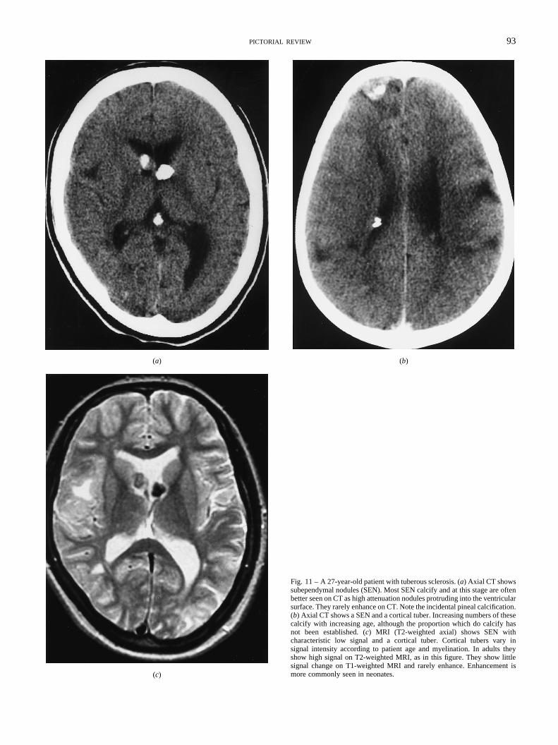
Fig. 11 – A 27-year-old patient with tuberous sclerosis. (a) Axial CT showssubependymal nodules (SEN). Most SEN calcify and at this stage are oftenbetter seen on CT as high attenuation nodules protruding into the ventricularsurface. They rarely enhance on CT. Note the incidental pineal calcification.(b) Axial CT shows a SEN and a cortical tuber. Increasing numbers of thesecalcify with increasing age, although the proportion which do calcify hasnot been established. (c) MRI (T2-weighted axial) shows SEN withcharacteristic low signal and a cortical tuber. Cortical tubers vary insignal intensity according to patient age and myelination. In adults theyshow high signal on T2-weighted MRI, as in this figure. They show littlesignal change on T1-weighted MRI and rarely enhance. Enhancement ismore commonly seen in neonates.
(a) (b)
(c)

94 CLINICAL RADIOLOGY
Fig. 12 – An 8-year-old patient with tuberous sclerosis. (a) Enhanced axial CT shows a giant cell astrocytoma, large amount of vasogenic oedema and asurgical defect of the skull. Note calcified SENs. These periventricular masses lie close to the foramen of Munro. They tend to enhance and in one-thirdof casesprominent flow voids are present. (b) MRI (T2-weighted axial) shows the mixed signal of the giant cell astrocytoma and prominent vasogenic oedema. (c) MRI(enhanced T1-weighted sagittal) shows the brightly enhancing giant cell astrocytoma.
(a) (b)
(c)

PICTORIAL REVIEW 95
Fig. 13 – A 5-month-old patient with Sturge Weber Syndrome. (a) MRI (T2-weighted axial) shows hemiatropy, (b) MRI (enhanced T1-weighted) shows highsignal bilaterally in the leptomeninges and a large choroid plexus on the left.
(a) (b)
Fig. 14 – A 46-year-old patient with Sturge Weber Syndrome. Axial CT shows cortical calcification. This dense gyriform calcification adjacent to abnormalmeninges is best seen on CT but not before 1 year of age. The distribution is most often occipital (92%), parietal (61%), frontal (38%) and temporal (22%).

96 CLINICAL RADIOLOGY
Fig. 15 – A 53-year-old patient with Von Hippel Lindau Syndrome. (a) MRI (T2-weighted axial) shows multiple large cystic haemangioblastomas. A solidnodule is seen. (b) MRI (enhanced T1-weighted axial) shows the nodule enhancing avidly.
(a) (b)
Fig. 16 – A 54-year-old patient with Wyburn-Mason Syndrome. MRI (T2-weighted axial) shows signal voids due to bilateral AVMs.

PICTORIAL REVIEW 97
Fig. 17 – An 11-year-old patient with ataxia telangiectasia. (a) MRI (T2-weighted axial and coronal) shows marked cerebellar atrophy affecting mai ly theanterior vermis, with atrophy of the olives and the dentate nucleus. (b) MRI (T1-weighted sagittal) shows cerebellar atrophy.
(a)
(b)
n

asess of
of
L.
98 CLINICAL RADIOLOGY
Table 3 – Diagnostic criteria for tuberous sclerosis are [8]:
Primary featuresFacial angiofibromasMultiple ungual fibromasCortical tuber (histologically confirmed)Subependymal nodule or giant cell astrocytoma (histologically confirmed)Multiple calcified supependymal nodules protruding into the ventricle (radiographic evidence)Multiple retinal astrocytomas
Secondary featuresAffected first-degree relativeCardiac rhabdomyoma (histologic or radiographic confirmation)Other retinal hamartoma or achromic patchNoncalcified subependymal nodules (radiographic confirmation)Shagreen patchForehead plaquePulmonary lymphangiomyomatosis (histological confirmation)Renal angiomyolipoma (radiographic or histologic confirmation)Renal cysts (histologic confirmation)
Tertiary featuresHypomelanotic macules‘Confetti’ skin lesionsRenal cysts (radiographic evidence)Randomly distributed enamel pits in deciduous and/or permanent teethBone cysts (radiographic confirmation)Hamartomatous rectal polyps (histologic confirmation)Pulmonary lymphangiomyomatosis (radiographic evidence)Cerebral white matter migration tracts or heterotopias (radiographic evidence)Gingival fibromasHamartomas of other organs (histologic confirmation)Infantile spasms
Definite TS – either one primary and two secondary features, or one secondary and two teriary featuresProbable TS – either one secondary and one tertiary feature, or three tertiary featuresSuspect TS – either one secondary or two tertiary features
Table 4 – Diagnostic criteria for Von Hippel-Lindau syndrome are [12]:
In the presence of a family history of a retinal or CNS haemangioblastoma either:One haemangioblastomaOr one visceral lesion such as a renal tumour; pancreatic cyst or tumour; phaeochromocytoma or papillary cystadenoma of the epidydimis
In an isolated case where there is no clear family history:Two or more haemangioblastomasOne haemangioblastoma and one visceral manifestation as listed above
lesions are associated with lesions in the CNS. 59% of chave CNS lesions including cerebral atrophy, abnormalitiemigration, gliosis and white matter involvement.
sl
g.
ousental
of
D.me.
barres.
E.
romes.
REFERENCES
1 Barkovich AJ.Pediatric Neuroimaging. 2nd edn. New York, RavenPress, 1995; 277–320.
2 Smirniotopoulos JG, Murphy FM. The phakomatoses.Am J Neuro-radiol 1992;13:725–746.
3 National Institutes of Health Consensus Development Conference.ArchNeurol1988;45:575–578.
4 Braffman BH, Bilaniuk LT, Zimmerman RA. MR for central nervousystem neoplasia of the phakomatoses.Semin Roentgeno1990;25:198–217.
5 Shy HH, Mirowitz SA, Wippold II. Neurofibromatosis: MR imaginfindings involving the head and spine.Am J Radiol1993;160:159–164
6 Riccardi VM. Neurofibromatosis.Neurol Clin1987;5:337–349.
7 Griffiths PD, Martland TR. Tuberous sclerosis complex: the roleneuroradiology.Neuropediatrics1997;28:224–252.
8 Roach ES, Smith M, Huttenlocher P, Bhat M, Alcorn D, HawleyDiagnostic criteria: tuberous sclerosis complex.J Child Neurol1992;7:221–223.
9 Shepard CW, Houser OW, Gomez MR. MR findings in tubersclerosis complex and correlation with seizure development and mimpairment.Am J Neuroradiol1995;16: 145–155.
10 Griffiths PD. Sturge-Weber syndrome revisited: the roleneuroradiology.Neuropediatrics1996;27:284–294.
11 Griffiths PD, Blaser S, Boodram MB, Armstrong D, Harwood-NashChoroid plexus size in young children with Sturge-Weber SyndroAm J Neuroradiol1996;17:175–180.
12 Choyke PL, Glenn GM, Walther MM, Patronas NJ, Lineham WM, ZB. Von Hippel-Lindau disease: genetic, clinical and imaging featuRadiology1995;194:629–642.
13 Sato Y, Waziri M, Smith W, Frey E, Yuh W, Hanson J, FrankenHippel-Lindau disease: MR imaging.Radiology1998;166:241–246.
14 Roach ES. Diagnosis and management of neurocutaneous syndSemin Neurol1988;1:83–96.