Intimate Partner Violence and Injury Prediction From ...
12
Intimate Partner Violence and Injury Prediction From Radiology Reports Irene Y. Chen 1* , Emily Alsentzer 2 , Hyesun Park 3 , Richard Thomas 4 , Babina Gosangi 5 , Rahul Gujrathi 3 , and Bharti Khurana 3,6 1 Electrical Engineering and Computer Science, Massachusetts Institute of Technology, Cambridge, MA 02139, USA 2 Health Sciences and Technology, Massachusetts Institute of Technology, Cambridge, MA 02139, USA 3 Department of Radiology, Brigham and Women’s Hospital, Boston, MA 02115, USA 4 Department of Radiology, Lahey Health Medical Center, Burlington, MA 01805, USA 5 Department of Radiology, Yale University, New Haven, CT 06510, USA 6 Department of Radiology, Harvard Medical School, Boston, MA 02115, USA Intimate partner violence (IPV) is an urgent, prevalent, and under-detected public health is- sue. We present machine learning models to assess patients for IPV and injury. We train the predictive algorithms on radiology reports with 1) IPV labels based on entry to a violence prevention program and 2) injury labels provided by emergency radiology fellowship-trained physicians. Our dataset includes 34,642 radiology reports and 1479 patients of IPV victims and control patients. Our best model predicts IPV a median of 3.08 years before violence prevention program entry with a sensitivity of 64% and a specificity of 95%. We conduct error analysis to determine for which patients our model has especially high or low perfor- mance and discuss next steps for a deployed clinical risk model. Keywords : intimate partner violence; radiology; risk stratification; natural language pro- cessing; contextual word embeddings. 1. Introduction Intimate partner violence (IPV) is defined as physical, sexual, psychological, or economic violence that occurs between former or current intimate partners. While men can also be affected, IPV is a gendered phenomenon largely perpetrated against women by male partners. 1 The Centers for Disease Control report that more than 1 in 3 women, and 1 in 10 men in the U.S. will experience physical violence, sexual violence, psychological violence, and/or stalking by an intimate partner during their lifetime. 2 IPV victims have a greater risk of health problems including higher rates of mental health illnesses, chronic pain, reproductive difficulties, and generally poorer health. 3–5 According to the United Nations, half of the women * Corresponding author email: [email protected]. c 2020 The Authors. Open Access chapter published by World Scientific Publishing Company and distributed under the terms of the Creative Commons Attribution Non-Commercial (CC BY-NC) 4.0 License. Pacific Symposium on Biocomputing 26:55-66 (2021) 55
Transcript of Intimate Partner Violence and Injury Prediction From ...

Intimate Partner Violence and Injury Prediction From Radiology Reports
Irene Y. Chen1∗, Emily Alsentzer2, Hyesun Park3, Richard Thomas4, Babina Gosangi5,
Rahul Gujrathi3, and Bharti Khurana3,6
1Electrical Engineering and Computer Science, Massachusetts Institute of Technology,Cambridge, MA 02139, USA
2Health Sciences and Technology, Massachusetts Institute of Technology,Cambridge, MA 02139, USA
3Department of Radiology, Brigham and Women’s Hospital, Boston, MA 02115, USA
4Department of Radiology, Lahey Health Medical Center, Burlington, MA 01805, USA
5Department of Radiology, Yale University, New Haven, CT 06510, USA
6Department of Radiology, Harvard Medical School, Boston, MA 02115, USA
Intimate partner violence (IPV) is an urgent, prevalent, and under-detected public health is-sue. We present machine learning models to assess patients for IPV and injury. We train thepredictive algorithms on radiology reports with 1) IPV labels based on entry to a violenceprevention program and 2) injury labels provided by emergency radiology fellowship-trainedphysicians. Our dataset includes 34,642 radiology reports and 1479 patients of IPV victimsand control patients. Our best model predicts IPV a median of 3.08 years before violenceprevention program entry with a sensitivity of 64% and a specificity of 95%. We conducterror analysis to determine for which patients our model has especially high or low perfor-mance and discuss next steps for a deployed clinical risk model.
Keywords: intimate partner violence; radiology; risk stratification; natural language pro-cessing; contextual word embeddings.
1. Introduction
Intimate partner violence (IPV) is defined as physical, sexual, psychological, or economicviolence that occurs between former or current intimate partners. While men can also beaffected, IPV is a gendered phenomenon largely perpetrated against women by male partners.1
The Centers for Disease Control report that more than 1 in 3 women, and 1 in 10 menin the U.S. will experience physical violence, sexual violence, psychological violence, and/orstalking by an intimate partner during their lifetime.2 IPV victims have a greater risk ofhealth problems including higher rates of mental health illnesses, chronic pain, reproductivedifficulties, and generally poorer health.3–5 According to the United Nations, half of the women
∗Corresponding author email: [email protected].
c© 2020 The Authors. Open Access chapter published by World Scientific Publishing Company anddistributed under the terms of the Creative Commons Attribution Non-Commercial (CC BY-NC)4.0 License.
Pacific Symposium on Biocomputing 26:55-66 (2021)
55

who are intentionally killed globally are killed by their intimate partners or family members.6
It is essential to detect IPV victims early to provide timely intervention.Healthcare providers have the opportunity to screen patients for IPV, but several barriers
at both patient and provider levels limit the effectiveness. IPV victims often seek treatmentwithin healthcare settings;7 however, despite its high prevalence, IPV is substantially under-diagnosed due to underreporting of violence by the victim to health care providers. BecauseIPV victims generally do not present with obvious trauma, even in emergency departments,8
they do not readily receive IPV-specific resources.Imaging studies provide an objective measurement of patient status, especially for vulnera-
ble individuals who are not forthcoming.9 In a prior observational study, researchers identifiedIPV-related injury patterns including soft-tissue and musculoskeletal injuries from imagingstudies of victims who visited the emergency department. They also found that IPV victimsreceive more radiology studies than a comparable control cohort.10
In this work, we present algorithms to predict IPV and injury from radiology reports. Wepredict IPV from a dataset of 24,131 radiology reports from 262 IPV victims who enrolled intoa violence prevention support program and 794 controls from the same hospital who were ageand sex-matched based on a subset of the IPV victims. We demonstrate strong quantitativeresults with our best model achieves a mean area under the received operator curve (AUC)of 0.852. With a sensitivity of 64% and a specificity of 95%, we are able to predict IPV amedian of 3.08 years in advance of entry into the violence prevent support program. To betterdetect severe forms of IPV, we predict injury from a dataset of radiology reports from onlyIPV victims with labels from four emergency radiology fellowship-trained radiologists. Ourbest model achieves a mean AUC of 0.887.
We analyze our models for validity and usability. Because IPV can manifest differentlyacross race,11 gender,12 age,13 and marital status,14 we present error analysis comparing accu-racy, sensitivity, and specificity across these groups using demographic information extractedfrom the clinical record. As IPV continues to affect vulnerable individuals—especially in timesof great crisis15,16—we demonstrate how automated predictive algorithms can be used to iden-tify patients at high risk of IPV and injury.
2. Related Work
2.1. Intimate partner violence
Early detection in IPV is critical to facilitate early intervention in the cycle of abuse, therebypreventing worsening health conditions,3–5 life threatening injuries, and potentially homi-cides.17 The main obstacle to early intervention is underreporting by the patient due to varietyof factors including shame, economic dependency, or lack of trust in healthcare providers.18
Automated screening can help physicians identify high risk individuals—potentially from ra-diology studies,19 substance abuse disorders,20 or other clinical data—and intervene quickly.Prior work has focused on analyzing associative patterns among IPV victims.10 To our knowl-edge, we present the first work to present an algorithm for IPV and injury prediction.
Pacific Symposium on Biocomputing 26:55-66 (2021)
56
2.2. Clinical prediction
Machine learning methods can assess patients and other individuals for different levels of riskto allocate resources and improve clinical workflows.21,22 The strength of machine learning liesin its ability to learn latent patterns from observational data and make robust predictionson new and previously unseen patients. Researchers have shown promising results about theuse of machine learning on chronic diseases like diabetes,23 diagnosis from radiology reports,24
rare conditions like preterm infant illnesses,25 and public health concerns like child welfare.26,27
In particular, supervised learning models excel in structured settings with large datasets andclearly defined labels, e.g. radiology report text and whether the patient ultimate enters aviolence prevention program.
2.3. Natural language processing
Natural language processing (NLP) techniques can extract information from unstructuredtext.28 In healthcare settings, researchers have leveraged NLP on clinical text such as nursingnotes, discharge summaries, and radiology and pathology reports for disease surveillance,24,29
cohort creation,30,31 prediction of adverse events,32–34 and diagnosis.35,36
A promising new area of natural language processing research is the use of contextualword embeddings. Whereas traditional approaches represent text as a non-sequential bag ofwords or a sequence of static word embeddings, more recent approaches construct unique rep-resentations for each word (or sub-word) depending on its surrounding context. For instance,the abbreviation “MS” may refer to mitral stenosis or multiple sclerosis depending on thesurrounding context. BERT,37 RoBERTa,38 AlBERT,39 and numerous other recent modelsare pretrained on large amounts of text using language modelling objectives and then fine-tuned on a smaller task-specific dataset. Among other examples, large open-source clinicaldatasets40 have enabled researchers to release clinical contextual word embedding models.ClinicalBERT is a publicly available BERT model initialized from BioBERT41 and furthertrained on intensive care unit notes.42
3. Dataset
We predict IPV using a dataset of IPV victims and age-matched control patients. We predictinjury using a dataset of only IPV victims, with labels from emergency radiologists.
3.1. IPV patient selection
The study cohort consisted of victims who were referred to a large academic hospital’s violenceprevention support program between January 2013 and June 2018. For the early detection ofIPV through IPV prediction, we randomly selected 265 women reporting physical abuse. Weexcluded all victims without any radiological studies from both groups because our algorithmseeks to predict IPV from radiology reports. The final IPV dataset consists of 262 patients.
For injury prediction, we examine a wider set of patients from two groups of victims referredto a large academic hospital’s violence prevention support program between January 2013 andJune 2018. For the first group, we randomly selected 940 victims out of 2948 reporting any
Pacific Symposium on Biocomputing 26:55-66 (2021)
57

type of IPV-physical, psychosocial, or sexual. The second group comprised of all 308 IPVvictims (including 265 women) reporting physical abuse. We excluded all victims without anyradiological studies from both groups. The final IPV dataset consists of 530 patients.
3.2. Control group selection
We age-matched against 265 women with physical abuse and filtered for patients with at leastone radiology study that was not canceled. We selected the first 795 of the resulting 1006patients to build our control cohort. Note that the control cohort was matched against the265 female IPV victims and does not contain any men.
3.3. Injury labels
The full set of radiological studies and reports of the injury prediction patient cohort were ana-lyzed for the presence of injury for each study. Any radiological findings unrelated to potentialphysical injury such as pancreatitis, malignancy, subarachnoid hemorrhage due to aneurysmrupture, etc. were not recorded as “injury”. All images were reviewed by four emergency radi-ology fellowship-trained radiologists who were aware of history of IPV but were blinded to thedate of identification of IPV and clinical notes. The readers had full access to the radiologyreports. The radiologists also recorded any injuries such as soft tissue swelling, rib fracture,etc. which might be overlooked or not mentioned in the original radiology reports. Each reportwas reviewed separately and labeled with an injury or not. Of the 15,639 radiology reportsreviewed, 2.57% of them were found to have an injury.
3.4. Data cleaning
For each radiology report, we remove extraneous information to improve clarity for the pre-dictive models. We remove all header and footer information, punctuation, and line breaks.We change the text to lowercase and create tokens from each word through bag of words orclinicalBERT.42 Radiology reports that lack meaningful information after this cleaning areremoved from the dataset. Patients who do not have any radiology reports after this step areremoved from the dataset completely.
3.5. Demographic data
We extract demographic data from IPV victims and controls including age, gender, race, andmarital status. To structure free-form responses for some fields, we consolidate each field intoseveral categories. For age, we discretize the field into < 30, 30-50, 51-65, and 66+. The averageage of patients in dataset is 43.8±18.5, with IPV victims average age at 40.9±13.3 and controlpopulation average age at 46.3±4.7. For race, we consider white, Black, Hispanic, and “other”categories with patients allowed to belong to more than one group. For marital status, wecategorize single, married, and other. Note that because our control population was sex andage-matched against a cohort of female IPV victims, our control population contains no men.We do not use demographic information for predictions and use only radiology reports. Forsummary statistics about the dataset, see Table 1.
Pacific Symposium on Biocomputing 26:55-66 (2021)
58

Table 1. Summary statistics for dataset, with percentages of radiology reports.
IPV Prediction Injury PredictionTotal IPV Control Total Injury No Injury
# Patients 1,056 262 794 530 135 395# Radiology Reports 24,131 5,127 19,004 10,009 172 9,837
Age < 30 6.8% 14.4% 4.8% 9.8% 10.5% 7.6%30-50 32.0% 47.4% 27.8% 53.6% 58.7% 58.6%51-65 37.2% 32.1% 38.5% 29.3% 21.5% 25.7%66+ 23.9% 6.1% 28.7% 7.3% 9.3% 8.1%
Gender Female 100.0% 100.0% 100.0% 93.4% 90.7% 94.8%Male 0.0% 0.0% 0.0% 6.6% 9.3% 5.2%
Race Black 50.8% 34.6% 55.2% 28.3% 29.1% 23.6%Hispanic 12.0% 24.7% 8.6% 22.2% 19.2% 21.9%White 10.0% 29.4% 4.8% 38.7% 40.7% 43.6%Other 27.6% 11.6% 32.0% 11.6% 11.6% 12.0%
Marital Status Single 45.0% 56.6% 41.8% 50.8% 57.0% 47.9%Married 36.1% 19.4% 40.7% 29.7% 27.3% 36.2%Other 18.9% 24.0% 17.5% 19.5% 15.7% 15.9%
4. Methodology
4.1. Experiment setup
We train our models on 60% of the patients, validate and select hyperparameters based on 20%of the patients, and report test performance on 20% of the patients. To avoid data leakage,we split our data based on patient rather than radiology study. Once a patient is assigned totrain, validation, or test dataset, we assign all radiology reports and labels for that patient tothe corresponding dataset. We perform analysis on five trials with shuffled splits of the data.All models are compared against the same five dataset splits.
4.2. Models
We compare two tasks and five models. We predict IPV and injury based on collected labels.We consider data from extracted demographic data, radiology reports, and a combination ofthe two. We use logistic regression, random forest, gradient boosted trees, neural network withbag of words representation, and neural network with clinicalBERT42 representation.
For logistic regression, we search over hyperparameters including regularization constantC = [0.001, 0.01, 0.1, 1., 2., 5.] and regularization type of L1 or L2. For random forest, we searchover maximum depth of trees of 10, 50, 100, 500, or no maximum depth. For gradient boostedtrees, we search over hyperparameters learning rate of 0.01, 0.1, 0.5, or 1 and maximum depth
Pacific Symposium on Biocomputing 26:55-66 (2021)
59
of 2, 3, and 4. We use the sklearn-learn Python package43 with otherwise default settings.We train two neural network models using the AllenNLP library.44 Both models contain
an embedding layer followed by two feed forward layers with rectified linear unit function andlinear activations. The first model represents each note as a vector of word frequencies (“Bagof Words”) projected down to a lower dimensional vector while the second model leveragesclinicalBERT’s contextual word embeddings to represent each note.
To facilitate more rapid training on CPUs, we freeze the clinicalBERT embeddings andonly train the feed forward layers. The first model was trained for 40 epochs with an earlystopping patient of 5 epochs, and the second model was trained for 10 epochs due to computa-tional constraints. Gradient norms were rescaled to a max of 5.0, and training examples werebatched by note length to minimize excess padding. Hyperparameters were selected accordingto validation set performance, resulting in a learning rate of 0.001, weight decay of 0.0001 andbatch size of 32 for both models.
4.3. Evaluation
4.3.1. Prediction and predictive features
We report the predictive performance as the area under the receiver operator curve (AUC)on the same train, validation, and test datasets for all models compared. We compute AUCmeans and standard deviations for the test datasets of the five shuffled splits of the data.
We present predictive features by finding words with high feature importance. Becausemany compared models are non-linear, it is difficult to use interpretability methods to findpredictive words. As logistic regression performance is comparable to that of other other non-linear methods (see Table 2), we present linear coefficients of the logistic regression across fivetest sets of the shuffled splits of the data.
4.3.2. Error analysis
As clinical models face high stakes decisions, it is important that machine learning reducehealth disparities45 rather than amplify existing biases.46 We audit our best prediction modelfor IPV and injury by comparing accuracy, sensitivity, and specificity for different subgroupsincluding age, race, gender, and marital status.47–49 We compute means and standard devi-ations of performance metrics for each subgroup with model sensitivity set to 0.95 becausethe clinical healthcare system can accommodate many false positives—e.g. offering a conver-sation with a social worker—whereas false negatives can be more dire—e.g. not providing anIPV victim with additional resources for help. Predicted probabilities are computed for testdatasets and compared to the true labels for the five shuffled splits of the data.
4.3.3. Report-program date gap
One practical measure of IPV prediction is how much earlier does our model predict IPVcompared to the date of patient entry into a violence prevention program. Although somepatients may be reluctant to seek clinical assistance,18 earlier detection and appropriate triageof IPV victims can help empower clinicians to intervene and provide better care for victims.19
Pacific Symposium on Biocomputing 26:55-66 (2021)
60

Table 2. Model AUC means and standard deviations over five datasplits for IPV and injury prediction using radiology reports. Bold rowsindicate best performance for task.
Model IPV Injury
Logistic Regression 0.841 ± 0.033 0.866 ± 0.016Random Forest 0.852 ± 0.022 0.887 ± 0.019Gradient Boosted Trees 0.842 ± 0.027 0.858 ± 0.030Neural Network (Bag of Words) 0.849 ± 0.026 0.879 ± 0.010Neural Network (clinicalBERT42) 0.843 ± 0.022 0.852 ± 0.021
For each radiology report, we compare the radiology report date with the entry date intothe program. We call this difference in dates the report-program date gap, or simply the dategap. Negative date gaps denote reports that occur before program entry. A radiology reportwith a large magnitude date gap is one that occurs long before program entry whereas a lowmagnitude date gap occurs shortly before program entry. A model that can make predictionswith a large magnitude date gap per IPV victim would allow us to allocate resources andsupport to high risk individuals more efficiently. For each IPV victim, we compute the largestdate gap for which the model predicts IPV above the chosen threshold.
We select the prediction threshold to satisfy specificity constraints. A trivial way to max-imize the early IPV detection would be to predict IPV for every patient in the dataset. Thissimplification would yield redundant results and a high sensitivity (true positive rate). Ac-cordingly, we fix our specificity level (true negative rate) to be at least 95% and compute thecorresponding model threshold. We report the median earliest date gap for all IPV victimsfor whom the model predicts correctly.
5. Results
5.1. IPV and injury prediction and predictive features
We are able to predict IPV (best mean AUC of 0.852, random forest classifier) and injury (bestmean AUC of 0.887, random forest classifier). For more results, see Table 2. We find that wordsthat are most predictive for IPV and injury match clinical literature in IPV injury patternsfrom radiology reports. In Table 3, we show words with highest feature importance fromlogistic regression for both tasks. Findings include soft-tissue abnormalities such as swellingand hematomas and musculoskeletal injuries such as fractures. These findings reflect priorresearch on IPV injury patterns.10
5.2. Error analysis
We find differences in performance in subgroups of age, gender, race in our error analysis(see Table 4). We focus on sensitivity because in cases of IPV and injury, it is much moreimportant to detect all true positives. In particular, older patients (51-65, 66+) have lowersensitivity for both IPV and injury prediction. Other groups have low sensitivity for either
Pacific Symposium on Biocomputing 26:55-66 (2021)
61

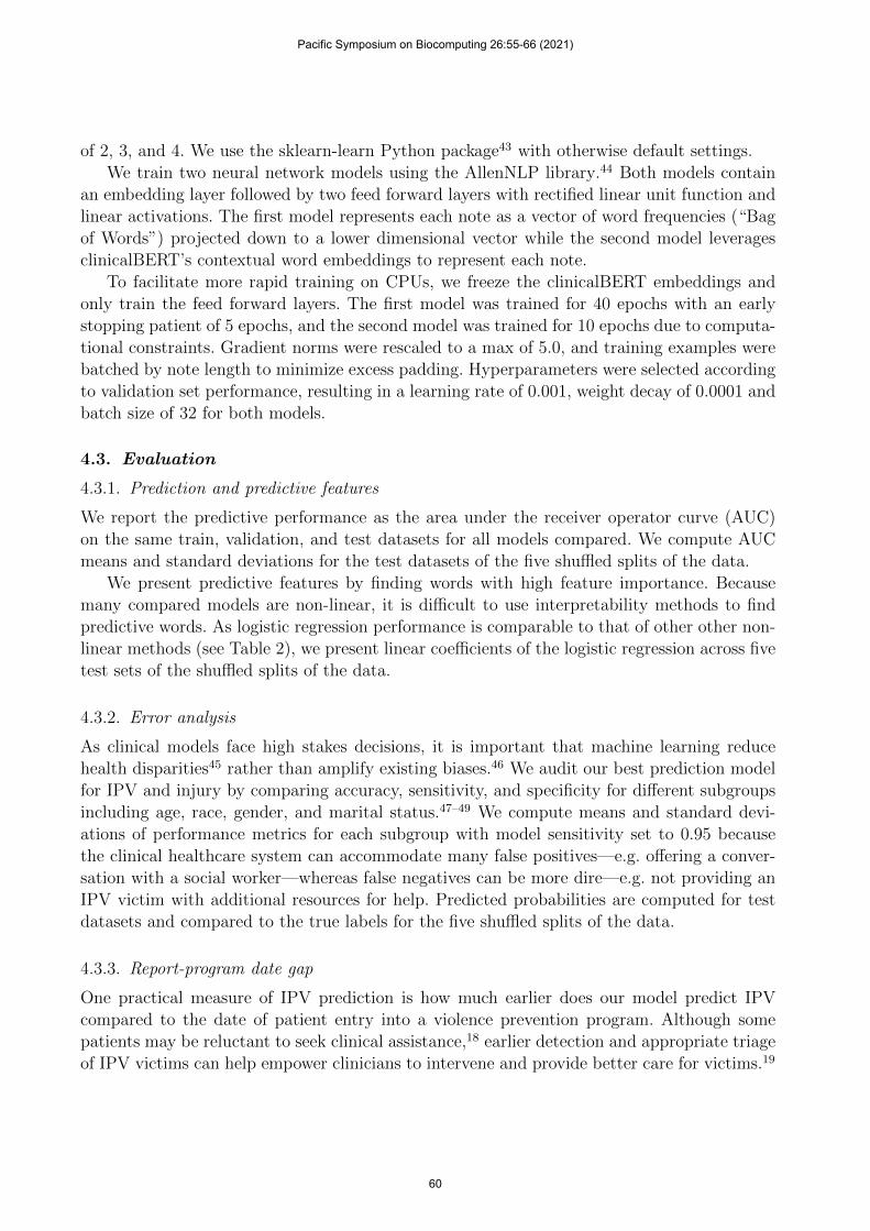
20 10 0Earliest possible date gap (years)
25
20
15
10
5
0
5
Ear
liest
pre
dict
ed d
ate
gap
(yea
rs)
30 20 10 0 10Report-program date gap (years)
0.0
0.2
0.4
0.6
0.8
1.0
IPV
pred
ictio
n pr
obab
ility
Fig. 1. Scatterplots and marginal histograms for random forest classifier for IPV prediction. Left:Earliest possible report-program date gap per patient (x-axis) compared to earliest predicted dategap (y-axis) with sensitivity of 64% and specificity of 95%. Right: Report-program date gap (x-axis)and IPV prediction probability (y-axis) for all radiology reports of IPV victims.
Table 3. Predictive words for IPV and injury averaged across five trialsbased on linear coefficients of logistic regression. Underline indicates wordsconsistent with clinical literature.10
Task Predictive words
IPV ordering, final, trauma, hematoma, technique, swelling, cell, fracture,type, fractures, lymphoma, electronically, male, pancreatitis, reason,gms, implants, unresponsive, assault, none, cancer, pregnancy, mca
Injury hematoma, fracture, fractures, swelling, trauma, subchorionic, for-eign, ankle, third, hand, nondisplaced, fall, stab, phalanx, finger, de-formity, skullbase, fifth, wound, laceration, sob, digit, measuring
IPV or injury prediction, but not both. For example, Black patients have lower sensitivityfor injury prediction. White patients have low sensitivity for IPV prediction. It appears thatpatients who are not single or married (e.g. widowed, separated) have lower sensitivity forinjury whereas married patients have lower sensitivity for IPV prediction.
5.3. Report-program date gap
We can detect IPV from radiology reports much earlier than a patient’s entry into a violenceprevention program. We compute the report-program date gap with specificity threshold ofthe random forest classifier set to 95% and find a median date gap of 3.08 years, compared tothe median earliest possible date gap of 5.83 years. For visual representations of the predicteddate gap compared to the earliest possible date gap and the prediction scores, see Figure 1.
Pacific Symposium on Biocomputing 26:55-66 (2021)
62

Table 4. Error analysis for IPV and injury predictions from random forest classifier. Means and standarddeviations of accuracy, sensitivity (TPR), and specificity (TNR) computed over 5 data splits with overall modelsensitivity set to 0.95. Bold indicates subgroups with particularly low metrics.
IPV Prediction Injury PredictionAccuracy TPR TNR Accuracy TPR TNR
Age < 30 83.6 ± 4% 97.7 ± 1% 53.6 ± 11% 62.5 ± 11% 93.9 ± 9% 61.2 ± 11%30-50 87.4 ± 1% 96.3 ± 1% 49.6 ± 5% 54.1 ± 12% 94.8 ± 1% 52.9 ± 13%51-65 71.8 ± 5% 92.5 ± 2% 49.1 ± 2% 41.4 ± 18% 89.5 ± 3% 40.4 ± 19%66+ 60.9 ± 5% 84.4 ± 2% 45.2 ± 9% 33.5 ± 16% 98.0 ± 4% 31.1 ± 17%
Gender Female 77.2 ± 1% 94.6 ± 1% 48.4 ± 4% 50.0 ± 15% 93.4 ± 1% 48.9 ± 15%Male — — — 31.7 ± 21% 96.2 ± 4% 28.8 ± 21%
Race Black 72.3 ± 2% 95.6 ± 0% 41.4 ± 7% 47.8 ± 14% 88.3 ± 3% 46.5 ± 14%Hispanic 91.1 ± 2% 97.9 ± 0% 51.9 ± 11% 58.0 ± 13% 96.9 ± 3% 57.4 ± 13%White 84.6 ± 1% 90.5 ± 2% 43.0 ± 5% 41.6 ± 18% 95.1 ± 3% 39.8 ± 18%Other 68.7 ± 3% 98.0 ± 0% 55.1 ± 5% 58.3 ± 13% 95.0 ± 6% 57.4 ± 13%
Marital Status Single 81.5 ± 2% 95.4 ± 0% 45.5 ± 7% 49.6 ± 13% 95.3 ± 1% 48.1 ± 14%Married 70.6 ± 1% 92.2 ± 2% 49.8 ± 3% 49.2 ± 18% 92.6 ± 7% 48.5 ± 19%Other 83.4 ± 2% 95.4 ± 2% 49.8 ± 9% 46.5 ± 16% 88.7 ± 3% 45.4 ± 17%
6. Discussion and conclusion
We present a range of findings on the use of prediction algorithms to address IPV in theclinical setting through the analysis of radiology reports. Our results demonstrate several maintakeaways. First, with a dataset of 34,642 reports and 1,479 patients, we are able accuratelypredict IPV and injury with AUCs of 0.852 and 0.887, respectively. Second, while our algorithmdemonstrates some bias in the form of differences in accuracy, sensitivity, and specificity withrespect to age, gender, race, and marital status, we are able to predict a median report-programdate gap of over 3.08 years with sensitivity of 64% and specificity of 95%.
Our work leads naturally to many directions for future research. One limitation of ourcurrent work is that we consider one radiology report at a time for IPV and injury predictionand exclude clinical history. Because IPV victims seek greater medical care from clinical set-tings like the emergency department,7,8 patient data including previous visits, clinical notes,and diagnoses could yield more accurate predictions and therefore earlier detection.19 Ad-ditionally, predictive algorithms can help identify the best intervention for an IPV victim.Currently screening programs for IPV vary in execution and effect,50 and once screened, IPVvictims face many obstacles before leaving an abusive relationship.51 Deeper understanding oftargeted interventions could provide a crucial contribution to patient advocacy.
Deployment of a predictive model for IPV and injury detection faces several practicalchallenges. As with many machine learning algorithms in clinical settings, question of general-ization across hospitals22 and across subgroups47 raise concerns about robustness and fairness.
Pacific Symposium on Biocomputing 26:55-66 (2021)
63

Moreover, better understanding of physician reliance on, distrust of, and confusion towardspredictive models in clinical settings is an active area of research.52 We have shown in ouranalysis that automated detection through machine learning can predict IPV and injury fromradiology reports. We look forward to future work towards the deployment of an IPV earlydetection model in a clinical setting.
References
1. E. Fulu, R. Jewkes, T. Roselli, C. Garcia-Moreno et al., Prevalence of and factors associatedwith male perpetration of intimate partner violence: findings from the un multi-country cross-sectional study on men and violence in asia and the pacific, The lancet global health 1, e187(2013).
2. M. Black, K. Basile, M. Breiding, S. Smith, M. Walters, M. Merrick, J. Chen and M. Stevens,National intimate partner and sexual violence survey: 2010 summary report (2011).
3. J. C. Campbell, Health consequences of intimate partner violence, The lancet 359, 1331 (2002).4. K. Tollestrup, D. Sklar, F. J. Frost, L. Olson, J. Weybright, J. Sandvig and M. Larson, Health
indicators and intimate partner violence among women who are members of a managed careorganization, Preventive medicine 29, 431 (1999).
5. M. Ellsberg, H. A. Jansen, L. Heise, C. H. Watts, C. Garcia-Moreno et al., Intimate partnerviolence and women’s physical and mental health in the who multi-country study on women’shealth and domestic violence: an observational study, The lancet 371, 1165 (2008).
6. U. N. O. on Drugs and Crime, Global Study on Homicide: Gender-related Killing of Women andGirls (UNODC, United Nations Office on Drugs and Crime, 2018).
7. C. Wisner, T. Gilmer, L. Saltzman and T. Zink, Intimate partner violence against women,Journal of family practice 48, 439 (1999).
8. S. R. Dearwater, J. H. Coben, J. C. Campbell, G. Nah, N. Glass, E. McLoughlin and B. Beke-meier, Prevalence of intimate partner abuse in women treated at community hospital emergencydepartments, Jama 280, 433 (1998).
9. A. Russo, A. Reginelli, M. Pignatiello, F. Cioce, G. Mazzei, O. Fabozzi, V. Parlato, S. Cappabi-anca and S. Giovine, Imaging of violence against the elderly and the women, in Seminars inUltrasound, CT and MRI , (1)2019.
10. E. George, C. H. Phillips, N. Shah, A. Lewis-O’Connor, B. Rosner, H. M. Stoklosa and B. Khu-rana, Radiologic findings in intimate partner violence, Radiology 291, 62 (2019).
11. S. Lipsky, R. Caetano, C. A. Field and G. L. Larkin, The role of intimate partner violence, race,and ethnicity in help-seeking behaviors, Ethnicity and Health 11, 81 (2006).
12. P. Tjaden and N. Thoennes, Prevalence and consequences of male-to-female and female-to-maleintimate partner violence as measured by the national violence against women survey, Violenceagainst women 6, 142 (2000).
13. C. M. Rennison, Intimate partner violence and age of victim, 1993-99 (US Department of Justice,Office of Justice Programs, Bureau of Justice . . . , 2001).
14. A. Salomon, S. S. Bassuk and N. Huntington, The relationship between intimate partner violenceand the use of addictive substances in poor and homeless single mothers, Violence AgainstWomen 8, 785 (2002).
15. N. Van Gelder, A. Peterman, A. Potts, M. O’Donnell, K. Thompson, N. Shah and S. Oertelt-Prigione, Covid-19: Reducing the risk of infection might increase the risk of intimate partnerviolence, EClinicalMedicine 21 (2020).
16. B. Gosangi, H. Park, R. Thomas, R. Gujrathi, C. P. Bay, A. S. Raja, S. E. Seltzer, M. C. Balcom,M. L. McDonald, D. P. Orgill et al., Exacerbation of physical intimate partner violence duringcovid-19 lockdown, Radiology , p. 202866 (2020).
Pacific Symposium on Biocomputing 26:55-66 (2021)
64

17. H. Stockl, K. Devries, A. Rotstein, N. Abrahams, J. Campbell, C. Watts and C. G. Moreno,The global prevalence of intimate partner homicide: a systematic review, The Lancet 382, 859(2013).
18. D. Hien and L. Ruglass, Interpersonal partner violence and women in the united states: Anoverview of prevalence rates, psychiatric correlates and consequences and barriers to help seeking,International journal of law and psychiatry 32, p. 48 (2009).
19. B. Khurana, S. E. Seltzer, I. S. Kohane and G. W. Boland, Making the ‘invisible’visible: trans-forming the detection of intimate partner violence, BMJ quality & safety 29, 241 (2020).
20. F. L. Kraanen, E. Vedel, A. Scholing and P. M. Emmelkamp, Prediction of intimate partnerviolence by type of substance use disorder, Journal of substance abuse treatment 46, 532 (2014).
21. M. Ghassemi, T. Naumann, P. Schulam, A. L. Beam, I. Y. Chen and R. Ranganath, A reviewof challenges and opportunities in machine learning for health, AMIA Summits on TranslationalScience Proceedings 2020, p. 191 (2020).
22. M. Ghassemi, T. Naumann, P. Schulam, A. L. Beam, I. Y. Chen and R. Ranganath, Practicalguidance on artificial intelligence for health-care data, The Lancet Digital Health 1, e157 (2019).
23. N. Razavian, S. Blecker, A. M. Schmidt, A. Smith-McLallen, S. Nigam and D. Sontag,Population-level prediction of type 2 diabetes from claims data and analysis of risk factors,Big Data 3, 277 (2015).
24. K. L. Kehl, H. Elmarakeby, M. Nishino, E. M. Van Allen, E. M. Lepisto, M. J. Hassett, B. E.Johnson and D. Schrag, Assessment of deep natural language processing in ascertaining oncologicoutcomes from radiology reports, JAMA oncology 5, 1421 (2019).
25. S. Saria, A. K. Rajani, J. Gould, D. Koller and A. A. Penn, Integration of early physiologicalresponses predicts later illness severity in preterm infants, Science translational medicine 2,48ra65 (2010).
26. A. Chouldechova, D. Benavides-Prado, O. Fialko and R. Vaithianathan, A case study ofalgorithm-assisted decision making in child maltreatment hotline screening decisions, in Con-ference on Fairness, Accountability and Transparency , 2018.
27. A. Brown, A. Chouldechova, E. Putnam-Hornstein, A. Tobin and R. Vaithianathan, Towardalgorithmic accountability in public services: A qualitative study of affected community perspec-tives on algorithmic decision-making in child welfare services, in Proceedings of the 2019 CHIConference on Human Factors in Computing Systems, 2019.
28. S. Wu, K. Roberts, S. Datta, J. Du, Z. Ji, Y. Si, S. Soni, Q. Wang, Q. Wei, Y. Xiang et al., Deeplearning in clinical natural language processing: a methodical review, Journal of the AmericanMedical Informatics Association 27, 457 (2020).
29. S. Gao, M. T. Young, J. X. Qiu, H.-J. Yoon, J. B. Christian, P. A. Fearn, G. D. Tourassi andA. Ramanthan, Hierarchical attention networks for information extraction from cancer pathologyreports, Journal of the American Medical Informatics Association 25, 321 (2018).
30. L. Chen, Y. Gu, X. Ji, C. Lou, Z. Sun, H. Li, Y. Gao and Y. Huang, Clinical trial cohortselection based on multi-level rule-based natural language processing system, Journal of theAmerican Medical Informatics Association 26, 1218 (2019).
31. N. Afzal, V. P. Mallipeddi, S. Sohn, H. Liu, R. Chaudhry, C. G. Scott, I. J. Kullo and A. M.Arruda-Olson, Natural language processing of clinical notes for identification of critical limbischemia, International journal of medical informatics 111, 83 (2018).
32. C. Poulin, B. Shiner, P. Thompson, L. Vepstas, Y. Young-Xu, B. Goertzel, B. Watts, L. Flashmanand T. McAllister, Predicting the risk of suicide by analyzing the text of clinical notes, PloS one9, p. e85733 (2014).
33. K. Huang, J. Altosaar and R. Ranganath, Clinicalbert: Modeling clinical notes and predictinghospital readmission, arXiv preprint arXiv:1904.05342 (2019).
34. A. Rumshisky, M. Ghassemi, T. Naumann, P. Szolovits, V. Castro, T. McCoy and R. Perlis,
Pacific Symposium on Biocomputing 26:55-66 (2021)
65

Predicting early psychiatric readmission with natural language processing of narrative dischargesummaries, Translational psychiatry 6, e921 (2016).
35. A.-D. Pham, A. Neveol, T. Lavergne, D. Yasunaga, O. Clement, G. Meyer, R. Morello andA. Burgun, Natural language processing of radiology reports for the detection of thromboembolicdiseases and clinically relevant incidental findings, BMC bioinformatics 15, 1 (2014).
36. W. Boag, D. Doss, T. Naumann and P. Szolovits, What’s in a note? unpacking predictive value inclinical note representations, AMIA Summits on Translational Science Proceedings 2018, p. 26(2018).
37. J. Devlin, M.-W. Chang, K. Lee and K. Toutanova, Bert: Pre-training of deep bidirectionaltransformers for language understanding, arXiv preprint arXiv:1810.04805 (2018).
38. Y. Liu, M. Ott, N. Goyal, J. Du, M. Joshi, D. Chen, O. Levy, M. Lewis, L. Zettlemoyerand V. Stoyanov, Roberta: A robustly optimized bert pretraining approach, arXiv preprintarXiv:1907.11692 (2019).
39. Z. Lan, M. Chen, S. Goodman, K. Gimpel, P. Sharma and R. Soricut, Albert: A lite bert forself-supervised learning of language representations, arXiv preprint arXiv:1909.11942 (2019).
40. A. E. Johnson, T. J. Pollard, L. Shen, H. L. Li-Wei, M. Feng, M. Ghassemi, B. Moody,P. Szolovits, L. A. Celi and R. G. Mark, Mimic-iii, a freely accessible critical care database,Scientific data 3, 1 (2016).
41. J. Lee, W. Yoon, S. Kim, D. Kim, S. Kim, C. H. So and J. Kang, Biobert: a pre-trained biomedicallanguage representation model for biomedical text mining, Bioinformatics 36, 1234 (2020).
42. E. Alsentzer, J. R. Murphy, W. Boag, W.-H. Weng, D. Jin, T. Naumann and M. McDermott,Publicly available clinical bert embeddings, arXiv preprint arXiv:1904.03323 2019 (2019).
43. F. Pedregosa, G. Varoquaux, A. Gramfort, V. Michel, B. Thirion, O. Grisel, M. Blondel, P. Pret-tenhofer, R. Weiss, V. Dubourg, J. Vanderplas, A. Passos, D. Cournapeau, M. Brucher, M. Perrotand E. Duchesnay, Scikit-learn: Machine learning in Python, Journal of Machine Learning Re-search 12, 2825 (2011).
44. M. Gardner, J. Grus, M. Neumann, O. Tafjord, P. Dasigi, N. F. Liu, M. Peters, M. Schmitz andL. S. Zettlemoyer, Allennlp: A deep semantic natural language processing platform2017.
45. I. Y. Chen, S. Joshi and M. Ghassemi, Treating health disparities with artificial intelligence,Nature Medicine 26, 16 (2020).
46. A. Rajkomar, M. Hardt, M. D. Howell, G. Corrado and M. H. Chin, Ensuring fairness in machinelearning to advance health equity, Annals of internal medicine 169, 866 (2018).
47. I. Y. Chen, P. Szolovits and M. Ghassemi, Can ai help reduce disparities in general medical andmental health care?, AMA journal of ethics 21, 167 (2019).
48. I. Y. Chen, M. Agrawal, S. Horng and D. Sontag, Robustly extracting medical knowledge fromehrs: A case study of learning a health knowledge graph, in Pac Symp Biocomput , 2020.
49. M. Hardt, E. Price and N. Srebro, Equality of Opportunity in Supervised Learning, in Proceed-ings of the 30th International Conference on Neural Information Processing Systems, NIPS’16(Curran Associates Inc., USA, June 2016). Barcelona, Spain.
50. L. O’Doherty, K. Hegarty, J. Ramsay, L. L. Davidson, G. Feder and A. Taft, Screening women forintimate partner violence in healthcare settings, Cochrane database of systematic reviews (2015).
51. J. Kim and K. A. Gray, Leave or stay? battered women’s decision after intimate partner violence,Journal of Interpersonal Violence 23, 1465 (2008).
52. P. Tschandl, C. Rinner, Z. Apalla, G. Argenziano, N. Codella, A. Halpern, M. Janda, A. Lallas,C. Longo, J. Malvehy et al., Human–computer collaboration for skin cancer recognition, NatureMedicine , 1 (2020).
Pacific Symposium on Biocomputing 26:55-66 (2021)
66