
Intestinal absorption of BCS class II drugs administered ...
16
doi: https://dx.doi.org/10.5599/admet.881 375 ADMET & DMPK 8(4) (2020) 375-390; doi: https://dx.doi.org/10.5599/admet.881 Open Access : ISSN : 1848-7718 http://www.pub.iapchem.org/ojs/index.php/admet/index Review Intestinal absorption of BCS class II drugs administered as nanoparticles: A review based on in vivo data from intestinal perfusion models David Dahlgren, Erik Sjögren and Hans Lennernäs* Department of Pharmaceutical Biosciences, Translational Drug Discovery and Development, Uppsala University, Sweden *Corresponding Author: E-mail: [email protected]; Tel.: +46 – 18 471 4317; Fax: +46 – 18 471 4223 Received: June 26, 2020; Revised: September 10, 2020; Published: September 17, 2020 Abstract An established pharmaceutical strategy to increase oral drug absorption of low solubility–high permeability drugs is to create nanoparticles of them. Reducing the size of the solid-state particles increases their dissolution and transport rate across the mucus barrier and the aqueous boundary layer. Suspensions of nanoparticles also sometimes behave differently than those of larger particles in the fed state. This review compares the absorption mechanisms of nano- and larger particles in the lumen at different prandial states, with an emphasis on data derived from in vivo models. Four BSC class II drugs—aprepitant, cyclosporine, danazol and fenofibrate—are discussed in detail based on information from preclinical intestinal perfusion models. Keywords Nano particle; nano drug delivery; nanomedicines; intestinal perfusion; pharmaceutical development. Introduction Rate and extent of drug absorption and bioavailability are critical pharmacokinetic (PK) parameters for oral pharmaceutical products. These parameters are determined and characterized in drug discovery and in preclinical and clinical development of a drug product. Successful design and development of any oral drug product requires understanding the complex interplay between the pharmacokinetic parameters, manufacturing methods, pharmaceutical excipients, and biopharmaceutics, as well as potency (the major driver for drug discovery), pharmacology, safety, and toxicity. Gastrointestinal (GI) absorption and bioavailability are both affected by a number of pharmaceutical, biopharmaceutical and physiological processes. Despite comprehensive knowledge about drug-product related and physiological properties in GI absorption, many drug candidates and drugs have suboptimal biopharmaceutical properties for oral dosing [1,2]. Several challenging drug candidates are listed in the biopharmaceutical classification system (BCS) as class II, III, or IV, for which absorption is limited by solubility properties in GI luminal fluids and/or intestinal permeability [3]. Limitations in any one of the main BCS parameters may reduce the rate and fraction dose absorbed (f abs ) from conventional, oral dosage forms intended for immediate release in the GI lumen. Drugs with low solubility and/or low intestinal permeability also tend to have highly variable plasma exposure
Transcript of Intestinal absorption of BCS class II drugs administered ...

doi: https://dx.doi.org/10.5599/admet.881 375
ADMET & DMPK 8(4) (2020) 375-390; doi: https://dx.doi.org/10.5599/admet.881
Open Access : ISSN : 1848-7718
http://www.pub.iapchem.org/ojs/index.php/admet/index
Review
Intestinal absorption of BCS class II drugs administered as nanoparticles: A review based on in vivo data from intestinal perfusion models
David Dahlgren, Erik Sjögren and Hans Lennernäs*
Department of Pharmaceutical Biosciences, Translational Drug Discovery and Development, Uppsala University, Sweden
*Corresponding Author: E-mail: [email protected]; Tel.: +46 – 18 471 4317; Fax: +46 – 18 471 4223
Received: June 26, 2020; Revised: September 10, 2020; Published: September 17, 2020
Abstract
An established pharmaceutical strategy to increase oral drug absorption of low solubility–high permeability drugs is to create nanoparticles of them. Reducing the size of the solid-state particles increases their dissolution and transport rate across the mucus barrier and the aqueous boundary layer. Suspensions of nanoparticles also sometimes behave differently than those of larger particles in the fed state. This review compares the absorption mechanisms of nano- and larger particles in the lumen at different prandial states, with an emphasis on data derived from in vivo models. Four BSC class II drugs—aprepitant, cyclosporine, danazol and fenofibrate—are discussed in detail based on information from preclinical intestinal perfusion models.
Keywords
Nano particle; nano drug delivery; nanomedicines; intestinal perfusion; pharmaceutical development.
Introduction
Rate and extent of drug absorption and bioavailability are critical pharmacokinetic (PK) parameters for
oral pharmaceutical products. These parameters are determined and characterized in drug discovery and in
preclinical and clinical development of a drug product. Successful design and development of any oral drug
product requires understanding the complex interplay between the pharmacokinetic parameters,
manufacturing methods, pharmaceutical excipients, and biopharmaceutics, as well as potency (the major
driver for drug discovery), pharmacology, safety, and toxicity. Gastrointestinal (GI) absorption and
bioavailability are both affected by a number of pharmaceutical, biopharmaceutical and physiological
processes. Despite comprehensive knowledge about drug-product related and physiological properties in GI
absorption, many drug candidates and drugs have suboptimal biopharmaceutical properties for oral dosing
[1,2]. Several challenging drug candidates are listed in the biopharmaceutical classification system (BCS) as
class II, III, or IV, for which absorption is limited by solubility properties in GI luminal fluids and/or intestinal
permeability [3]. Limitations in any one of the main BCS parameters may reduce the rate and fraction dose
absorbed (fabs) from conventional, oral dosage forms intended for immediate release in the GI lumen. Drugs
with low solubility and/or low intestinal permeability also tend to have highly variable plasma exposure

H. Lennernäs et al. ADMET & DMPK 8(4) (2020) 375-390
376
profiles within and between individuals [4,5].
In the design and development of oral drug dosage forms, a PK assessment is fundamental for
distinguishing between fabs and bioavailability (F or BA) [6]. In general pharmacology, F represents the
fraction of an administered dose of a drug that reaches the systemic circulation in intact form (parent drug)
and it is one of the primary PK properties of any drug product. It is assumed that the bioavailability for any
drug product given intravenously is 100 % and the bioavailability of any orally administered dosage form is
measured relative to this value, calculated as the ratio of dose-normalized plasma exposure.
Many of the drug candidates currently generated in early development are lipophilic. This is probably a
result of the drug discovery lead-finding techniques applied, which rely on in silico and in vitro screening of
receptor-ligand interactions [7-9]. These molecular screening tools propose candidates with a high
potential for interacting with designated target receptors. As these receptors are usually associated with
lipophilic drugs, the tools invariably select lipophilic candidates [10-12]. While lipophilic active
pharmaceutical ingredients (APIs) tend to have more than sufficiently high effective permeability across the
apical membrane of the enterocytes, they suffer from limited solubility and dissolution in the GI lumen [13].
The dissolution process of a solid API particle can briefly be described in two steps. First, the drug
molecules are released from the particle surface to the surrounding dissolution media, which creates a
saturated, stagnant layer adjacent to the solid surface of the particle. Thereafter, the released drug diffuses
into the bulk of the solvent from regions of high, to regions of low, drug concentration (schematically
displayed in Figure 1).
Figure 1. Dissolution of a solid API releases the drug molecule, shown here for an unrestricted volume.
The rate of drug dissolution (dM/dt) is traditionally described by the Noyes-Whitney/Nernst-Brunner
equation 1 [14-18]:
s( )d
dtD A C CM
t h
(1)
where dM/dt is the dissolution rate (i.e., the change in dissolved drug concentration with time), A is the
surface area (4πr2) of the particle available for dissolution, D is the diffusion rate constant, h is the thickness
of an aqueous boundary layer (ABL) with limited convection surrounding the particle, Cs is the saturation

ADMET & DMPK 8(4) (2020) 375-390 Intestinal absorption of BCS class II drugs nanoparticles
doi: https://dx.doi.org/10.5599/admet.881 377
solubility of the drug at the particle-dissolution media interface, and Ct the dissolved drug concentration in
the bulk at time t. The equation assumes that the diffusion of an API monomer through the ABL is the
slowest (i.e., rate-limiting) step in this series of events. This assumption allows the incorporation of Fick’s
law of diffusion to quantify the dissolution rate of a solid in its own solution (Figure 1) [19].
The Noyes-Whitney/Nernst-Brunner equation (Equation 1) predicts the dissolution of particles greater
than a few micrometres; however, this equation may not be optimal for nanoparticles [16]. In addition to
increasing total surface area, it should also be mentioned that particle size reduction (down to about 1 µm
in diameter) will affect the ABL surrounding each particle, both according to the Prandtl equation (Equation
2) but also by a reduced influence of convection [20,21].
3 ( )
LH k
V (2)
In Equation (2) H stands for the hydrodynamic boundary layer thickness, k is a constant, L is the length of
the particle surface in the flow direction, and V is the relative velocity of the liquid surrounding the particle.
Theoretically, a reduced particle size also increases the saturation solubility, according to the Kelvin effect
[20]. The theorem originates from the description of vapour pressure over a droplet, which increases with
droplet curvature. This theorem originates from the description of vapour pressure over a droplet, which
increases with droplet curvature. The true implication of this effect is however difficult to assess. First, as
the effect apply to particles below 10 nm only a fraction (<1 %) of the original mass of a conventional
nanosized (~100 nm) system remains to be dissolved. Secondly, in polydisperse particles systems, the
gained dissolution of small particles may be countered by growth of larger particles, according to the
Ostwald ripening principles.
Dissolution is typically the key parameter for the assessment of the onset of action of an oral dosage
form with BCS class II drugs. The dissolution rate of any oral drug product is determined by the
physicochemical properties of the substance, dosage form composition and functions, as well as by the
physiological GI conditions (Table 1). The latter varies within and between subjects as well as between the
fasted and fed state.
Table 1. Overview of the pharmaceutical, physicochemical and the physiological properties that influence intestinal drug dissolution.
Properties
Affected by
Physicochemical parameters
Pharmaceutical parameters
Physiological parameters
Drug particle surface area
Wettability, particle size, aggregation, surfactnts
Deaggregation by surfactants in gastric juice and intestinal fluid
(from bile)
Drug diffusion Molecular size
(Stokes–Einstein equation)
Viscosity of GI luminal contents
(fasted/fed) (Stokes–Einstein equation)
Diffusion layer Surface energy Particle size Motility patterns and luminal
flow rate
Drug solubility Lipophilicity, pKa,
melting point
Crystal form, solubilisation, precipitation, pharmaceutical
excipients, impurities
Luminal pH, buffer capacity, bile and food composition
Amount of drug dissolved
See above See above Intestinal permeability
Volume of solvent available
GI secretion, fluid absorption, co-
administered fluids

H. Lennernäs et al. ADMET & DMPK 8(4) (2020) 375-390
378
A range of formulation strategies to increase the dissolution rate have been invented and established
for oral drug products with APIs of low solubility [16]. For acidic and basic APIs, a common approach is to
form a salt with the charged form of the API. Use of a counter-ion increases the solubility and dissolution
rate in the diffusion layer around the solid drug particles [22]. Three other strategies are to: (i) predissolve
the drug in a lipid-based formulation for circumventing the slow dissolution step; (ii) change the solid state
properties of the APIs from stable crystalline state(s) to metastable amorphous structures with lower
intermolecular cohesive forces; or (iii) reduce the particle size, which increases the surface area of the API
and consequently the dissolution rate [23-25].
Two established pharmaceutical strategies to reduce the size of the solid state particles are to create
micro- or nanosuspensions of them [26]. Particle size reduction (i.e., micronization) enhance the dissolution
rate (Equation 1) and may consequently increase the intestinal absorption and bioavailability of low
solubility drug compounds. However, for BCS II APIs, nanosuspensions appear to offer additional
advantages over microsuspensions; the enhancement of the in vivo dissolution rate required to increase
the GI drug absorption and bioavailability for low solubility drug candidates is often greater than what a
nanosuspension can achieve [27]. This is shown for a nanosuspension of a test drug with low molecular
mass 450, acid pKa 4.7, log P of 5 and a solubility of 2 μM at pH 6.8 [28]. Interestingly, with these nano-
technologies for solid-state particles, it is apparent that additional intraluminal mechanism(s) than
dissolution must be operating [29].
Despite the advantages with nanoparticles, a number of hurdles need to be resolved with these oral
drug delivery systems. For instance, instability related to aggregation of nanoparticles and/or to a change in
the solid-state properties of the API may reduce their applicability in pharmaceutical development [30].
Still, in oral product development, nanotechnologies have been successful to develop products to a clinical
stage based on solubility, but mainly by enhancing dissolution. However, there is a need to fully understand
the contribution from enhancement of solubility and/or dissolution as well as additional absorption
mechanisms, such as particle drifting in the mucus layer [31]. There are even strategies in which
nanoparticles may increase absorption of low permeability compounds through increased transepithelial
transport [32]. Obviously, more formulation research and development beyond the established
pharmaceutical strategies are needed, to better understand the mechanisms, physiological possibilities,
and limitations in the in vivo GI absorption of orally administered nanoparticles.
This review focuses on nanoparticle-based oral drug delivery systems and their in vivo performance
based on experimental studies in single-pass intestinal perfusion (SPIP) models. In particular, the intestinal
absorption mechanisms will be discussed. These data may contribute to a future framework for enabling
oral formulation strategies for mainly BCS class II drugs. This review looks at the role of nanoparticles of
four low solubility-high permeability drugs: aprepitant, fenofibrate, cyclosporine, and danazol. A summary
of data from both fasted and fed simulated conditions is included, because prandial state has a strong
effect on rate and extent of intestinal drug absorption for some drug products. Prandial state also
illustrates some of the different intraluminal dissolution and absorption mechanisms that come into play.
Intraluminal transport of nanoparticles and drugs in the intestine
Pharmaceutical development of oral drug delivery systems for low solubility drugs frequently uses
particle size reduction as a formulation method. Development of particle sizes in the nano-range (diameter
<1000 nm) for oral products has increased as a result of improvements in pharmaceutical processing and
manufacturing [33]. As discussed above, oral nanosuspensions dissolve faster than microsuspensions, and
accordingly increase intestinal absorption and bioavailability of BCS II compounds [28,34-37]. Theoretically,

ADMET & DMPK 8(4) (2020) 375-390 Intestinal absorption of BCS class II drugs nanoparticles
doi: https://dx.doi.org/10.5599/admet.881 379
API particle size reduction reaches a point at which dissolution is no the longer the rate-limiting step in in
vivo absorption. At that point, further size reduction is not expected to increase intestinal absorption as it
has become solubility controlled [8]. However, experimental observations indicate otherwise. Absorption
rates continue to increase with decreasing particle size, which suggests that additional absorption-
promoting intestinal processes are taking place simultaneously [29,38,39]. It has been proposed that
nanoparticles are crossing the epithelial mucus layer to a greater extent than larger particles [40]. Mucus is
predominately comprised of the glycoprotein mucin and water. This gives it hydrophilic and hydrophobic
domains, a negative net charge, and high porosity and pore interconnectivity. In addition, mucus is a
dynamic, semipermeable transport barrier that is continuously secreted, shed, and digested.
Mucus thickness is about 120 μm, 480 μm and 830 μm in the jejunum, ileum and colon, respectively,
with a turnover time of a few hours [41,42]. The small intestinal mucus contains a single mucus layer that is
not a rate-limiting step in the absorption, not even for lipophilic high-permeability drugs and/or high
permeability drugs transported by an efficient carrier-mediated (CM) transport route [43-45]. Based on in
vivo jejunal perfusion at different rates in humans, the resistance of the mucus layer to the intestinal
absorption of highly permeable solutes is markedly overestimated. Instead, intestinal absorption in humans
seems to be membrane controlled for both low- and high-permeability compounds, irrespective of
transport mechanism [46]. It is well-recognized that nanoparticles interact with mucus by different
mechanisms and that nanoparticles may acquire different biological and physicochemical properties as a
consequence of adsorption of intraluminal GI biomolecules to their surface (i.e., corona formation).
Commercially available oral nanoparticles are typically around 100–300 nm, which should allow them to
pass faster than larger API particles across the gel-like, mucin-based mesh structure composing the mucus
layer. A continuous mucus layer exists throughout the small intestine, and is highly stratified adjacent to
the epithelium. Experiments show that mucin pore size is consistent with free diffusion of 100 nm particles,
but limited diffusion of 500 nm particles [40,47]. This is due to increased diffusivity for a spherical particle
with smaller radius, which then establishes a high local drug concentration available for permeation
adjacent to the apical enterocyte membrane [31,48]. The primary mechanism of transport through this
layer is diffusion, as opposed to the intestinal lumen, where the primary transport mechanism is convection
[49]. The combined effects of increased dissolution, mucus penetration, and mucus layer diffusivity for
small particles seem to explain the increased intestinal absorption of nanoparticles compared to larger
ones.
Method of in vivo absorption: single-pass intestinal perfusion model and absorption calculations
This review discusses mainly data from the rat and pig single-pass intestinal perfusion (SPIP) models in
relation to other relevant in vivo data [50-53]. The SPIP model has been extensively used in preclinical
studies to investigate and determine epithelial effective intestinal permeability (Peff), as well as other
absorption parameters for drugs and drug delivery systems, such as suspensions and lipid-based ones
(Figure 2). Intestinal Peff, absorption flux (Japp), and fraction dose absorbed (fabs) are the three key
biopharmaceutical variables that describe the absorption and transport properties of a drug across the
intestinal barrier in the SPIP model [3,54]. This model is used to investigate oral drug delivery and
characterize pre-formulations, as part of the development process of a pharmaceutical product. The SPIP
model is also useful for investigating regional intestinal permeability and transport mechanisms involved
during in vivo–relevant conditions, and for determining the BCS classification of any API [54]. This model is
also used to investigate the interplay between in vivo dissolution of various dosage forms, such as nano-
formulations and various pharmaceutical excipients, and intestinal permeability, during both fed and fasted
conditions [55].
H. Lennernäs et al. ADMET & DMPK 8(4) (2020) 375-390
380
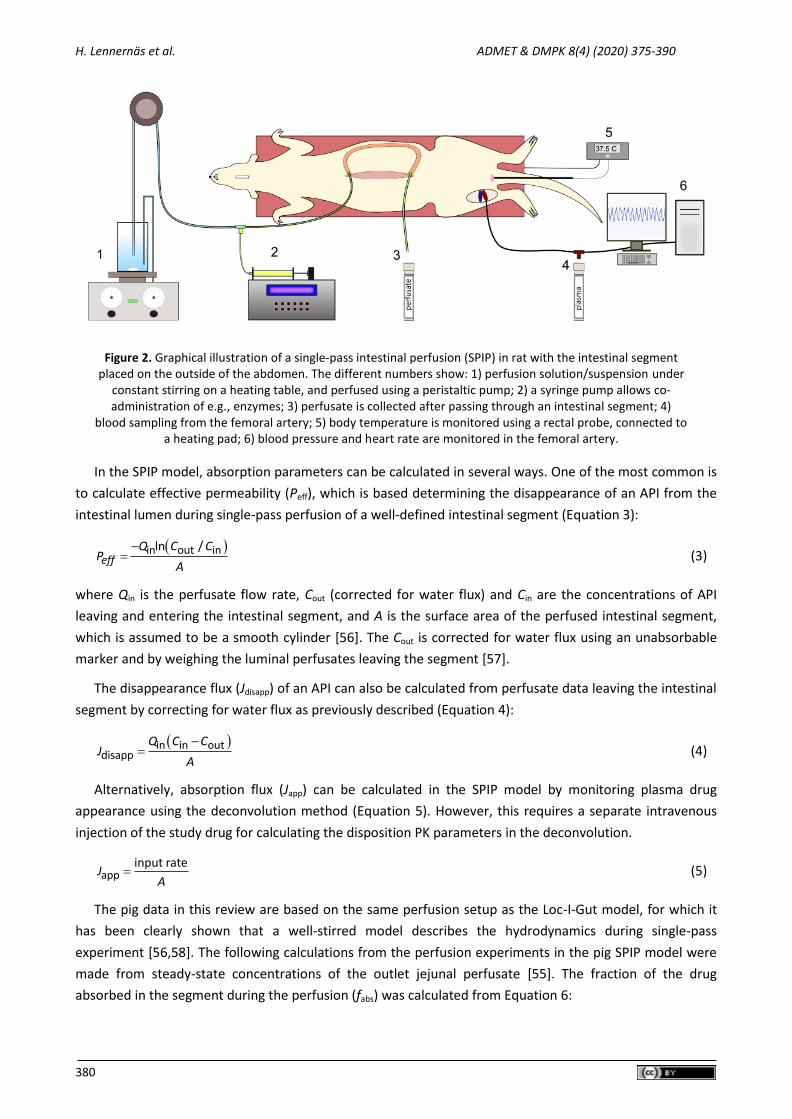
Figure 2. Graphical illustration of a single-pass intestinal perfusion (SPIP) in rat with the intestinal segment placed on the outside of the abdomen. The different numbers show: 1) perfusion solution/suspension under
constant stirring on a heating table, and perfused using a peristaltic pump; 2) a syringe pump allows co-administration of e.g., enzymes; 3) perfusate is collected after passing through an intestinal segment; 4)
blood sampling from the femoral artery; 5) body temperature is monitored using a rectal probe, connected to a heating pad; 6) blood pressure and heart rate are monitored in the femoral artery.
In the SPIP model, absorption parameters can be calculated in several ways. One of the most common is
to calculate effective permeability (Peff), which is based determining the disappearance of an API from the
intestinal lumen during single-pass perfusion of a well-defined intestinal segment (Equation 3):
in out inln /eff
Q C CP
A
(3)
where Qin is the perfusate flow rate, Cout (corrected for water flux) and Cin are the concentrations of API
leaving and entering the intestinal segment, and A is the surface area of the perfused intestinal segment,
which is assumed to be a smooth cylinder [56]. The Cout is corrected for water flux using an unabsorbable
marker and by weighing the luminal perfusates leaving the segment [57].
The disappearance flux (Jdisapp) of an API can also be calculated from perfusate data leaving the intestinal
segment by correcting for water flux as previously described (Equation 4):
in in outdisapp
Q C CJ
A
(4)
Alternatively, absorption flux (Japp) can be calculated in the SPIP model by monitoring plasma drug
appearance using the deconvolution method (Equation 5). However, this requires a separate intravenous
injection of the study drug for calculating the disposition PK parameters in the deconvolution.
appinput rate
JA
(5)
The pig data in this review are based on the same perfusion setup as the Loc-I-Gut model, for which it
has been clearly shown that a well-stirred model describes the hydrodynamics during single-pass
experiment [56,58]. The following calculations from the perfusion experiments in the pig SPIP model were
made from steady-state concentrations of the outlet jejunal perfusate [55]. The fraction of the drug
absorbed in the segment during the perfusion (fabs) was calculated from Equation 6:

ADMET & DMPK 8(4) (2020) 375-390 Intestinal absorption of BCS class II drugs nanoparticles
doi: https://dx.doi.org/10.5599/admet.881 381
inabs
out
1
out
in
C PEGf
C PEG
(6)
where Cin and Cout are the concentrations of the study drug, and PEGin and PEGout are the non-absorbable
volume correction marker ( [14C]-PEG 4000), entering and leaving the jejunal segment, respectively. The
jejunal Peff of each drug is calculated according to a well-mixed tank model, as shown in Equation 7 [59]:
in outineff
in
A
C CQP
C
(7)
where A is the cylindrical area of the perfused jejunal segment (A).
In vivo absorption of four model drugs, under different luminal conditions
This review focuses on the in vivo, small intestine absorption of four BCS class II drugs: aprepitant,
cyclosporine, danazol, and fenofibrate. A range of physicochemical properties of these drugs are presented
in Table 2. They have been investigated in the small intestine as nano-formulations under different luminal
conditions.
Table 2. Physicochemical descriptors of danazol, cyclosporine, aprepitant, and fenofibrate.
danazol [55,60] cyclosporine [55] aprepitant [60] fenofibrate [60]
Molecular mass (g/mol)
337 1202 535 361
Water solubility at 37 °C (µg/ml)
0.5 7 0.37 0.25
log P 3.7 3 4.7 6.9
Papp (·10-6
cm/s) 14.15 2.6 170 220
Aprepitant is an orally administered neurokinin NK-1 receptor antagonist, used clinically to prevent
acute and delayed chemotherapy-induced nausea and vomiting [61]. The molecular mass is 534 Da. It has a
basic pKa of 2.4, an acidic pKa of 9.2, and is primarily uncharged at a jejunal pH of 6.5. The aqueous
solubility is poor (0.37 μg/mL) and it has a high apparent permeability (Papp) in Caco-2 cells (1.7·10−4 cm/s),
which predicts a high Peff in both small and large intestine [62]. In fact, the rat small intestinal Peff is
reported to be 1.7·10−4 cm/s [63]. Aprepitant is mainly metabolized by gut and hepatic CYP3A4, which
means that plasma pharmacokinetics (i.e. F) can be affected by differences in small intestinal absorption
rate [64]. The oral product with aprepitant (Emend; Merck & Co., Inc., NJ) is based on nanoparticles of the
API and has an average particle diameter below 200 nm. These particles are coated onto larger cellulose
beads and encapsulated [65].
Cyclosporine is an immunosuppressant used to prevent organ transplant rejection and danazol is a
steroid used to treat endometriosis. As evident by their BCS class II classification, both cyclosporine and
danazol have high intestinal permeability values and low aqueous solubility.
Both cyclosporine and danazol are metabolized in the small intestine and liver by CYP3A4, where the gut
wall and liver first-pass extraction might be affected by differences in small intestinal absorption [66-68].
The size of the nanoparticles of cyclosporine and danazol used in the experiments discussed below was 650
and 150 nm, respectively. To ensure that the nanosuspension was homogenous and stabilized, cyclosporine
and danazol were added to an aqueous solution containing small amounts of water-soluble polymer and
surfactant as stabilizers/dispersants (PEG-4000 0.13 mg/mL, monoolein 4 mg/mL, sodium taurocholate
20 mg/mL, oleic acid 20 mg/mL, and phosphatidylcholine 6 mg/mL). Both model drugs were single-passed
H. Lennernäs et al. ADMET & DMPK 8(4) (2020) 375-390
382
perfused through pig jejunum in isotonic fluid alone with and without a P-gp inhibitor, and with dietary and
endogenous lipids. To determine the effect of food on the in vivo dissolution of cyclosporine and danazol,
saturated drug solutions in isotonic fluid containing lipids were also perfused in pig.
Fenofibrate is mainly used for primary hypercholesterolemia or mixed dyslipidemia. Fenofibrate is a low
molecular mass (360.4 Da) BCS class II drug that is very lipophilic (log P = 5.24), with an aqueous solubility of
<0.1 mg/mL. Fenofibrate is an ester of fenofibric acid. After oral administration in humans, it is completely
converted to its active metabolite, fenofibric acid. Fenofibric acid and fenofibric acid glucuronide are then
excreted into urine (60 % of the dose) and feces (25 %) [69].
Comparison between micro- and nanosuspensions of BCS class II drugs
Experimental data and simulations show that small intestinal absorption of larger particles of danazol
(226 µm), griseofulvin (118 µm), and aprepitant (26 µm), is dissolution rate-controlled [70]. Takano et al.
also show that reduction of particle size increases in vitro dissolution and improves intestinal absorption in
dogs. Increasing the dissolution rate by using of smaller particles of these three selected model drugs does
not improve their small intestinal absorption because their absorption is limited by their solubility during
non-sink luminal conditions [70]. However, for some drug formulated as nanoparticles in the range of 50–
200 nm, the absorption and plasma exposure increased. The increased absorption rate is often explained by
enhanced drug dissolution rate [28,34,71]. These differences in experimental in vivo data and their
interpretation will be discussed below.
The intestinal absorption rate of aprepitant increases by increasing the concentration of nanoparticles
(200 µM versus 20 µM) in the perfusion suspension passed along the small intestine segment in the rat SPIP
model [5]. Aprepitant permeates the apical membrane as a monomer very swiftly and has a small intestinal
Peff of 1.7 · 10-4 cm/s. Both the drifting and deposition of nanoparticles with aprepitant in the ABL, as well as
colloidal structures, may contribute to an increase in intestinal absorption. Absorption of nano- and
microsuspensions has been compared in the rat SPIP model when the small intestinal segment was
perfused with buffer, FaSSIF, and FeSSIF. In with single-pass perfusions of jejunum, the plasma
concentration time–curves clearly show that fed conditions, i.e. FeSSIF, increase the intestinal absorption of
aprepitant as microsuspensions, but not as nanosuspensions. This difference is in line with previous data
from both dog and human, in which nanosuspensions seem to prevent variation in plasma exposure
between different prandial states, while microsuspensions show significant food-effects [72-75].
The mucus layer also influences absorption of drugs delivered orally as nanoparticles [76]. For example,
the hydrophobicity, electrostatic properties, and steric hindrance, of mucus are key feature that prevents
the hydrophilic pancreatic proteases from acting on, and thereby injuring, the intestinal epithelium [77]. To
quantify the jejunal flux of aprepitant across the enterocytes, Japp was calculated according to Eq. 4 (Table
3). There were no significant differences for micro- and nanosuspensions, but a large, albeit non-significant
one, for the microsuspensions in FeSSIF compared to buffer and FASSIF (64 and 4.8 times higher,
respectively). For the nanosuspensions, the Japp were all within a 1.6-fold range, reiterating the effective
elimination of a luminal food effect by the nanoformulations. The high variability of the data in this paper
demonstrates that studies with more animals are needed to verify these observed trends for nano- and
microsuspensions (Table 3).
ADMET & DMPK 8(4) (2020) 375-390 Intestinal absorption of BCS class II drugs nanoparticles
doi: https://dx.doi.org/10.5599/admet.881 383
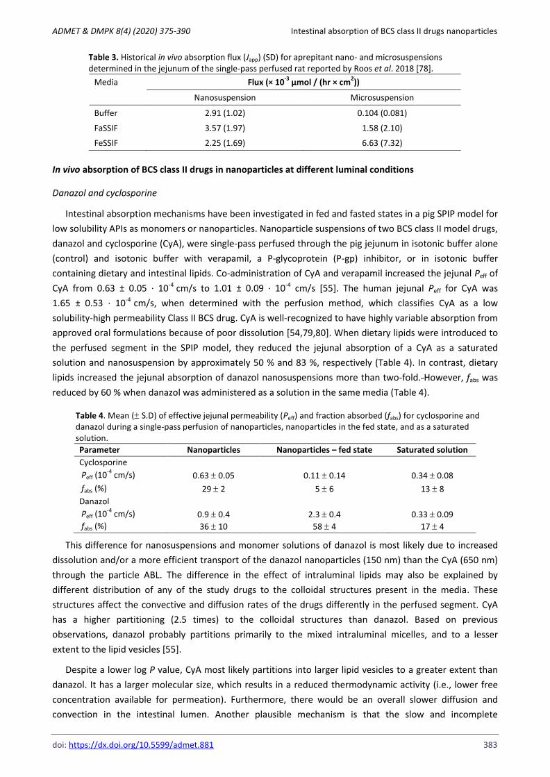
Table 3. Historical in vivo absorption flux (Japp) (SD) for aprepitant nano- and microsuspensions determined in the jejunum of the single-pass perfused rat reported by Roos et al. 2018 [78].
Media Flux (× 10-3
µmol / (hr × cm2))
Nanosuspension Microsuspension
Buffer 2.91 (1.02) 0.104 (0.081)
FaSSIF 3.57 (1.97) 1.58 (2.10)
FeSSIF 2.25 (1.69) 6.63 (7.32)
In vivo absorption of BCS class II drugs in nanoparticles at different luminal conditions
Danazol and cyclosporine
Intestinal absorption mechanisms have been investigated in fed and fasted states in a pig SPIP model for
low solubility APIs as monomers or nanoparticles. Nanoparticle suspensions of two BCS class II model drugs,
danazol and cyclosporine (CyA), were single-pass perfused through the pig jejunum in isotonic buffer alone
(control) and isotonic buffer with verapamil, a P-glycoprotein (P-gp) inhibitor, or in isotonic buffer
containing dietary and intestinal lipids. Co-administration of CyA and verapamil increased the jejunal Peff of
CyA from 0.63 ± 0.05 · 10-4 cm/s to 1.01 ± 0.09 · 10-4 cm/s [55]. The human jejunal Peff for CyA was
1.65 ± 0.53 · 10-4 cm/s, when determined with the perfusion method, which classifies CyA as a low
solubility-high permeability Class II BCS drug. CyA is well-recognized to have highly variable absorption from
approved oral formulations because of poor dissolution [54,79,80]. When dietary lipids were introduced to
the perfused segment in the SPIP model, they reduced the jejunal absorption of a CyA as a saturated
solution and nanosuspension by approximately 50 % and 83 %, respectively (Table 4). In contrast, dietary
lipids increased the jejunal absorption of danazol nanosuspensions more than two-fold. However, fabs was
reduced by 60 % when danazol was administered as a solution in the same media (Table 4).
Table 4. Mean ( S.D) of effective jejunal permeability (Peff) and fraction absorbed (fabs) for cyclosporine and danazol during a single-pass perfusion of nanoparticles, nanoparticles in the fed state, and as a saturated solution.
Parameter Nanoparticles Nanoparticles – fed state Saturated solution
Cyclosporine
Peff (10-4
cm/s) 0.63 0.05 0.11 0.14 0.34 0.08
fabs (%) 29 2 5 6 13 8
Danazol
Peff (10-4
cm/s) 0.9 0.4 2.3 0.4 0.33 0.09
fabs (%) 36 10 58 4 17 4
This difference for nanosuspensions and monomer solutions of danazol is most likely due to increased
dissolution and/or a more efficient transport of the danazol nanoparticles (150 nm) than the CyA (650 nm)
through the particle ABL. The difference in the effect of intraluminal lipids may also be explained by
different distribution of any of the study drugs to the colloidal structures present in the media. These
structures affect the convective and diffusion rates of the drugs differently in the perfused segment. CyA
has a higher partitioning (2.5 times) to the colloidal structures than danazol. Based on previous
observations, danazol probably partitions primarily to the mixed intraluminal micelles, and to a lesser
extent to the lipid vesicles [55].
Despite a lower log P value, CyA most likely partitions into larger lipid vesicles to a greater extent than
danazol. It has a larger molecular size, which results in a reduced thermodynamic activity (i.e., lower free
concentration available for permeation). Furthermore, there would be an overall slower diffusion and
convection in the intestinal lumen. Another plausible mechanism is that the slow and incomplete

H. Lennernäs et al. ADMET & DMPK 8(4) (2020) 375-390
384
partitioning from the vesicles to the aqueous-based chime leads to a subsequent rapid intestinal absorption
of CyA. A temporarily unsaturated aqueous phase might form along the perfused jejunal segment and
reduce thermodynamic activity. It is also clear that drug-solubilization is a more important factor in fed
state for CyA than P-gp inhibition for food–drug interaction. Drug partitioning into different luminal CS, and
their fate in the lumen and across the ABL, are important processes to consider in designing nano-based
oral formulations of poorly soluble drugs.
Both the Peff and fabs were higher for danazol when administered as a nanosuspension in lipid-containing
media than in the control buffer. However, due to the rapid dissolution of the nanoparticles, the difference
in absorption did not correspond to previously observed increases in bioavailability of danazol administered
with food. Administration of danazol in the same media, but as a solution, decreased both Peff and fabs. This
shows the importance of the rapid dissolution of the drug nanoparticles. When danazol is administered as a
solution, thermodynamic activity may decrease because absorption of the drug might create a state of
undersaturation.
Aprepitant
The solubility of aprepitant in biorelevant media, e.g., fasted-state simulated intestinal fluid (FaSSIF), is
approximately 60 times higher than in buffer alone [60]. This indicates that aprepitant readily partitions to
luminal colloidal structures. The influence of these colloidal structures on the in vivo absorption of
aprepitant was investigated in the rat SPIP model. When colloidal structures were added in the perfusion
medium, the absorption was indeed affected as the observed small intestinal absorption rate increased.
However, the experimentally determined increase in small intestinal absorption rate cannot be explained
only by an increased luminal dissolution rate. Rather, the increased absorption rate may be a consequence
of changes to the total effective diffusion from the bulk to the epithelial membrane of aprepitant, i.e.,
transport as free monomers and monomers partitioned to colloidal structures, and as particles [63]. Total
effective diffusion may be an appropriate parameter to describe these experimental observations and to
translate the SPIP data on nanoparticle formulations to in vivo performance. These specific data have been
used to evaluate a mathematical model simulating the absorption-promoting effects of nanoparticle
formulations. This model included interlinked descriptions of hydrodynamics, particle dissolution, and
particle diffusion for both the API and luminal colloidal structures. This mathematical, mechanistic model
adequately describes the above discussed in vivo absorption data of nano-formulated aprepitant. It also
supports the proposed mechanism by which contributing effects of the diffusion across the mucus layer of
both aprepitant nanoparticles and colloidal structures into which the drug had partitioned [81]. This
exemplifies a model in which representation of physiology and description of mechanistic formulation-
physiology relationships was pivotal to establish the role of formulation for effective drug absorption. The
need of such physiologically based biopharmaceutics models (PBBM) for predictions and assessments of
formulations in vivo performance has recently been highlighted with an increased scientific activity in the
area [1,82].
Fenofibrate and other drugs
In contrast to these absorption-promoting results for nanoparticles aprepitant and danazol in both
fasted and fed conditions in the SPIP model, only the fed state reduces the intestinal absorption of nano-
sized drugs such as fenofibrate, CsA, pafenolol [37,55,83,84]. In a regional absorption study in humans with
a site-specific delivery system (Enterion capsule), fenofibrate were given as single, equimolar doses into the
stomach, proximal small bowel, distal small bowel, and colon. The bioavailability (related to an intravenous
dose of fenofibric acid) in the stomach, proximal small bowel, distal small bowel, and colon was 69 %, 73 %,

ADMET & DMPK 8(4) (2020) 375-390 Intestinal absorption of BCS class II drugs nanoparticles
doi: https://dx.doi.org/10.5599/admet.881 385
66 %, and 22 %, respectively [85]. This clearly shows that fenofibrate is absorbed from the small intestine.
However, the solubility and dissolution conditions in the colon—less fluid and limited amounts of bile acids
and colloidal structures—prevent efficient fenofibrate absorption from this region.
An in vivo study in rats compared in vitro dissolution, solubility and bioavailability of three different oral
nanosystems with fenofibrate powder. The three systems included PVP nanospheres, HP-β-CD
nanocapsules, and gelatin nanocapsules [86]. the most improved apparent solubility and oral bioavailability
in fasted rats was with fenofibrate nanoencapsualted with gelatin at a ratio of 1:8 (w/w). The authors of the
study proposed that the improved dissolution rate and subsequent intestinal absorption was due to: (i) a
higher solubility of fenofibrate as it converted into the amorphous form or nanocrystalline state; (ii) a large
surface area for dissolution of drug; (iii) improved wetting of fenofibrate by the polymeric matrix in the
formulation; and (iv) reduction in the crystalline intensity.
A method to investigate in vivo-relevant mechanisms of drug absorption in humans is by GI fluid
sampling [52,87]. GI intubation can explore directly the dynamic interplay of drug release, dissolution,
precipitation, and absorption of the drug from a dosage form. The dissolved drug and solid-state
concentration–time profiles from different segments in the GI tract can be obtained. In addition, direct
infusion of a suspension into the duodenum allows patient control of the therapeutic system [88,89].
One study compared nano- and microsuspensions of fenofibrate administered orally in fed state in
humans. The nanosuspensions gave higher local concentrations than the microsuspensions in the proximal
small intestinal lumen and resulted in higher absorption and plasma exposure–time profile of the drug [37].
In fed conditions, the duodenal concentrations of fenofibrate were higher for both oral formulations, but
there was no increase in plasma exposure of fenofibric acid. Micellar encapsulation of the fenofibrate in the
lumen may have limited potential to permeate from colloidal structures in fed-state intestinal fluids.
Absorption can also decrease in the presence of micelle-forming lipids due to lower thermodynamic activity
[39]. The absorption of BCS class II drugs does not necessarily increase when administered with food, as
proposed by others [90]. Taken together, these in vivo data demonstrate that increasing the intestinal
luminal concentration of BCS class II drugs in complex intestinal fluids does not always result in more rapid
and extensive intestinal absorption. Especially in fed state, the encapsulation of a lipophilic drug in micelles
and vesicles in the intestinal lumen may reduce the potential for intestinal flux.
Conclusion
The use of oral, nanosystems for drug delivery can increase intestinal absorption and bioavailability,
reduce the risk for food–drug interactions, and pH-dependent intestinal absorption. However, for some
drugs, the interaction with colloidal structures in the lumen may prevent absorption even if the oral
nanosystems enhance dissolution in vitro compared to the standard oral formulation. Nevertheless,
nanoparticle formulations offer an important strategy in the development of new drugs and for currently
difficult poorly soluble drugs.
Conflict of interest: Conflict of interest does not exist.
References
[1] B. Abrahamsson, M. McAllister, P. Augustijns, P. Zane, J. Butler, R. Holm, P. Langguth, A. Lindahl, A. Müllertz, X. Pepin. Six years of progress in the oral biopharmaceutics area–a summary from the IMI OrBiTo project. European Journal of Pharmaceutics and Biopharmaceutics (2020).
H. Lennernäs et al. ADMET & DMPK 8(4) (2020) 375-390
386
[2] E. Sjögren, B. Abrahamsson, P. Augustijns, D. Becker, M.B. Bolger, M. Brewster, J. Brouwers, T. Flanagan, M. Harwood, C. Heinen, R. Holm, H. Juretschke, M. Kubbinga, A. Lindahl, V. Lukacova, U. Münster, S. Neuhoff, M. Nguyen, A. Peer, C. Reppas, A. Hodjegan, C. Tannergren, W. Weitschies, C. Wilson, P. Zane, H. Lennernäs, P. Langguth. In vivo methods for drug absorption–comparative physiologies, model selection, correlations with in vitro methods (IVIVC), and applications for formulation/API/excipient characterization including food effects. European Journal of Pharmaceutical Sciences 57 (2014) 99-151.
[3] G.L. Amidon, H. Lennernas, V.P. Shah, J.R. Crison. A theoretical basis for a biopharmaceutic drug classification: the correlation of in vitro drug product dissolution and in vivo bioavailability. Pharmaceutical Research 12 (1995) 413-420.
[4] E.T. Hellriegel, T.D. Bjornsson. Interpatient variability in bioavailability is related to the extent of absorption: Implications for bioavailability and bioequivalence studies. Clinical Pharmacology and Therapeutics 60 (1996) 601-607.
[5] M. Sugihara, S. Takeuchi, M. Sugita, K. Higaki, M. Kataoka, S. Yamashita. Analysis of Intra-and Intersubject Variability in Oral Drug Absorption in Human Bioequivalence Studies of 113 Generic Products. Molecular Pharmaceutics 12 (2015) 4405-4413.
[6] C.Y. Wu, L.Z. Benet, M.F. Hebert, S.K. Gupta, M. Rowland, D.Y. Gomez, V.J. Wacher. Differentiation of absorption and first‐pass gut and hepatic metabolism in humans: studies with cyclosporine. Clinical Pharmacology and Therapeutics 58 (1995) 492-497.
[7] H.H. Refsgaard, B.F. Jensen, P.B. Brockhoff, S.B. Padkjær, M. Guldbrandt, M.S. Christensen. In silico prediction of membrane permeability from calculated molecular parameters. J Med Chem 48 (2005) 805-811.
[8] E. Sjögren, J. Westergren, I. Grant, G. Hanisch, L. Lindfors, H. Lennernäs, B. Abrahamsson, C. Tannergren. In silico predictions of gastrointestinal drug absorption in pharmaceutical product development: application of the mechanistic absorption model GI-Sim. European journal of pharmaceutical sciences 49 (2013) 679-698.
[9] H.D. Williams, N.L. Trevaskis, S.A. Charman, R.M. Shanker, W.N. Charman, C.W. Pouton, C.J. Porter. Strategies to address low drug solubility in discovery and development. Pharmacological Reviews 65 (2013) 315-499.
[10] C.A. Lipinski. Drug-like properties and the causes of poor solubility and poor permeability. Journal of pharmacological and toxicological methods 44 (2000) 235-249.
[11] P.W. Kenny. The nature of ligand efficiency. Journal of Cheminformatics 11 (2019) 1-18.
[12] M.M. Hann, A.R. Leach, G. Harper. Molecular complexity and its impact on the probability of finding leads for drug discovery. Journal of Chemical Information and Computer Sciences 41 (2001) 856-864.
[13] C. Lipinski. Poor aqueous solubility—an industry wide problem in drug discovery. Am Pharm Rev 5 (2002) 82-85.
[14] A.A. Noyes, W.R. Whitney. The rate of solution of solid substances in their own solutions. Journal of the American Chemical Society 19 (1897) 930-934.
[15] E. Brunner. Reaktionsgeschwindigkeit in heterogenen Systemen. Zeitschrift für physikalische Chemie 47 (1904) 56-102.
[16] D.A. Shah, S.B. Murdande, R.H. Dave. A review: pharmaceutical and pharmacokinetic aspect of nanocrystalline suspensions. Journal of Pharmaceutical Sciences 105 (2016) 10-24.
[17] J.B. Dressman, G.L. Amidon, C. Reppas, V.P. Shah. Dissolution testing as a prognostic tool for oral drug absorption: immediate release dosage forms. Pharmaceutical Research 15 (1998) 11-22.
[18] B. Shekunov, E.R. Montgomery. Theoretical analysis of drug dissolution: I. Solubility and intrinsic dissolution rate. Journal of Pharmaceutical Sciences 105 (2016) 2685-2697.
[19] J. Siepmann, F. Siepmann. Mathematical modeling of drug dissolution. International Journal of Pharmaceutics 453 (2013) 12-24.
[20] V.B. Junyaprasert, B. Morakul. Nanocrystals for enhancement of oral bioavailability of poorly water-soluble drugs. asian journal of pharmaceutical sciences 10 (2015) 13-23.

ADMET & DMPK 8(4) (2020) 375-390 Intestinal absorption of BCS class II drugs nanoparticles
doi: https://dx.doi.org/10.5599/admet.881 387
[21] R.H. Müller, K. Peters. Nanosuspensions for the formulation of poorly soluble drugs: I. Preparation by a size-reduction technique. International Journal of Pharmaceutics 160 (1998) 229-237.
[22] A.T. Serajuddin. Salt formation to improve drug solubility. Adv Drug Deliv Rev 59 (2007) 603-616.
[23] P. Kanaujia, P. Poovizhi, W. Ng, R. Tan. Amorphous formulations for dissolution and bioavailability enhancement of poorly soluble APIs. Powder Technology 285 (2015) 2-15.
[24] G.G. Liversidge, K.C. Cundy. Particle size reduction for improvement of oral bioavailability of hydrophobic drugs: I. Absolute oral bioavailability of nanocrystalline danazol in beagle dogs. International Journal of Pharmaceutics 125 (1995) 91-97.
[25] L.C. Alskär, C.J. Porter, C.A. Bergström. Tools for early prediction of drug loading in lipid-based formulations. Molecular Pharmaceutics 13 (2016) 251-261.
[26] B. Van Eerdenbrugh, G. Van den Mooter, P. Augustijns. Top-down production of drug nanocrystals: nanosuspension stabilization, miniaturization and transformation into solid products. International Journal of Pharmaceutics 364 (2008) 64-75.
[27] E.R. Cooper. Nanoparticles: a personal experience for formulating poorly water soluble drugs. Journal of Controlled Release 141 (2010) 300-302.
[28] K. Sigfridsson, A.J. Lundqvist, M. Strimfors. Particle size reduction for improvement of oral absorption of the poorly soluble drug UG558 in rats during early development. Drug development and industrial pharmacy 35 (2009) 1479-1486.
[29] K. Sugano. Possible reduction of effective thickness of intestinal unstirred water layer by particle drifting effect. Int J Pharm 387 (2010) 103-109.
[30] S.B. Murdande, D.A. Shah, R.H. Dave. Impact of nanosizing on solubility and dissolution rate of poorly soluble pharmaceuticals. Journal of Pharmaceutical Sciences 104 (2015) 2094-2102.
[31] F. Kesisoglou, M. Wang, K. Galipeau, P. Harmon, G. Okoh, W. Xu. Effect of amorphous nanoparticle size on bioavailability of anacetrapib in dogs. Journal of Pharmaceutical Sciences 108 (2019) 2917-2925.
[32] D. Babadi, S. Dadashzadeh, M. Osouli, M.S. Daryabari, A. Haeri. Nanoformulation strategies for improving intestinal permeability of drugs: A more precise look at permeability assessment methods and pharmacokinetic properties changes. Journal of Controlled Release (2020).
[33] S. Colombo, M. Brisander, J. Haglöf, P. Sjövall, P. Andersson, J. Østergaard, M. Malmsten. Matrix effects in nilotinib formulations with pH-responsive polymer produced by carbon dioxide-mediated precipitation. International Journal of Pharmaceutics 494 (2015) 205-217.
[34] J.-i. Jinno, N. Kamada, M. Miyake, K. Yamada, T. Mukai, M. Odomi, H. Toguchi, G.G. Liversidge, K. Higaki, T. Kimura. Effect of particle size reduction on dissolution and oral absorption of a poorly water-soluble drug, cilostazol, in beagle dogs. Journal of controlled release 111 (2006) 56-64.
[35] E. Merisko-Liversidge, G.G. Liversidge, E.R. Cooper. Nanosizing: a formulation approach for poorly-water-soluble compounds. European journal of pharmaceutical sciences 18 (2003) 113-120.
[36] R. Müller, C. Jacobs, O. Kayser. Nanosuspensions as particulate drug formulations in therapy: rationale for development and what we can expect for the future. Adv Drug Deliv Rev 47 (2001) 3-19.
[37] B. Hens, J. Brouwers, M. Corsetti, P. Augustijns. Gastrointestinal behavior of nano-and microsized fenofibrate: in vivo evaluation in man and in vitro simulation by assessment of the permeation potential. European Journal of Pharmaceutical Sciences 77 (2015) 40-47.
[38] K. Sugano. Aqueous boundary layers related to oral absorption of a drug: from dissolution of a drug to carrier mediated transport and intestinal wall metabolism. Molecular Pharmaceutics 7 (2010) 1362-1373.
[39] G.E. Amidon, W.I. Higuchi, N.F. Ho. Theoretical and experimental studies of transport of micelle‐solubilized solutes. J Pharm Sci 71 (1982) 77-84.
[40] S.K. Lai, D.E. O'Hanlon, S. Harrold, S.T. Man, Y.-Y. Wang, R. Cone, J. Hanes. Rapid transport of large polymeric nanoparticles in fresh undiluted human mucus. Proceedings of the National Academy of Sciences 104 (2007) 1482-1487.

H. Lennernäs et al. ADMET & DMPK 8(4) (2020) 375-390
388
[41] K. Netsomboon, A. Bernkop-Schnürch. Mucoadhesive vs. mucopenetrating particulate drug delivery. European Journal of Pharmaceutics and Biopharmaceutics 98 (2016) 76-89.
[42] J. Leal, H.D. Smyth, D. Ghosh. Physicochemical properties of mucus and their impact on transmucosal drug delivery. International Journal of Pharmaceutics 532 (2017) 555-572.
[43] M.D. Levitt, A. Strocchi, D.G. Levitt. Human jejunal unstirred layer: evidence for extremely efficient luminal stirring. American Journal of Physiology-Gastrointestinal and Liver Physiology 262 (1992) G593-G596.
[44] M.D. Levitt, J.K. Furne. Shaking of the Intact Angulation Diminish Layer. Gastroenterology 103 (1992) 1460-1466.
[45] D. Dahlgren, M. Sjöblom, H. Lennernäs. Intestinal absorption-modifying excipients: A current update on preclinical in vivo evaluations. European Journal of Pharmaceutics and Biopharmaceutics 142 (2019) 411-420.
[46] U. Fagerholm, H. Lennernäs. Experimental estimation of the effective unstirred water layer thickness in the human jejunum, and its importance in oral drug absorption. European Journal of Pharmaceutical Sciences 3 (1995) 247-253.
[47] B.H. Bajka, N.M. Rigby, K.L. Cross, A. Macierzanka, A.R. Mackie. The influence of small intestinal mucus structure on particle transport ex vivo. Colloids and Surfaces B: Biointerfaces 135 (2015) 73-80.
[48] A. Einstein. On the motion of small particles suspended in liquids at rest required by the molecular-kinetic theory of heat. Annalen der physik 17 (1905) 549-560.
[49] Y. Wang, J.G. Brasseur. Three-dimensional mechanisms of macro-to-micro-scale transport and absorption enhancement by gut villi motions. Physical Review E 95 (2017) 062412.
[50] D. Dahlgren, C. Roos, P. Johansson, C. Tannergren, A. Lundqvist, P. Langguth, M. Sjöblom, E. Sjögren, H. Lennernas. The effects of three absorption-modifying critical excipients on the in vivo intestinal absorption of six model compounds in rats and dogs. International Journal of Pharmaceutics 547 (2018) 158-168.
[51] E. Sjödin, H. Fritsch, U.G. Eriksson, U. Logren, A. Nordgren, P. Forsell, L. Knutson, H. Lennernäs. Intestinal and hepatobiliary transport of ximelagatran and its metabolites in pigs. Drug metabolism and disposition 36 (2008) 1519-1528.
[52] L. Bønløkke, L. Hovgaard, H.G. Kristensen, L. Knutson, H. Lennernäs. Direct estimation of the in vivo dissolution of spironolactone, in two particle size ranges, using the single-pass perfusion technique (Loc-I-Gut®) in humans. European Journal of Pharmaceutical Sciences 12 (2001) 239-250.
[53] L. Bønløkke, F.N. Christensen, L. Knutson, H.G. Kristensen, H. Lennernäs. A new approach for direct in vivo dissolution studies of poorly soluble drugs. Pharmaceutical Research 14 (1997) 1490-1492.
[54] D. Dahlgren, C. Roos, E. Sjögren, H. Lennernäs. Direct In Vivo Human Intestinal Permeability (Peff) Determined with Different Clinical Perfusion and Intubation Methods. Journal of Pharmaceutical Sciences 104 (2014) 2702-2726.
[55] E.M. Persson, A. Nordgren, P. Forsell, L. Knutson, C. Öhgren, S. Forssén, H. Lennernäs, B. Abrahamsson. Improved understanding of the effect of food on drug absorption and bioavailability for lipophilic compounds using an intestinal pig perfusion model. European Journal of Pharmaceutical Sciences 34 (2008) 22-29.
[56] H. Lennernäs, Ö. Ahrenstedt, R. Hällgren, L. Knutson, M. Ryde, L.K. Paalzow. Regional jejunal perfusion, a new in vivo approach to study oral drug absorption in man. Pharmaceutical Research 9 (1992) 1243-1251.
[57] S.C. Sutton, M. Rinaldi, K. Vukovinsky. Comparison of the gravimetric, phenol red, and 14C-PEG-3350 methods to determine water absorption in the rat single-pass intestinal perfusion model. AAPS PharmSci 3 (2001) 93.
[58] H. Lennernäs, I.D. Lee, U. Fagerholm, G.L. Amidon. A Residence‐Time Distribution Analysis of the Hydrodynamics within the Intestine in Man during a Regional Single‐pass Perfusion with Loc‐I‐Gut: In‐vivo Permeability Estimation. Journal of Pharmacy and Pharmacology 49 (1997) 682-686.

ADMET & DMPK 8(4) (2020) 375-390 Intestinal absorption of BCS class II drugs nanoparticles
doi: https://dx.doi.org/10.5599/admet.881 389
[59] H. Lennernas. Human jejunal effective permeability and its correlation with preclinical drug absorption models. Journal of Pharmacy and Pharmacology 49 (1997) 627-638.
[60] E. Sjögren, J. Westergren, I. Grant, G. Hanisch, L. Lindfors, H. Lennernäs, B. Abrahamsson, C. Tannergren. In silico predictions of gastrointestinal drug absorption in pharmaceutical product development: Application of the mechanistic absorption model GI-Sim. European Journal of Pharmaceutical Sciences (2013).
[61] T.M. Dando, C.M. Perry. Aprepitant. Drugs 64 (2004) 777-794.
[62] C. Tannergren, A. Bergendal, H. Lennernäs, B. Abrahamsson. Toward an increased understanding of the barriers to colonic drug absorption in humans: implications for early controlled release candidate assessment. Molecular Pharmaceutics 6 (2009) 60-73.
[63] C. Roos, D. Dahlgren, S. Berg, J. Westergren, B. Abrahamsson, C. Tannergren, E. Sjögren, H. Lennernäs. In Vivo Mechanisms of Intestinal Drug Absorption from Aprepitant Nanoformulations. Molecular Pharmaceutics 14 (2017) 4233-4242.
[64] R.I. Sanchez, R.W. Wang, D.J. Newton, R. Bakhtiar, P. Lu, S.-H.L. Chiu, D.C. Evans, S.-E.W. Huskey. Cytochrome P450 3A4 is the major enzyme involved in the metabolism of the substance P receptor antagonist aprepitant. Drug metabolism and disposition 32 (2004) 1287-1292.
[65] R. Hargreaves, J.C.A. Ferreira, D. Hughes, J. Brands, J. Hale, B. Mattson, S. Mills. Development of aprepitant, the first neurokinin‐1 receptor antagonist for the prevention of chemotherapy‐induced nausea and vomiting. Annals of the New York Academy of Sciences 1222 (2011) 40-48.
[66] P.F. Augustijns, T.P. Bradshaw, L.-S.L. Gan, R.W. Hendren, D.R. Thakker. Evidence for a polarized efflux system in Caco-2 cells capable of modulating cyclosporine A transport. Biochemical and Biophysical Research Communications 197 (1993) 360-365.
[67] T. Saeki, K. Ueda, Y. Tanigawara, R. Hori, T. Komano. Human P-glycoprotein transports cyclosporin A and FK506. Journal of Biological Chemistry 268 (1993) 6077-6080.
[68] T. Kronbach, V. Fischer, U.A. Meyer. Cyclosporine metabolism in human liver: identification of a cytochrome P‐450III gene family as the major cyclosporine‐metabolizing enzyme explains interactions of cyclosporine with other drugs. Clinical Pharmacology and Therapeutics 43 (1988) 630-635.
[69] A. Munoz, J. Guichard, P. Reginault. Micronised fenofibrate. Atherosclerosis 110 (1994) S45-S48.
[70] R. Takano, K. Furumoto, K. Shiraki, N. Takata, Y. Hayashi, Y. Aso, S. Yamashita. Rate-limiting steps of oral absorption for poorly water-soluble drugs in dogs; prediction from a miniscale dissolution test and a physiologically-based computer simulation. Pharmaceutical Research 25 (2008) 2334-2344.
[71] L. Jia, H. Wong, C. Cerna, S.D. Weitman. Effect of nanonization on absorption of 301029: ex vivo and in vivo pharmacokinetic correlations determined by liquid chromatography/mass spectrometry. Pharmaceutical Research 19 (2002) 1091-1096.
[72] Y. Shono, E. Jantratid, F. Kesisoglou, C. Reppas, J.B. Dressman. Forecasting in vivo oral absorption and food effect of micronized and nanosized aprepitant formulations in humans. European Journal of Pharmaceutics and Biopharmaceutics 76 (2010) 95-104.
[73] Y. Wu, A. Loper, E. Landis, L. Hettrick, L. Novak, K. Lynn, C. Chen, K. Thompson, R. Higgins, U. Batra. The role of biopharmaceutics in the development of a clinical nanoparticle formulation of MK-0869: a Beagle dog model predicts improved bioavailability and diminished food effect on absorption in human. Int J Pharm 285 (2004) 135-146.
[74] A.K. Majumdar, L. Howard, M.R. Goldberg, L. Hickey, M. Constanzer, P.L. Rothenberg, T.M. Crumley, D. Panebianco, T.E. Bradstreet, A.J. Bergman. Pharmacokinetics of aprepitant after single and multiple oral doses in healthy volunteers. The Journal of Clinical Pharmacology 46 (2006) 291-300.
[75] F. Kesisoglou, S. Panmai, Y. Wu. Nanosizing—oral formulation development and biopharmaceutical evaluation. Adv Drug Deliv Rev 59 (2007) 631-644.
[76] P. Lundquist, P. Artursson. Oral absorption of peptides and nanoparticles across the human intestine: Opportunities, limitations and studies in human tissues. Advanced drug delivery reviews 106 (2016) 256-276.

H. Lennernäs et al. ADMET & DMPK 8(4) (2020) 375-390
390
[77] S.M. Sharpe, X. Qin, Q. Lu, E. Feketeova, D.C. Palange, W. Dong, S.U. Sheth, M.A. Lee, D. Reino, D.-Z. Xu. Loss of the intestinal mucus layer in the normal rat causes gut injury, but not toxic mesenteric lymph nor lung injury. Shock (Augusta, Ga.) 34 (2010) 475.
[78] C. Roos, D. Dahlgren, E. Sjögren, M. Sjöblom, M. Hedeland, H. Lennernäs. Jejunal absorption of aprepitant from nanosuspensions: Role of particle size, prandial state and mucus layer. European Journal of Pharmaceutics and Biopharmaceutics 132 (2018) 222-230.
[79] Y.-Y. Chiu, K. Higaki, B.L. Neudeck, J.L. Barnett, L.S. Welage, G.L. Amidon. Human jejunal permeability of cyclosporin A: influence of surfactants on P-glycoprotein efflux in Caco-2 cells. Pharmaceutical Research 20 (2003) 749-756.
[80] H. Lennernas. Intestinal permeability and its relevance for absorption and elimination. Xenobiotica 37 (2007) 1015-1051.
[81] C. Roos, J. Westergren, D. Dahlgren, H. Lennernäs, E. Sjögren. Mechanistic modelling of intestinal drug absorption–The in vivo effects of nanoparticles, hydrodynamics, and colloidal structures. European Journal of Pharmaceutics and Biopharmaceutics 133 (2018) 70-76.
[82] T. Heimbach, S. Suarez-Sharp, M. Kakhi, N. Holmstock, A. Olivares-Morales, X. Pepin, E. Sjögren, E. Tsakalozou, P. Seo, M. Li. Dissolution and Translational Modeling Strategies Toward Establishing an In Vitro-In Vivo Link—a Workshop Summary Report. Springer, 2019.
[83] H. Lennernäs, C.-G. Regårdh. Evidence for an interaction between the β-blocker pafenolol and bile salts in the intestinal lumen of the rat leading to dose-dependent oral absorption and double peaks in the plasma concentration–time profile. Pharmaceutical Research 10 (1993) 879-883.
[84] C. Regårdh, A. Heggelund, K. Kylberg‐Hanssen, P. Lundborg. Pharmacokinetics of pafenolol after iv and oral administration of three separate doses of different strength to man. Biopharmaceutics and Drug Disposition 11 (1990) 607-617.
[85] T. Zhu, J.C. Ansquer, M.T. Kelly, D.J. Sleep, R.S. Pradhan. Comparison of the gastrointestinal absorption and bioavailability of fenofibrate and fenofibric acid in humans. The Journal of Clinical Pharmacology 50 (2010) 914-921.
[86] A.M. Yousaf, D.W. Kim, Y.-K. Oh, C.S. Yong, J.O. Kim, H.-G. Choi. Enhanced oral bioavailability of fenofibrate using polymeric nanoparticulated systems: physicochemical characterization and in vivo investigation. International journal of nanomedicine 10 (2015) 1819.
[87] B. Borgström. Studies on intestinal cholesterol absorption in the human. The Journal of clinical investigation 39 (1960) 809-815.
[88] D. Nyholm, A. Johansson, S.-M. Aquilonius, E. Hellquist, H. Lennernäs, H. Askmark. Complexity of motor response to different doses of duodenal levodopa infusion in Parkinson disease. Clinical Neuropharmacology 35 (2012) 6-14.
[89] D. Nyholm, H. Askmark, C. Gomes–Trolin, T. Knutson, H. Lennernäs, C. Nyström, S.-M. Aquilonius. Optimizing levodopa pharmacokinetics: intestinal infusion versus oral sustained-release tablets. Clinical Neuropharmacology 26 (2003) 156-163.
[90] D. Fleisher, C. Li, Y. Zhou, L.-H. Pao, A. Karim. Drug, meal and formulation interactions influencing drug absorption after oral administration. Clinical Pharmacokinetics 36 (1999) 233-254.
©2020 by the authors; licensee IAPC, Zagreb, Croatia. This article is an open-access article distributed under the terms and conditions of the Creative Commons Attribution license (http://creativecommons.org/licenses/by/3.0/)

![Intestinal absorption of BCS class II drugs administered as ...1491849/...absorption, many drug candidates and drugs have suboptimal biopharmaceutical properties for oral dosing [1,2].](https://static.fdocuments.net/doc/165x107/60a032c4e469b924473bff7c/intestinal-absorption-of-bcs-class-ii-drugs-administered-as-1491849-absorption.jpg)


![[PPT]OBSTRUCCION INTESTINAL - semio2013 | This … · Web viewOBSTRUCCION INTESTINAL OBSTRUCCION INTESTINAL OBSTACULO AL TRANSITO DEL CONTENIDO INTESTINAL Adinámico o paralítico](https://static.fdocuments.net/doc/165x107/5b36ceb57f8b9a4a728b5103/pptobstruccion-intestinal-semio2013-this-web-viewobstruccion-intestinal.jpg)


