International Journal of Pharmaceutics - kanglab.net pessary.pdf · Sun et al./International...
8
Fabrication of non-dissolving analgesic suppositories using 3D printed moulds Yuanyuan Sun a,b , Xucong Ruan b , Hairui Li a , Himanshu Kathuria b , Guang Du a, *, Lifeng Kang b, * a Department of Pharmacy, Tongji Hospital, Tongji Medical College, Huazhong University of Science and Technology, Wuhan, 430030, China b Department of Pharmacy, National University of Singapore, Singapore, 117543, Republic of Singapore A R T I C L E I N F O Article history: Received 29 May 2016 Received in revised form 22 September 2016 Accepted 25 September 2016 Available online 26 September 2016 Keywords: Non-dissolving suppository Silicone elastomer Sustained release Three dimensional printing Moulding Analgesic A B S T R A C T Conventional suppositories sometimes fail in exerting their therapeutic activity as the base materials melt inside body cavities. Also they are not suitable to provide long term treatment. Biomedical grade silicone elastomers may be used to fabricate non-dissolvable suppositories to overcome these disadvantages. We kneaded 4 analgesics into the 2 kinds of silicone polymers at 1%, 5% and 10% drug loading, respectively, to test their mechanical properties and drug release profiles. The optimized drug- polymer combinations were used to fabricate suppositories, and three dimensional printing (3DP) was used to create the suppository moulds. Subsequently, the drug release profiles and biocompatibility of the suppositories were studied. It was found that, the mechanical properties of the drug laden silicone elastomers and the rate of drug release from the elastomers can be tuned by varying drug-polymer combinations. The silicone elastomers containing 1% (w/w) and 5% (w/w) diclofenac sodium were the optimal formulations with prolonged drug release and biocompatibility at cellular level. These properties, together with complex geometries offered by 3DP technique, potentially made the non- dissolving suppositories promising therapeutic agents for personalized medicine. ã 2016 Elsevier B.V. All rights reserved. 1. Introduction Suppositories are dosage forms intended for rectal, vaginal and urethral applications, to exert local or systemic effect (Leon Shargel, 2010). They are useful for children, the severely debilitated and those with difficulty taking conventional medications. The suppository base materials can be hydrophobic, hydrophilic and amphiphilic (Yarnykh et al., 2011). These materials melt at body temperature to release drugs to the site of application. The melted suppository debris may migrate up or leak out of the location of administration, and fail to achieve their therapeutic effect (Peppas et al., 2000). Besides, extended drug release is not possible by using these melting base materials. To this end, silicone elastomers may be a suitable alternative as suppository bases, owing to their biocompatibility and non- biodegradability ( Ríhová, 1996). These elastic materials can provide snug fit for clinical applications. The non-biodegradable materials can potentially prevent the suppository from melting and/or displacement inside human cavities. On the other hand, these elastomers can also be used to deliver drugs (Baum et al., 2012; Fu and Kao, 2010) sustainably for long-term treatment. Long term analgesic treatment with suppository is needed for patients who suffer from postpartum perineal pain, post-operative pain of gastrointestinal surgery and terminally ill cancer pain (Ali Ebrahim et al., 2014; Andrews et al., 2008; Davis et al., 2002; Dodd et al., 2004; Kirchhoff et al., 2010; Lowenstein et al., 2006; Macarthur and Macarthur, 2004; Petrowsky et al., 2004; Yoong et al., 1997; Wilasrusmee et al., 2008). For these patients, oral delivery of painkillers for long term treatment is either challenging or prohibitive, therefore, non-dissolvable analgesic suppositories can provide a useful alternative treatment option. The commenly used painkillers include lidocaine hydrochloride (LidoHCl) and non-steroidal anti-inflammatory agents (NSAIDs). The sodium salt forms of diclofenac and ibuprofen have been used in suppositories (Davis et al., 2002; James Barron, 1977; Rezaei et al., 2014). Different from diclofenac and ibuprofen, the free acid Abbreviations: Diclo, diclofenac sodium; Ibu, ibuprofen sodium; Keto, ketopro- fen; LidoHCl, lidocaine hydrochloride monohydrate; NSAIDs, non-steroidal anti- inflammatory dugs; PBS, phosphate buffered saline; 3DP, three dimensional printing. * Corresponding authors. E-mail addresses: [email protected] (G. Du), [email protected] (L. Kang). http://dx.doi.org/10.1016/j.ijpharm.2016.09.073 0378-5173/ã 2016 Elsevier B.V. All rights reserved. International Journal of Pharmaceutics 513 (2016) 717–724 Contents lists available at ScienceDirect International Journal of Pharmaceutics journal homepage: www.elsev ier.com/locate /ijpharm
Transcript of International Journal of Pharmaceutics - kanglab.net pessary.pdf · Sun et al./International...

International Journal of Pharmaceutics 513 (2016) 717–724
Fabrication of non-dissolving analgesic suppositories using 3D printedmoulds
Yuanyuan Suna,b, Xucong Ruanb, Hairui Lia, Himanshu Kathuriab, Guang Dua,*,Lifeng Kangb,*aDepartment of Pharmacy, Tongji Hospital, Tongji Medical College, Huazhong University of Science and Technology, Wuhan, 430030, ChinabDepartment of Pharmacy, National University of Singapore, Singapore, 117543, Republic of Singapore
A R T I C L E I N F O
Article history:Received 29 May 2016Received in revised form 22 September 2016Accepted 25 September 2016Available online 26 September 2016
Keywords:Non-dissolving suppositorySilicone elastomerSustained releaseThree dimensional printingMouldingAnalgesic
A B S T R A C T
Conventional suppositories sometimes fail in exerting their therapeutic activity as the base materialsmelt inside body cavities. Also they are not suitable to provide long term treatment. Biomedical gradesilicone elastomers may be used to fabricate non-dissolvable suppositories to overcome thesedisadvantages. We kneaded 4 analgesics into the 2 kinds of silicone polymers at 1%, 5% and 10% drugloading, respectively, to test their mechanical properties and drug release profiles. The optimized drug-polymer combinations were used to fabricate suppositories, and three dimensional printing (3DP) wasused to create the suppository moulds. Subsequently, the drug release profiles and biocompatibility ofthe suppositories were studied. It was found that, the mechanical properties of the drug laden siliconeelastomers and the rate of drug release from the elastomers can be tuned by varying drug-polymercombinations. The silicone elastomers containing 1% (w/w) and 5% (w/w) diclofenac sodium were theoptimal formulations with prolonged drug release and biocompatibility at cellular level. Theseproperties, together with complex geometries offered by 3DP technique, potentially made the non-dissolving suppositories promising therapeutic agents for personalized medicine.
ã 2016 Elsevier B.V. All rights reserved.
Contents lists available at ScienceDirect
International Journal of Pharmaceutics
journal homepage: www.elsev ier .com/locate / i jpharm
1. Introduction
Suppositories are dosage forms intended for rectal, vaginal andurethral applications, to exert local or systemic effect (LeonShargel, 2010). They are useful for children, the severely debilitatedand those with difficulty taking conventional medications. Thesuppository base materials can be hydrophobic, hydrophilic andamphiphilic (Yarnykh et al., 2011). These materials melt at bodytemperature to release drugs to the site of application. The meltedsuppository debris may migrate up or leak out of the location ofadministration, and fail to achieve their therapeutic effect (Peppaset al., 2000). Besides, extended drug release is not possible by usingthese melting base materials.
To this end, silicone elastomers may be a suitable alternative assuppository bases, owing to their biocompatibility and non-
Abbreviations: Diclo, diclofenac sodium; Ibu, ibuprofen sodium; Keto, ketopro-fen; LidoHCl, lidocaine hydrochloride monohydrate; NSAIDs, non-steroidal anti-inflammatory dugs; PBS, phosphate buffered saline; 3DP, three dimensionalprinting.* Corresponding authors.E-mail addresses: [email protected] (G. Du), [email protected] (L. Kang).
http://dx.doi.org/10.1016/j.ijpharm.2016.09.0730378-5173/ã 2016 Elsevier B.V. All rights reserved.
biodegradability (�Ríhová, 1996). These elastic materials canprovide snug fit for clinical applications. The non-biodegradablematerials can potentially prevent the suppository from meltingand/or displacement inside human cavities. On the other hand,these elastomers can also be used to deliver drugs (Baum et al.,2012; Fu and Kao, 2010) sustainably for long-term treatment.
Long term analgesic treatment with suppository is needed forpatients who suffer from postpartum perineal pain, post-operativepain of gastrointestinal surgery and terminally ill cancer pain (AliEbrahim et al., 2014; Andrews et al., 2008; Davis et al., 2002; Doddet al., 2004; Kirchhoff et al., 2010; Lowenstein et al., 2006;Macarthur and Macarthur, 2004; Petrowsky et al., 2004; Yoonget al., 1997; Wilasrusmee et al., 2008). For these patients, oraldelivery of painkillers for long term treatment is either challengingor prohibitive, therefore, non-dissolvable analgesic suppositoriescan provide a useful alternative treatment option.
The commenly used painkillers include lidocaine hydrochloride(LidoHCl) and non-steroidal anti-inflammatory agents (NSAIDs).The sodium salt forms of diclofenac and ibuprofen have been usedin suppositories (Davis et al., 2002; James Barron, 1977; Rezaeiet al., 2014). Different from diclofenac and ibuprofen, the free acid

718 Y. Sun et al. / International Journal of Pharmaceutics 513 (2016) 717–724
form of ketoprofen is often used in the clinical applications, ratherthan its sodium salts (Souza et al., 2013).
In this study, we investigate the interaction of drugs of differentphysicochemical properties and polymers of varying chain lengths.The mechanical properties of these drug laden elastomers and thedrug release from the elastomers were studied. Subsequently, non-dissolvable suppositories have been developed by using theelastomers. The suppository moulds were fabricated by usingthree dimensional printing (3DP). The novel approach can be usedto fabricate suppository moulds of various shapes and sizes to meetthe requirements of physicians and patients, potentially useful forpersonalized medicine (Goyanes et al., 2015).
2. Materials and methods
2.1. Cells
The cells used for the cytotoxicity testing were L-929 mousefibroblast (ATCC1 CCL-1TM). The cells were cultured in T-75 flasksat 37 �C, in an atmosphere of 5% CO2, and subcultured twice a week.The culture medium was Dulbecco's modified Eagle’s medium(DMEM) (Gibco1 by life technologyTM, USA) supplemented with10% (v/v) fetal bovine serum (FBS) and 1% (v/v) of 10,000 unit/mlpenicillin and 10,000 mg/ml streptomycin. Adherent cells weredetached with a mixture of 0.025% trypsin (Gibco1 by lifetechnologyTM, USA) and 0.02% ethylenediaminetetraacetic acid(EDTA), incubated for 7 min at 37 �C and used for cell inoculation.
2.2. Materials
Drug laden elastomers were fabricated by using Silastic1 Q7-4720 (Dow Corning, USA) and MED-4901 (NuSil, USA) siliconepolymers. Both polymers were supplied as two-component kits.Part A contained the platinum catalyst and part B contained thecross-linkers. The part A to B ratio was 10:1 for Silastic Q7-4720and 1:1 for MED-4901. Diclofenac sodium (Diclo) (CAS No.: 15307-79-6), Ibuprofen sodium (Ibu) (CAS No.: 31121-93-4), Lidocainehydrochloride (LidoHCl) (CAS No.: 6108-05-0) and Ketoprofen(Keto) (CAS No.: 22071-15-4) were supplied by Sigma-Aldrich
Fig. 1. Schematic representation of the c
(USA). 3DM Castable resin (Kudo3D Inc., USA) was used to fabricatethe suppository moulds. All the materials were used as suppliedwithout further purification.
2.3. Fabrication process for elastomer-drug interaction testing
For drug-laden elastomers, drugs and silicone polymers werekneaded together and pushed into (using a syringe) cylindricalpolypropylene mould (diameter = 2.8 mm) for preliminary test.These polymers were then cured for 18 h at 70 �C (Silasticelastomers) and 60 �C (MED-4901 elastomers) (Fig. 1). Drugdistribution within the elastomers were characterized using aSMZ1500 stereo microscope (Nikon, Japan).
The suppository moulds were designed and created withAutoCADã 2016 (San Rafael, USA) (Fig. 2). Then the computer-aideddesign (CAD) model was sliced into 100 mm horizontal layerimages by a slicing algorithm using the software CreationWorkshop (DataTree3D Inc., USA), the images were compressedinto a zip file and loaded onto Titan10s control software (Kudo3DInc., USA). The sliced layers were built up with 3DM Castable resinby the printer (Titan 1, Kudo3D Inc., USA) to create the threedimensional moulds layer by layer. The moulds were rinsed withIsopropyl Alcohol (IPA) with agitation for 15 min before beingsubjected to UV light for 2 h for post processing.
2.4. Mechanical tests
The drug-laden elastomers were first sectioned to 40 mmlength, and 20 mm were marked in the middle of the sample withnon-destructive ink. Precise measurement of the length was taken(L0) to calculate the strain. The diameters of the section were takenat three different points using a vernier caliper to calculate anaverage cross-section area for stress calculations. Specimens wereclamped at each end up to demarcated margins using Instron 5500Series Material Testing System and tested at 50 mm/min crossheadvelocity.
Tensile stress is defined as the load on the specimen per unitarea:
Tensile stress = load/(cross-sectional area) (1)
uring process of silicone elastomers.
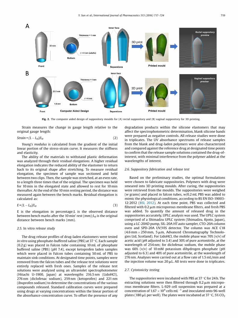
Fig. 2. The computer aided design of suppository moulds for (A) rectal suppository and (B) vaginal suppository for 3D printing.
Y. Sun et al. / International Journal of Pharmaceutics 513 (2016) 717–724 719
Strain measures the change in gauge length relative to theoriginal gauge length:
Strain = (L � L0)/L0 (2)
Young’s modulus is calculated from the gradient of the initiallinear portion of the stress-strain curve. It measures the stiffnessand elasticity.
The ability of the materials to withstand plastic deformationwas analyzed through their residual elongations. A higher residualelongation indicates the reduced ability of the elastomer to returnback to its original shape after stretching. To measure residualelongation, the specimen of sample was sectioned and heldbetween two clips. Then, the sample was stretched, at an even rate,to a length three times that of the original. The specimen was heldfor 10 min in the elongated state and allowed to rest for 10 minthereafter. At the end of the 10 min resting period, the distance wasmeasured again between the bench marks. Residual elongation iscalculated as:
E = (L � L0)/L0 (3)
E is the elongation in percentage;L is the observed distancebetween bench marks after the 10 min’ rest (mm);L0 is the originaldistance between bench marks (mm).
2.5. In vitro release study
The drug release profiles of drug-laden elastomers were testedin vitro using phosphate-buffered saline (PBS) at 37 �C. Each sample(0.2 g) was placed in Falcon tube containing 10 mL of phosphatebuffered saline (PBS) (pH 7.4), except ketoprofen laden sampleswhich were placed in Falcon tubes containing 50 mL of PBS tomaintain sink conditions. At designated time points, samples wereremoved from the falcon tubes and the release test solutions wereentirely replaced with fresh ones. Samples of the release testsolutions were analyzed using an ultraviolet spectrophotometer(Hitachi U-1900, Japan) at wavelengths 216.5 nm (LidoHCl),276 nm (diclofenac sodium), 259 nm (ketoprofen) and 221 nm(ibuprofen sodium) to determine the concentrations of the variouscompounds released. Standard calibration curves were preparedusing drugs at varying concentrations within the linear portion ofthe absorbance-concentration curve. To offset the presence of any
degradation products within the silicone elastomers that mayaffect the spectrophotometric determination, blank silicone bandswere prepared as negative controls. All release studies were donein triplicates. The UV absorbance spectrums of release samplesfrom the blank and drug-laden polymers were also characterizedand compared against the reference drug at designated time pointsto confirm that the release sample solutions contained the drug-of-interest, with minimal interference from the polymer added at thewavelengths of interest.
2.6. Suppository fabrication and release test
Based on the preliminary studies, the optimal formulationswere chosen to fabricate suppositories. Polymers with drug weresmeared into 3D printing moulds. After curing, the suppositorieswere retrieved from the moulds. The suppositories were weighed(w grams) and placed in falcon tubes, w/0.2 mL PBS was added tomimic the physiological conditions, according to BS EN ISO-19003-12:2012 (BSI, 2012). At each time point, PBS was collected andfiltered with 0.2 mm microporous membrane filters, and fresh PBSwas added. To quantify the amount of released drug in thesuppositories accurately, UPLC analysis was used. The UPLC systemcomprised of a Shimadzu UPLC system (Shimadzu, Kyoto, Japan),using a LC-20AD pump, SIL-20A HT auto sampler, CTO-20A columnoven and SPD-20A UV/VIS detector. The column was ACE C18(4.6 mm � 250 mm, 5 mm, Advanced Chromatography Technolo-gies Ltd, Scotland). For LidoHCl, the mobile phase was 70% (v/v) ofacetic acid (pH adjusted to 3.4) and 30% of pure acetonitrile, at thewavelength of 254 nm; for diclofenac sodium, the mobile phasewas 60% (v/v) of 10 mM potassium dihydrogen phosphate (pHadjusted to 6.3) and 40% of pure acetonitrile, at the wavelength of276 nm. Analyses were carried out at a flow rate of 1.5 mL/min andthe injection volume was 20 mL. All tests were done in triplicates.
2.7. Cytotoxicity testing
The suppositories were incubated with PBS at 37 �C for 24 h. Theextracting solutions were then filtered through 0.2 mm micropo-rous membrane filters. L-929 cell suspension was prepared at aconcentration of 1.67 � 104 cells mL�1 and inoculated onto 96-wellplates (180 mL per well). The plates were incubated at 37 �C, 5% CO2

720 Y. Sun et al. / International Journal of Pharmaceutics 513 (2016) 717–724
for 24 h. 20 mL of extract were pipetted into seeded wells, thesewells served as the experimental wells. 20 mL of sterile PBS wereadded onto remaining seeded wells, which served as the controls.Then, 20 mL of sterile PBS were pipetted in unseeded wells, whichserved as blank. The cells were incubated at the same conditionsstated above for up to 5 days. 22 mL of PrestoBlueTM ViabilityReagent (Thermo Fisher Scientific Inc., USA) was pipetted intotesting wells. The 96-well plates were then incubated for 30 min at37 �C, 5% CO2. Subsequently, the absorbance was analyzed withSpectraMax1 190 absorbance microplate reader (MolecularDevices, LLC., USA) at the wavelength of 570 nm with a referencewavelength of 600 nm.
2.8. Statistical analysis
All analyses were performed by using the Statistical Package forthe Social Sciences (SPSS) version 21.0 (IBM Corporation, USA) andOriginPro 2016 (OriginLab Corporation, USA). Both non parametricand parametric tests were used. A p-value of less than 0.05 wasconsidered significant.
3. Results
LidoHCl crystals existed as translucent drug crystals embeddedwithin the polydimethylsiloxane matrix (Fig. 3A). Diclofenacsodium, ibuprofen sodium and ketoprofen were present asspherical white particles uniformly dispersed within the polymer(Fig. 3B–D).
For Silastic, the stiffness and elasticity decreased whenincreasing drug loading of LidoHCl, while they remained compa-rable among the NSAIDs except ibuprofen sodium (Fig. 4A). Nosignificant difference in stiffness was observed in MED-4901elastomer at various drug loadings (Fig. 4A). Data from Ketoprofenladen MED-4901 polymers were excluded due to insufficientcuring. Compared to drug laden Silastic elastomers, MED-4901drug laden elastomers showed lower and more stable stiffness.
A reduction in the ability to withstand plastic deformation(reflected by the values of residual elongation) were observed atincreased LidoHCl laden Silastic elastomers (Fig. 4B). Similarly, theaddition of increasing concentrations of diclofenac sodium,ibuprofen sodium and ketoprofen also adversely affected themechanical properties Silastic elastomers in terms of the ability totolerate deformation (Fig. 4B). MED-4901 elastomers were betterable to resist plastic deformation compared to Silastic elastomersregardless of the concentration of LidoHCl, diclofenac sodium andibuprofen sodium added (p < 0.05) (Fig. 4B).
At 1%, 5% and 10% drug loading in Silastic elastomers,ketoprofen showed the fastest initial release, followed by LidoHCl,ibuprofen sodium and diclofenac sodium (Fig. 5A). LidoHClplateaued the earliest among the four drugs, within 19 h for both1% and 5% drug loading and 10 days for 10% drug loading in Silasticpolymers. During the 30-day drug release study, ketoprofen ladenSilastic polymers released the most drug, followed by ibuprofensodium, LidoHCl and lastly diclofenac sodium at 5% and 10% drugloading. Among MED-4901 polymers, LidoHCl showed the fastestinitial release at 1%, 5% and 10% drug loading, followed byibuprofen sodium and diclofenac sodium (Fig. 5B). LidoHCl releaseplateaued at 19 h for 1%, 2 days for 5% and 10 days for 10% drugloading. Between the three drugs tested, the cumulative release inMED-4901 elastomer was the highest for ibuprofen sodium,followed by diclofenac sodium and lastly LidoHCl at 1% and 5% drugloading.
MED-4901 loaded with 1%, 5% LidoHCl and diclofenac sodiumwere selected for suppository fabrication. At 1% and 5%, LidoHCland diclofenac sodium suppositories were loaded with 60 mg and300 mg of drugs, respectively. The initial release rate of LidoHCl
and diclofenac sodium showed no difference (p > 0.05). Both 1%LidoHCl and diclofenac sodium suppositories released about6.5 mg of loaded drugs, and 8.8 mg for 5% within 3 h. Thesuppositories containing 1% and 5% LidoHCl plateaued afterreleasing for 4 days, with the total amount of released LidoHClbeing 9.20 � 0.16 mg and 14.25 � 0.44 mg. For 1% diclofenacsodium suppository, cumulative release for up to 96% wasobserved in 30 days, which was 57.35 � 0.50 mg of diclofenacsodium; 5% diclofenac sodium suppository released96.18 � 1.30 mg of drug (Fig. 6).
In the cytotoxicity testing, exposure of L-929 cells to 5% LidoHClor diclofenac sodium suppositories extracts did not affect the cellsurvival rates in 5 days. No significant difference was foundbetween control group and experiment group (p > 0.05) (Fig. 7).
4. Discussion
Silicone polymers can be cured by two methods, namely,peroxide curing or platinum-catalyzed addition reactions. Bothsilicone polymers used in our study were cured by additionreactions. Polymers which utilized peroxide curing were notchosen since peroxide radicals may interact with the drug moietiesand degrade them during the curing process (de Buyl, 2001).Despite the inert nature of our chosen silicone polymers,incorporating drugs within the silicone matrix was challengingas the platinum catalyst may be ‘poisoned’ by compoundscontaining amine, tin, sulphur and carboxylic acid groups(Snorradottir et al., 2009). Ketoprofen laden MED-4901 elastomerswere unable to be completely cured in our study, possibly due tothe presence of such groups i.e., COOH, and this character may alsocause the insufficient curing of ketoprofen laden Silastic, whichfinally led to a high release of ketoprofen.
To incorporate the drugs into the elastomer, a possible solutionis to use its salt forms. Sodium salt was chosen in our study becauseit showed a higher degree of cure compared to the potassium saltin ibuprofen laden polymers (Snorradottir et al., 2009). Diclofenacsodium, despite holding a secondary amine group, this drug ladenpolymers were able to be cured, possibly one was due to thesodium salt composition, the other was because the formation ofintra-molecular hydrogen bonds between the carboxylate group ofdiclofenac sodium and the secondary amine, which prevented the–NH moiety from forming inter-molecular hydrogen bonds withthe siloxane backbone.
For Silastic elastomers, drug loading decreased the ability ofresisting plastic deformation. This may be associated with anincrease in the number of drug particles disrupting the orderedcross-links found within this polymer (Li et al., 2011). To this end,MED-4901 polymer was superior to Silastic polymer assuppository material, considering its ability to resist plasticdeformation regardless of the types of drugs and their concen-trations. At the same time, the pliable MED-4901 elastomers mayoffer a more comfortable fit for the patients as the suppositorybase.
From the release profiles of drug-laden elastomers, it seemsthat the release of drugs correlated with the permeability of thesilicone elastomer (Raul et al., 2006). Given the porous nature ofthese polymers and the molding method, drugs of smaller particlesize were able to diffuse out from the micro-channels present withless resistance (Golomb et al., 1990). We noted that ketoprofenelastomers released the most drug during the 30-day releasestudies, followed by ibuprofen sodium and diclofenac sodium. Thistrend correlated with the diameter of the drug particles, whichwere 6.67 mm, 10.00 mm and 13.33 mm for ketoprofen, ibuprofensodium and diclofenac sodium, respectively.
Interestingly, LidoHCl showed higher initial release thandiclofenac sodium despite having a much larger particle size of

Fig. 3. Cross section of cured drug laden silicone elastomers. Percentages indicate% loading of various drugs within the silicone polymers.
Y. Sun et al. / International Journal of Pharmaceutics 513 (2016) 717–724 721
150 mm. This could be due to the high solubility of LidoHCl in PBS(508.76 mg/ml) compared to diclofenac sodium (8.21 mg/ml).Given the propensity of LidoHCl to dissolve in PBS, the dissolution
of peripheral LidoHCl molecules created empty pores from whichPBS can enter, further solvating LidoHCl particles trapped deepwithin the convoluted micro-channels of the silicone polymer

Fig. 5. Cumulative release of 1%, 5% and 10% diclofenac sodium (Diclo), ibuprofen sodium (Ibu), ketoprofen (Keto) and LidoHCl from (A) Silastic and (B) MED-4901 elastomers.
Fig. 4. Comparison of mechanical properties between Silastic and MED-4901 polymers laden with different drugs at 1%, 5% and 10% drug loading (A) Young’s Modulus and (B)residual elongation.
722 Y. Sun et al. / International Journal of Pharmaceutics 513 (2016) 717–724
(Snorradottir et al., 2009). Once LidoHCl crystals were solvated,pore size no longer presents as a limiting factor to drug release.
Considering the drug-elastomer testing results, MED-4901were used for suppository fabrication, but not Silastic (Table 1). Interms of drug selection, LidoHCl, ibuprofen sodium and diclofenacsodium did not cause significant changes of the cured MED-4901elastomers. LidoHCl laden elastomers have fast onset of drugrelease, hence useful for immediate pain-relief. Clinically, theexcretion of diclofenac in human milk has been reported low,which is comparatively safer during breast-breeding for postpar-tum women with perineum pain (Rezaei et al., 2014). Moreover,due to the prolonged release profiles of diclofenac sodium from theelastomers, it can be useful for the treatment of patients who sufferfrom long term terminally ill cancer pain. It’s worth mentioningthat high concentrations of drugs (�10%) rendered viscous surfaces
of suppositories by moulding method. Therefore, only 1% and 5% ofLidoHCl and diclofenac sodium were used to fabricate thesuppositories for subsequent testing.
The initial drug release profiles of LidoHCl suppositories anddiclofenac suppositories were very similar. For diclofenac sodiumsuppositories, the cumulative release of drugs increased than thatin the preliminary testing. It could have been caused by thedifferent fabrication processes. In the preliminary testing, thepolymers were squeezed with syringes and likely formed densestructures. In contrast, suppositories were fabricated withoutsyringe extrusion, which may lead to larger pore sizes inside theelastomers, letting more diclofenac sodium diffusing out.
Compared with conventional suppositories, the ones wefabricated with bio-grade polymers will not melt at bodytemperature and have the ability of resisting plastic deformation

Fig. 7. Cytotoxicity testing of the suppository aqueous extracts.
Fig. 6. Drug release profiles from MED-4901 suppositories with 1%, 5% LidoHCl and diclofenac sodium (A) cumulative drug release amount (mg) and (B) release percentage.
Y. Sun et al. / International Journal of Pharmaceutics 513 (2016) 717–724 723
inside body cavities. These characters allow the suppositories tostay at site of administration to exert its therapeutic activity. Inaddition, it can be retrieved easily by pulling a thread affixed to thesuppositories to discontinue the medication (James Barron, 1977).
For current clinical practice, the suppositories of diclofenac areavailable in high dose and administered several times daily
Table 1Comparison of the drug laden elastomers in terms of mechanical properties and drug
Drugs aYoung’s modulus, MPa bResidua
Silastic MED Silastic
LidoHCl >0.5 <0.25 >5
Diclo >1.25 <0.25 >7
Ibu >1.75 <0.25 >7
Keto >0.75 – >15
a Young’s modulus represents the stiffness and elasticity.b Higher residual elongation indicates the reduced ability of elastomer to return bac
because of high protein binding (Setoguchi et al., 2013). The drugcontent has been reported to be 60 mg for lidocaine suppositories(Goluza et al., 2011) and 100 mg for diclofenac sodium supposito-ries (Lua et al., 2015). Azechi Y. et al. have reported two-fold longerhalf-life when controlled release suppository was administered(Azechi et al., 2000). Schneeweis, A. et al. have also reported theincrease in mean residence time by controlling the release rate ofthe diclofenac (Schneeweis and Muller-Goymann, 1997). There-fore, the use of suppository providing the prolonged release willimprove the therapeutic effect.
In this study, we tested 60 mg (1%) and 300 mg (5%) for bothdrugs, aimed at prolonged release to minimize the potential sideeffects (Irwin et al., 1995) and to reduce the dosing frequency. Forvaginal administration these suppositories may provide prolongedtherapeutic effects for up to a few weeks potentially.
Three dimensional printing, which emerges as a powerful toolin fabricating drug delivery systems recently, has been used as themethod to fabricate suppository moulds in our study (Bandyo-padhyay et al., 2015; Jonathan and Karim, 2016). The geometricalfeatures were designed according to the shapes and sizes of humanbody cavities. This can be useful especially for women sufferingfrom different degrees of vaginal relaxation syndrome or posteriorprolapse (exacerbated by childbirth, especially multiple pregnan-cies and deliveries, and the vaginal atrophy). In this condition,muscles ligaments and fascia that hold and support the vaginabecome stretched and weakened. Thus, the appearance and size ofthe vaginal opening can vary, demanding customized vaginalsuppositories (Goodman et al., 2010; Kegel, 1956; Lee, 2014). It hasbeen reported that women had their own preference over theshape, size and firmness of the vaginal suppositories (Li et al.,
release.
l elongation, % Releasing period, days
MED Silastic MED
<0.25 <10 <10<0.25 >30 �30<0.25 >30 �30– <20 –
k to its original shape.

724 Y. Sun et al. / International Journal of Pharmaceutics 513 (2016) 717–724
2013). Therefore, customized elastic vaginal suppositories made by3DP could meet the needs of female patients, for instance, to fit inthe cavities firmly to prevent migration and to offer acceptablesensory owing to the elastic nature of the materials.
Admittedly, 3DP is not confined to build the moulds ofsuppository. The pressure-assisted micro-syringes printing, oneof 3DP methods, is a promising way to create complex drugdelivery systems, potentially useful to fabricate elastic suppositorydirectly (Khaled et al., 2014). This technology is based on extrudinga viscous semi-liquid material from syringe to achieve a desirable3D shape (Chia and Wu, 2015). However, extensive studies areneeded to investigate the rheological properties of inks, thematerial stability and the drying process associated with thisapproach (Aho et al., 2015; Goyanes et al., 2015; Yu et al., 2007).
5. Conclusion
The mechanical properties of the drug laden silicone elastomersand the rate of drug release from the elastomers are tunable. MED-4901 is a promising base material to make non-dissolvablesuppository for sustained drug release. In addition, 3DP ispotentially useful in making personalized suppository moulds tomeet the requirements and preferences of physicians and patients.
Acknowledgement
The work is supported by a Singapore MOE AcRF Tier 1 grant (R-148-000-207-112). The authors thank the China ScholarshipCouncil to support Sun Y.
References
Aho, J., Boetker, J.P., Baldursdottir, S., Rantanen, J., 2015. Rheology as a tool forevaluation of melt processability of innovative dosage forms. Int. J. Pharm. 494,623–642.
Ali Ebrahim, R.M., Nadia, Bani-Hashem, Ali, Jabbari, 2014. Early post-operative reliefof pain and shivering using diclofenac suppository versus intravenous pethidinein spinal anesthesia. J. Anaesthesiol. Clin. Pharmacol. 30, 6.
Andrews, V., Thakar, R., Sultan, A.H., Jones, P.W., 2008. Evaluation of postpartumperineal pain and dyspareunia – a prospective study. Eur. J. Obstet. Gynecol.Reprod. Biol. 137, 152–156.
Azechi, Y., Ishikawa, K., Mizuno, N., Takahashi, K., 2000. Sustained release ofdiclofenac from polymer-containing suppository and the mechanism involved.Drug Dev. Ind. Pharm. 26, 1177–1183.
BSI, 2012. BS EN ISO 10993-12:2012, Biological Evaluation of Medical Devices Part 1:Sample Preparation and Reference Materials. The British Standards Institution,pp. 2.
Bandyopadhyay, A., Bose, S., Das, S., 2015. 3D printing of biomaterials. MRS Bull. 40,108–115.
Baum, M.M., Butkyavichene, I., Gilman, J., Kennedy, S., Kopin, E., Malone, A.M.,Nguyen, C., Smith, T.J., Friend, D.R., Clark, M.R., Moss, J.A., 2012. An intravaginalring for the simultaneous delivery of multiple drugs. J. Pharm. Sci. 101, 2833–2843.
Chia, H.N., Wu, B.M., 2015. Recent advances in 3D printing of biomaterials. J. Biol.Eng. 9, 4.
Davis, M.P., Walsh, D., LeGrand, S.B., Naughton, M., 2002. Symptom control in cancerpatients: the clinical pharmacology and therapeutic role of suppositories andrectal suspensions. Support. Care Cancer 10, 117–138.
de Buyl, F., 2001. Silicone sealants and structural adhesives. Int. J. Adhes. Adhes. 21,411–422.
Dodd, J.M., Hedayati, H., Pearce, E., Hotham, N., Crowther, C.A., 2004. Rectalanalgesia for the relief of perineal pain after childbirth: a randomised controlledtrial of diclofenac suppositories. BJOG 111, 1059–1064.
Fu, Y., Kao, W.J., 2010. Drug release kinetics and transport mechanisms of non-degradable and degradable polymeric delivery systems. Expert Opin. DrugDeliv. 7, 429–444.
Golomb, G., Fisher, P., Rahamim, E., 1990. The relationship between drug releaserate, particle size and swelling of silicone matrices. J. Control. Release 12, 121–132.
Goluza, E., Hudolin, T., Kastelan, Z., Peric, M., Murselovic, T., Sosic, H., 2011. Lidocainesuppository for transrectal ultrasound-guided biopsy of the prostate: aprospective, double-blind, randomized study. Urol. Int. 86, 315–319.
Goodman, M.P., Placik, O.J., Benson 3rd, R.H., Miklos, J.R., Moore, R.D., Jason, R.A.,Matlock, D.L., Simopoulos, A.F., Stern, B.H., Stanton, R.A., Kolb, S.E., Gonzalez, F.,2010. A large multicenter outcome study of female genital plastic surgery. J. Sex.Med. 7, 1565–1577.
Goyanes, A., Robles Martinez, P., Buanz, A., Basit, A.W., Gaisford, S., 2015. Effect ofgeometry on drug release from 3D printed tablets. Int. J. Pharm. 494, 657–663.
Irwin, M.G., Roulson, C.J., Jones, R.D., Cheng, I.K., Visram, A.R., Chan, Y.M., 1995. Peri-operative administration of rectal diclofenac sodium: the effect on renalfunction in patients undergoing minor orthopaedic surgery. Eur. J. Anaesthesiol.12, 403–406.
James Barron, J.R.B., 1977. Anorectal pain relief by use of local silastic suppositories.Dis. Colon Rectum 20, 2.
Jonathan, G., Karim, A., 2016. 3D printing in pharmaceutics: a new tool for designingcustomized drug delivery systems. Int. J. Pharm. 499, 376–394.
Kegel, A.H., 1956. Early genital relaxation: new technic of diagnosis and nonsurgicaltreatment. Obstet. Gynecol. 8, 6.
Khaled, S.A., Burley, J.C., Alexander, M.R., Roberts, C.J., 2014. Desktop 3D printing ofcontrolled release pharmaceutical bilayer tablets. Int. J. Pharm. 461, 105–111.
Kirchhoff, P., Clavien, P.A., Hahnloser, D., 2010. Complications in colorectal surgery:risk factors and preventive strategies. Patient Saf. Surg. 4, 5.
Lee, M.S., 2014. Treatment of vaginal relaxation syndrome with an erbium: YAGlaser using 90� and 360� scanning scopes: a pilot study & short-term results.Laser Ther. 23, 8.
Leon Shargel, I.K., 2010. Generic Drug. Product Development: Specialty DosageForms. 26.
Li, H., Jiang, B., Yan, J., Yang, Z., Chen, Y., Zhang, W., Choy, A., Lee, C.-Y., Kang, L., 2011. Adrug-laden elastomer for surgical treatment of anal fistula. Drug Deliv. Transl.Res. 1, 439–447.
Li, B., Zaveri, T., Ziegler, G.R., Hayes, J.E., 2013. Shape of vaginal suppositories affectswillingness-to-try and preference. Antiviral Res. 97, 280–284.
Lowenstein, L., Granot, M., Tamir, A., Glik, A., Deutsch, M., Jakobi, P., Zimmer, E.Z.,2006. Efficacy of suppository analgesia in postabortion pain reduction.Contraception 74, 345–348.
Lua, G.W., Muthukaruppan, R., Menon, J., 2015. Can rectal diclofenac prevent postendoscopic retrograde cholangiopancreatography pancreatitis? Dig. Dis. Sci. 60,3118–3123.
Macarthur, A.J., Macarthur, C., 2004. Incidence, severity, and determinants ofperineal pain after vaginal delivery: a prospective cohort study. Am. J. Obstet.Gynecol. 191, 1199–1204.
Peppas, N.A., Bures, P., Leobandung, W., Ichikawa, H., 2000. Hydrogels inpharmaceutical formulation. Eur. J. Pharm. Biopharm. 50, 20.
Petrowsky, H., Demartines, N., Rousson, V., Clavien, P.-A., 2004. Evidence-basedvalue of prophylactic drainage in gastrointestinal surgery. Ann. Surg. 240, 1074–1085.
Raul, V.A., Schalau, G.K., Starch, M., 2006. Controlled release of active ingredientsfrom cross-linked silicones. Cosmet. Toiletries 121, 69–72.
Rezaei, Z., Haghighi, Z., Haeri, G., Hekmatdoust, A., 2014. A comparative study onrelieving post-episiotomy pain with diclofenac and indomethacin suppositoriesor placebo. J. Obstet. Gynaecol. 34, 293–296.
�Ríhová, B., 1996. Biocompatibility of biomaterials: hemocompatibility,immunocompatiblity and biocompatibility of solid polymeric materials andsoluble targetable polymeric carriers. Adv. Drug Deliv. Rev. 21, 157–176.
Schneeweis, A., Muller-Goymann, C.C., 1997. In vivo and in vitro diclofenac sodiumevaluation after rectal application of soft gelatine capsules enabling applicationinduced transformation (AIT) into a semisolid system of liquid crystals (SSLC)for controlled release. Pharm. Res. 14, 1726–1729.
Setoguchi, N., Takamura, N., Fujita, K., Ogata, K., Tokunaga, J., Nishio, T., Chosa, E.,Arimori, K., Kawai, K., Yamamoto, R., 2013. A diclofenac suppository-nabumetone combination therapy for arthritic pain relief and a monitoringmethod for the diclofenac binding capacity of HSA site II in rheumatoid arthritis.Biopharm. Drug Dispos. 34, 125–136.
Snorradottir, B.S., Gudnason, P.I., Scheving, R., Thorsteinsson, F., Masson, M., 2009.Release of anti-inflammatory drugs from a silicone elastomer matrix system.Pharmazie 64, 19–25.
Souza, J., Meira, A., Volpato, N.M., Mayorga, P., Gottfried, C., 2013. Effect ofphonophoresis on skin permeation of commercial anti-inflammatory gels:sodium diclofenac and ketoprofen. Ultrasound Med. Biol. 39, 1623–1630.
Wilasrusmee, S., Chittachareon, A., Jirasiritum, S., Srisangchai, P., 2008. Naproxensuppository for perineal pain after vaginal delivery. Int. J. Gynaecol. Obstet. 102,19–22.
Yarnykh, T.G., Tolochko, E.V., Chushenko, V.N., 2011. Drug synthesis methods andmanufacturing technology: studing an assortment of suppository bases(review). Pharm. Chem. J. 44, 6.
Yoong, W.C., Biervliet, F., Nagrani, R., 1997. The prophylactic use of diclofenac(Voltarol) suppositories in perineal pain after episiotomy: a random allocationdouble-blind study. J. Obstet. Gynaecol. 17, 3.
Yu, D.G., Yang, X.L., Huang, W.D., Liu, J., Wang, Y.G., Xu, H., 2007. Tablets withmaterial gradients fabricated by three-dimensional printing. J. Pharm. Sci. 96,2446–2456.