INTERNATIONAL JOURNAL OF PHARMACEUTICAL ... 757.pdfpolymers like Poloxamer 188 and Poloxamer 407...
19
Research Article CODEN: IJPRNK IMPACT FACTOR: 4.278 ISSN: 2277-8713 Mansi Shah, IJPRBS, 2014; Volume 3(6): 1-19 IJPRBS Available Online at www.ijprbs.com 1 FORMULATION AND EVALUATION OF IN-SITU SOL-GEL OCULAR DRUG DELIVERY SYSTEM USING MOXIFLOXACIN HCL MANSI SHAH, SANKET SHAH Pioneer Pharmacy Degree College, Sayajipura, Ajwa-Nimeta Road, Vadodara. Accepted Date: 28/06/2014; Published Date: 27/12/2014 Abstract: Ophthalmic drug delivery is extremely interesting and highly challenging endeavors. In recent scenario, most eye-diseases are treated with topical application of eye-drops which have two major problems. 1) It needs frequent administration at every 4 hours or 1 hour if the infection is severe and 2) Formation of crystalline deposits on cornea due to its pH-dependent solubility which is very low. In order to provide the solution to above problems, the present work has been done to formulate and evaluate the ophthalmic drug delivery system using Moxifloxacin-HCl, the 4 th generation flouroquinolones, the antibacterial agent, containing polymers like Poloxamer 188 and Poloxamer 407 which exhibit temp-triggered reversible in-situ sol to gel phase transition in combination with different grades of HPMC (Methocel E50 LV) as viscosifying agent. Different formulations were made and evaluated regarding their clarity, pH, drug content, viscosity, gelling capacity, rheological characteristics, sterility testing, in-vitro drug release behavior and antimicrobial study. The developed formulation provided better drug product effectiveness, reliability, stability, safety, non-irritancy and prolonged release. The formulated gels were transparent, uniform in consistency & had spreadability in the pH range of 6.8 to 7.4. Thus, it’s a viable alternative to conventional eye-drops by virtue of its ability to enhance bioavailability through its longer pre-corneal residence time & ability to provide prolonged drug release up to 8 hours. The main important factor is the reduced frequency & the ease of installation resulting in better patient acceptance. Keywords: Temp-triggered system, ocular in-situ sol-gel phase transition, prolonged release, Moxifloxacin-HCl, HPMC, Poloxamer 188 and 407. INTERNATIONAL JOURNAL OF PHARMACEUTICAL RESEARCH AND BIO-SCIENCE PAPER-QR CODE Corresponding Author: MS. MANSI SHAH Access Online On: www.ijprbs.com How to Cite This Article: Mansi Shah, IJPRBS, 2014; Volume 3(6): 1-19
Transcript of INTERNATIONAL JOURNAL OF PHARMACEUTICAL ... 757.pdfpolymers like Poloxamer 188 and Poloxamer 407...

Research Article CODEN: IJPRNK IMPACT FACTOR: 4.278 ISSN: 2277-8713 Mansi Shah, IJPRBS, 2014; Volume 3(6): 1-19 IJPRBS
Available Online at www.ijprbs.com 1
FORMULATION AND EVALUATION OF IN-SITU SOL-GEL OCULAR DRUG
DELIVERY SYSTEM USING MOXIFLOXACIN HCL
MANSI SHAH, SANKET SHAH
Pioneer Pharmacy Degree College, Sayajipura, Ajwa-Nimeta Road, Vadodara.
Accepted Date: 28/06/2014; Published Date: 27/12/2014
Abstract: Ophthalmic drug delivery is extremely interesting and highly challenging endeavors. In recent
scenario, most eye-diseases are treated with topical application of eye-drops which have two major problems. 1)
It needs frequent administration at every 4 hours or 1 hour if the infection is severe and 2) Formation of
crystalline deposits on cornea due to its pH-dependent solubility which is very low. In order to provide the
solution to above problems, the present work has been done to formulate and evaluate the ophthalmic drug
delivery system using Moxifloxacin-HCl, the 4th
generation flouroquinolones, the antibacterial agent, containing
polymers like Poloxamer 188 and Poloxamer 407 which exhibit temp-triggered reversible in-situ sol to gel phase
transition in combination with different grades of HPMC (Methocel E50 LV) as viscosifying agent. Different
formulations were made and evaluated regarding their clarity, pH, drug content, viscosity, gelling capacity,
rheological characteristics, sterility testing, in-vitro drug release behavior and antimicrobial study. The developed
formulation provided better drug product effectiveness, reliability, stability, safety, non-irritancy and prolonged
release. The formulated gels were transparent, uniform in consistency & had spreadability in the pH range of 6.8
to 7.4. Thus, it’s a viable alternative to conventional eye-drops by virtue of its ability to enhance bioavailability
through its longer pre-corneal residence time & ability to provide prolonged drug release up to 8 hours. The main
important factor is the reduced frequency & the ease of installation resulting in better patient acceptance.
Keywords: Temp-triggered system, ocular in-situ sol-gel phase transition, prolonged release, Moxifloxacin-HCl,
HPMC, Poloxamer 188 and 407.
INTERNATIONAL JOURNAL OF
PHARMACEUTICAL RESEARCH AND BIO-SCIENCE
PAPER-QR CODE
Corresponding Author: MS. MANSI SHAH
Access Online On:
www.ijprbs.com
How to Cite This Article:
Mansi Shah, IJPRBS, 2014; Volume 3(6): 1-19

Research Article CODEN: IJPRNK IMPACT FACTOR: 4.278 ISSN: 2277-8713 Mansi Shah, IJPRBS, 2014; Volume 3(6): 1-19 IJPRBS
Available Online at www.ijprbs.com 2
INTRODUCTION
Eye drops that are conventional ophthalmic delivery systems often result in poor
bioavailability and therapeutic response because high tear fluid turnover and dynamics
cause rapid precorneal elimination of the drug. A high frequency of eye drop instillation
is associated with patient non-compliance. Inclusion of excess drug in the formulation in
an attempt to overcome bioavailability problem is potentially dangerous if the drug
solution drained from the eye is systemically absorbed from the nasolacrimal duct.[1,2]
Various ophthalmic vehicles such as inserts, ointments, Suspensions, and aqueous gels,
have been developed in order to lengthen the residence time of instilled dose and
enhance the ophthalmic bioavailability.[3,4] These ocular drug delivery systems, however,
have not been used extensively because of some drawbacks such as blurred vision from
ointments or low patient compliance from inserts.[5,6] Several insitu gel forming system
have been developed to prolong the precorneal residence time of a drug and improve
ocular bioavailability. These systems consist of polymers that exhibit sol- gel phase
tansititions due to change in specific physico chemical parameter (pH, temperature), in
their environment, the cul-de-sac in this case.[4,6] Depending on the method employed to
cause sol-to-gel phase transition on the eye surface, the following three types of
systems are recognized. pH triggered system, temperature dependant system[7] and ion
activated system.[8] Using these three methods above in-situ gelling ophthalmic delivery
system is developed. However most of the systems require the use of high
concentration of polymers. For instance, it needs 25% (w/v) to 30% (w/v) of Poloxamer
to from stiff gel upon instillation in the eye. As the concentration of Poloxamer
increases in the vehicle, its acidic nature may cause stimulation to the eye tissue.[9,10,11]
The present study aim was to develop the in-situ gelling ophthalmic delivery system of
Moxifloxacin hydrochloride, a fourth-generation fluoroquinolone derivative used in
external infections of eye such as acute and sub acute conjunctivitis, bacterial keratitis
and keratoconjunctivitis.[12,13,1]
MATERIALS AND METHODS
Materials
Moxifloxacin hydrochloride was obtained from TRC, Ahmedabad, India. HPMC E50 was
obtained from Colorcon Asia Pvt. Ltd; Goa and Poloxamer 188 & Poloxamer 407 were
obtained from BASF, Mumbai. Calcium chloride, Hydrochloric acid, Benzalkonium chloride,
Methanol, PEG 400, Potassium dihydrogen phosphate, Sodium bicarbonate, Sodium
chloride and Sodium hydroxide were purchased from Merck, Mumbai.

Research Article CODEN: IJPRNK IMPACT FACTOR: 4.278 ISSN: 2277-8713 Mansi Shah, IJPRBS, 2014; Volume 3(6): 1-19 IJPRBS
Available Online at www.ijprbs.com 3
METHODS
1. Preformulation studies
A. Determination of melting point
Melting point of Moxifloxacin Hydrochloride was determined by Melting point apparatus.
B. Solubility
Solubility is an important consideration in ophthalmic formulations as clarity of the
solution is an essential requirement. The solubility of Moxifloxacin Hydrochloride was
tested in various solvents such as distilled water, ethyl alcohol, 2-propanol and acetone.
C. IR Spectroscopy
The FT-IR spectrum of the obtained sample of the drug was compared with the
standard FT-IR spectra of the pure drug.
2. Compatibility studies
FT-IR spectroscopy was carried out to check the compatibility between drug and
polymer. The FT-IR spectra of drug with polymers were compared with the standard FT-
IR spectrum of the pure drug.
3. Selection of the Vehicle
The solubility of Moxifloxacin Hydrochloride was tested in various buffers such as acetate
buffer I.P. (pH 6.0 & 6.5), citrophosphate buffer B.P. (pH 6.0 and 6.2) and phosphate
buffer USP (pH 7.2 and 7.4) in order to select a suitable vehicle. Solutions of
moxifloxacin in the above buffers were prepared to test its solubility at the dosage level
desired (0.5%, w/v).
4. Standard calibration curve of Moxifloxacin
Accurately weighed 10 mg Moxifloxacin Hydrochloride was dissolved in 100ml of
Phosphate buffer pH 7.4 to get the stock solution of 100µg/ml. From this stock solution
aliquots of 0.2, 0.4, 0.6, 0.8, 1.0, 1.2 & 1.4ml were withdrawn and further diluted to 10
ml with buffer to obtain a concentrations range of 2 to 14 µg/ml. The absorbance of
the solutions was measured at 288 nm by using UV-Vis spectrophotometer. A graph of
Concentration vs. Absorbance was plotted.
5. Optimized method for temperature-triggered in situ hydrogel preparation [14]
Poloxamer in situ forming gels were prepared by the modified cold method. Briefly, both
the types of Poloxamer (P188) and (P407) were slowly added to the calculated amount
of cold purified water with continuous mixing using a thermostatically controlled
magnetic stirrer. The partially dissolved poloxamer solutions were stored in a refrigerator

Research Article CODEN: IJPRNK IMPACT FACTOR: 4.278 ISSN: 2277-8713 Mansi Shah, IJPRBS, 2014; Volume 3(6): 1-19 IJPRBS
Available Online at www.ijprbs.com 4
and stirred periodically until clear homogenous solutions were obtained (approximately
24 h). Then, the required amounts of HPMC E50 LV (0.5, 1 and 1.5%) were slowly
added and stirred continuously until dissolved completely. The ophthalmic formulations
were prepared by dissolving the appropriate amount of moxifloxacin-HCl (0.5% w/v), in
the calculated amount of 0.1 N NaOH separately. Then stabilizers i.e. Tween 20 and PEG
400 were added in polymer solution and then both the solutions were mixed with
continuous stirring until homogenous solution is obtained.
6. Experimental design
The experimental design to formulate the ocular in-situ sol-gel controlled release gel
having moxifloxacin HCl is given below. The design was employed for formulations
containing the combination of two different gelling agents Poloxamer 188 and Poloxamer
407 having a viscofying agent HPMC E50 LV. (Table 1)
EVALUATION PARAMETERS
Appearance
pH
In vitro gelation studies
Rheological studies
Drug content
In vitro drug release study
Sr.
No.
Ingredients Quantity of the ingredients (%W/V)
MF1 MF2 MF3 MF4 MF5 MF6 MF7 MF8 MF9
1 Moxifloxacin HCl 0.5 0.5 0.5 0.5 0.5 0.5 0.5 0.5 0.5
2 Poloxamer 188 12 12 12 15 15 15 18 18 18
3 Poloxamer 407 15 15 15 15 15 15 15 15 15
4 HPMC E50LV 0.5 1 1.5 0.5 1 1.5 0.5 1 1.5
5 Tween 20 0.2 0.2 0.2 0.2 0.2 0.2 0.2 0.2 0.2
6 PEG 400 0.1 0.1 0.1 0.1 0.1 0.1 0.1 0.1 0.1
7 BKCl QS QS QS QS QS QS QS QS QS
8 HCl QS QS QS QS QS QS QS QS QS
9 NaOH QS QS QS QS QS QS QS QS QS

Research Article CODEN: IJPRNK IMPACT FACTOR: 4.278 ISSN: 2277-8713 Mansi Shah, IJPRBS, 2014; Volume 3(6): 1-19 IJPRBS
Available Online at www.ijprbs.com 5
Sterility (Antimicrobial testing)
Stability study
Isotonicity study
1. Appearance
Clarity is one of the most important characteristic features of ophthalmic preparations.
All developed formulations were evaluated for clarity by visual observation against a
black and white background.
2. pH
The pH of ophthalmic formulation should be such that the formulation will be stable at
that pH and at the same time there would be no irritation to the patient upon
administration of the formulation. Ophthalmic formulations should have pH range in
between 5 to 7.4. The developed formulations were evaluated for pH by using Elico
India Systronics digital pH meter.
3. In-vitro gelation studies
The gelling capacity was determined by placing a drop of the system in a vial containing
2 ml of artificial tear fluid freshly prepared and equilibrated at 37ºC and visually
assessing the gel formation and noting the time for gelation and the time taken for the
gel formed to dissolve. The composition of artificial tear fluid used was sodium chloride
0.670 g, sodium bicarbonate 0.200g, calcium chloride 2H2O 0.008 g, purified water Q.S.
100.0 g.
4. Rheological studies
Viscosity of instilled formulation is an important factor in determining residence time of
drug in the eye. The developed formulations were poured into the small sample adaptor
of the Brookfield Synchrolectric viscometer and the angular velocity increased gradually
from 0.5 to 50 rpm. The hierarchy of the angular velocity was reversed. The average of the
two readings was used to calculate the viscosity.
5. Drug content
Uniform distribution of active ingredient is important to achieve dose uniformity. The
drug content was determined by diluting 0.5 ml of the formulation to 25 ml with
phosphate buffer solution pH 7.4. Aliquot of 2.5 ml was withdrawn and further diluted
to 25 ml with PBS. Moxifloxacin hydrochloride concentration was then determined at
288 nm by using UV-Vis spectrophotometer.

Research Article CODEN: IJPRNK IMPACT FACTOR: 4.278 ISSN: 2277-8713 Mansi Shah, IJPRBS, 2014; Volume 3(6): 1-19 IJPRBS
Available Online at www.ijprbs.com 6
6. In vitro drug release studies
The in vitro release of Moxifloxacin Hydrochloride from the formulations was studied
through cellophane membrane using a modified USP XXIII dissolution testing apparatus.
The dissolution medium used was artificial tear fluid freshly prepared (pH 7.4).
Cellophane membrane, previously soaked overnight in the dissolution medium, was tied
to one end of a specifically designed glass cylinder (open at both ends and of 5 cm
diameter). A 1-ml volume of the formulation was accurately pipette into this assembly.
The cylinder was attached to the metallic driveshaft and suspended in 50 ml of
dissolution medium maintained at 37± 1°C so that the membrane just touched the
receptor medium surface. The dissolution medium was stirred at 50 rpm using magnetic
stirrer. Aliquots, each of 1-ml volume, were withdrawn at hourly intervals and replaced
by an equal volume of the receptor medium. The aliquots were diluted with the
receptor medium and analyzed by UV- Vis spectrophotometer at 288 nm.
7. Sterility (Antimicrobial testing)
All ophthalmic preparations should be sterile therefore the test for sterility is very
important evaluation parameter. The sterility test was performed according to Indian
Pharmacopoeia. Direct inoculation method was used. 2 ml of liquid from test container was
removed with a sterile pipette or with a sterile syringe or a needle. The test liquid was
aseptically transferred to fluid thioglycolate medium (20 ml) and soyabean-casein digest
medium (20 ml) separately. The liquid was mixed with the media. The inoculated media were
incubated for not less than 14 days at 30°C to 35°C in the case of fluid thioglycolate medium
and 20°C to 25°C in the case of soyabean-casein digest medium and compared with marketed
formulation.
8. Stability study
Stability is defined as the extent to which a product retains, within specified limits and
throughout its period of storage and use (i.e. its shelf life), the same properties and
characteristics that it possessed at the time of its manufacture. Stability testing is
performed to ensure that drug products retain their fitness for use until the end of
their expiration dates. All the formulations are sealed with aluminium foil and were
subjected to stability studies at ambient humidity conditions at 2°C to 8°C and 40±1°C
for a period of one month. The samples were withdrawn after 7, 15 and 30 days and
were evaluated for following parameters.
1. Clarity
2. pH
3. Gelling capacity

Research Article CODEN: IJPRNK IMPACT FACTOR: 4.278 ISSN: 2277-8713 Mansi Shah, IJPRBS, 2014; Volume 3(6): 1-19 IJPRBS
Available Online at www.ijprbs.com 7
4. Drug content
5. In vitro drug release
9. Isotonicity evaluation
Isotonicity is the most important characteristic of the ophthalmic formulation. Isotonicity
has to be maintained to prevent the tissue damage or the irritation of the eye.
Formulations were mixed with few drops of blood and observed under microscope at
45X magnification and compared with standard marketed ophthalmic formulation
containing Moxifloxacin.
RESULT AND DISCUSSION
1. Preformulation studies
I. Identification of the API
A. Determination of melting point
Melting point of Moxifloxacin Hydrochloride was found to be in the range of 254- 259ºC
in triplicate as reported in literature, thus indicating purity of the drug sample.
B. Solubility
Moxifloxacin was found to be freely soluble in water and in ethyl alcohol. It was also soluble in
2 propanol and acetone.
C. IR Spectroscopy (compatibility study)
The IR spectrum of the pure Moxifloxacin Hydrochloride sample recorded by FTIR
spectrometer is shown in Figure 1 which was compared with standard functional group
frequencies of Moxifloxacin as shown in Table 1 which indicates that the obtained
sample of moxifloxacin was pure.
Table 1- Reported and observed IR frequencies of Moxifloxacin Hydrochloride
Functional group Reported frequencies (cm-1) Observed frequencies (cm-1)
F 1400-1000 1045
C=O 1725-1680 1708
N-H 3500-3310 3472
-OH 3550-3450 3531

Research Article CODEN: IJPRNK IMPACT FACTOR: 4.278 ISSN: 2277-8713 Mansi Shah, IJPRBS, 2014; Volume 3(6): 1-19 IJPRBS
Available Online at www.ijprbs.com 8
Figure 1- Infrared spectrum of Moxifloxacin HCl
2. Compatibility studies
Preformulation studies were carried out to study the compatibility of pure drug
Moxifloxacin with the polymers HPMC E50 LV, Poloxamer 188 and Poloxamer 407 prior
to the preparation of ophthalmic in situ hydrogel. The individual IR spectra of the pure
drug and polymers as well as the combination spectra of the drug and polymer are
shown in the Figure 2-7, which indicate no interaction between moxifloxacin and
polymers when compared with infrared spectrum of pure drug as all functional group
frequencies were present.
Figure 2- Infrared spectrum of HPMC E50 LV Figure 3- Infrared spectrum of Poloxamer 188

Research Article CODEN: IJPRNK IMPACT FACTOR: 4.278 ISSN: 2277-8713 Mansi Shah, IJPRBS, 2014; Volume 3(6): 1-19 IJPRBS
Available Online at www.ijprbs.com 9
Figure 4- Infrared spectrum of Poloxamer 407 Figure 5- IR spectrum of Mox. HCl and HPMC E50 LV
Figure 6- IR spectrum of Mox. HCl and Poloxamer 188 Figure 7- IR spectrum of Mox. HCl and Poloxamer 407
3. Selection of Vehicle
Buffers play a pivotal role in formulating ophthalmic drops. Hence the importance of selecting
a suitable buffer ensures product stability and desired drug solubility. The studies in
various buffer solutions indicated the drug was soluble in acetate buffers of pH 6 & 6.5,
citrophosphate buffers of pH 6.0 & 6.2 and phosphate buffers of pH 7.2 & 7.4 at the
dosage level desired (0.5%, w/v).
4. Standard calibration curve of Moxifloxacin HCl
Table 2 shows the absorbance of Moxifloxacin standard solutions containing 2-14 µg/ml
of drug in phosphate buffer pH 7.4. Figure 8 shows a representative standard calibration
curve with slope, regression coefficient, and intercept (Table 3). The curve was found to
be linear in the range of 2-14 µg/ml at λmax 288 nm.

Research Article CODEN: IJPRNK IMPACT FACTOR: 4.278 ISSN: 2277-8713 Mansi Shah, IJPRBS, 2014; Volume 3(6): 1-19 IJPRBS
Available Online at www.ijprbs.com 10
Table 2- Standard calibration table for Moxifloxacin Hydrochloride in pH 7.4 PBS
Conc. of drug µg/ml Absorbance
0 0.000
2 0.1852
4 0.3532
6 0.5216
8 0.7043
10 0.8543
12 1.0199
14 1.1823
Figure 8- Calibration curve of Moxifloxacin HCl
Table 3- Statistical values of Mox. HCl
5. Optimized method for in situ hydrogel preparation
Poloxamer is a temperature induced gelling agent. At room temperature, it is in the solution
form but transferred to stiff gel when touched to eye cavity. Purified water was used as
a vehicle & HPMC as a viscosity induced polymer in the poloxamer in situ hydrogel.
y = 0.0841x + 0.0136 R² = 0.9995
0
0.5
1
1.5
0 5 10 15
Ab
sorb
ance
Conc. in ppm
Calibration curve of Moxifloxacin HCl
Formula Value
Slope 11.8786
Regression coefficient 0.9995
Intercept 0.2774

Research Article CODEN: IJPRNK IMPACT FACTOR: 4.278 ISSN: 2277-8713 Mansi Shah, IJPRBS, 2014; Volume 3(6): 1-19 IJPRBS
Available Online at www.ijprbs.com 11
6. Experimental design
Here all the factors were studied at all the possible combinations, as it is considered to
be most efficient in estimating the influence of individual variables using minimum
experimentation.
7. Evaluation parameters
7.1. Appearance
Clarity of all the formulations was found to be satisfactory as given in table 4. Terminal
sterilization by autoclaving had no effect on the clarity and other physicochemical
properties of the formulations. The haziness that was observed after autoclaving (due to
precipitation of HPMC at elevated temperature) was found to disappear and the original
clarity was regained after overnight standing.
Table 4- Appearance of the formulations
Formulation code Clarity Colour
MF 1 Clear Light yellow
MF 2 Clear Light yellow
MF 3 Clear Light yellow
MF 4 Clear Light yellow
MF 5 Clear Light yellow
MF 6 Clear Light yellow
MF 7 Clear Light yellow
MF 8 Clear Light yellow
MF 9 Clear Light yellow
7.2. pH
The pH of the formulations was found to be satisfactory and was in the range of 6-7.4.
(Table 5). Terminal sterilization by autoclaving had no effect on the pH.
Table 5- pH of the formulations
Formulation code pH
MF 1 6.92
MF 2 7.11
MF 3 6.85
MF 4 7.38
MF 5 7.23

Research Article CODEN: IJPRNK IMPACT FACTOR: 4.278 ISSN: 2277-8713 Mansi Shah, IJPRBS, 2014; Volume 3(6): 1-19 IJPRBS
Available Online at www.ijprbs.com 12
MF 6 6.96
MF 7 7.6
MF 8 7.52
MF 9 7.18
7.3. In vitro gelation studies
It is the main prerequisite of an in situ gelling system i.e. Speed and extent of gelation.
The formulation should have an optimum viscosity that will allow easy instillation into
the eye as a liquid (drops), which would undergo a rapid sol-to-gel transition. Table 6
shows the gelling capacity of all formulations and is depicted as + (gels after few
minutes i.e. 20 minutes and dissolves rapidly), ++ (gelation immediate i.e. within 5 minutes,
remains for few hours) and +++ (gelation immediate i.e. Within 2 minutes, remains for
extended period). Here gelling capacity gets affected at higher concentration of viscofying
agent (0.5 %w/v).
Table 6- Gelling capacity of the formulations
All the formulations except MF 2, MF 3 and MF 6 showed instantaneous gelation when
contacted with simulated tear fluid (STF). However, the nature of the gel formed
depended on the concentration of polymers used. The formation of instantaneous gels
can be attributed to the buffering capacity of the simulated tear fluid. Formulation MF
2, MF 3 and MF 6 showed the formation of gel after a few minutes which dissolved
rapidly. Formulation MF 1, MF 7, and MF 8 showed immediate gelation and remained for
few hours, where as the formulation MF 4, MF 5 and MF 9 showed immediate gelation
and remained for extended period.
Formulation code Gelling capacity Viscosity
MF 1 ++ 1342.7
MF 2 + 908.1
MF 3 + 719.4
MF 4 +++ 1572.4
MF 5 +++ 1610.8
MF 6 + 1107.2
MF 7 ++ 2157.9
MF 8 ++ 1946.6
MF 9 +++ 1521.9

Research Article CODEN: IJPRNK IMPACT FACTOR: 4.278 ISSN: 2277-8713 Mansi Shah, IJPRBS, 2014; Volume 3(6): 1-19 IJPRBS
Available Online at www.ijprbs.com 13
7.4. Rheological Studies.
Table 6 shows the viscosity (cp) of formulations from MF 1 to MF 12 at 20 rpm. The
viscosity measured at 20 rpm was used for purpose of comparative evaluation. Here the
viscosity increased in proportion with viscofying agent both at lower and higher
concentration of gelling agent, i.e. gelling agent had a little effect on viscosity. On the
basis of gelling capacity formulations MF 4, MF 5 and MF 9 showed optimum results
within the desired range. Hence, these three formulations were subjected for further
evaluation parameters.
Table 7 shows the viscosity values obtained for formulations MF 4, MF 5 and MF 9
using Brookfield DV-111+ rheomoter at different angular velocity. Formulations were
shear thinning and an increase in shear stress was observed with increase in angular
velocity (pseudoplastic rheology). The results obtained from the rheological study of
prepared in situ gelling system MF 4, MF 5 and MF 9 revealed that the viscosity
decreases as the angular velocity increases.
Table 7- Rheological profile of the In-situ gelling systems
Angular velocity (rpm) Viscosity (x 100 cP)
MF 4 MF 5 MF 9
0.5 89.245 91.214 90.553
1.5 75.562 78.231 73.117
2.5 62.879 66.735 60.324
5 41.451 42.892 39.173
10 26.815 29.764 25.431
20 15.724 16.108 15.213
30 12.821 13.917 12.543
40 11.249 12.155 11.103
50 11.118 11.254 10.986
The rheological profile of prepared in situ gelling systems of Moxifloxacin Hydrochloride
is shown in Figure 9.
Research Article CODEN: IJPRNK IMPACT FACTOR: 4.278 ISSN: 2277-8713 Mansi Shah, IJPRBS, 2014; Volume 3(6): 1-19 IJPRBS
Available Online at www.ijprbs.com 14
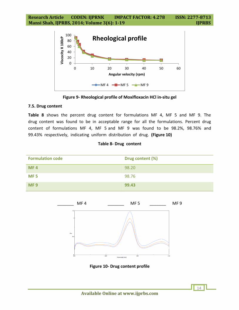
Figure 9- Rheological profile of Moxifloxacin HCl in-situ gel
7.5. Drug content
Table 8 shows the percent drug content for formulations MF 4, MF 5 and MF 9. The
drug content was found to be in acceptable range for all the formulations. Percent drug
content of formulations MF 4, MF 5 and MF 9 was found to be 98.2%, 98.76% and
99.43% respectively, indicating uniform distribution of drug. (Figure 10)
Table 8- Drug content
_______ MF 4 _______ MF 5 _______ MF 9
Figure 10- Drug content profile
0
20
40
60
80
100
0 10 20 30 40 50 60
Vis
coci
ty X
10
0cP
Angular velocity (rpm)
Rheological profile
MF 4 MF 5 MF 9
Formulation code Drug content (%)
MF 4 98.20
MF 5 98.76
MF 9 99.43

Research Article CODEN: IJPRNK IMPACT FACTOR: 4.278 ISSN: 2277-8713 Mansi Shah, IJPRBS, 2014; Volume 3(6): 1-19 IJPRBS
Available Online at www.ijprbs.com 15
7.6. In vitro drug release studies
The three in situ gelling formulations of Moxifloxacin Hydrochloride, MF 4, MF 5 and MF
9, were subjected to in vitro release studies. These in vitro release studies were carried
out using simulated tear fluid (STF) of pH 7.4 as the dissolution medium. The drug
release data obtained for formulations MF 4, MF 5 and MF 9 is tabulated in Table 9.
Figure 11 shows the plot of cumulative percent drug released as a function of time for
formulation MF 4, MF 5 and MF 9…
Table 9- drug release data of MF 4, MF 5 and MF 9
Figure 11- Cumulative percent drug released vs. time
It was found that cumulative percent drug release was 91.98%, 87.12% and 93.78% for
formulation MF 4, MF 5 and MF 9 respectively after 8 hours. The in vitro release data
indicated that the formulation MF 9 showed better sustained effect than other two
formulations. All the three formulations showed an initial burst release. The prolonged
release in the later stage can be attributed to the slow diffusion of the drug through
polymer matrix. The initial burst release of the drug can be explained by the fact that,
0
20
40
60
80
100
0 2 4 6 8 10
% c
um
ula
tive
dru
g re
leas
e
time (hr)
% Cumulative drug release vs. time
MF 4
MF 5
MF 9
Time Abs CDR % CDR Abs CDR % CDR Abs CDR % CDR
(hr) MF 4 MF 5 MF 9
1 0.126 1.34 24.12 0.1108 1.15 20.7 0.1202 1.26 22.68
2 0.165 1.81 32.58 0.1467 1.58 28.44 0.1563 1.69 30.42
3 0.225 2.52 45.36 0.1943 2.14 38.52 0.2021 2.24 40.32
4 0.270 3.05 54.9 0.2461 2.76 49.68 0.2611 2.94 52.92
5 0.315 3.59 64.62 0.2939 3.33 59.94 0.3049 3.46 62.28
6 0.367 4.21 75.78 0.3517 4.02 72.36 0.3625 4.17 75.06
7 0.406 4.67 84.06 0.3999 4.59 82.62 0.4134 4.75 85.51
8 0.443 5.11 91.98 0.4213 4.84 87.12 0.4522 5.21 93.78
Research Article CODEN: IJPRNK IMPACT FACTOR: 4.278 ISSN: 2277-8713 Mansi Shah, IJPRBS, 2014; Volume 3(6): 1-19 IJPRBS
Available Online at www.ijprbs.com 16
the in situ gelling system is formulated in water and hence the polymer was completely
hydrated. When they come in contact with STF, gelation occurs and a prehydrated matrix
is formed in which hydration and water penetration no longer limit drug release, leading
to an apparent diffusion-controlled release.
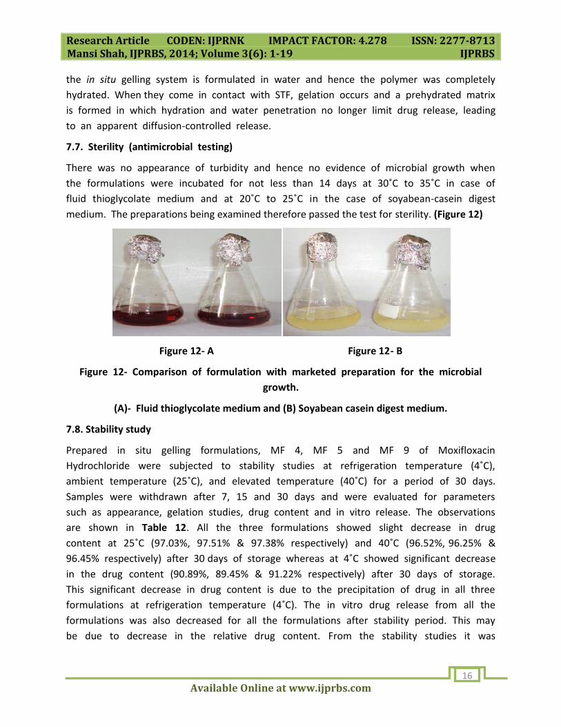
7.7. Sterility (antimicrobial testing)
There was no appearance of turbidity and hence no evidence of microbial growth when
the formulations were incubated for not less than 14 days at 30˚C to 35˚C in case of
fluid thioglycolate medium and at 20˚C to 25˚C in the case of soyabean-casein digest
medium. The preparations being examined therefore passed the test for sterility. (Figure 12)
Figure 12- A Figure 12- B
Figure 12- Comparison of formulation with marketed preparation for the microbial
growth.
(A)- Fluid thioglycolate medium and (B) Soyabean casein digest medium.
7.8. Stability study
Prepared in situ gelling formulations, MF 4, MF 5 and MF 9 of Moxifloxacin
Hydrochloride were subjected to stability studies at refrigeration temperature (4˚C),
ambient temperature (25˚C), and elevated temperature (40˚C) for a period of 30 days.
Samples were withdrawn after 7, 15 and 30 days and were evaluated for parameters
such as appearance, gelation studies, drug content and in vitro release. The observations
are shown in Table 12. All the three formulations showed slight decrease in drug
content at 25˚C (97.03%, 97.51% & 97.38% respectively) and 40˚C (96.52%, 96.25% &
96.45% respectively) after 30 days of storage whereas at 4˚C showed significant decrease
in the drug content (90.89%, 89.45% & 91.22% respectively) after 30 days of storage.
This significant decrease in drug content is due to the precipitation of drug in all three
formulations at refrigeration temperature (4˚C). The in vitro drug release from all the
formulations was also decreased for all the formulations after stability period. This may
be due to decrease in the relative drug content. From the stability studies it was

Research Article CODEN: IJPRNK IMPACT FACTOR: 4.278 ISSN: 2277-8713 Mansi Shah, IJPRBS, 2014; Volume 3(6): 1-19 IJPRBS
Available Online at www.ijprbs.com 17
confirmed that in situ gelling formulations of Moxifloxacin hydrochloride remained stable
at ambient temperature (25˚C) and humidity.
Table 12- Stability studies: Characterization of Formulations
Formulation
code
Storage
condition
Parameters evaluated
After 7 days After 14 days After 21 days
Drug
content
% CDR Drug
content
% CDR Drug
content
% CDR
MF 4 4°C 98.09 89.57 94.60 88.69 90.89 87.35
25°C 98.12 85.27 97.64 85.22 97.03 84.98
40°C 98.10 88.85 97.16 88.52 96.52 87.32
MF 5 4°C 98.42 87.69 93.89 87.35 89.45 86.59
25°C 98.66 84.98 98.17 84.85 97.51 84.11
40°C 98.59 85.35 97.46 85.30 96.25 85.02
MF 9 4°C 98.78 89.65 95.35 89.34 91.22 88.57
25°C 98.91 90.87 98.21 90.48 97.38 89.52
40°C 98.82 89.32 97.98 89.10 96.45 88.97
7.9. Isotonicity Evaluation
All the three formulations were subjected to the Isotonicity study and the results were
studied under the optical microscope using 45X magnification and the results were as
shown in figure 13.
A B C D
Figure 13- Isotonicity studies of different formulations.
A- Marketed preparation C- MF 5
B- MF 9 D- MF 9
CONCLUSION
Infrared spectroscopy studies of Moxifloxacin Hydrochloride, Poloxamer 188 and 407 with
HPMC E50 LV alone and their physical mixture revealed that, Moxifloxacin Hydrochloride

Research Article CODEN: IJPRNK IMPACT FACTOR: 4.278 ISSN: 2277-8713 Mansi Shah, IJPRBS, 2014; Volume 3(6): 1-19 IJPRBS
Available Online at www.ijprbs.com 18
is compatible with all the polymers used. Ophthalmic in situ gelling system was
successfully formulated using different gelling agents’ viz. Poloxamer 188 and Poloxamer
407 as temperature sensitive along with HPMC E50 LV as viscosity enhancing agent. The
clarity of the prepared formulations was found satisfactory. The pH of all formulations
was found to be satisfactory in the range of 6 - 7.4. The drug content of the prepared
formulation was within the acceptable range, and ensures dose uniformity. The
formulation MF 9 showed maximum drug content. All the formulations except MF 2, MF
3 and MF 6 showed instantaneous gelation when contacted with simulated tear fluid
(STF). Formulations MF 4, MF 5 and MF 9 were shear thinning and an increase in shear
stress was observed with increase in angular velocity (pseudoplastic rheology).
Formulation MF 4, MF 5 and MF 9 showed sustained drug release for a period of 8
hour. Formulation MF 9 showed most sustained drug release. Results of sterility test
confirmed that all the formulations were sterile. From the stability studies it was
confirmed that in situ gelling formulations of Moxifloxacin Hydrochloride remained more
stable at ambient temperature (25°C) and humidity. The maximum instability of in situ
gelling formulations was observed at 40±1°C and 4°C (significant decrease in drug
content and in vitro drug release). Present work was a satisfactory preliminary study in
developing in situ gelling system of Moxifloxacin Hydrochloride.
REFERENCES
1. Mundada AS, Avari JG, Mehta SP, Pandit SS, Patil AT. Recent advances in ophthalmic drug
delivery system. Pharm Rev 2008;6(1).
2. Khurana AK. Comprehensive ophthalmology; 4th ed. Age International (P) Ltd Pub. 2007.
3. Shoenwald RD. Ocular drug delivery: pharmacokinetic considerations. Clin.Pharmacokinet
1998; 18: 255-269.
4. Lee VHL. Drugs and pharmaceutical sciences. In: Swarbrick J editor. Precorneal, corneal and
post corneal factors. New York: marcel Dekker Inc; 58, 1993. p. 59-81.
5. Macha S, Mitra AK. Ophthalmic drug delivery systems; second edition revised and expanded.
Chapter 1 Overview of Ocular Drug Delivery. p 1-3.
6. Le Bourlais, C. A., Treupel-Acar, L., Rhodes, C. T., Sado, P. A., & Leverge, R. New ophthalmic
drug delivery systems. Drug Development & Industrial Pharmacy (1995), 21(1), 19-59.
7. Mishima, S., Gasset, A., Klyce, S. D., Jr., & Baum, J. L. Determination of tear volume and tear
flow. Investigative Ophthalmology, (1966), 5(3), 264-276.

Research Article CODEN: IJPRNK IMPACT FACTOR: 4.278 ISSN: 2277-8713 Mansi Shah, IJPRBS, 2014; Volume 3(6): 1-19 IJPRBS
Available Online at www.ijprbs.com 19
8. Attia MA, Kassem MA, Safwat S. In-vivo performance of dexamethasone ophthalmic film
delivery system in the rabbit eye. International J Pharm 1988; 47:21-30.
9. Meqi SA, Deshpande SG. Ocular drug delivery: Controlled and novel drug delivery. New
delhi:CBS Publishers; 2002. 82-84.
10. Eva M, Amo D, Urtti A. Current and future ophthalmic drug delivery systems. A shift to the
posterior segment. Drug Discov Today 2004; 13:135-143.
11. http://www.eyecaresource.com/healthyvisionforahealthylife.
12. Le Bourlais C, Acar L, Zia H, Sado PA, Needham T, Leverge R. Ophthalmic drug
deliverysystem- Recent Advances, Progress in Retinal and Eye Research 1998: 1733-58.
13. Rathore K. S., Nema R. K., An Insight Into Ophthalmic Drug Delivery System, International
Journal of Pharmaceutical Sciences and Drug Research 2009; 1(1):1-5
14. Manjappa AS, Nanjawade BK, Manvi FV, Murthy SR. Sustained ophthalmic in situ gel of
ketorolac tromethamine: rheology and in vivo studies. Drug Deliv Res 2009;69:1–8.