International Journal of Pharma and Bio Sciences ISSN 0975 … · Bismarck Brown Y was selected as...
12
Int J Pharm Bio Sci 2013 July; 4(3): (B) 862 - 873 This article can be downloaded from www.ijpbs.net B - 862 Research Article Microbiology International Journal of Pharma and Bio Sciences ISSN 0975-6299 MYCOREMEDIATION OF BISMARCK BROWN Y BY INDIGENOUS FUNGAL ISOLATE ALTERNARIA BRASSICAE TSF – 07 AND OPTIMIZATION OF CULTURAL CONDITIONS TO ENHANCE ITS DECOLOURIZATION KANYAKUMARI P. SHINDE *1 AND P. R. THORAT 2 1 Department of Biotechnology, Walchand College of Arts and Science, Solapur, MS, India 2 Department of Microbiology and Research Center, Shri Shivaji Mahavidyalaya, Barshi-413 411 Dist-Solapur, MS, India ABSTRACT Bismarck Brown Y was selected as model textile dye. The fungal strain Alternaria brassicae TSF – 07 isolated from textile wastewater sludge was screened for decolourization of Bismarck Brown Y. Optimization was carried out with respect to carbon source, nitrogen source, pH, inoculums size, incubation period and temperature. It was found that the optimum inoculum size, pH and temperature were 2.5%, 5.0 and 35 0 C respectively. Fructose at 1.0 % and yeast extract at the concentration of 0.1 % was found to give maximum decolourization. Further, it was found that the culture has brought about almost 96.03% decolourization of a recalcitrant dye, Bismarck Brown Y (100 mgL -1 ) at shaking condition within 3 days. This isolate also showed the degradation as evidenced from the reduction in the term of COD mg/L to the extent of 68.09 % within three days. The biodegradation was monitored by UV–Vis and FTIR spectroscopy.This study revealed the enormous biodegradation abilities of indigenous microbial flora. KEYWORDS: Mycoremediation, Alternaria brassicae, optimization, biodecolourization, FTIR, COD *Corresponding author KANYAKUMARI P. SHINDE Department of Biotechnology, Walchand College of Arts and Science, Solapur, MS, India
Transcript of International Journal of Pharma and Bio Sciences ISSN 0975 … · Bismarck Brown Y was selected as...

Int J Pharm Bio Sci 2013 July; 4(3): (B) 862 - 873
This article can be downloaded from www.ijpbs.net
B - 862
Research Article Microbiology
International Journal of Pharma and Bio Sciences ISSN
0975-6299
MYCOREMEDIATION OF BISMARCK BROWN Y BY INDIGENOUS FUNGAL
ISOLATE ALTERNARIA BRASSICAE TSF – 07 AND OPTIMIZATION OF
CULTURAL CONDITIONS TO ENHANCE ITS DECOLOURIZATION
KANYAKUMARI P. SHINDE*1
AND P. R. THORAT 2
1Department of Biotechnology, Walchand College of Arts and Science, Solapur, MS, India
2Department of Microbiology and Research Center, Shri Shivaji Mahavidyalaya,
Barshi-413 411 Dist-Solapur, MS, India
ABSTRACT
Bismarck Brown Y was selected as model textile dye. The fungal strain Alternaria brassicae TSF – 07 isolated from textile wastewater sludge was screened for decolourization of Bismarck Brown Y. Optimization was carried out with respect to carbon source, nitrogen source, pH, inoculums size, incubation period and temperature. It was found that the optimum inoculum size, pH and temperature were 2.5%, 5.0 and 350C respectively. Fructose at 1.0 % and yeast extract at the concentration of 0.1 % was found to give maximum decolourization. Further, it was found that the culture has brought about almost 96.03% decolourization of a recalcitrant dye, Bismarck Brown Y (100 mgL-1) at shaking condition within 3 days. This isolate also showed the degradation as evidenced from the reduction in the term of COD mg/L to the extent of 68.09 % within three days. The biodegradation was monitored by UV–Vis and FTIR spectroscopy.This study revealed the enormous biodegradation abilities of indigenous microbial flora.
KEYWORDS: Mycoremediation, Alternaria brassicae, optimization, biodecolourization, FTIR, COD
*Corresponding author
KANYAKUMARI P. SHINDE
Department of Biotechnology, Walchand College of Arts and Science, Solapur, MS, India
Int J Pharm Bio Sci 2013 July; 4(3): (B) 862 - 873
This article can be downloaded from www.ijpbs.net
B - 863
INTRODUCTION
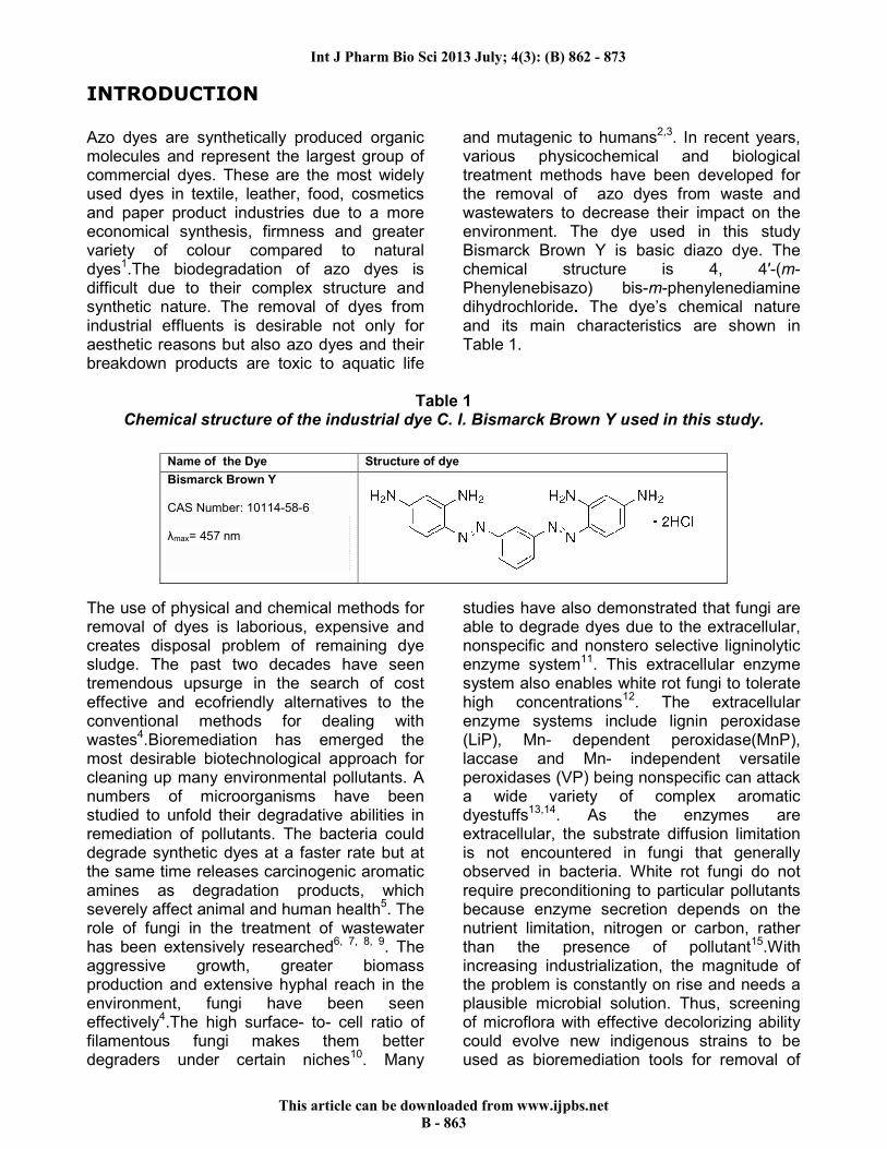
Azo dyes are synthetically produced organic molecules and represent the largest group of commercial dyes. These are the most widely used dyes in textile, leather, food, cosmetics and paper product industries due to a more economical synthesis, firmness and greater variety of colour compared to natural dyes1.The biodegradation of azo dyes is difficult due to their complex structure and synthetic nature. The removal of dyes from industrial effluents is desirable not only for aesthetic reasons but also azo dyes and their breakdown products are toxic to aquatic life
and mutagenic to humans2,3. In recent years, various physicochemical and biological treatment methods have been developed for the removal of azo dyes from waste and wastewaters to decrease their impact on the environment. The dye used in this study Bismarck Brown Y is basic diazo dye. The chemical structure is 4, 4′-(m-Phenylenebisazo) bis-m-phenylenediamine dihydrochloride. The dye’s chemical nature and its main characteristics are shown in Table 1.
Table 1
Chemical structure of the industrial dye C. I. Bismarck Brown Y used in this study.
The use of physical and chemical methods for removal of dyes is laborious, expensive and creates disposal problem of remaining dye sludge. The past two decades have seen tremendous upsurge in the search of cost effective and ecofriendly alternatives to the conventional methods for dealing with wastes4.Bioremediation has emerged the most desirable biotechnological approach for cleaning up many environmental pollutants. A numbers of microorganisms have been studied to unfold their degradative abilities in remediation of pollutants. The bacteria could degrade synthetic dyes at a faster rate but at the same time releases carcinogenic aromatic amines as degradation products, which severely affect animal and human health5. The role of fungi in the treatment of wastewater has been extensively researched6, 7, 8, 9. The aggressive growth, greater biomass production and extensive hyphal reach in the environment, fungi have been seen effectively4.The high surface- to- cell ratio of filamentous fungi makes them better degraders under certain niches10. Many
studies have also demonstrated that fungi are able to degrade dyes due to the extracellular, nonspecific and nonstero selective ligninolytic enzyme system11. This extracellular enzyme system also enables white rot fungi to tolerate high concentrations12. The extracellular enzyme systems include lignin peroxidase (LiP), Mn- dependent peroxidase(MnP), laccase and Mn- independent versatile peroxidases (VP) being nonspecific can attack a wide variety of complex aromatic dyestuffs13,14. As the enzymes are extracellular, the substrate diffusion limitation is not encountered in fungi that generally observed in bacteria. White rot fungi do not require preconditioning to particular pollutants because enzyme secretion depends on the nutrient limitation, nitrogen or carbon, rather than the presence of pollutant15.With increasing industrialization, the magnitude of the problem is constantly on rise and needs a plausible microbial solution. Thus, screening of microflora with effective decolorizing ability could evolve new indigenous strains to be used as bioremediation tools for removal of
Name of the Dye Structure of dye
Bismarck Brown Y CAS Number: 10114-58-6 λmax= 457 nm

Int J Pharm Bio Sci 2013 July; 4(3): (B) 862 - 873
This article can be downloaded from www.ijpbs.net
B - 864
azo dyes16. The aim of the present study to investigate the decolourization and degradation potential of a deuteromycete fungal strain of Alternaria brassicae TSF - 07 for the removal of Bismarck Brown Y from an aqueous solution. Dye decolourization by fungal culture was optimized with respect to various nutritional sources (carbon, nitrogen) and environmental parameters (pH, temperature and inoculum size). The IR spectrum of Bismarck Brown Y after biological treatment with the tested organism was also studied to study its biodegradation.
MATERIALS AND METHODS
Dyes and chemicals Bismarck Brown Y (C18H18N8 · 2HCl, M.W. 419.31 g/mol) synthetic dye was procured from Sigma-Aldrich (St. Louis, MO, USA).Dye solution was prepared by dissolving it in distilled water before each experiment. Nutrient media and all other chemicals were obtained from Hi media. Soil samples were collected from the nearby effluent disposal area of textile units located at Sunil Nagar and Neelam Nagar, Akkalkot MIDC, Solapur, MS, India. Isolation and screening of decolorizing microorganisms The collected soil samples were used for isolation of fungal strains capable of decolorizing dyes by subjecting it to serial dilutions using sterile D/W. 0.1 ml aliquot from
each dilution of 10-3 & 10-4 was spread inoculated on sterile Sabouraud’s agar plates containing Bismarck Brown Y dye at a final concentration of 100 mg L-1. All plates were incubated at room temperature for 24-72 hours. Dye decolourization ability of microorganisms was confirmed by presence of clear zone of decolourization around the colonies. The colony showing prominent zone of decolourization of the dye on the agar plate was designated as TSF- 07and was selected for further studies. Decolourization of dye Bismarck Brown Y The selected fungal isolate was inoculated in 50 ml sterilized Sabouraud’s glucose broth containing 5 mg of Bismarck Brown Y dye. It was incubated at ambient temperature at both static & shaking (150 rpm in an orbital Shaker) conditions for 3 days. The decolorizing activity was measured on each day and was expressed as Percent decolourization. The growth of fungus was studied by measuring the fresh and dry weight of the mycelium. Measurement of decolourization (Decolourization assay) Bismarck Brown Y decolorizing activity was determined by measuring the decrease in the color intensity as absorbency at 457 nm on each day by using U.V.Visible spectrophotometer (Shimadzu, UV-3600). Percent decolourization was calculated by using following formula as-
Optimization of culture medium composition Decolourization of Bismarck Brown Y (100 mg L-1) in Sabouraud’s broth by selected fungal isolate TSF -07 was optimized with respect to the effect at 1% of different carbon sources (glucose, maltose, fructose, sucrose, lactose and no carbon i.e. dye itself), 0.1% organic nitrogen sources (mycological peptone and yeast extract) and inorganic nitrogen sources (ammonium sulphate, ammonium nitrate,
ammonium chloride and urea), pH (3, 4, 5, 6, 7 and 8) temperature ( 250C, 300C, 350C and 40°C) and inoculum size (1, 2.5, 5 and 10 ml.). All the flasks were inoculated with culture of TSF -07 and were incubated under shaking conditions for 3 days. Percent COD reduction The COD of the dye solution was estimated before and after its biological treatment using

Int J Pharm Bio Sci 2013 July; 4(3): (B) 862 - 873
This article can be downloaded from www.ijpbs.net
B - 865
dichromate reflux method and percent COD reduction was determined by TSF -07. UV- Vis spectral analysis of the decolorized products The samples were collected before and after the decolourization process and filtered through 0.2 µm membrane filters. The filtrates were then scanned in the UV- Vis Spectrophotometer (Shimadzu UV- Vis 3600, Japan) within the range of 200-650 nm. Appropriate blank was also subjected to the scanning process. The band width was set to1 nm during scanning program. The absorbance was noted at the respective characteristic peak area (λmax) for the interpretation of results. FTIR analysis Biodegradation of Bismarck Brown Y was monitored by FTIR spectroscopy. For this, 100 ml sample was taken after decolourization. The centrifugation was carried out at 10,000 rpm and the metabolites were extracted from supernatant using equal volume of Dichloromethane. The extract was dried over anhydrous Na2SO4 and evaporated to dryness in a rotary vacuum flash evaporator. The treated Bismarck Brown Y dye was characterized by Fourier Transform Infrared
Spectrometer (Perkin Elmer Spectrum 65) and compared with control (before treatment) dye. The samples were mixed with spectroscopically pure KBr in the ratio of 1:100 and pressed to obtain IR- transparent pellet. The pellet was placed in sample holder and the analysis was carried out in the mid IR region of 450-4000 cm -1 with 16 scan speed8.
RESULTS AND DISCUSSION
Isolation, identification and phylogenetic analysis The fungal isolate designated as TSF- 07 was selected as good candidate for decolourization in this study based on its ability to form clear halos on Sabouraud’s Agar plate containing dye. Molecular identification was carried out of this isolate based on 18s rRNA sequencing. The phylogenetic tree was constructed by using Neighbour joining method by Kimura – 2 parameter with 1000 replicates in MEGA 4.0 (Figure 1). According to sequencing similarities and multiple alignments the isolate TSF-07 was found to be in a close relation to the Alternaria brassicae. The partial sequence of 18S r RNA of this isolate TSF-07 has been deposited into Gene Bank under accession number HF913433.
Figure 1
Phylogenic tree based on ITS region gene sequences showing relationships among strain TSF-07 and the most close type strain species of Alternaria brassicae. Numbers at nodes indicate percentages of bootstrap support based on a Neighbour-joining analysis of 1,000 resampled datasets. Bar 0.2 substitutions per nucleotide position.

Int J Pharm Bio Sci 2013 July; 4(3): (B) 862 - 873
This article can be downloaded from www.ijpbs.net
B - 866
Percent decolourization of dye The promising isolate TSF- 07was evaluated for its ability to decolorize the dye Bismarck Brown Y in Sabouraud’s medium containing 100 mg L-1 concentration of dye. The decolourization of Bismarck Brown Y was found to be 65.53% within 72 hours at static condition while 89.91% at shaking condition. The percent decolourization at static and shaking condition for Bismarck Brown Y is given Graph 1.The results showed that shaking favoured higher decolourization and faster biodegradation of Bismarck Brown Y when compared to static cultures. This may be due to an increase in the mass and oxygen transfer between cells and the medium,
factors that optimise the action of oxidative enzymes17. The pH was 5.20 before treatment that was found to be 4.94 after treatment. The growth of mycelium obtained as biomass gets calculated as 3.940 gm/ 50 mL and 1.687gm/ 50 mL of medium on wet weight and dry weight basis respectively. The biodecolourization of Bismarck Brown Y by microorganisms has been investigated by a very few authors. It is reported that 75.7 mg of Bismarck Brown Y biosorption was obtained by per gram of dried magnetically modified cells of Kluyveromyces fragilis.18 Basidiomycota sp. L-168 showed98.53% decolorization of Remazol Brilliant Blue R (RBBR) in 5 days of incubation19.
Graph 1
Percent decolourization for Bismarck Brown Y by at static and shaking condition.
Optimization of culture medium for decolourization To enhance the decolourization of Bismarck Brown Y by Alternaria brassicae TSF-07 experiments were conducted for optimization of carbon source, nitrogen source, pH, temperature and inoculum size. Effect of carbon source There are various influencing factors involved in the decolourization process by microorganisms. The effect of various carbon sources was assessed. It is seen from the Graph 2 that the supplementation of 1% fructose recorded significant decolourization yield (84.40 %). The 1 % lactose was found to be the next best co – substrate with 80.97% decolourization. It is noteworthy to mention that in absence of carbon source also, the TSF -07 culture has shown 71.05 % decolourization of dye under study. It indicates
that the culture has utilized dye as a carbon source. Basically, a carbon source is necessary for providing electrons for the reduction of the xenobiotics20. Similar results were reported that fructose was the most effective carbon source for maximum decolourization of Brown GR accounting 82% decolourization of the textile dye21. The metabolism of reducing sugars resulted in the production of reduced nucleotides like NAOH, FAOH which led to enhanced decolourization efficiency. These reduced nucleotides have been reported to be redox mediators involved in reduction of azo bond. The observations draw support from findings for Phanerochaete chrysosporium22 and Citeromyces sp. WR-43-6 and showed that the reducing sugar in culture broth rapidly decreased with decreasing color intensity23. It was also concluded that the utilization of organic

Int J Pharm Bio Sci 2013 July; 4(3): (B) 862 - 873
This article can be downloaded from www.ijpbs.net
B - 867
nitrogen and carbon source (reducing sugar) has a critical role in decolourization24. Effect of Nitrogen source The supply of various organic and inorganic nitrogen sources further increased decolourization. The effect of types and concentrations of nitrogen sources on decolourisation of Bismarck Brown Y by Alternaria brassicae TSF - 07 culture is shown in Graph 3.Out of six nitrogen sources screened, yeast extract recorded significantly greater decolourization (94.76%). The next best nitrogen source was 0.1% sodium nitrate (91.66%) followed by mycological peptone (90.45%).Ammonium sulphate recorded the least decolourization. Our findings are in conformity with the findings of Singh et al.21, who also observed 76 % decolourization of Brown GR with 1 % yeast extract by Aspergillus sp. There is one of evidence also showed similar result for nitrogen optimization study in which Aspergillus flavus was found to be the most effective decolourizer and yeast extract was the most effective nitrogen source for maximum decolourization (97.82 %) of true blue dye25.The addition of yeast extract as nitrogen source was observed to have a significant effect on dye colour reduction. Effect of pH The effect of different pH on the decolourization of dye by Alternaria brassicae TSF – 07 is shown in Graph 4. The significant decolourization was observed in the pH range 4.0 to 6.0.The results further revealed the highest decolourization 96.19% was obtained when pH of was adjusted to 5.0 by TSF-07 isolate. However, decolorizing activity decreased significantly when the pH was higher than 6.0. Similarly it was reported that, ascomycetes yeast strain showed maximum decolourization of azo dyes in the acidic range and optimum pH depends upon the dye structure26. The pH significantly influenced the dye biosorption properties of fungi27. Higher uptake obtain at lower pH value may be due to the electrostatic attraction between negatively charged dye anions and positively charged cell surface6.The optimum pH was 4.0 and 5.0 in the dye removal study of Mehna et al. (1995)28.
Effect of Temperature It is observed that TSF -07 culture showed highest decolourization activity at a temperature 350C about 92.18 % decolourization followed by 87.46% at 300C(Graph 5). The results of present study showed that, dye removal was influenced by fluctuating given temperatures. The optimum temperature was reported for color removal of red azo dye was 300C and 35°C29. The colour reduction increased with temperature due to higher respiration and substrate metabolism at the elevated temperature30. Effect of Inoculum size The effect of inoculum size of homogenized culture of Alternaria brassicae TSF – 07 (1, 2, 5 and 10 %) on decolourization efficiency of fungal strains of dye Bismarck Brown Y. The relationship between inoculum size and decolourization yield is shown in Graph 6.The highest decolourization was observed with 2.5 % inoculum about 94.81 %. Beyond 2.5%, the decolourization yield did not vary significantly. Inoculum size from 1 to 10 % has an adverse effect on the dye removal efficiency and it was 94 to 59% with Alternaria brassicae TSF – 07. The color reduction was found to increase from 45 to 80% when the inoculum concentration was increased from 0.5 to 5.0 g l-1 and leveled off beyond that27. The present results are in agreement with Erum et al., 2011 who found 2 % inoculum for maximum decolourization of AR 151 for studied fungal isolates of Aspergillus sp. and further increase in the inoculum level has shown in decrease in decolourisation efficiency of isolates and in the same context reported that the utilization of minimal nutrients and oxygen by fungus and the rate of accumulation of fungal metabolites in media not supported by initial massive fungal inoculum can participate in the dye removal. From the results of optimisation of various parameters of growth conditions, the medium composed of Fructose 1.0%, yeast extract 0.1%, pH adjusted to 5.0 of dye was prepared. It is inoculated with 2.5% inoculum of TSF-07 isolate and incubated at temperature 350C. It is observed that Alternaria brassicae TSF – 07 isolate has brought about 96.03% decolourisation of Bismarck Brown Y within 72 hrs. of incubation period.

Int J Pharm Bio Sci 2013 July; 4(3): (B) 862 - 873
This article can be downloaded from www.ijpbs.net
B - 868
Graph 2 Effect of carbon- source on % decolourization of Bismarck Brown Y dye.
Graph 3 Effect of nitrogen- source on % decolourization of Bismarck Brown Y dye.
Graph 4 Effect of pH on % decolourization of Bismarck Brown Y dye.

Int J Pharm Bio Sci 2013 July; 4(3): (B) 862 - 873
This article can be downloaded from www.ijpbs.net
B - 869
Graph 5 Effect of temperature on % decolourization of Bismarck Brown Y dye.
Graph 6 Effect of inoculum size on % decolourization of Bismarck Brown Y dye.
Percent COD reduction To evaluate the biodegradation of Bismarck Brown Y by Alternaria brassicae TSF -07, the determination of percentage of mineralization (represented by COD removal) was carried out by measuring the initial and final organic content. Upon 3 days of treatment of dye by TSF- 07 culture with optimized cultural conditions, percent COD reduction was obtained to the extent of 68.09. UV- Vis spectral analysis The biodegradation of the dye Bismarck Brown Y was monitored by UV-Vis
Spectrophotometric analysis. For the untreated dye, as shown in Figure 2 (A), the large absorbance peaks was found at 457 nm while 226 nm, 259nm and 278 nm for the treated dye as in Figure 2 (B). After biodecolourization in the aerobic condition, the absorbance peaks in the visible region i.e. the absorbance peak at 457 nm was completely disappeared, indicating the complete decolourization and deformation of the structural confirmation that was responsible for the color.

Int J Pharm Bio Sci 2013 July; 4(3): (B) 862 - 873
This article can be downloaded from www.ijpbs.net
B - 870
Figure 2 Absorption Spectra of Bismarck Brown Y dye before and after decolourization with Alternaria brassicae TSF - 07 (A – Native dye Bismarck Brown Y; B - after decolourization).
Biodegradation analysis The difference in FTIR spectrum of Bismarck Brown Y and metabolites obtained after its decolourization resulted in biodegradation. The FTIR spectrum of control Bismarck Brown Y showed the specific peaks in fingerprint region (1500 to 500 cm-1) for the mono-substituted and para-disubstituted benzene rings which is supporting to the peak at 1585.97 cm-1 for the N=N stretching of the benzene ring. It was also supported by the peak at 733.62 cm-1 for aromatic C – C out of plane vibration,830.97 cm-1 that corresponds to N-H weak out of plane bending vibration, 1243.92 cm-1 corresponds to N- H bending vibrations, 1436.48 cm-1 for stretching of N - H, 1503.75 cm-1 for C=N ring stretching
vibrations, 1523.99cm-1, 1610.49cm-1and 1633.99cm-1 for C=C stretching vibrations of aromatic benzene ring, 3142.55 cm-1 for NH2 group while 3306.05 cm-1 for NH stretching vibrations.(Figure 3A). The FTIR spectrum of metabolites obtained after decolourization of Bismarck Brown Y by TSF-07 isolate showed peaks at 751.04cm-1 for N-H bending vibrations, 1268.01cm-1for N-H stretching vibrations, 1667.79 cm-1 for C=O stretching vibrations and 3502.50 cm-1 for OH stretching vibrations (Figure 3B). The absence of peak at 1585.97 cm-1 for N=N stretching vibrations in the FTIR spectrum of metabolites obtained after decolourization of Bismarck Brown Y because of the cleavage of azo bond.

Int J Pharm Bio Sci 2013 July; 4(3): (B) 862 - 873
This article can be downloaded from www.ijpbs.net
B - 871
Figure 3 FTIR analyses of Bismarck Brown Y for its biodegradation by TSF - 07 isolate – A) FTIR spectrum of control dye Bismarck Brown Y and B) metabolites obtained after decolourization of Bismarck Brown Y.
CONCLUSION
Based upon these findings, it can be predicted that fungi present in the vicinity of discharged effluent possess a great potential for the use in bioremediation of textile dyes. Decolourization or degradation dyes has been mostly studied with white rot fungi, however, this newly isolated and screened fungal strain for dye decolourization that belonging to a deuteromycete showed great ability to grow and degrade Bismarck Brown Y. To the best of our knowledge, this is the first report with respect to the fungal degradation of Bismarck
Brown Y by Alternaria brassicae strain. The present study has revealed that optimization for nutrients viz. carbon and nitrogen as well as physical parameters (pH, temperature and inoculum size) had significantly increased dye decolourization. Moreover, results showed the decolourization of dye under shaking condition comparatively very fast i.e. within 72 hours. The present study indicate that the isolate Alternaria brassicae is an excellent strain for the decolourization of azo textile dyes effluents and it might be a practical alternative

Int J Pharm Bio Sci 2013 July; 4(3): (B) 862 - 873
This article can be downloaded from www.ijpbs.net
B - 872
in dyeing wastewater treatment. The results of this study will form the basis for development of a cost effective and robust indigenous process for bioremediation of textile dyes- based effluent. Further studies in this domain are valuable to use this microorganism for the decolourization of real dye wastewaters (which are complex mixtures of many dyes) and to know about the nature and fate of the degradation products.
ACKNOWLEDGEMENT
The authors would like to thank National Centre for Cell Sciences, University of Pune campus for the 18s rRNA sequencing of the isolate. Our sincere thanks to Principal, Walchand College of Arts and Science, Solapur for providing the Laboratory facilities.
REFERENCES
1. X. Lu and R. Liu. Treatment of Azo dye- containing wastewater using integrated processes. In: H. Atacag Erkurt (ed.), The Handbook of Environmental Chemistry Volume 9, Biodegradation of Azo dyes, Springer Heidelberg Dordrecht London, New York, 2009, pp.133-135.
2. Chung KT, Stevens SEJ, Degradation of azo dyes by environmental organisms and helminthes, Environ Toxicol Chem, 12: 2121-2132, (1993).
3. Weisburger JH, Comments on the history and importance of the aromatic and heterocyclic – amines in public health. Mutat Res, 506-507:9-20, (2002).
4. Asamudo NU, Daba AS and Ezeronye OU (2005), Bioremediation of textile effluent using Phaneochaete chrysosporium, Afr J Biotechnol, 4 (13): 1548 – 1553, (2005).
5. Brown M A and DeVito SC, Predicting azo dye toxicity, Crit Rev Environ Sci Technol, 23: 249-324, (1993).
6. Erum S And Ahmed S, Comparision of dye decolourization efficiencies of indigenous fungal isolates, Afr J Biotechnol, 10 (17): 3399- 3411, (2011).
7. Saraswathy RN, Shanmugapriya S, Shakthipriyadarshini S, Sadasivam S and Shanmugaprakash M, Decolourization of textile dyes by Aspergillus tamarii, mixed fungal culture and Penicillium purpurogenum, J Sci Ind Res, 69: 151- 153, (2010).
8. Shinde KP, Thorat PR, Biodecolourization of Diazo dye Direct Red 28 By Aspergillus aculeatus TSF – 05, Bionano Frontier, 6 (1): 45 -49, (2013).
9. Sathiya MP, Periyar SS, Sasikalaveni A, Murugesan K and Kalaichelvan PT, Decolourization of textile dyes and their effluents using white rot fungi, Afr J Biotechnol, 6 (4): 424-429, (2007).
10. C. Ashoka, K. Manjunath, S.B. Sullia.
Biological treatment of combined textile dye effluent. In: D.J. Bhat, S. Raghukumar (eds.), Ecology of fungi, 2002, pp. 63- 67.
11. Park C, Lee M, Lee B, Kim S W, Chase HA, Lee J, Kim S, Biodegradation and biosorption for decolourization of synthetic dyes by Funalia trogii, Biochem Eng J, 36 (1): 59-65, (2006).
12. Knapp JS, Zhang F, Tapley NK, Decolourization of Orange II by a wood- rotting fungus, J Chem Technol Biotechnol, 69:289-296, (1997).
13. Cripps C, Bumpus JA and Aust SD, Biodegradation of azo and heterocyclic dyes by Phanerochaete chrysosporium, Appl Environ Microbiol, 56 (4): 1114 – 1118, (1990).
14. Camarero S, Ibarra D, Martinez MJ, and Martinez AT, Lignin- derived compounds as efficient laccase mediators for decolourization of different types of recalcitrant dyes, Appl Environ Microbiol, 71 (4):1775- 1784, (2005).
15. Jin X, Liu G, Xu Z, Decolourization of a dye industry effluent by Aspergillus fumigatus XC 6, Appl Microbiol Biotechnol, 74: 239-243, (2007).
16. Sawney R, Kumar A, Direct Red 28 (Azo dye) decolourization by local isolate VT-II inhabiting dye- effluent exposed soil, Int J Envt Sci, 1(6): 1261-1267, (2011).

Int J Pharm Bio Sci 2013 July; 4(3): (B) 862 - 873
This article can be downloaded from www.ijpbs.net
B - 873
17. Bhattacharya S, Das A , Mangai G, Vignesh K, Sangeetha J, Mycoremediation of Direct red 28 dye by filamentous fungi, Brazilian J Microbiol , 42:1526-1536, (2011).
18. Safarik I, Rego L F T, Borovska M, Mosiniewicz-Szablewska E, Weyda F, Safarikova M, New magnetically responsive yeast-based biosorbent for the efficient removal of water-soluble dyes, Enzyme Microb Technol, 40 (6): 1551- 1556, (2007).
19. Narkhede M, Mahajan R and Narkhede K, Ligninolytic enzyme production and Remazol Brilliant Blue R (RBBR) decolorization by a newly isolated white rot fungus:Basidiomycota spp. L- 168, Int J Pharm Bio Sci, 4(1): (B) 220-228, 2013.
20. Naik NM, Jagadeesh KS and Patil SG, Decolourization of spentwash by marine fungi: Optimization of process parameters, Asian J Microbiol Biotech Env Sci, 11(3): 621-624, (2009).
21. Singh AK, Prakash D, and Shahi S K, Decolourization of the textile dye (Brown GR) by isolated Aspergillus strain from Meerut region, Int Res J Environment Sci, 2 (2): 25- 29,(2013).
22. Guimaraes C, Porto P, Oliveira R, Mota M., Continuous decolourization of a sugar refiner wastewater in a modified rotating biological contactor with Phanerochaete chrysosporium immobilized on polyurethane foam disks, Process Biochem, 40:535-540, (2005).
23. Sirianuntapiboon S, Zohsalam P and Ohmomo S, Decolourization of molasses wastewater by Citeromyces sp. WR- 43-6, Process Biochem, 39:917-924, (2004).
24. Adikane HV, Dange MN and Selvakumari K, Optimization of anaerobically digested distillery molasses spent wash decolourization using soil as inoculum in the absence of additional carbon and nitrogen source, Bioresour Technol , 97: 2131-2135, (2006).
25. Ponraj M, Jamunarani P, Zambare V, Isolation and Optimization of Culture Conditions for Decolourization of True Blue Using Dye Decolorizing Fungi, Asian J Exp Biol Sci, 2(2): 270-76, (2011).
26. Patricica RA, Helena CM, Cavaco PA, Teresa R, Characterization of azo reduction activity in a novel ascomycete yeast strain, Appl Environ Microbiol, 4 (0090): 2279-2288, (2004).
27. Ali N, Hameed A, Ahmed S, Khan AG, Decolourization of structurally different textile dyes by Aspergillus niger SA1, World J Microbiol Biotechnol, 24: 1067- 1072, (2007).
28. Mehna A, Bajpai P, Bajpai PK, Studies on decolourization of effluent from a small pulp mill utilizing agri residues with Tremetes versicolor, Enzyme Microb Technol, 17: 18-22, (1995).
29. Chen M, Liu H, Wang J, Wang Y, Influence of supplemental nutrient on aerobic decolourization of acid red 14 inactivated sludge, J Int Microb Biotechnol, 23(1): 686-692, (2002).
30. Thongchai, PM, Worrawit L, Decolourization of reactive dyes with different molecular structures under different environmental conditions, Water Res, 34: 4177-4184, (2000).