International Journal of Infectious Diseases - core.ac.uk · Review Update on pulmonary disease due...
12
Review Update on pulmonary disease due to non-tuberculous mycobacteria Jason E. Stout a,1, *, Won-Jung Koh b,1 , Wing Wai Yew c a Division of Infectious Diseases and International Health, Department of Medicine, Duke University Medical Center, Box 102359-DUMC, Durham, NC 27710, USA b Division of Pulmonary and Critical Care Medicine, Department of Medicine, Samsung Medical Center, Sungkyunkwan University School of Medicine, Seoul, South Korea c Stanley Ho Centre for Emerging Infectious Diseases, The Chinese University of Hong Kong, Hong Kong, China 1. Introduction Pulmonary disease due to non-tuberculous mycobacteria (NTM) has emerged as an increasingly prevalent clinical entity in the past two to three decades. Advances in imaging and microbiological techniques, particularly molecular techniques, have significantly enhanced our understanding of this disease, but many uncertainties remain, especially in epidemiology and clinical management. The aims of this concise and practical review are threefold: (1) to provide an update regarding the small proportion of the approximately 160 known species of NTM that are commonly associated with lung disease in humans; 1,2 (2) to elucidate the clinical approach to these pulmonary infections; and (3) to focus attention on areas of NTM pulmonary disease in which further research is urgently required. 2. Global epidemiology Over the past two decades, improving microbiological techni- ques have enhanced the recovery of NTM from the respiratory tract, and there has been a growing appreciation of their clinical significance. Perhaps driven by these phenomena, but also in addition to them, there seems to have been a genuine increase in the prevalence of pulmonary disease due to these organisms. 3 Interestingly, the increase in proportion of pulmonary disease caused by NTM seems to be associated with a simultaneous decrease in the incidence of tuberculosis. 4 However, obtaining an accurate picture of the epidemiology of NTM disease is compro- mised by the fact that these infections are not reportable in most of the world. Much of the available epidemiological data on pulmonary NTM therefore come from sentinel surveillance and microbiology laboratory-based studies, with the attendant limita- tions of those study designs. 5 Furthermore, describing the epidemiology of NTM pulmonary disease is complicated by several challenges: (1) case ascertainment (e.g., patients are variably symptomatic, and the diagnosis often depends on computed International Journal of Infectious Diseases 45 (2016) 123–134 A R T I C L E I N F O Article history: Received 20 January 2016 Received in revised form 6 March 2016 Accepted 8 March 2016 Corresponding Editor: Eskild Petersen, Aarhus, Denmark. Keywords: Non-tuberculous mycobacteria Lung diseases Epidemiology Therapy Immunocompetent S U M M A R Y Non-tuberculous mycobacteria (NTM) are emerging worldwide as significant causes of chronic pulmonary infection, posing a number of challenges for both clinicians and researchers. While a number of studies worldwide have described an increasing prevalence of NTM pulmonary disease over time, population-based data are relatively sparse and subject to ascertainment bias. Furthermore, the disease is geographically heterogeneous. While some species are commonly implicated worldwide (Mycobacterium avium complex, Mycobacterium abscessus), others (e.g., Mycobacterium malmoense, Mycobacterium xenopi) are regionally important. Thoracic computed tomography, microbiological testing with identification to the species level, and local epidemiology must all be taken into account to accurately diagnose NTM pulmonary disease. A diagnosis of NTM pulmonary disease does not necessarily imply that treatment is required; a patient-centered approach is essential. When treatment is required, multidrug therapy based on appropriate susceptibility testing for the species in question should be used. New diagnostic and therapeutic modalities are needed to optimize the management of these complicated infections. ß 2016 The Authors. Published by Elsevier Ltd on behalf of International Society for Infectious Diseases. This is an open access article under the CC BY-NC-ND license (http://creativecommons.org/licenses/by- nc-nd/4.0/). * Corresponding author. fax: +1 919 681 7494. E-mail address: [email protected] (J.E. Stout). 1 Jason E. Stout and Won-Jung Koh were equal contributors to the manuscript. Contents lists available at ScienceDirect International Journal of Infectious Diseases jou r nal h o mep ag e: w ww .elsevier .co m /loc ate/ijid http://dx.doi.org/10.1016/j.ijid.2016.03.006 1201-9712/ß 2016 The Authors. Published by Elsevier Ltd on behalf of International Society for Infectious Diseases. This is an open access article under the CC BY-NC-ND license (http://creativecommons.org/licenses/by-nc-nd/4.0/).
-
Upload
truongcong -
Category
Documents
-
view
214 -
download
0
Transcript of International Journal of Infectious Diseases - core.ac.uk · Review Update on pulmonary disease due...
International Journal of Infectious Diseases 45 (2016) 123–134
Review
Update on pulmonary disease due to non-tuberculous mycobacteria
Jason E. Stout a,1,*, Won-Jung Koh b,1, Wing Wai Yew c
a Division of Infectious Diseases and International Health, Department of Medicine, Duke University Medical Center, Box 102359-DUMC, Durham, NC 27710,
USAb Division of Pulmonary and Critical Care Medicine, Department of Medicine, Samsung Medical Center, Sungkyunkwan University School of Medicine, Seoul,
South Koreac Stanley Ho Centre for Emerging Infectious Diseases, The Chinese University of Hong Kong, Hong Kong, China
A R T I C L E I N F O
Article history:
Received 20 January 2016
Received in revised form 6 March 2016
Accepted 8 March 2016
Corresponding Editor: Eskild Petersen,
Aarhus, Denmark.
Keywords:
Non-tuberculous mycobacteria
Lung diseases
Epidemiology
Therapy
Immunocompetent
S U M M A R Y
Non-tuberculous mycobacteria (NTM) are emerging worldwide as significant causes of chronic
pulmonary infection, posing a number of challenges for both clinicians and researchers. While a number
of studies worldwide have described an increasing prevalence of NTM pulmonary disease over time,
population-based data are relatively sparse and subject to ascertainment bias. Furthermore, the
disease is geographically heterogeneous. While some species are commonly implicated worldwide
(Mycobacterium avium complex, Mycobacterium abscessus), others (e.g., Mycobacterium malmoense,
Mycobacterium xenopi) are regionally important. Thoracic computed tomography, microbiological
testing with identification to the species level, and local epidemiology must all be taken into account to
accurately diagnose NTM pulmonary disease. A diagnosis of NTM pulmonary disease does not
necessarily imply that treatment is required; a patient-centered approach is essential. When treatment
is required, multidrug therapy based on appropriate susceptibility testing for the species in question
should be used. New diagnostic and therapeutic modalities are needed to optimize the management of
these complicated infections.
� 2016 The Authors. Published by Elsevier Ltd on behalf of International Society for Infectious Diseases.
This is an open access article under the CC BY-NC-ND license (http://creativecommons.org/licenses/by-
nc-nd/4.0/).
Contents lists available at ScienceDirect
International Journal of Infectious Diseases
jou r nal h o mep ag e: w ww .e lsev ier . co m / loc ate / i j id
1. Introduction
Pulmonary disease due to non-tuberculous mycobacteria(NTM) has emerged as an increasingly prevalent clinical entityin the past two to three decades. Advances in imaging andmicrobiological techniques, particularly molecular techniques,have significantly enhanced our understanding of this disease, butmany uncertainties remain, especially in epidemiology and clinicalmanagement. The aims of this concise and practical review arethreefold: (1) to provide an update regarding the small proportionof the approximately 160 known species of NTM that arecommonly associated with lung disease in humans;1,2 (2) toelucidate the clinical approach to these pulmonary infections; and(3) to focus attention on areas of NTM pulmonary disease in whichfurther research is urgently required.
* Corresponding author. fax: +1 919 681 7494.
E-mail address: [email protected] (J.E. Stout).1 Jason E. Stout and Won-Jung Koh were equal contributors to the manuscript.
http://dx.doi.org/10.1016/j.ijid.2016.03.006
1201-9712/� 2016 The Authors. Published by Elsevier Ltd on behalf of International So
license (http://creativecommons.org/licenses/by-nc-nd/4.0/).
2. Global epidemiology
Over the past two decades, improving microbiological techni-ques have enhanced the recovery of NTM from the respiratorytract, and there has been a growing appreciation of their clinicalsignificance. Perhaps driven by these phenomena, but also inaddition to them, there seems to have been a genuine increase inthe prevalence of pulmonary disease due to these organisms.3
Interestingly, the increase in proportion of pulmonary diseasecaused by NTM seems to be associated with a simultaneousdecrease in the incidence of tuberculosis.4 However, obtaining anaccurate picture of the epidemiology of NTM disease is compro-mised by the fact that these infections are not reportable in most ofthe world. Much of the available epidemiological data onpulmonary NTM therefore come from sentinel surveillance andmicrobiology laboratory-based studies, with the attendant limita-tions of those study designs.5 Furthermore, describing theepidemiology of NTM pulmonary disease is complicated by severalchallenges: (1) case ascertainment (e.g., patients are variablysymptomatic, and the diagnosis often depends on computed
ciety for Infectious Diseases. This is an open access article under the CC BY-NC-ND
J.E. Stout et al. / International Journal of Infectious Diseases 45 (2016) 123–134124
tomography, which is not universally available and/or used); (2) thepresence of the organism in the environment, which clouds thesignificance of a positive culture in an individual patient; (3) adisease definition based on scant evidence; and (4) reporting of thedisease is not required or performed in many jurisdictions, resultingin spotty population-based data. As a result of these challenges,many authors focus on reporting disease prevalence (defined as theproportion of individuals in a given setting/region with diseaseaccording to a standardized definition, often but not always based onthe American Thoracic Society/Infectious Diseases Society ofAmerica (ATS/IDSA) guidelines).6 Another commonly used measureis isolate incidence (number of individuals with NTM newly isolatedfrom a respiratory source during a time period, without regard todisease status). NTM disease incidence (defined as the number ofpersons with a new diagnosis of NTM pulmonary disease by astandardized definition) is rarely reported because it is challengingto meaningfully describe given uncertainties in timing of diseaseonset and variable timing and rate of ascertainment after onset.
In Ontario, Canada, the annual prevalence of NTM isolationfrom respiratory specimens (without considering whether truedisease was present) has recently ranged from 14.1 to 22.2 per100 000 population.7,8 In one study, the prevalence of disease wasestimated to be 9.8 per 100 000 in 2010.8 With the exclusion ofMycobacterium gordonae, Mycobacterium avium complex (MAC)was found to be the most common species both isolated from therespiratory tract and associated with clinical lung infection,followed by Mycobacterium xenopi and the rapidly growingmycobacteria (RGM).
In Oregon, USA, the estimated prevalence of pulmonary NTMdisease was 8.6 per 100 000.9 A population-based study in thesame state highlighted increasing pulmonary NTM incidence from4.8 per 100 000 in 2007 to 5.6 per 100 000 in 2012.10 In other partsof the country, using laboratory surveillance complemented byelectronic medical record review within four health systems, theprevalence of pulmonary disease due to NTM was estimated to be1.4 to 6.6 per 100 000.11 In 2007, using data from ICD-9 coding(International Classification of Diseases ninth revision), a diseaseprevalence of approximately 47 per 100 000 was observed amongadults aged �65 years in the USA, although there was quite markedvariance in the regional prevalence of NTM pulmonary disease indifferent parts of the country.12
The available information from Central and South America hasbeen limited, with significant potential for selection bias that callsthe generalizability of the data into question.13 The estimatedprevalence of NTM lung disease, as reported, was around 1 per100 000 or even less. MAC was generally the most common speciesisolated, followed by Mycobacterium kansasii and the RGM.13,14
In Europe, due to varying study methodologies and differencesin underlying populations studied, the reported prevalence ofisolation of NTM from respiratory specimens and the reportedprevalence of such disease have been discrepant. For example, inthe UK, Greece, and the Netherlands, NTM isolation rates ofapproximately 2.9 per 100 000, 7.0 per 100 000, and 6.3 per100 000, respectively, have been found, and the prevalence of NTMpulmonary disease has been estimated to be 1.7 per 100 000,0.7 per 100 000, and 1.4 per 100 000, respectively.15–17 For thiscontinent, recent data have also revealed marked geographicvariability in the species isolated from patients.18 MAC wasisolated more frequently in Northern Europe (44% among all NTM)than in Southern Europe (31% of all NTM), with M. avium being thepredominant subspecies. M. xenopi was more frequently isolated inSouthern Europe (21% of all NTM isolates) than in Northern Europe(6% only).
In Africa, there has been recent enthusiasm to search for NTM inpatients with suspected pulmonary tuberculosis.19,20 While thesestudies have not categorically classified NTM lung disease, their
findings suggest that a proportion of patients with suspectedtuberculosis (3.7–15%) might actually have NTM disease instead oftuberculosis. Likewise, other studies have suggested that asignificant proportion of patients with suspected multidrug-resistant tuberculosis (18% in each of two studies) might haveNTM pulmonary disease instead.21,22
In Asia, there has been no population-based study regarding theepidemiology of NTM pulmonary isolates and NTM pulmonarydisease to enable in-depth understanding of the size of theproblem. The available data come from studies undertaken in somecountries and geographical areas of Eastern Asia, notably Japan,South Korea, India, China, Thailand, and Taiwan.23 A recent studyfrom Japan estimated the national prevalence of NTM lung diseaseto be 33 to 65 per 100 000 in 2005, with most of the cases due toMAC.24 The leading role of MAC in the pulmonary isolates of NTMwas also observed in most of the other countries in Eastern Asia.23
The other frequently isolated species included RGM and M.
kansasii. Mycobacterium scrofulaceum and Mycobacterium szulgai
were also occasionally found in respiratory specimens. A studyfrom Taiwan reported an estimated prevalence of 7.94 per100 000 inpatients in 2008, with the disease occurring largely inelderly subjects.25 In some Asian countries where the mainstay oftuberculosis diagnosis is the acid-fast smear, there are concernsthat a number of patients diagnosed with tuberculosis, especiallyputative drug-resistant tuberculosis, might actually have NTMpulmonary disease (30.7% of isolates that tested resistant toisoniazid and rifampicin and 4% of tuberculosis retreatment casesin one study from China, similar to the African data mentionedpreviously).26,27 For example, a study from China demonstratedthat 3.4% of smear-positive sputum specimens grew NTM,primarily MAC.28
In Australia and New Zealand, there have been a few robustpopulation-based studies to address the epidemiology of pulmo-nary NTM isolation and disease.29–31 The most recent data havesuggested a rising disease incidence/prevalence (it is oftenchallenging to distinguish the two) that reached 3 per100 000. MAC has consistently been the most commonly isolatedpulmonary NTM species associated with pulmonary disease.29
Thus the available data, especially those derived from popula-tion-based studies undertaken in countries in North America,Europe, and Australia, have suggested a continuing rise in theprevalence of pulmonary NTM isolates and NTM disease in thesecontinents. Studies from some countries and geographical areas inEastern Asia, such as Japan, South Korea, and Taiwan, have echoedthis phenomenon.5
The increasing prevalence of pulmonary disease due to NTM isespecially notable among the elderly, particularly in the context ofaging populations in many countries.11 The gender predominanceis frequently confounded by the differential prevalence ofsmoking-associated lung damage between men and women.5
NTM are ubiquitous organisms found in environmental sourcesthat include drinking and natural water, as well as soil and dust.Human subjects can inhale or ingest NTM in water, aerosols, or dust.NTM are quite resistant to water disinfectants in common use, suchas chlorine.32 This resistance likely contributes to reports describingfrequent NTM detection in potable water within Australia and theUSA.32,33 The ability of NTM to persist in urban water supplies maytherefore be contributing to the increasing prevalence of pulmonaryNTM disease in many countries. The relationships betweenenvironmental conditions, exposure, and development of diseaseare poorly understood and bear further study.
3. Host susceptibility and disease pathogenesis
The early concepts of NTM in the respiratory tract described adichotomous scenario of colonization versus invasion.34 In all

J.E. Stout et al. / International Journal of Infectious Diseases 45 (2016) 123–134 125
likelihood the situation is more complicated than a simpledichotomy and is more of a spectrum, as modeled inFigure 1. The outcome of respiratory exposure to NTM likelydepends on a complex interplay between exposure-related factors(particle size, number of organisms, duration) and host-relatedfactors (immune status, genetic background, presence of localizedor generalized lung damage). Most of this interplay is poorlyunderstood at present, but some recent data have begun to revealcertain of the key factors at work.
The microbial factors and the host susceptibility factors arediscussed in the subsections below.
3.1. Clinical relevance of NTM species
The pathogenicities of the various NTM species vary widely.Figure 2 shows the most important species that cause pulmonarydisease. These pathogens include slowly growing mycobacteria,notably Mycobacterium malmoense, M. szulgai, M. kansasii, and M.
xenopi, as well as the RGM, especially Mycobacterium abscessus
(subsp. abscessus and bolletii).17,35 MAC account for the plurality ofpulmonary isolates as well as disease worldwide.12,24,25,35,36
Interestingly, the clinical relevance of NTM isolation fromrespiratory specimens appears to vary by geographic region,presumably due to variability in both environmental microbialdistribution and the prevalence of host risk factors.
3.2. Exposure dose of NTM
Aside from pathogenicity of the NTM species, the quantitativeexposure to mycobacteria appears to be associated with thelikelihood of progression to clinical respiratory disease.37 Howev-er, the critical exposure dose remains unknown, and almost
Figure 1. Interactions between NTM and the host that determine the clinical
outcome (NTM, non-tuberculous mycobacteria).
Figure 2. Clinically relevant species of NTM arranged according to relative
pathogenicity.
certainly varies by host susceptibility. At an extreme end of thespectrum, hot tub lung caused by NTM (which afflicts immuno-competent hosts) occurs in the setting of indoor hot tubs, withpresumed exposure to large numbers of organisms due to acombination of high organism burden in the water and concen-tration of aerosols within closed indoor spaces.37 The relationshipbetween quantitative mycobacterial exposure and disease in othersettings is not well understood.
3.3. Impairment of host local defenses
A number of diseases associated with structural lung damagehave been cited as predisposing to NTM pulmonary infection.Chronic obstructive pulmonary disease (COPD) has frequentlybeen associated with NTM lung disease.12,15,38,39 Pneumoconiosessuch as silicosis have also been traditionally mentioned as riskfactors,40,41 although the relative importance of these conditions isdiminishing as their prevalence diminishes worldwide. Althoughthe available evidence is limited, there is some suggestion thattobacco smoke and alcohol (whose mechanism may not be relatedto impairment of local host defenses) may be cofactors in thedevelopment of pulmonary involvement by NTM in the humanhost.42,43
Cystic fibrosis (CF) has frequently been associated with NTMrespiratory colonization and disease, and NTM infection has beenincreasingly relevant in CF patients as medical advances haveprolonged the life-spans of these patients.44 NTM have beenisolated from between 4% and 32% of respiratory specimensobtained from CF patients.45–48 Interested readers are directed torecent references that touch on the epidemiology and transmissionof NTM in this population.49–51
In recent years, the relationship between NTM and non-CFbronchiectasis has been further elucidated.52 Some authors haveposited that gastroesophageal reflux may cause bronchiectasis andpredispose to subsequent NTM pulmonary disease due to chronicaspiration, but published associations have not been consis-tent.52,53 There is a reciprocal relationship between bronchiectasisand NTM pulmonary disease. NTM can cause bronchiectasis bydestroying the bronchial anatomy, and bronchiectasis can predis-pose to NTM colonization/disease due to impaired host localdefenses.54,55 NTM pulmonary disease in bronchiectasis can alsobe complicated by co-infection by other bacteria, especiallyPseudomonas aeruginosa,56 and fungi, including Aspergillus spe-cies.57 Bronchiectasis and other forms of lung damage caused byconcurrent or prior Mycobacterium tuberculosis are also associatedwith NTM infection; whether this predisposition is due to localimpairment in host defenses due to lung damage or to a commonunderlying immunological defect is unknown.43,58 NTM arefrequently found in respiratory specimens from patients with M.
tuberculosis pulmonary disease, but the clinical significance of theNTM in this setting is unclear.59,60 In a systematic review, theoverall prevalence of NTM among patients with bronchiectasis was9.3%. Subgroup analysis revealed that the prevalence was higher instudies with a larger sample size, time of study at 2002 or beyond,and Asian locations.61
A number of miscellaneous disorders associated with pulmo-nary abnormalities have been associated with pulmonary NTM. Inpatients with ciliary dyskinesia, 10% of adult patients had NTMisolated from their sputum.62 In addition, some patients initiallythought to have idiopathic bronchiectasis were found to have CFtransmembrane conductance regulator (CFTR) gene mutations;63
whether a proportion of them might have had a ‘forme fruste’ of CFis currently not known. Alpha-1-antitrypsin deficiency andpulmonary alveolar proteinosis have also been reported aspredisposing conditions, presumably due to a combination ofanatomical abnormalities and impaired macrophage function.64,65

Figure 3. Nodular bronchiectatic form of pulmonary disease due to Mycobacterium
abscessus in a 62-year-old female patient. Chest CT shows severe bronchiectasis in
the right middle lobe and the lingular segment of the left upper lobe. Note the
multiple bilateral small nodules and tree-in-bud appearance suggesting
bronchiolitis.
J.E. Stout et al. / International Journal of Infectious Diseases 45 (2016) 123–134126
3.4. Impairment of host immunity
Immunosuppressed hosts are at increased risk of NTM disease,more commonly disseminated than pulmonary.66 Immunosup-pressive conditions associated with NTM disease include HIVinfection, hematological and lymphoproliferative malignancy,stem cell and solid organ transplant, and inflammatory disorderstreated with biologicals.66 Among the anti-tumor necrosis factoralpha (anti-TNF-a) agents, infliximab and adalimumab seem topose a greater risk than etanercept.67 A recent population-basedstudy has also suggested that inhaled corticosteroids increase therisk of NTM lung disease among patients with chronic respiratorydiseases (notably those with asthma and COPD).68 The proportionof NTM disease involving the lungs varies widely among variouscategories of immunocompromised patients, ranging from lessthan 5% in patients with HIV (who primarily develop disseminatedNTM) to close to 70% among patients on biological agents such asTNF inhibitors.66 Complicating evaluation, NTM are not infre-quently isolated from respiratory specimens among persons withHIV, but most of these are not associated with clinical disease. Forexample, a study of patients in Southeast Asia demonstrated that21% of 1060 HIV-infected persons evaluated had NTM isolatedfrom respiratory specimens, but only 19 (2%) had NTM disease,only nine of whom had pulmonary disease (the remainder haddisseminated disease).69 An evidence-based case definition ofpulmonary NTM disease is definitely needed to clarify thedifferences between colonization and disease in HIV-infectedpersons.
A number of rare inherited and acquired immunodeficienciespredispose to NTM infection. NTM disease afflicts persons withdefects in the interleukin-12/interferon-gamma axis, chronicgranulomatous disease, and common variable immunodeficiency;the former two are commonly associated with disseminateddisease.65,66 There is also some evidence for an associationbetween MAC pulmonary disease and polymorphisms in thenatural-resistance-associated macrophage protein 1 gene.70
3.5. Host susceptibility of unknown cause
Pulmonary disease due to NTM was first reported in elderly,non-smoking, thin women with nodular bronchiectasis, initiallyreferred to (somewhat disparagingly) as ‘Lady Windermeresyndrome’.71,72 In a study of six families, clustering of pulmonaryNTM disease was noted, and the affected women similarly had lowbody mass.73 In addition, 51% of patients had scoliosis, 11% pectusexcavatum, and 9% mitral valve prolapse.74 Subsequent studieshave confirmed an increased prevalence of these characteristicsamong both patients with NTM pulmonary disease and theirrelatives.75,76 Stimulated cytokine production in the commonlyincriminated pathways of primary immunodeficiency was normal,but interestingly 36% of patients had CFTR mutations. Recent datahave extended these observations, noting an increased prevalenceof mutations in genes controlling ciliary function, connectivetissue, immune function, and the CFTR in patients with pulmonaryNTM disease compared with population controls; the investigatorstermed the combination of these mutations ‘Mendelian suscepti-bility to mycobacterial disease’.77 These data strongly suggest thatpulmonary NTM in otherwise healthy persons is a manifestation ofa complex genetic disorder determined by interactions amongmultiple genes, as well as environmental exposures.
4. Diagnosis
The diagnosis of NTM pulmonary disease is complicated by thepresence of the organisms in the environment and the widespectrum of clinical manifestations. Accurately assigning this
diagnosis relies on a constellation of clinical features, radiographicfindings, and microbiology studies.
4.1. Symptoms and clinical features
The diagnosis of NTM pulmonary disease is frequently delayedbecause the symptoms are non-specific and often occur in thesetting of pre-existing lung disease.15,55,61 Furthermore, patientswith radiographically minimal disease (e.g., solitary or few nodules)may be entirely asymptomatic, and the bronchiectasis or nodulesthat lead to the NTM diagnosis may only be detected incidentally viaimaging studies performed for another purpose.78 Historically,significant overlap between the symptoms of NTM pulmonarydisease and tuberculosis has been described,79 probably becauseclinicians were looking for a disease resembling tuberculosis. As thespectrum of NTM pulmonary disease has been elucidated over time,it appears that most patients present with chronic cough, oftenwithout symptoms such as fever or weight loss, which tend to bepresent only in patients with advanced disease. 71,80,81 Whilepatients without pre-existing lung disease tend to have certainmorphological features (tall, thin, pectus excavatum, kyphoscoliosis,and mitral valve prolapse),74,76 these features do not have adequatepredictive value to be diagnostically useful.
4.2. Radiographic studies
Once the diagnosis of NTM pulmonary disease is entertained,the initial diagnostic tool is often a radiographic study. Typicalradiographic findings include a spectrum from a solitary nodule tomultiple nodules with bronchiectasis to extensive fibrocavitarydisease (Figures 3 and 4). The preferred radiographic study for theevaluation of suspected NTM pulmonary disease is a high-resolution computed tomography (CT) scan, although recent datasuggest that thoracic magnetic resonance imaging (MRI) orpositron emission tomography (PET)-CT are emerging modalitiesthat may be useful in the future.82,83 CT scans are significantlymore sensitive than plain chest radiography for the detection ofbronchiectasis and pulmonary nodules.84 The finding of nodulesassociated with areas of bronchiectasis has been deemedparticularly suggestive of NTM pulmonary disease, but the positivepredictive value of this finding has ranged from 34% to 53%.85–87
Furthermore, nodules associated with bronchiectasis are muchmore commonly associated with NTM pulmonary disease thantuberculosis,88 so this finding may be helpful in sorting through the
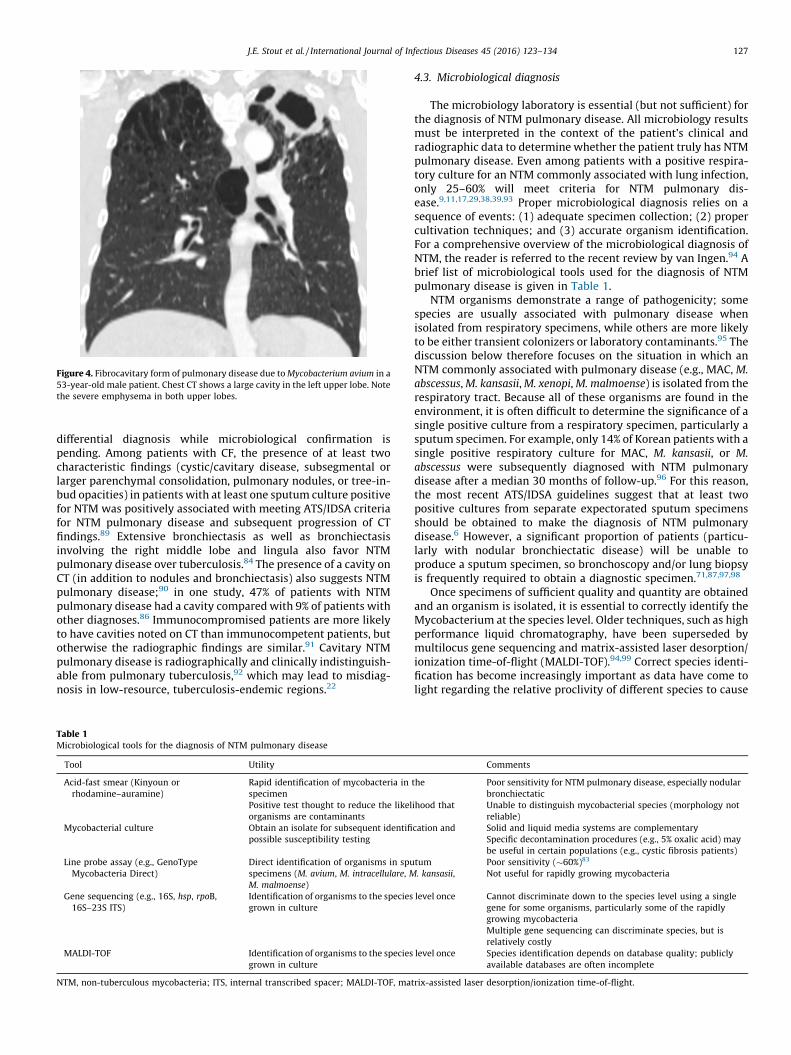
Figure 4. Fibrocavitary form of pulmonary disease due to Mycobacterium avium in a
53-year-old male patient. Chest CT shows a large cavity in the left upper lobe. Note
the severe emphysema in both upper lobes.
J.E. Stout et al. / International Journal of Infectious Diseases 45 (2016) 123–134 127
differential diagnosis while microbiological confirmation ispending. Among patients with CF, the presence of at least twocharacteristic findings (cystic/cavitary disease, subsegmental orlarger parenchymal consolidation, pulmonary nodules, or tree-in-bud opacities) in patients with at least one sputum culture positivefor NTM was positively associated with meeting ATS/IDSA criteriafor NTM pulmonary disease and subsequent progression of CTfindings.89 Extensive bronchiectasis as well as bronchiectasisinvolving the right middle lobe and lingula also favor NTMpulmonary disease over tuberculosis.84 The presence of a cavity onCT (in addition to nodules and bronchiectasis) also suggests NTMpulmonary disease;90 in one study, 47% of patients with NTMpulmonary disease had a cavity compared with 9% of patients withother diagnoses.86 Immunocompromised patients are more likelyto have cavities noted on CT than immunocompetent patients, butotherwise the radiographic findings are similar.91 Cavitary NTMpulmonary disease is radiographically and clinically indistinguish-able from pulmonary tuberculosis,92 which may lead to misdiag-nosis in low-resource, tuberculosis-endemic regions.22
Table 1Microbiological tools for the diagnosis of NTM pulmonary disease
Tool Utility
Acid-fast smear (Kinyoun or
rhodamine–auramine)
Rapid identification of mycobacteria in
specimen
Positive test thought to reduce the likel
organisms are contaminants
Mycobacterial culture Obtain an isolate for subsequent identifi
possible susceptibility testing
Line probe assay (e.g., GenoType
Mycobacteria Direct)
Direct identification of organisms in spu
specimens (M. avium, M. intracellulare, M
M. malmoense)
Gene sequencing (e.g., 16S, hsp, rpoB,
16S–23S ITS)
Identification of organisms to the species
grown in culture
MALDI-TOF Identification of organisms to the species
grown in culture
NTM, non-tuberculous mycobacteria; ITS, internal transcribed spacer; MALDI-TOF, mat
4.3. Microbiological diagnosis
The microbiology laboratory is essential (but not sufficient) forthe diagnosis of NTM pulmonary disease. All microbiology resultsmust be interpreted in the context of the patient’s clinical andradiographic data to determine whether the patient truly has NTMpulmonary disease. Even among patients with a positive respira-tory culture for an NTM commonly associated with lung infection,only 25–60% will meet criteria for NTM pulmonary dis-ease.9,11,17,29,38,39,93 Proper microbiological diagnosis relies on asequence of events: (1) adequate specimen collection; (2) propercultivation techniques; and (3) accurate organism identification.For a comprehensive overview of the microbiological diagnosis ofNTM, the reader is referred to the recent review by van Ingen.94 Abrief list of microbiological tools used for the diagnosis of NTMpulmonary disease is given in Table 1.
NTM organisms demonstrate a range of pathogenicity; somespecies are usually associated with pulmonary disease whenisolated from respiratory specimens, while others are more likelyto be either transient colonizers or laboratory contaminants.95 Thediscussion below therefore focuses on the situation in which anNTM commonly associated with pulmonary disease (e.g., MAC, M.
abscessus, M. kansasii, M. xenopi, M. malmoense) is isolated from therespiratory tract. Because all of these organisms are found in theenvironment, it is often difficult to determine the significance of asingle positive culture from a respiratory specimen, particularly asputum specimen. For example, only 14% of Korean patients with asingle positive respiratory culture for MAC, M. kansasii, or M.
abscessus were subsequently diagnosed with NTM pulmonarydisease after a median 30 months of follow-up.96 For this reason,the most recent ATS/IDSA guidelines suggest that at least twopositive cultures from separate expectorated sputum specimensshould be obtained to make the diagnosis of NTM pulmonarydisease.6 However, a significant proportion of patients (particu-larly with nodular bronchiectatic disease) will be unable toproduce a sputum specimen, so bronchoscopy and/or lung biopsyis frequently required to obtain a diagnostic specimen.71,87,97,98
Once specimens of sufficient quality and quantity are obtainedand an organism is isolated, it is essential to correctly identify theMycobacterium at the species level. Older techniques, such as highperformance liquid chromatography, have been superseded bymultilocus gene sequencing and matrix-assisted laser desorption/ionization time-of-flight (MALDI-TOF).94,99 Correct species identi-fication has become increasingly important as data have come tolight regarding the relative proclivity of different species to cause
Comments
the
ihood that
Poor sensitivity for NTM pulmonary disease, especially nodular
bronchiectatic
Unable to distinguish mycobacterial species (morphology not
reliable)
cation and Solid and liquid media systems are complementary
Specific decontamination procedures (e.g., 5% oxalic acid) may
be useful in certain populations (e.g., cystic fibrosis patients)
tum
. kansasii,
Poor sensitivity (�60%)83
Not useful for rapidly growing mycobacteria
level once Cannot discriminate down to the species level using a single
gene for some organisms, particularly some of the rapidly
growing mycobacteria
Multiple gene sequencing can discriminate species, but is
relatively costly
level once Species identification depends on database quality; publicly
available databases are often incomplete
rix-assisted laser desorption/ionization time-of-flight.

J.E. Stout et al. / International Journal of Infectious Diseases 45 (2016) 123–134128
NTM pulmonary disease. For example, in a cohort of patients withMAC isolated from respiratory specimens, patients with M. avium
or Mycobacterium intracellulare were significantly (2–3-fold) morelikely to meet criteria for NTM pulmonary disease than patientswith Mycobacterium chimaera.100 Other authors have foundrelative differences in the likelihood of M. avium and M.
intracellulare or even M. avium subspecies to be associated withpulmonary disease when isolated from respiratory speci-mens.101,102
4.4. Immunological diagnosis
Given the non-specific symptoms of NTM pulmonary disease,the limitations of radiography, and the fact that invasiveprocedures are frequently required to make the diagnosis, otherdiagnostic modalities are sorely needed. Skin testing using NTMantigens has not been useful for the diagnosis of NTM disease. Skintest reactivity to NTM antigens is common among healthy persons,and there is immunological cross-reactivity among NTM andbetween NTM and tuberculosis.103,104 Interferon gamma releaseassays have been used to diagnose tuberculosis infection, but noneare commercially available for NTM. These assays are unlikely tobe helpful for NTM diagnosis; patients with MAC pulmonarydisease actually demonstrate less interferon gamma release inresponse to MAC antigens than persons without disease who havea positive skin test response to MAC sensitin.105 Serodiagnosticassays that detect antibodies against specific mycobacterialantigens are increasingly being tested for MAC and M.
abscessus. One assay that has been examined uses an enzymeimmunoassay that detects IgA antibodies that bind to glycopepti-dolipid, which is found in the MAC cell wall. The reportedsensitivity of this serodiagnostic assay for MAC pulmonary diseaseranges from 70% to 92%, depending on the population and cut-offvalue studied, and there appears to be a positive associationbetween antibody levels and disease activity (although there iscross-reactivity with M. abscessus as well, so the assay is notcompletely specific for MAC).106–113 A second serodiagnostic testexamines IgG antibodies to mycobacterial antigen A60 as adiagnostic for M. abscessus pulmonary disease. A60 antibody titerswere found to be relatively sensitive (86.7%) and specific (95.1%)for M. abscessus pulmonary disease among patients with CF, andseemed to correlate with disease activity.114 Unfortunately, A60antigen is not specific for M. abscessus, and these antibodies may be
Table 2Conditions that predispose to NTM pulmonary disease
Category Condition
Structural lung disease Alpha-1-antitrypsin deficiency
Bronchiectasis
COPD
Ciliary dyskinesia
Cystic fibrosis
Pulmonary alveolar proteinosis
Tuberculosis
Immune deficiencies Chronic granulomatous disease
Common variable immunodeficiency
Hematological malignancy
HIV
Interleukin-12/interferon-gamma axis defects
Inhaled corticosteroids
Mendelian susceptibility to mycobacterial disease
Transplant (stem cell or solid organ)
NTM, non-tuberculous mycobacteria; COPD, chronic obstructive pulmonary disease.
elevated in other mycobacterial diseases such as tuberculosis.115
While neither of these serological tests is ready for clinical use,they represent a line of research that could significantly advancethe diagnosis of NTM pulmonary disease (Tables 2 and 3).
5. Treatment
5.1. Treat or not
A diagnosis of NTM lung disease alone does not obligate theimmediate initiation of treatment directed against the NTMpathogen. Instead, this decision should be based on the potentialrisks and benefits of a prolonged course of multiple antibiotics forthe individual patient, taking into consideration age, comorbidmedical conditions, and disease type.6,116 Patients with fibroca-vitary disease usually require immediate treatment becausecavitary disease is associated with a higher rate of mortality dueto NTM lung disease.117,118 Conversely, nodular bronchiectaticdisease tends to occur in the absence of significant comorbidityand often progresses slowly.119 Therefore, early treatment of mildand indolent nodular bronchiectatic disease may not be advisablebecause of the adverse effects of the long-term use of multipleantibiotics.6,116 The decision may also be aided by molecularanalyses, because specific mycobacterial genotypes have beenshown to be predictive of disease progression or treatmentresponse in patients with NTM lung disease.120,121 Becausemicrobiological cure can be difficult to achieve in a substantialproportion of patients, other treatment goals, including improve-ment in quality of life, may be more appropriate.122,123
Once the decision has been made to initiate treatment for NTMlung disease, the treatment regimens should be formulatedaccording to established guidelines, understanding that a substan-tial proportion of current guidelines rely upon expert opinionrather than randomized clinical trials.124 However, adherence tothe current guidelines for treating NTM lung disease is poor, andsuboptimal or potentially harmful antibiotic regimens arecommonly prescribed in practice.125
5.2. Antibiotic therapy
5.2.1. MAC lung disease
Newer azalide/macrolide drugs such as azithromycin andclarithromycin (subsequently both referred to as macrolides here)
Comments
May be predisposing or result of NTM infection
Presence of cystic fibrosis transmembrane conductance regulator gene
polymorphisms without cystic fibrosis disease may predispose to NTM
infection by increasing the risk of bronchiectasis
Impaired macrophage function in this disorder may augment the risk over that
conferred by structural abnormalities
May cause bronchiectasis or other airway abnormalities; possible shared
underlying genetic vulnerability with NTM
Both of these are more likely to present with disseminated than pulmonary
disease
NTM infection relatively uncommon compared to disseminated disease; M.
kansasii and M. xenopi more likely to cause pulmonary infection
Disseminated disease more than pulmonary; genetic disorders rare; acquired
autoantibodies to interferon-gamma increasingly recognized
Local immunosuppression
Likely under-recognized underlying deficit in ‘idiopathic’ NTM pulmonary
infection—complex, polygenetic disorder
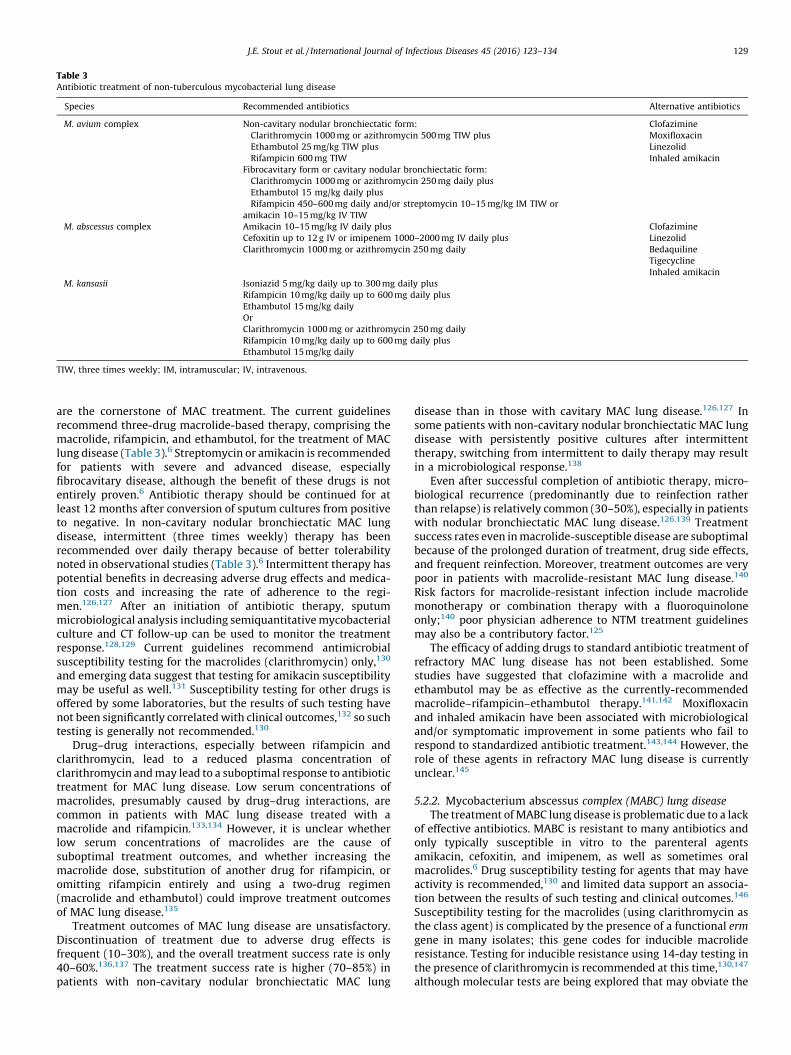
Table 3Antibiotic treatment of non-tuberculous mycobacterial lung disease
Species Recommended antibiotics Alternative antibiotics
M. avium complex Non-cavitary nodular bronchiectatic form:
Clarithromycin 1000 mg or azithromycin 500 mg TIW plus
Ethambutol 25 mg/kg TIW plus
Rifampicin 600 mg TIW
Fibrocavitary form or cavitary nodular bronchiectatic form:
Clarithromycin 1000 mg or azithromycin 250 mg daily plus
Ethambutol 15 mg/kg daily plus
Rifampicin 450–600 mg daily and/or streptomycin 10–15 mg/kg IM TIW or
amikacin 10–15 mg/kg IV TIW
Clofazimine
Moxifloxacin
Linezolid
Inhaled amikacin
M. abscessus complex Amikacin 10–15 mg/kg IV daily plus
Cefoxitin up to 12 g IV or imipenem 1000–2000 mg IV daily plus
Clarithromycin 1000 mg or azithromycin 250 mg daily
Clofazimine
Linezolid
Bedaquiline
Tigecycline
Inhaled amikacin
M. kansasii Isoniazid 5 mg/kg daily up to 300 mg daily plus
Rifampicin 10 mg/kg daily up to 600 mg daily plus
Ethambutol 15 mg/kg daily
Or
Clarithromycin 1000 mg or azithromycin 250 mg daily
Rifampicin 10 mg/kg daily up to 600 mg daily plus
Ethambutol 15 mg/kg daily
TIW, three times weekly; IM, intramuscular; IV, intravenous.
J.E. Stout et al. / International Journal of Infectious Diseases 45 (2016) 123–134 129
are the cornerstone of MAC treatment. The current guidelinesrecommend three-drug macrolide-based therapy, comprising themacrolide, rifampicin, and ethambutol, for the treatment of MAClung disease (Table 3).6 Streptomycin or amikacin is recommendedfor patients with severe and advanced disease, especiallyfibrocavitary disease, although the benefit of these drugs is notentirely proven.6 Antibiotic therapy should be continued for atleast 12 months after conversion of sputum cultures from positiveto negative. In non-cavitary nodular bronchiectatic MAC lungdisease, intermittent (three times weekly) therapy has beenrecommended over daily therapy because of better tolerabilitynoted in observational studies (Table 3).6 Intermittent therapy haspotential benefits in decreasing adverse drug effects and medica-tion costs and increasing the rate of adherence to the regi-men.126,127 After an initiation of antibiotic therapy, sputummicrobiological analysis including semiquantitative mycobacterialculture and CT follow-up can be used to monitor the treatmentresponse.128,129 Current guidelines recommend antimicrobialsusceptibility testing for the macrolides (clarithromycin) only,130
and emerging data suggest that testing for amikacin susceptibilitymay be useful as well.131 Susceptibility testing for other drugs isoffered by some laboratories, but the results of such testing havenot been significantly correlated with clinical outcomes,132 so suchtesting is generally not recommended.130
Drug–drug interactions, especially between rifampicin andclarithromycin, lead to a reduced plasma concentration ofclarithromycin and may lead to a suboptimal response to antibiotictreatment for MAC lung disease. Low serum concentrations ofmacrolides, presumably caused by drug–drug interactions, arecommon in patients with MAC lung disease treated with amacrolide and rifampicin.133,134 However, it is unclear whetherlow serum concentrations of macrolides are the cause ofsuboptimal treatment outcomes, and whether increasing themacrolide dose, substitution of another drug for rifampicin, oromitting rifampicin entirely and using a two-drug regimen(macrolide and ethambutol) could improve treatment outcomesof MAC lung disease.135
Treatment outcomes of MAC lung disease are unsatisfactory.Discontinuation of treatment due to adverse drug effects isfrequent (10–30%), and the overall treatment success rate is only40–60%.136,137 The treatment success rate is higher (70–85%) inpatients with non-cavitary nodular bronchiectatic MAC lung
disease than in those with cavitary MAC lung disease.126,127 Insome patients with non-cavitary nodular bronchiectatic MAC lungdisease with persistently positive cultures after intermittenttherapy, switching from intermittent to daily therapy may resultin a microbiological response.138
Even after successful completion of antibiotic therapy, micro-biological recurrence (predominantly due to reinfection ratherthan relapse) is relatively common (30–50%), especially in patientswith nodular bronchiectatic MAC lung disease.126,139 Treatmentsuccess rates even in macrolide-susceptible disease are suboptimalbecause of the prolonged duration of treatment, drug side effects,and frequent reinfection. Moreover, treatment outcomes are verypoor in patients with macrolide-resistant MAC lung disease.140
Risk factors for macrolide-resistant infection include macrolidemonotherapy or combination therapy with a fluoroquinoloneonly;140 poor physician adherence to NTM treatment guidelinesmay also be a contributory factor.125
The efficacy of adding drugs to standard antibiotic treatment ofrefractory MAC lung disease has not been established. Somestudies have suggested that clofazimine with a macrolide andethambutol may be as effective as the currently-recommendedmacrolide–rifampicin–ethambutol therapy.141,142 Moxifloxacinand inhaled amikacin have been associated with microbiologicaland/or symptomatic improvement in some patients who fail torespond to standardized antibiotic treatment.143,144 However, therole of these agents in refractory MAC lung disease is currentlyunclear.145
5.2.2. Mycobacterium abscessus complex (MABC) lung disease
The treatment of MABC lung disease is problematic due to a lackof effective antibiotics. MABC is resistant to many antibiotics andonly typically susceptible in vitro to the parenteral agentsamikacin, cefoxitin, and imipenem, as well as sometimes oralmacrolides.6 Drug susceptibility testing for agents that may haveactivity is recommended,130 and limited data support an associa-tion between the results of such testing and clinical outcomes.146
Susceptibility testing for the macrolides (using clarithromycin asthe class agent) is complicated by the presence of a functional erm
gene in many isolates; this gene codes for inducible macrolideresistance. Testing for inducible resistance using 14-day testing inthe presence of clarithromycin is recommended at this time,130,147
although molecular tests are being explored that may obviate the

J.E. Stout et al. / International Journal of Infectious Diseases 45 (2016) 123–134130
need for such testing.148,149 Current guidelines recommend aregimen comprising two parenteral agents and a macrolide for 2 to4 months (Table 3).6 The reported treatment success rate rangesfrom 50% to 70%.146,150–153 However, these studies were retro-spective, involved various regimens with different treatmentdurations, and many patients also underwent surgery.154 There-fore, the results cannot be generalized to all patients, and theseregimens should be applied with caution in individual patientswith MABC lung disease.
MABC lung disease can be further subdivided by causativeorganism: M. abscessus subsp. abscessus (hereafter M. abscessus), M.
abscessus subsp. massiliense (hereafter M. massiliense), and M.
abscessus subsp. bolletii (hereafter M. bolletii).155,156 However, thetaxonomic status of MABC is still a subject of debate.147 It isimportant to identify the exact species, because M. massiliense hasbeen consistently associated with a higher probability of treatmentsuccess than other MABC organisms. For example, one studyobserved sustained negative cultures in 88% of patients with M.
massiliense lung disease, compared with 25% of patients with M.
abscessus lung disease.157 Similar results have been reported inother studies.158–161 These differences in treatment outcomes areattributable to differential macrolide susceptibility among mem-bers of the MABC. M. abscessus and M. bolletii display induciblemacrolide resistance conferred by the ribosomal methyl transfer-ase erm(41) gene.162–164 In contrast, M. massiliense strains do notshow inducible resistance because of a large deletion in theerm(41) gene that renders it non-functional.162–164 However, apolymorphism is present at position 28 of erm(41) (T or C) in M.
abscessus.165 The majority of M. abscessus isolates are of the T28sequevar and exhibit inducible macrolide resistance, but 7–18% areof the C28 sequevar and susceptible to macrolides because of lossof function of the erm(41) gene.149,166,167 Thus, both thesequencing of the erm(41) gene of M. abscessus as well as preciseidentification of MABC are important for predicting treatmentoutcomes.149,166,167
None of the commonly used MABC treatment regimens iscategorically safe, well-tolerated, and effective. The prolonged useof parenteral amikacin is limited by nephrotoxicity and ototoxicity.Inhaled amikacin can be considered as it might have lower toxicitythan parenteral therapy, albeit with limited efficacy data.144
Clofazimine, linezolid, bedaquiline, and tigecycline show in vitroactivities against most isolates of MABC, and two-drug combina-tions, such as clofazimine and amikacin, clofazimine andtigecycline, and clofazimine and bedaquiline, exert synergisticeffects.168–174 Treatment with an antibiotic regimen containingtigecycline for >1 month was shown to result in an improvementin >60% of patients with treatment-refractory M. abscessus lungdisease. However, 90% of patients experienced adverse reactions,largely gastrointestinal in nature.175 Bedaquiline as salvagetherapy for advanced M. abscessus lung disease was also shownto result in improvement in six out 10 patients.176 Therefore,experts currently believe that curative therapy is possible only inpatients with limited disease using a combination of surgicalresection and chemotherapy, particularly for M. abscessus lungdisease.177–179 Suppressive therapy using periodic parenteralantibiotics or oral antibiotics to control symptoms and progressionof disease may be appropriate in some patients.6
5.2.3. Mycobacterium kansasii lung disease
Rifampicin is the most important drug for the treatment of M.
kansasii lung disease because it is associated with a high cultureconversion rate and low long-term relapse rate.6 The guidelinestherefore recommend a rifampicin-containing regimen in combi-nation with isoniazid and ethambutol (Table 3).6 This regimen hasshown a high sputum culture conversion rate and a low long-termrecurrence rate. Treatment for this disease is recommended to
continue for at least 12 months after negative sputum conversion.Because antibiotic treatment shows an excellent outcome, surgicalresection is not recommended for this disease. Substitution of anewer macrolide for isoniazid was recently recommended becauseof the efficacy of new macrolides against M. kansasii; the limiteddata on this regimen are encouraging, but additional study isneeded.180,181 Fluoroquinolones may help in the presence ofrifampicin-resistant M. kansasii lung disease.182
5.2.4. Other NTM
M. xenopi infection is commonly associated with underlyinglung conditions such as COPD, and the prognosis is poor.183–185 Therecommended treatment regimen is likely a combination ofclarithromycin, ethambutol, and rifampicin, with or without aninitial course of streptomycin. Moxifloxacin can be substituted forone of the recommended drugs, as guided by drug susceptibilitytesting.6,186 Regarding the treatment of M. malmoense lung disease,previous studies have suggested combination therapy of isoniazid,rifampicin, and ethambutol, with or without fluoroquinolones/macrolides.187,188 Most M. szulgai isolates are susceptible in vitroto many anti-tuberculosis drugs. Treatment with isoniazid,rifampicin, and ethambutol or macrolide, rifampicin, and etham-butol may lead to favorable outcomes without microbiologicalrecurrence.189–191 The optimal antibiotic regimen for Mycobacte-
rium simiae lung disease is unknown. Although combinationtherapy with clarithromycin, ethambutol, and rifampicin issometimes recommended,6 treatment outcomes in M. simiae lungdisease are typically poor due to high rates of resistance to thesedrugs and a likely lack of rifampicin–ethambutol synergy.192–194 Atreatment regimen including both a macrolide and moxifloxacin,and one or two additional drugs based on drug susceptibility testresults, or adding clofazimine to an amikacin-containing regimen,has been suggested recently due to their synergistic activity.169,193
M. scrofulaceum lung disease is associated with old tuberculosisscarring and silicosis,195 and there appears to be no optimaltreatment regimen. Currently, rifampicin, ethambutol, isoniazid,and perhaps macrolides/fluoroquinolones, can be considered withsome guidance from drug susceptibility testing, similar to thesituation for members of the M. simiae complex.6,196
5.3. Surgical therapy
Because the success rates of combination antibiotic treatmentfor NTM lung disease are unsatisfactory, adjuvant surgicalresection could be considered in selected patients with localizedrefractory NTM lung disease who can tolerate the procedure.Although the sputum culture conversion rate among patients whoundergo resection is >90% with acceptable morbidity andmortality rates in many reported series, postoperative complica-tions including bronchopleural fistula have been reported,especially in patients with fibrocavitary NTM pulmonary dis-ease.197–199 The optimal selection criteria, timing, and duration ofpostoperative medical therapy require further evaluation.
6. Epilogue
There remain considerable gaps in our knowledge regarding themanagement of NTM lung disease. Microbiological eradicationusing currently available antibiotic regimens is difficult andmicrobiological recurrence is common even after successfulcompletion of treatment of NTM lung disease. The role of drugsusceptibility testing and therapeutic drug monitoring has notbeen fully established. Discrepancies between in vitro drugsusceptibility and clinical outcomes and the lack of pharmacoki-netic and pharmacodynamic indices for the currently used drugsmake the selection of the optimal antibiotic and determination of
J.E. Stout et al. / International Journal of Infectious Diseases 45 (2016) 123–134 131
the appropriate dosage difficult.200,201 The increasing rate ofantibiotic resistance to commonly used drugs for NTM lungdisease, such as macrolides, is a challenging problem.202,203 Thereare currently only limited data on the efficacy of repurposed andnew agents. Perhaps most importantly, our understanding ofpatient-relevant outcomes for the treatment of NTM pulmonarydisease is still quite premature. Given that survival among patientswith NTM pulmonary disease is primarily determined byunderlying comorbidities, rather than by the NTM disease or itstreatment,117,118,204 studies focusing on the impact of NTMpulmonary disease and its treatment on patient function andquality of life are of paramount importance.122 Such data areparticularly relevant given that preliminary work suggests weakassociations between traditional ‘hard’ endpoints such as micro-biological culture conversion and radiographic improvement andpatient-centered endpoints such as quality of life.123,203 To thisend, quality clinical trials focused on such patient-centeredoutcomes are needed to delineate new regimens or approachesin the management of NTM lung diseases.
Ethical approval: Not required.Conflict of interest: Jason E. Stout, no conflicts to declare; Won-
Jung Koh, no conflicts to declare; Wing Wai Yew, no conflicts todeclare.
References
1. Tortoli E. The new mycobacteria: an update. FEMS Immunol Med Microbiol2006;48:159–78.
2. Tortoli E. Microbiological features and clinical relevance of new species of thegenus Mycobacterium. Clin Microbiol Rev 2014;27:727–52.
3. Kendall BA, Winthrop KL. Update on the epidemiology of pulmonary nontu-berculous mycobacterial infections. Semin Respir Crit Care Med 2013;34:87–94.
4. Brode SK, Daley CL, Marras TK. The epidemiologic relationship betweentuberculosis and non-tuberculous mycobacterial disease: a systematic review.Int J Tuberc Lung Dis 2014;18:1370–7.
5. Prevots DR, Marras TK. Epidemiology of human pulmonary infection withnontuberculous mycobacteria: a review. Clin Chest Med 2015;36:13–34.
6. Griffith DE, Aksamit T, Brown-Elliott BA, Catanzaro A, Daley C, Gordin F, et al.An official ATS/IDSA statement: diagnosis, treatment, and prevention ofnontuberculous mycobacterial diseases. Am J Respir Crit Care Med 2007;175:367–416.
7. Al Houqani M, Jamieson F, Chedore P, Mehta M, May K, Marras TK. Isolationprevalence of pulmonary nontuberculous mycobacteria in Ontario in 2007.Can Respir J 2011;18:19–24.
8. Marras TK, Mendelson D, Marchand-Austin A, May K, Jamieson FB. Pulmonarynontuberculous mycobacterial disease, Ontario, Canada, 1998–2010. EmergInfect Dis 2013;19:1889–91.
9. Winthrop KL, McNelley E, Kendall B, Marshall-Olson A, Morris C, Cassidy M,et al. Pulmonary nontuberculous mycobacterial disease prevalence and clini-cal features: an emerging public health disease. Am J Respir Crit Care Med2010;182:977–82.
10. Henkle E, Hedberg K, Schafer S, Novosad S, Winthrop KL. Population-basedincidence of pulmonary nontuberculous mycobacterial disease in Oregon2007 to 2012. Ann Am Thorac Soc 2015;12:642–7.
11. Prevots DR, Shaw PA, Strickland D, Jackson LA, Raebel MA, Blosky MA, et al.Nontuberculous mycobacterial lung disease prevalence at four integratedhealth care delivery systems. Am J Respir Crit Care Med 2010;182:970–6.
12. Adjemian J, Olivier KN, Seitz AE, Holland SM, Prevots DR. Prevalence ofnontuberculous mycobacterial lung disease in U.S. Medicare beneficiaries.Am J Respir Crit Care Med 2012;185:881–6.
13. Pedro Hda S, Pereira MI, Goloni Mdo R, Ueki SY, Chimara E. Nontuberculousmycobacteria isolated in Sao Jose do Rio Preto, Brazil between 1996 and 2005. JBras Pneumol 2008;34:950–5.
14. de Mello KG, Mello FC, Borga L, Rolla V, Duarte RS, Sampaio EP, et al. Clinicaland therapeutic features of pulmonary nontuberculous mycobacterial disease,Brazil, 1993–2011. Emerg Infect Dis 2013;19:393–9.
15. Henry MT, Inamdar L, O’Riordain D, Schweiger M, Watson JP. Nontuberculousmycobacteria in non-HIV patients: epidemiology, treatment and response. EurRespir J 2004;23:741–6.
16. Gerogianni I, Papala M, Kostikas K, Petinaki E, Gourgoulianis KI. Epidemiologyand clinical significance of mycobacterial respiratory infections in CentralGreece. Int J Tuberc Lung Dis 2008;12:807–12.
17. van Ingen J, Bendien SA, de Lange WC, Hoefsloot W, Dekhuijzen PN, Boeree MJ,et al. Clinical relevance of non-tuberculous mycobacteria isolated in theNijmegen-Arnhem region, the Netherlands. Thorax 2009;64:502–6.
18. Hoefsloot W, van Ingen J, Andrejak C, Angeby K, Bauriaud R, Bemer P, et al. Thegeographic diversity of nontuberculous mycobacteria isolated from pulmo-nary samples: an NTM-NET collaborative study. Eur Respir J 2013;42:1604–13.
19. Aliyu G, El-Kamary SS, Abimiku A, Brown C, Tracy K, Hungerford L, et al.Prevalence of non-tuberculous mycobacterial infections among tuberculosissuspects in Nigeria. PLoS One 2013;8:e63170.
20. Asiimwe BB, Bagyenzi GB, Ssengooba W, Mumbowa F, Mboowa G, Wajja A,et al. Species and genotypic diversity of non-tuberculous mycobacteria iso-lated from children investigated for pulmonary tuberculosis in rural Uganda.BMC Infect Dis 2013;13:88.
21. Newman MJ, Addo KK, Aboagye S, Bonsu FA, Caulley P, Hesse IF, et al. Culture andsensitivity of mycobacterial isolates from cases of pulmonary tuberculosis classi-fied as treatment failures in a teaching hospital. West Afr J Med 2007;26:131–3.
22. Maiga M, Siddiqui S, Diallo S, Diarra B, Traore B, Shea YR, et al. Failure torecognize nontuberculous mycobacteria leads to misdiagnosis of chronicpulmonary tuberculosis. PLoS One 2012;7:e36902.
23. Simons S, van Ingen J, Hsueh PR, Van Hung N, Dekhuijzen PN, Boeree MJ, et al.Nontuberculous mycobacteria in respiratory tract infections, eastern Asia.Emerg Infect Dis 2011;17:343–9.
24. Morimoto K, Iwai K, Uchimura K, Okumura M, Yoshiyama T, Yoshimori K, et al.A steady increase in nontuberculous mycobacteriosis mortality and estimatedprevalence in Japan. Ann Am Thorac Soc 2014;11:1–8.
25. Lai CC, Tan CK, Chou CH, Hsu HL, Liao CH, Huang YT, et al. Increasing incidenceof nontuberculous mycobacteria, Taiwan, 2000–2008. Emerg Infect Dis2010;16:294–6.
26. Jing H, Wang H, Wang Y, Deng Y, Li X, Liu Z, et al. Prevalence of nontuberculousmycobacteria infection, China, 2004–2009. Emerg Infect Dis 2012;18:527–8.
27. Khann S, Mao ET, Rajendra YP, Satyanarayana S, Nagaraja SB, Kumar AM.Linkage of presumptive multidrug resistant tuberculosis (MDR-TB) patients todiagnostic and treatment services in Cambodia. PLoS One 2013;8:e59903.
28. Shao Y, Chen C, Song H, Li G, Liu Q, Li Y, et al. The epidemiology and geographicdistribution of nontuberculous mycobacteria clinical isolates from sputumsamples in the eastern region of China. PLoS Negl Trop Dis 2015;9:e0003623.
29. Thomson RM. Changing epidemiology of pulmonary nontuberculous myco-bacteria infections. Emerg Infect Dis 2010;16:1576–83.
30. Haverkort F. National atypical mycobacteria survey, 2000. Commun Dis Intell QRep 2003;27:180–9.
31. Freeman J, Morris A, Blackmore T, Hammer D, Munroe S, McKnight L. Incidenceof nontuberculous mycobacterial disease in New Zealand, 2004. N Z Med J2007;120:U2580.
32. Thomson RM, Carter R, Tolson C, Coulter C, Huygens F, Hargreaves M. Factorsassociated with the isolation of nontuberculous mycobacteria (NTM) from alarge municipal water system in Brisbane, Australia. BMC Microbiol 2013;13:89.
33. Donohue MJ, Mistry JH, Donohue JM, O’Connell K, King D, Byran J, et al.Increased frequency of nontuberculous mycobacteria detection at potablewater taps within the United States. Environ Sci Technol 2015;49:6127–33.
34. Wolinsky E. Nontuberculous mycobacteria and associated diseases. Am RevRespir Dis 1979;119:107–59.
35. Bodle EE, Cunningham JA, Della-Latta P, Schluger NW, Saiman L. Epidemiologyof nontuberculous mycobacteria in patients without HIV infection, New YorkCity. Emerg Infect Dis 2008;14:390–6.
36. Al-Mahruqi SH, van-Ingen J, Al-Busaidy S, Boeree MJ, Al-Zadjali S, Patel A, et al.Clinical relevance of nontuberculous mycobacteria. Oman Emerg Infect Dis2009;15:292–4.
37. Johnson MM, Odell JA. Nontuberculous mycobacterial pulmonary infections. JThorac Dis 2014;6:210–20.
38. Marras TK, Mehta M, Chedore P, May K, Al Houqani M, Jamieson F. Non-tuberculous mycobacterial lung infections in Ontario, Canada: clinical andmicrobiological characteristics. Lung 2010;188:289–99.
39. Andrejak C, Thomsen VO, Johansen IS, Riis A, Benfield TL, Duhaut P, et al.Nontuberculous pulmonary mycobacteriosis in Denmark: incidence and prog-nostic factors. Am J Respir Crit Care Med 2010;181:514–21.
40. Sonnenberg P, Murray J, Glynn JR, Thomas RG, Godfrey-Faussett P, Shearer S.Risk factors for pulmonary disease due to culture-positive M. tuberculosis ornontuberculous mycobacteria in South African gold miners. Eur Respir J2000;15:291–6.
41. McGrath EE, Bardsley P. An association between Mycobacterium malmoenseand coal workers’ pneumoconiosis. Lung 2009;187:51–4.
42. Gitti Z, Mantadakis E, Maraki S, Samonis G. Clinical significance and antibioticsusceptibilities of nontuberculous mycobacteria from patients in Crete,Greece. Future Microbiol 2011;6:1099–109.
43. Matveychuk A, Fuks L, Priess R, Hahim I, Shitrit D. Clinical and radiologicalfeatures of Mycobacterium kansasii and other NTM infections. Respir Med2012;106:1472–7.
44. Leung JM, Olivier KN. Nontuberculous mycobacteria: the changing epidemi-ology and treatment challenges in cystic fibrosis. Curr Opin Pulm Med2013;19:662–9.
45. Rodman DM, Polis JM, Heltshe SL, Sontag MK, Chacon C, Rodman RV, et al. Latediagnosis defines a unique population of long-term survivors of cystic fibrosis.Am J Respir Crit Care Med 2005;171:621–6.
46. Adjemian J, Olivier KN, Prevots DR. Nontuberculous mycobacteria amongpatients with cystic fibrosis in the United States: screening practices andenvironmental risk. Am J Respir Crit Care Med 2014;190:581–6.
47. Binder AM, Adjemian J, Olivier KN, Prevots DR. Epidemiology of nontubercu-lous mycobacterial infections and associated chronic macrolide use amongpersons with cystic fibrosis. Am J Respir Crit Care Med 2013;188:807–12.
48. Olivier KN, Weber DJ, Wallace Jr RJ, Faiz AR, Lee JH, Zhang Y, et al. Non-tuberculous mycobacteria. I: multicenter prevalence study in cystic fibrosis.Am J Respir Crit Care Med 2003;167:828–34.

J.E. Stout et al. / International Journal of Infectious Diseases 45 (2016) 123–134132
49. Qvist T, Pressler T, Hoiby N, Katzenstein TL. Shifting paradigms of nontuber-culous mycobacteria in cystic fibrosis. Respir Res 2014;15:41.
50. Martiniano SL, Nick JA. Nontuberculous mycobacterial infections in cysticfibrosis. Clin Chest Med 2015;36:101–15.
51. Aitken ML, Limaye A, Pottinger P, Whimbey E, Goss CH, Tonelli MR, et al.Respiratory outbreak of Mycobacterium abscessus subspecies massiliense in alung transplant and cystic fibrosis center. Am J Respir Crit Care Med2012;185:231–2.
52. Mirsaeidi M, Hadid W, Ericsoussi B, Rodgers D, Sadikot RT. Non-tuberculousmycobacterial disease is common in patients with non-cystic fibrosis bron-chiectasis. Int J Infect Dis 2013;17:e1000–4.
53. Thomson RM, Armstrong JG, Looke DF. Gastroesophageal reflux disease, acidsuppression, and Mycobacterium avium complex pulmonary disease. Chest2007;131:1166–72.
54. Griffith DE, Aksamit TR. Bronchiectasis and nontuberculous mycobacterialdisease. Clin Chest Med 2012;33:283–95.
55. Bonaiti G, Pesci A, Marruchella A, Lapadula G, Gori A, Aliberti S. Nontubercu-lous mycobacteria in noncystic fibrosis bronchiectasis. Biomed Res Int2015;2015:197950.
56. Wickremasinghe M, Ozerovitch LJ, Davies G, Wodehouse T, Chadwick MV,Abdallah S, et al. Non-tuberculous mycobacteria in patients with bronchiec-tasis. Thorax 2005;60:1045–51.
57. Kunst H, Wickremasinghe M, Wells A, Wilson R. Nontuberculous mycobacte-rial disease and Aspergillus-related lung disease in bronchiectasis. Eur Respir J2006;28:352–7.
58. Yew WW, Chiang CY, Lumb R, Islam T. Are pulmonary non-tuberculousmycobacteria of concern in the Western Pacific Region? Int J Tuberc LungDis 2015;19:499–500.
59. Hwang SM, Lim MS, Hong YJ, Kim TS, Park KU, Song J, et al. Simultaneousdetection of Mycobacterium tuberculosis complex and nontuberculous myco-bacteria in respiratory specimens. Tuberculosis (Edinb) 2013;93:642–6.
60. Damaraju D, Jamieson F, Chedore P, Marras TK. Isolation of non-tuberculousmycobacteria among patients with pulmonary tuberculosis in Ontario,Canada. Int J Tuberc Lung Dis 2013;17:676–81.
61. Chu H, Zhao L, Xiao H, Zhang Z, Zhang J, Gui T, et al. Prevalence of nontu-berculous mycobacteria in patients with bronchiectasis: a meta-analysis. ArchMed Sci 2014;10:661–8.
62. Noone PG, Leigh MW, Sannuti A, Minnix SL, Carson JL, Hazucha M, et al.Primary ciliary dyskinesia: diagnostic and phenotypic features. Am J Respir CritCare Med 2004;169:459–67.
63. Bienvenu T, Sermet-Gaudelus I, Burgel PR, Hubert D, Crestani B, Bassinet L, et al.Cystic fibrosis transmembrane conductance regulator channel dysfunction innon-cystic fibrosis bronchiectasis. Am J Respir Crit Care Med 2010;181:1078–84.
64. Chan ED, Kaminska AM, Gill W, Chmura K, Feldman NE, Bai X, et al. Alpha-1-antitrypsin (AAT) anomalies are associated with lung disease due to rapidlygrowing mycobacteria and AAT inhibits Mycobacterium abscessus infection ofmacrophages. Scand J Infect Dis 2007;39:690–6.
65. Honda JR, Knight V, Chan ED. Pathogenesis and risk factors for nontuberculousmycobacterial lung disease. Clin Chest Med 2015;36:1–11.
66. Henkle E, Winthrop KL. Nontuberculous mycobacteria infections in immuno-suppressed hosts. Clin Chest Med 2015;36:91–9.
67. Winthrop KL, Chang E, Yamashita S, Iademarco MF, LoBue PA. Nontuberculousmycobacteria infections and anti-tumor necrosis factor-alpha therapy. EmergInfect Dis 2009;15:1556–61.
68. Andrejak C, Nielsen R, Thomsen VO, Duhaut P, Sorensen HT, Thomsen RW.Chronic respiratory disease, inhaled corticosteroids and risk of non-tubercu-lous mycobacteriosis. Thorax 2013;68:256–62.
69. McCarthy KD, Cain KP, Winthrop KL, Udomsantisuk N, Lan NT, Sar B, et al.Nontuberculous mycobacterial disease in patients with HIV in Southeast Asia.Am J Respir Crit Care Med 2012;185:981–8.
70. Sapkota BR, Hijikata M, Matsushita I, Tanaka G, Ieki R, Kobayashi N, et al.Association of SLC11A1 (NRAMP1) polymorphisms with pulmonary Mycobac-terium avium complex infection. Hum Immunol 2012;73:529–36.
71. Prince DS, Peterson DD, Steiner RM, Gottlieb JE, Scott R, Israel HL, et al.Infection with Mycobacterium avium complex in patients without predispos-ing conditions. N Engl J Med 1989;321:863–8.
72. Reich JM, Johnson RE. Mycobacterium avium complex pulmonary diseasepresenting as an isolated lingular or middle lobe pattern. The Lady Wind-ermere syndrome. Chest 1992;101:1605–9.
73. Colombo RE, Hill SC, Claypool RJ, Holland SM, Olivier KN. Familial clustering ofpulmonary nontuberculous mycobacterial disease. Chest 2010;137:629–34.
74. Kim RD, Greenberg DE, Ehrmantraut ME, Guide SV, Ding L, Shea Y, et al.Pulmonary nontuberculous mycobacterial disease: prospective study of adistinct preexisting syndrome. Am J Respir Crit Care Med 2008;178:1066–74.
75. Leung JM, Fowler C, Smith C, Adjemian J, Frein C, Claypool RJ, et al. A familialsyndrome of pulmonary nontuberculous mycobacteria infections. Am J RespirCrit Care Med 2013;188:1373–6.
76. Kartalija M, Ovrutsky AR, Bryan CL, Pott GB, Fantuzzi G, Thomas J, et al. Patientswith nontuberculous mycobacterial lung disease exhibit unique body andimmune phenotypes. Am J Respir Crit Care Med 2013;187:197–205.
77. Szymanski EP, Leung JM, Fowler CJ, Haney C, Hsu AP, Chen F, et al. Pulmonarynontuberculous mycobacterial infection. A multisystem, multigenic disease.Am J Respir Crit Care Med 2015;192:618–28.
78. Alpert JB, Fantauzzi JP, Melamud K, Greenwood H, Naidich DP, Ko JP. Clinicalsignificance of lung nodules reported on abdominal CT. AJR Am J Roentgenol2012;198:793–9.
79. Evans SA, Colville A, Evans AJ, Crisp AJ, Johnston ID. Pulmonary Mycobacteriumkansasii infection: comparison of the clinical features, treatment and outcomewith pulmonary tuberculosis. Thorax 1996;51:1248–52.
80. Griffith DE, Girard WM, Wallace Jr RJ. Clinical features of pulmonary diseasecaused by rapidly growing mycobacteria. An analysis of 154 patients. Am RevRespir Dis 1993;147:1271–8.
81. Lee AR, Lee J, Choi SM, Seong MW, Kim SA, Kim M, et al. Phenotypic,immunologic, and clinical characteristics of patients with nontuberculousmycobacterial lung disease in Korea. BMC Infect Dis 2013;13:558.
82. Chung JH, Huitt G, Yagihashi K, Hobbs SB, Faino AV, Bolster Jr BD, et al. Protonmagnetic resonance imaging for initial assessment of isolated Mycobacteriumavium complex pneumonia. Ann Am Thorac Soc 2016;13:49–57.
83. Del Giudice G, Bianco A, Cennamo A, Santoro G, Bifulco M, Marzo C, et al. Lungand nodal involvement in nontuberculous mycobacterial disease: PET/CT role.Biomed Res Int 2015;2015:353202.
84. Lynch DA, Simone PM, Fox MA, Bucher BL, Heinig MJ. CT features of pulmonaryMycobacterium avium complexinfection. J Comput Assist Tomogr 1995;19:353–60.
85. Swensen SJ, Hartman TE, Williams DE. Computed tomographic diagnosis ofMycobacterium avium-intracellulare complex in patients with bronchiectasis.Chest 1994;105:49–52.
86. Koh WJ, Lee KS, Kwon OJ, Jeong YJ, Kwak SH, Kim TS. Bilateral bronchiectasisand bronchiolitis at thin-section CT: diagnostic implications in nontubercu-lous mycobacterial pulmonary infection. Radiology 2005;235:282–8.
87. Tanaka E, Amitani R, Niimi A, Suzuki K, Murayama T, Kuze F. Yield of computedtomography and bronchoscopy for the diagnosis of Mycobacterium aviumcomplex pulmonary disease. Am J Respir Crit Care Med 1997;155:2041–6.
88. Primack SL, Logan PM, Hartman TE, Lee KS, Muller NL. Pulmonary tuberculosisand Mycobacterium avium-intracellulare: a comparison of CT findings. Radiol-ogy 1995;194:413–7.
89. Olivier KN, Weber DJ, Lee JH, Handler A, Tudor G, Molina PL, et al. Nontu-berculous mycobacteria. II: nested-cohort study of impact on cystic fibrosislung disease. Am J Respir Crit Care Med 2003;167:835–40.
90. Marras TK, Wagnetz U, Jamieson FB, Patsios DA. Chest computed tomographypredicts microbiological burden and symptoms in pulmonary Mycobacteriumxenopi. Respirology 2013;18:92–101.
91. Lee Y, Song JW, Chae EJ, Lee HJ, Lee CW, Do KH, et al. CT findings of pulmonarynon-tuberculous mycobacterial infection in non-AIDS immunocompromisedpatients: a case-controlled comparison with immunocompetent patients. Br JRadiol 2013;86:20120209.
92. Kim YK, Hahn S, Uh Y, Im DJ, Lim YL, Choi HK, et al. Comparable characteristicsof tuberculous and non-tuberculous mycobacterial cavitary lung diseases. Int JTuberc Lung Dis 2014;18:725–9.
93. Koh WJ, Kwon OJ, Jeon K, Kim TS, Lee KS, Park YK, et al. Clinical significance ofnontuberculous mycobacteria isolated from respiratory specimens in Korea.Chest 2006;129:341–8.
94. van Ingen J. Microbiological diagnosis of nontuberculous mycobacterial pul-monary disease. Clin Chest Med 2015;36:43–54.
95. Kim J, Seong MW, Kim EC, Han SK, Yim JJ. Frequency and clinical implications ofthe isolation of rare nontuberculous mycobacteria. BMC Infect Dis 2015;15:9.
96. Koh WJ, Chang B, Ko Y, Jeong BH, Hong G, Park HY, et al. Clinical significance ofa single isolation of pathogenic nontuberculous mycobacteria from sputumspecimens. Diagn Microbiol Infect Dis 2013;75:225–6.
97. Huang JH, Kao PN, Adi V, Ruoss SJ. Mycobacterium avium-intracellulare pulmo-nary infection in HIV-negative patients without preexisting lung disease:diagnostic and management limitations. Chest 1999;115:1033–40.
98. Kennedy TP, Weber DJ. Nontuberculous mycobacteria. An underappreciatedcause of geriatric lung disease. Am J Respir Crit Care Med 1994;149:1654–8.
99. Lin CS, Su CC, Hsieh SC, Lu CC, Wu TL, Jia JH, et al. Rapid identification ofMycobacterium avium clinical isolates by matrix-assisted laser desorption/ionization time-of-flight mass spectrometry. J Microbiol Immunol Infect2015;48:205–12.
100. Boyle DP, Zembower TR, Reddy S, Qi C. Comparison of clinical features,virulence, and relapse among Mycobacterium avium complex species. Am JRespir Crit Care Med 2015;191:1310–7.
101. Stout JE, Hopkins GW, McDonald JR, Quinn A, Hamilton CD, Reller LB, et al.Association between 16S–23S internal transcribed spacer sequence groups ofMycobacterium avium complex and pulmonary disease. J Clin Microbiol2008;46:2790–3.
102. Tran QT, Han XY. Subspecies identification and significance of 257 clinicalstrains of Mycobacterium avium. J Clin Microbiol 2014;52:1201–6.
103. von Reyn CF, Horsburgh CR, Olivier KN, Barnes PF, Waddell R, Warren C, et al.Skin test reactions to Mycobacterium tuberculosis purified protein derivativeand Mycobacterium avium sensitin among health care workers and medicalstudents in the United States. Int J Tuberc Lung Dis 2001;5:1122–8.
104. Bierrenbach AL, Cunha SS, Barreto ML, Pereira SM, Rodrigues LC. Skin testreactivity to mycobacterial antigens parallels the phylogenetic structure oftheir genus. Int J Tuberc Lung Dis 2001;5:656–63.
105. Vankayalapati R, Wizel B, Samten B, Griffith DE, Shams H, Galland MR, et al.Cytokine profiles in immunocompetent persons infected with Mycobacteriumavium complex. J Infect Dis 2001;183:478–84.
106. Kitada S, Levin A, Hiserote M, Harbeck RJ, Czaja CA, Huitt G, et al. Serodiagnosisof Mycobacterium avium complex pulmonary disease in the USA. Eur Respir J2013;42:454–60.
107. Kitada S, Yoshimura K, Miki K, Miki M, Hashimoto H, Matsui H, et al. Validationof a commercial serodiagnostic kit for diagnosing pulmonary Mycobacteriumavium complex disease. Int J Tuberc Lung Dis 2015;19:97–103.

J.E. Stout et al. / International Journal of Infectious Diseases 45 (2016) 123–134 133
108. Kitada S, Maekura R, Toyoshima N, Fujiwara N, Yano I, Ogura T, et al. Ser-odiagnosis of pulmonary disease due to Mycobacterium avium complex withan enzyme immunoassay that uses a mixture of glycopeptidolipid antigens.Clin Infect Dis 2002;35:1328–35.
109. Kitada S, Maekura R, Toyoshima N, Naka T, Fujiwara N, Kobayashi M, et al. Useof glycopeptidolipid core antigen for serodiagnosis of Mycobacterium aviumcomplex pulmonary disease in immunocompetent patients. Clin Diagn LabImmunol 2005;12:44–51.
110. Kitada S, Nishiuchi Y, Hiraga T, Naka N, Hashimoto H, Yoshimura K, et al.Serological test and chest computed tomography findings in patients withMycobacterium avium complex lung disease. Eur Respir J 2007;29:1217–23.
111. Kitada S, Kobayashi K, Nishiuchi Y, Fushitani K, Yoshimura K, Tateishi Y, et al.Serodiagnosis of pulmonary disease due to Mycobacterium avium complexproven by bronchial wash culture. Chest 2010;138:236–7.
112. Kitada S, Kobayashi K, Ichiyama S, Takakura S, Sakatani M, Suzuki K, et al.Serodiagnosis of Mycobacterium avium-complex pulmonary disease using anenzyme immunoassay kit. Am J Respir Crit Care Med 2008;177:793–7.
113. Jeong BH, Kim SY, Jeon K, Lee SY, Shin SJ, Koh WJ. Serodiagnosis of Mycobac-terium avium complex and Mycobacterium abscessus complex pulmonarydisease by use of IgA antibodies to glycopeptidolipid core antigen. J ClinMicrobiol 2013;51:2747–9.
114. Ferroni A, Sermet-Gaudelus I, Le Bourgeois M, Pierre-Audigier C, Offredo C,Rottman M, et al. Measurement of immunoglobulin G against mycobacterialantigen A60 in patients with cystic fibrosis and lung infection due to Myco-bacterium abscessus. Clin Infect Dis 2005;40:58–66.
115. Bukhary ZA. Evaluation of anti-A60 antigen IgG enzyme-linked immunosor-bent assay for serodiagnosis of pulmonary tuberculosis. Ann Thorac Med2007;2:47–51.
116. Thomson RM, Yew WW. When and how to treat pulmonary non-tuberculousmycobacterial diseases. Respirology 2009;14:12–26.
117. Ito Y, Hirai T, Maekawa K, Fujita K, Imai S, Tatsumi S, et al. Predictors of 5-yearmortality in pulmonary Mycobacterium avium-intracellulare complex disease.Int J Tuberc Lung Dis 2012;16:408–14.
118. Hayashi M, Takayanagi N, Kanauchi T, Miyahara Y, Yanagisawa T, Sugita Y.Prognostic factors of 634 HIV-negative patients with Mycobacterium aviumcomplex lung disease. Am J Respir Crit Care Med 2012;185:575–83.
119. Lee G, Lee KS, Moon JW, Koh WJ, Jeong BH, Jeong YJ, et al. Nodular bronch-iectatic Mycobacterium avium complex pulmonary disease. Natural course onserial computed tomographic scans. Ann Am Thorac Soc 2013;10:299–306.
120. Kikuchi T, Kobashi Y, Hirano T, Tode N, Santoso A, Tamada T, et al. Mycobacte-rium avium genotype is associated with the therapeutic response to lunginfection. Clin Microbiol Infect 2014;20:256–62.
121. Shin SJ, Choi GE, Cho SN, Woo SY, Jeong BH, Jeon K, et al. Mycobacterialgenotypes are associated with clinical manifestation and progression of lungdisease caused by Mycobacterium abscessus and Mycobacterium massiliense.Clin Infect Dis 2013;57:32–9.
122. Satta G, McHugh TD, Mountford J, Abubakar I, Lipman M. Managing pulmo-nary nontuberculous mycobacterial infection. Time for a patient-centeredapproach. Ann Am Thorac Soc 2014;11:117–21.
123. Czaja CA, Levin AR, Cox CW, Vargas D, Daley CL, Cott GR. Improvement inquality of life after therapy for Mycobacterium abscessus group lung infection.A prospective cohort study. Ann Am Thorac Soc 2016;13:40–8.
124. Marras TK, Prevots DR, Jamieson FB, Winthrop KL. Variable agreement amongexperts regarding Mycobacterium avium complex lung disease. Respirology2015;20:348–51.
125. Adjemian J, Prevots DR, Gallagher J, Heap K, Gupta R, Griffith D. Lack ofadherence to evidence-based treatment guidelines for nontuberculous my-cobacterial lung disease. Ann Am Thorac Soc 2014;11:9–16.
126. Wallace Jr RJ, Brown-Elliott BA, McNulty S, Philley JV, Killingley J, Wilson RW,et al. Macrolide/azalide therapy for nodular/bronchiectatic Mycobacteriumavium complex lung disease. Chest 2014;146:276–82.
127. Jeong BH, Jeon K, Park HY, Kim SY, Lee KS, Huh HJ, et al. Intermittent antibiotictherapy for nodular bronchiectatic Mycobacterium avium complex lung dis-ease. Am J Respir Crit Care Med 2015;191:96–103.
128. Griffith DE, Adjemian J, Brown-Elliott BA, Philley JV, Prevots DR, Gaston C, et al.Semiquantitative culture analysis during therapy for Mycobacterium aviumcomplex lung disease. Am J Respir Crit Care Med 2015;192:754–60.
129. Lee G, Kim HS, Lee KS, Koh WJ, Jeon K, Jeong BH, et al. Serial CT findings ofnodular bronchiectatic Mycobacterium avium complex pulmonary diseasewith antibiotic treatment. AJR Am J Roentgenol 2013;201:764–72.
130. Woods GL, Brown-Elliott BA, Conville PS, Desmond EP, Hall GS, Lin G, et al.Susceptibility Testing of Mycobacteria, Nocardiae, and Other Aerobic Actino-mycetes; Approved Standard.2nd edition CLSI document No. M24-A2 Wayne,PA: Clinical Laboratory Standards Institute; 2011.
131. Brown-Elliott BA, Iakhiaeva E, Griffith DE, Woods GL, Stout JE, Wolfe CR, et al.In vitro activity of amikacin against isolates of Mycobacterium avium complexwith proposed MIC breakpoints and finding of a 16S rRNA gene mutation intreated isolates. J Clin Microbiol 2013;51:3389–94.
132. van Ingen J, Hoefsloot W, Mouton JW, Boeree MJ, van Soolingen D. Synergisticactivity of rifampicin and ethambutol against slow-growing nontuberculousmycobacteria is currently of questionable clinical significance. Int J AntimicrobAgents 2013;42:80–2.
133. van Ingen J, Egelund EF, Levin A, Totten SE, Boeree MJ, Mouton JW, et al. Thepharmacokinetics and pharmacodynamics of pulmonary Mycobacteriumavium complex disease treatment. Am J Respir Crit Care Med 2012;186:559–65.
134. Koh WJ, Jeong BH, Jeon K, Lee SY, Shin SJ. Therapeutic drug monitoring in thetreatment of Mycobacterium avium complex lung disease. Am J Respir Crit CareMed 2012;186:797–802.
135. Miwa S, Shirai M, Toyoshima M, Shirai T, Yasuda K, Yokomura K, et al. Efficacyof clarithromycin and ethambutol for Mycobacterium avium complex pulmo-nary disease. A preliminary study. Ann Am Thorac Soc 2014;11:23–9.
136. Field SK, Fisher D, Cowie RL. Mycobacterium avium complex pulmonary diseasein patients without HIV infection. Chest 2004;126:566–81.
137. Xu HB, Jiang RH, Li L. Treatment outcomes for Mycobacterium avium complex:a systematic review and meta-analysis. Eur J Clin Microbiol Infect Dis2014;33:347–58.
138. Koh WJ, Jeong BH, Jeon K, Park HY, Kim SY, Huh HJ, et al. Response to switchfrom intermittent therapy to daily therapy for refractory nodular bronchiec-tatic Mycobacterium avium complex lung disease. Antimicrob Agents Chemother2015;59:4994–6.
139. Lee BY, Kim S, Hong Y, Lee SD, Kim WS, Kim DS, et al. Risk factors for recurrenceafter successful treatment of Mycobacterium avium complex lung disease.Antimicrob Agents Chemother 2015;59:2972–7.
140. Griffith DE, Brown-Elliott BA, Langsjoen B, Zhang Y, Pan X, Girard W, et al.Clinical and molecular analysis of macrolide resistance in Mycobacteriumavium complex lung disease. Am J Respir Crit Care Med 2006;174:928–34.
141. Field SK, Cowie RL. Treatment of Mycobacterium avium-intracellulare complexlung disease with a macrolide, ethambutol, and clofazimine. Chest2003;124:1482–6.
142. Jarand J, Davis JP, Cowie RL, Field SK, Fisher DA. Long term follow up ofMycobacterium avium complex lung disease in patients treated with regimensincluding clofazimine and/or rifampin. Chest 2015 Oct 29. http://dx.doi.org/10.1378/chest.15-0543 [Epub ahead of print].
143. Koh WJ, Hong G, Kim SY, Jeong BH, Park HY, Jeon K, et al. Treatment ofrefractory Mycobacterium avium complex lung disease with a moxifloxacin-containing regimen. Antimicrob Agents Chemother 2013;57:2281–5.
144. Olivier KN, Shaw PA, Glaser TS, Bhattacharyya D, Fleshner M, Brewer CC, et al.Inhaled amikacin for treatment of refractory pulmonary nontuberculousmycobacterial disease. Ann Am Thorac Soc 2014;11:30–5.
145. Jo KW, Kim S, Lee JY, Lee SD, Kim WS, Kim DS, et al. Treatment outcomes ofrefractory MAC pulmonary disease treated with drugs with unclear efficacy. JInfect Chemother 2014;20:602–6.
146. Jeon K, Kwon OJ, Lee NY, Kim BJ, Kook YH, Lee SH, et al. Antibiotic treatment ofMycobacterium abscessus lung disease: a retrospective analysis of 65 patients.Am J Respir Crit Care Med 2009;180:896–902.
147. Griffith DE, Brown-Elliott BA, LB, Wallace Jr RJ. Mycobacterium abscessus‘‘pleased to meet you, hope you guess my name’’. Ann Am Thorac Soc2015;12:436–9.
148. Hanson KE, Slechta ES, Muir H, Barker AP. Rapid molecular detection ofinducible macrolide resistance in Mycobacterium chelonae and M. abscessusstrains: a replacement for 14-day susceptibility testing? J Clin Microbiol2014;52:1705–7.
149. Brown-Elliott BA, Vasireddy S, Vasireddy R, Iakhiaeva E, Howard ST, Nash K,et al. Utility of sequencing the erm(41) gene in isolates of Mycobacteriumabscessus subsp. abscessus with low and intermediate clarithromycin MICs. JClin Microbiol 2015;53:1211–5.
150. Lyu J, Jang HJ, Song JW, Choi CM, Oh YM, Lee SD, et al. Outcomes in patientswith Mycobacterium abscessus pulmonary disease treated with long-terminjectable drugs. Respir Med 2011;105:781–7.
151. Jarand J, Levin A, Zhang L, Huitt G, Mitchell JD, Daley CL. Clinical andmicrobiologic outcomes in patients receiving treatment for Mycobacteriumabscessus pulmonary disease. Clin Infect Dis 2011;52:565–71.
152. Huang YC, Liu MF, Shen GH, Lin CF, Kao CC, Liu PY, et al. Clinical outcome ofMycobacterium abscessus infection and antimicrobial susceptibility testing. JMicrobiol Immunol Infect 2010;43:401–6.
153. Tung YJ, Bittaye SO, Tsai JR, Lin CY, Huang CH, Chen TC, et al. Risk factors formicrobiologic failure among Taiwanese adults with Mycobacterium abscessuscomplex pulmonary disease. J Microbiol Immunol Infect 2015;48:437–45.
154. Koh WJ, Stout JE, Yew WW. Advances in the management of pulmonarydisease due to Mycobacterium abscessus complex. Int J Tuberc Lung Dis2014;18:1141–8.
155. Cho YJ, Yi H, Chun J, Cho SN, Daley CL, Koh WJ, et al. The genome sequence of‘Mycobacterium massiliense’ strain CIP 108297 suggests the independenttaxonomic status of the Mycobacterium abscessus complex at the subspecieslevel. PLoS One 2013;8:e81560.
156. Sassi M, Drancourt M. Genome analysis reveals three genomospecies inMycobacterium abscessus. BMC Genomics 2014;15:359.
157. Koh WJ, Jeon K, Lee NY, Kim BJ, Kook YH, Lee SH, et al. Clinical significance ofdifferentiation of Mycobacterium massiliense from Mycobacterium abscessus.Am J Respir Crit Care Med 2011;183:405–10.
158. Kim HS, Lee KS, Koh WJ, Jeon K, Lee EJ, Kang H, et al. Serial CT findings ofMycobacterium massiliense pulmonary disease compared with Mycobacteriumabscessus disease after treatment with antibiotic therapy. Radiology2012;263:260–70.
159. Harada T, Akiyama Y, Kurashima A, Nagai H, Tsuyuguchi K, Fujii T, et al. Clinicaland microbiological differences between Mycobacterium abscessus and Myco-bacterium massiliense lung diseases. J Clin Microbiol 2012;50:3556–61.
160. Lyu J, Kim BJ, Kim BJ, Song JW, Choi CM, Oh YM, et al. A shorter treatmentduration may be sufficient for patients with Mycobacterium massiliense lungdisease than with Mycobacterium abscessus lung disease. Respir Med2014;108:1706–12.
J.E. Stout et al. / International Journal of Infectious Diseases 45 (2016) 123–134134
161. Roux AL, Catherinot E, Soismier N, Heym B, Bellis G, Lemonnier L, et al.Comparing Mycobacterium massiliense and Mycobacterium abscessus lunginfections in cystic fibrosis patients. J Cyst Fibros 2015;14:63–9.
162. Nash KA, Brown-Elliott BA, Wallace Jr RJ. A novel gene, erm(41), confersinducible macrolide resistance to clinical isolates of Mycobacterium abscessusbut is absent from Mycobacterium chelonae. Antimicrob Agents Chemother2009;53:1367–76.
163. Kim HY, Kim BJ, Kook Y, Yun YJ, Shin JH, Kim BJ, et al. Mycobacteriummassiliense is differentiated from Mycobacterium abscessus and Mycobacteriumbolletii by erythromycin ribosome methyltransferase gene (erm) and clari-thromycin susceptibility patterns. Microbiol Immunol 2010;54:347–53.
164. Choi GE, Shin SJ, Won CJ, Min KN, Oh T, Hahn MY, et al. Macrolide treatment forMycobacterium abscessus and Mycobacterium massiliense infection and induc-ible resistance. Am J Respir Crit Care Med 2012;186:917–25.
165. Bastian S, Veziris N, Roux AL, Brossier F, Gaillard JL, Jarlier V, et al. Assessmentof clarithromycin susceptibility in strains belonging to the Mycobacteriumabscessus group by erm(41) and rrl sequencing. Antimicrob Agents Chemother2011;55:775–81.
166. Lee SH, Yoo HK, Kim SH, Koh WJ, Kim CK, Park YK, et al. Detection andassessment of clarithromycin inducible resistant strains among Korean My-cobacterium abscessus clinical strains: PCR methods. J Clin Lab Anal2014;28:409–14.
167. Shallom SJ, Moura NS, Olivier KN, Sampaio EP, Holland SM, Zelazny AM. Newreal-time PCR assays for detection of inducible and acquired clarithromycinresistance in the Mycobacterium abscessus group. J Clin Microbiol2015;53:3430–7.
168. Shen GH, Wu BD, Hu ST, Lin CF, Wu KM, Chen JH. High efficacy of clofazimineand its synergistic effect with amikacin against rapidly growing mycobacteria.Int J Antimicrob Agents 2010;35:400–4.
169. van Ingen J, Totten SE, Helstrom NK, Heifets LB, Boeree MJ, Daley CL. In vitrosynergy between clofazimine and amikacin in treatment of nontuberculousmycobacterial disease. Antimicrob Agents Chemother 2012;56:6324–7.
170. Huang CW, Chen JH, Hu ST, Huang WC, Lee YC, Huang CC, et al. Synergisticactivities of tigecycline with clarithromycin or amikacin against rapidlygrowing mycobacteria in Taiwan. Int J Antimicrob Agents 2013;41:218–23.
171. Oh CT, Moon C, Park OK, Kwon SH, Jang J. Novel drug combination forMycobacterium abscessus disease therapy identified in a Drosophila infectionmodel. J Antimicrob Chemother 2014;69:1599–607.
172. Singh S, Bouzinbi N, Chaturvedi V, Godreuil S, Kremer L. In vitro evaluation of anew drug combination against clinical isolates belonging to the Mycobacteri-um abscessus complex. Clin Microbiol Infect 2014;20:O1124–7.
173. Kim SY, Kim CK, Bae IK, Jeong SH, Yim JJ, Jung JY, et al. The drug susceptibilityprofile and inducible resistance to macrolides of Mycobacterium abscessus andMycobacterium massiliense in Korea. Diagn Microbiol Infect Dis 2015;81:107–11.
174. Obregon-Henao A, Arnett KA, Henao-Tamayo M, Massoudi L, Creissen E, AndriesK, et al. Susceptibility of Mycobacterium abscessus to antimycobacterial drugs inpreclinical models. Antimicrob Agents Chemother 2015;59:6904–12.
175. Wallace Jr RJ, Dukart G, Brown-Elliott BA, Griffith DE, Scerpella EG, Marshall B.Clinical experience in 52 patients with tigecycline-containing regimens forsalvage treatment of Mycobacterium abscessus and Mycobacterium chelonaeinfections. J Antimicrob Chemother 2014;69:1945–53.
176. Philley JV, Wallace Jr RJ, Benwill JL, Taskar V, Brown-Elliott BA, Thakkar F, et al.Preliminary results of bedaquiline as salvage therapy for patients with non-tuberculous mycobacterial lung disease. Chest 2015;148:499–506.
177. Benwill JL, Wallace Jr RJ. Mycobacterium abscessus: challenges in diagnosis andtreatment. Curr Opin Infect Dis 2014;27:506–10.
178. Kasperbauer SH, De Groote MA. The treatment of rapidly growing mycobac-terial infections. Clin Chest Med 2015;36:67–78.
179. Lee MR, Sheng WH, Hung CC, Yu CJ, Lee LN, Hsueh PR. Mycobacterium abscessuscomplex infections in humans. Emerg Infect Dis 2015;21:1638–46.
180. Philley JV, Griffith DE. Treatment of slowly growing mycobacteria. Clin ChestMed 2015;36:79–90.
181. Moon SM, Park HY, Jeon K, Kim SY, Chung MJ, Huh HJ, et al. Clinical significanceof Mycobacterium kansasii isolates from respiratory specimens. PLoS One2015;10:e0139621.
182. Guna R, Munoz C, Dominguez V, Garcia-Garcia A, Galvez J, de Julian-Ortiz JV,et al. In vitro activity of linezolid, clarithromycin and moxifloxacin against
clinical isolates of Mycobacterium kansasii. J Antimicrob Chemother 2005;55:950–3.
183. van Ingen J, Boeree MJ, de Lange WC, Hoefsloot W, Bendien SA, Magis-EscurraC, et al. Mycobacterium xenopi clinical relevance and determinants, theNetherlands. Emerg Infect Dis 2008;14:385–9.
184. Marusic A, Katalinic-Jankovic V, Popovic-Grle S, Jankovic M, Mazuranic I, PuljicI, et al. Mycobacterium xenopi pulmonary disease—epidemiology and clinicalfeatures in non-immunocompromised patients. J Infect 2009;58:108–12.
185. Andrejak C, Lescure FX, Pukenyte E, Douadi Y, Yazdanpanah Y, Laurans G, et al.Mycobacterium xenopi pulmonary infections: a multicentric retrospectivestudy of 136 cases in north-east France. Thorax 2009;64:291–6.
186. Andrejak C, Almeida DV, Tyagi S, Converse PJ, Ammerman NC, Grosset JH.Improving existing tools for Mycobacterium xenopi treatment: assessment ofdrug combinations and characterization of mouse models of infection andchemotherapy. J Antimicrob Chemother 2013;68:659–65.
187. Murray MP, Laurenson IF, Hill AT. Outcomes of a standardized triple-drugregimen for the treatment of nontuberculous mycobacterial pulmonary in-fection. Clin Infect Dis 2008;47:222–4.
188. Hoefsloot W, van Ingen J, de Lange WC, Dekhuijzen PN, Boeree MJ, vanSoolingen D. Clinical relevance of Mycobacterium malmoense isolation inthe Netherlands. Eur Respir J 2009;34:926–31.
189. van Ingen J, Boeree M, Janssen M, Ullmann E, de Lange W, de Haas P, et al.Pulmonary Mycobacterium szulgai infection and treatment in a patient receivinganti-tumor necrosis factor therapy. Nat Clin Pract Rheumatol 2007;3:414–9.
190. van Ingen J, Boeree MJ, de Lange WC, de Haas PE, Dekhuijzen PN, van SoolingenD. Clinical relevance of Mycobacterium szulgai in the Netherlands. Clin Infect Dis2008;46:1200–5.
191. Yoo H, Jeon K, Kim SY, Jeong BH, Park HY, Ki CS, et al. Clinical significance ofMycobacterium szulgai isolates from respiratory specimens. Scand J Infect Dis2014;46:169–74.
192. van Ingen J, van der Laan T, Dekhuijzen R, Boeree M, van Soolingen D. In vitrodrug susceptibility of 2275 clinical non-tuberculous Mycobacterium isolates of49 species in the Netherlands. Int J Antimicrob Agents 2010;35:169–73.
193. van Ingen J, Totten SE, Heifets LB, Boeree MJ, Daley CL. Drug susceptibilitytesting and pharmacokinetics question current treatment regimens in Myco-bacterium simiae complex disease. Int J Antimicrob Agents 2012;39:173–6.
194. van Ingen J, Boeree MJ, Dekhuijzen PN, van Soolingen D. Clinical relevance ofMycobacterium simiae in pulmonary samples. Eur Respir J 2008;31:106–9.
195. Corbett EL, Hay M, Churchyard GJ, Herselman P, Clayton T, Williams BG, et al.Mycobacterium kansasii and M. scrofulaceum isolates from HIV-negative SouthAfrican gold miners: incidence, clinical significance and radiology. Int J TubercLung Dis 1999;3:501–7.
196. Rastogi N, Goh KS, Bryskier A, Devallois A. Spectrum of activity of levofloxacinagainst nontuberculous mycobacteria and its activity against the Mycobacte-rium avium complex in combination with ethambutol, rifampin, roxithromy-cin, amikacin, and clofazimine. Antimicrob Agents Chemother 1996;40:2483–7.
197. Shiraishi Y. Surgical treatment of nontuberculous mycobacterial lung disease.Gen Thorac Cardiovasc Surg 2014;62:475–80.
198. Mitchell JD. Surgical approach to pulmonary nontuberculous mycobacterialinfections. Clin Chest Med 2015;36:117–22.
199. Kang HK, Park HY, Kim D, Jeong BH, Jeon K, Cho JH, et al. Treatment outcomesof adjuvant resectional surgery for nontuberculous mycobacterial lung dis-ease. BMC Infect Dis 2015;15:76.
200. van Ingen J, Ferro BE, Hoefsloot W, Boeree MJ, van Soolingen D. Drug treatmentof pulmonary nontuberculous mycobacterial disease in HIV-negativepatients: the evidence. Expert Rev Anti Infect Ther 2013;11:1065–77.
201. Egelund EF, Fennelly KP, Peloquin CA. Medications and monitoring in non-tuberculous mycobacteria infections. Clin Chest Med 2015;36:55–66.
202. Tang SS, Lye DC, Jureen R, Sng LH, Hsu LY. Rapidly growing mycobacteria inSingapore, 2006–2011. Clin Microbiol Infect 2015;21:236–41.
203. Lam PK, Griffith DE, Aksamit TR, Ruoss SJ, Garay SM, Daley CL, et al. Factorsrelated to response to intermittent treatment of Mycobacterium avium com-plex lung disease. Am J Respir Crit Care Med 2006;173:1283–9.
204. Kotilainen H, Valtonen V, Tukiainen P, Poussa T, Eskola J, Jarvinen A. Clinicalfindings in relation to mortality in non-tuberculous mycobacterial infections:patients with Mycobacterium avium complex have better survival than patientswith other mycobacteria. Eur J Clin Microbiol Infect Dis 2015;34:1909–18.