International Journal of Current Research in Ponkumar, et al.pdfP. Saranya et al. (2017) / Isolation...
11
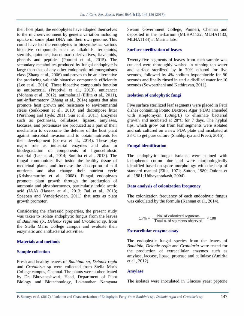
Int. J. Curr. Res. Biosci. Plant Biol. 4(11), 146-156 (2017) P. Saranya et al. (2017) / Isolation and Characterization of Endophytic Fungi from Bauhinia sp., Delonix regia and Crotalaria sp. 146 International Journal of Current Research in Biosciences and Plant Biology Volume 4 ● Number 11 (November-2017) ● ISSN: 2349-8080 (Online) Journal homepage: www.ijcrbp.com Original Research Article doi: https://doi.org/10.20546/ijcrbp.2017.411.013 Isolation and Characterization of Endophytic Fungi from Bauhinia sp., Delonix regia and Crotalaria sp. Saranya Ponkumar, Aruna Sharmili Sundararaj* and Anbumalarmathi Jeybaskaran Department of Biotechnology, Stella Maris College (Autonomous), Chennai 600 086, India *Corresponding author. Abstract Article Info Endophytic fungi are a group of unexplored microorganisms with an abundant source of natural bioactive compounds of industrial and pharmaceutical importance. The current study reports a total of five different endophytic fungi viz., Phoma sp., Phomopsis sp. (brown colonies), Phomopsis sp. (white colonies), Drechslera biseptata, Nigrospora sphaerica isolates obtained from the leaf of Bauhinia sp., Delonix regia and Crotalaria sp. The highest colonization frequency was observed in Phoma sp. (75%) followed by Drechslera biseptata (61%). Extra hyphal clearing was observed for all the isolates grown on media containing glucose as the sole carbon source indicating the presence of amylase whereas Phoma sp. and Nigrospora sphaerica were able to produce laccase. Phoma sp. and Phomopsis sp. (white) were able to produce protease. The isolates also produced ammonia. Klebsiella pneumoniae and Pseudomonas aeruginosa was inhibited by Phoma sp., Phomopsis sp. (white), Phomopsis sp. (brown), Drechslera biseptata and Nigrospora sphaerica at a concentration of 5mg/ml. Proteus mirabilis was inhibited by all the isolates except Drechslera biseptata. Interspecific activities of the isolated endophytic fungi indicated Nigrospora sphaerica was the dominant organism. Accepted: 31 October 2017 Available Online: 06 November 2017 Keywords Antibacterial activity Bauhinia sp. Crotalaria sp. Delonix regia Endophytic fungus Enzymes Introduction Endophytes are microorganisms (bacteria, fungi, yeast) that colonize the intercellular tissues of healthy plant tissues without causing any symptoms of disease (Tan and Zhou, 2001). They have been associated with plants for over 400 million years and their association can be obligate or facultative without causing harm to their host plants and also mutualism and antagonism with their hosts (Krings et al., 2007; Kumaresan and Suryanarayanan, 2002). Several endophytic fungi have mutualistic relationship with the host plant thereby limit the damage caused by pathogens by inducing resistance intrinsic to the host plant (Eaton et al., 2011). In the case of symbiotic association there is an increase in biomass as well as better development of aerial parts (Das et al., 2012). They are important components of the plant micro-ecosystem (Rodriguez et al., 2009; Zhang et al., 2006; Tan and Zhou, 2001) and are ubiquitous, colonize all plants and have been isolated from mosses (Davey and Currah, 2006), lichens (Li et al., 2007 a,b; Suryanarayanan et al., 2005) shrubs (Petrini et al., 1982), ferns (Swatzell et al., 1996), grasses (Muller and Krauss, 2005; Su et al., 2010), coniferous trees(Sun et al., 2011; Albrectsen et al., 2010; Mohamed et al., 2010). In the long co-evolution of the endophytes and
Transcript of International Journal of Current Research in Ponkumar, et al.pdfP. Saranya et al. (2017) / Isolation...
Int. J. Curr. Res. Biosci. Plant Biol. 4(11), 146-156 (2017)
P. Saranya et al. (2017) / Isolation and Characterization of Endophytic Fungi from Bauhinia sp., Delonix regia and Crotalaria sp.
146
International Journal of Current Research in
Biosciences and Plant Biology
Volume 4 ● Number 11 (November-2017) ● ISSN: 2349-8080 (Online)
Journal homepage: www.ijcrbp.com
Original Research Article doi: https://doi.org/10.20546/ijcrbp.2017.411.013
Isolation and Characterization of Endophytic Fungi from Bauhinia sp.,
Delonix regia and Crotalaria sp.
Saranya Ponkumar, Aruna Sharmili Sundararaj* and Anbumalarmathi Jeybaskaran
Department of Biotechnology, Stella Maris College (Autonomous), Chennai 600 086, India
*Corresponding author.
A b s t r a c t
A r t i c l e I n f o
Endophytic fungi are a group of unexplored microorganisms with an abundant source of
natural bioactive compounds of industrial and pharmaceutical importance. The current
study reports a total of five different endophytic fungi viz., Phoma sp., Phomopsis sp.
(brown colonies), Phomopsis sp. (white colonies), Drechslera biseptata, Nigrospora
sphaerica isolates obtained from the leaf of Bauhinia sp., Delonix regia and Crotalaria sp.
The highest colonization frequency was observed in Phoma sp. (75%) followed by
Drechslera biseptata (61%). Extra hyphal clearing was observed for all the isolates grown
on media containing glucose as the sole carbon source indicating the presence of amylase
whereas Phoma sp. and Nigrospora sphaerica were able to produce laccase. Phoma sp.
and Phomopsis sp. (white) were able to produce protease. The isolates also produced
ammonia. Klebsiella pneumoniae and Pseudomonas aeruginosa was inhibited by Phoma
sp., Phomopsis sp. (white), Phomopsis sp. (brown), Drechslera biseptata and Nigrospora
sphaerica at a concentration of 5mg/ml. Proteus mirabilis was inhibited by all the isolates
except Drechslera biseptata. Interspecific activities of the isolated endophytic fungi
indicated Nigrospora sphaerica was the dominant organism.
Accepted: 31 October 2017
Available Online: 06 November 2017
K e y w o r d s
Antibacterial activity
Bauhinia sp.
Crotalaria sp.
Delonix regia
Endophytic fungus
Enzymes
Introduction
Endophytes are microorganisms (bacteria, fungi, yeast)
that colonize the intercellular tissues of healthy plant
tissues without causing any symptoms of disease (Tan
and Zhou, 2001). They have been associated with plants
for over 400 million years and their association can be
obligate or facultative without causing harm to their host
plants and also mutualism and antagonism with their
hosts (Krings et al., 2007; Kumaresan and
Suryanarayanan, 2002). Several endophytic fungi have
mutualistic relationship with the host plant thereby limit
the damage caused by pathogens by inducing resistance
intrinsic to the host plant (Eaton et al., 2011). In the case
of symbiotic association there is an increase in biomass
as well as better development of aerial parts (Das et al.,
2012). They are important components of the plant
micro-ecosystem (Rodriguez et al., 2009; Zhang et al.,
2006; Tan and Zhou, 2001) and are ubiquitous, colonize
all plants and have been isolated from mosses (Davey
and Currah, 2006), lichens (Li et al., 2007 a,b;
Suryanarayanan et al., 2005) shrubs (Petrini et al.,
1982), ferns (Swatzell et al., 1996), grasses (Muller and
Krauss, 2005; Su et al., 2010), coniferous trees(Sun et
al., 2011; Albrectsen et al., 2010; Mohamed et al.,
2010). In the long co-evolution of the endophytes and
Int. J. Curr. Res. Biosci. Plant Biol. 4(11), 146-156 (2017)
P. Saranya et al. (2017) / Isolation and Characterization of Endophytic Fungi from Bauhinia sp., Delonix regia and Crotalaria sp.
147
their host plant, the endophytes have adapted themselves
to the microenvironment by genetic variation including
uptake of some plant DNA into their own genome. This
could have led the endophytes to biosynthesize various
bioactive compounds such as alkaloids, terpenoids,
steroids, quinones, isocoumarin derivatives, flavanoids,
phenols and peptides (Poorani et al., 2015). The
secondary metabolites produced by fungal endophyte is
large than that of any other endophytic microorganisms
class (Zhang et al., 2006) and proves to be an alternative
for producing valuable bioactive compounds efficiently
(Lee et al., 2014). These bioactive compounds function
as antibacterial (Praptiwi et al., 2013), anticancer
(Mohana et al., 2012), antimalarial (Elfita et al., 2011),
anti-inflammatory (Zhang et al., 2014) agents that also
promote host growth and resistance to environmental
stress (Saikkonen et al., 2010) and decompose litter
(Purahong and Hyde, 2011; Sun et al., 2011). Enzymes
such as pectinases, cellulases, lipases, amylases,
laccases, and proteinases are produced as a part of their
mechanism to overcome the defense of the host plant
against microbial invasion and to obtain nutrients for
their development (Correa et al., 2014). These play
major role as industrial enzymes and also in
biodegradation of components of lignocellulosic
material (Lee et al., 2014; Sunitha et al., 2013). The
fungal communities live inside the healthy tissue of
medicinal plants and increase the absorption of soil
nutrients and also change their nutrient cycle
(Krishnamurthy et al., 2008). Fungal endophytes
promote plant growth through the production of
ammonia and phytohormones, particularly indole acetic
acid (IAA) (Hassan et al., 2013; Bal et al., 2013;
Spaepen and Vanderleyden, 2011) that acts as plant
growth promoter.
Considering the aforesaid properties, the present study
was taken to isolate endophytic fungus from the leaves
of Bauhinia sp., Delonix regia and Crotalaria sp. from
the Stella Maris College campus and evaluate their
enzymatic and antibacterial activities.
Materials and methods
Sample collection
Fresh and healthy leaves of Bauhinia sp, Delonix regia
and Crotalaria sp were collected from Stella Maris
College campus, Chennai. The plants were authenticated
by Dr. Bhuvaneshwari, Head, Department of Plant
Biology and Biotechnology, Lokanathan Narayana
Swami Government College, Ponneri, Chennai and
deposited in the herbarium (MLHA1132, MLHA1133,
MLHA1134) at Marina labs.
Surface sterilization of leaves
Twenty five segments of leaves from each sample was
cut and were thoroughly washed in running tap water
and surface sterilized by in 70% ethanol for five
seconds, followed by 4% sodium hypochloride for 90
seconds and finally rinsed in sterile distilled water for 10
seconds (Sowparthani and Kathiravan, 2011).
Isolation of endophytic fungi
Five surface sterilized leaf segments were placed in Petri
dishes containing Potato Dextrose Agar (PDA) amended
with streptomycin (50mg/L) to eliminate bacterial
growth and incubated at 28°C for 7 days. The hyphal
tips, which grow out from leaf segments were isolated
and sub cultured on a new PDA plate and incubated at
28°C to get pure culture (Shubhpriya and Preeti, 2015).
Fungal identification
The endophytic fungal isolates were stained with
lactophenol cotton blue and were morphologically
identified based on spore morphology with the help of
standard manual (Ellis, 1971; Sutton, 1980; Onions et
al., 1981; Udhayaprakash, 2004).
Data analysis of colonization frequency
The colonization frequency of each endophytic fungus
was calculated by the formula (Kannan et al., 2014).
CF% = No. of colonized segments
× 100 Total n. of segments observed
Extracellular enzyme assay
The endophytic fungal species from the leaves of
Bauhinia, Delonix regia and Crotalaria were tested for
the production of extracellular enzymes such as
amylase, laccase, lipase, protease and cellulase (Amirita
et al., 2012).
Amylase
The isolates were inoculated in Glucose yeast peptone
Int. J. Curr. Res. Biosci. Plant Biol. 4(11), 146-156 (2017)
P. Saranya et al. (2017) / Isolation and Characterization of Endophytic Fungi from Bauhinia sp., Delonix regia and Crotalaria sp.
148
(GYP) agar medium supplemented with 2% soluble
starch. After 3-5 days of incubation at 37°C, the fully
formed cultures were flooded with 1% iodine. The plates
were observed for the formation of halos around the
colony.
Laccase
The isolates were inoculated in GYP agar medium
supplemented with 0.005% 1-naphthol and incubated for
3-5 days at 37°C. The change in the colour of the
medium was observed.
Lipase
The lipase activity was tested by growing the isolates on
the peptone agar media amended with sterile 10ml of
1% (v/v) Tween 20 and incubated for 5-7 days at 37°C.
The plates were observed for the formation of halos
around the colony.
Protease
The protease activity was determined by growing the
isolates on 1000ml of GYP agar media supplemented
with 0.4% gelatin. Eight grams of gelatin was dissolved
separately in 100ml distilled water and sterilized. This
was added to the media prepared earlier. The plates were
inoculated with the fungal isolates and incubated for 3-5
days at 37°C. The culture was flooded with saturated
aqueous ammonium sulphate solution. The plates were
observed for the formation of clear zone around the
culture.
Cellulase
The isolates were grown on yeast extract peptone agar
medium amended with 0.5% Na-carboxymethyl
cellulose and incubated for 5-7days at 37°C. After
incubation the plate were flooded with 0.1% Congo red
and destained with 1M sodium chloride for 15 min. The
plates were observed for the formation of clear zone
around the culture.
Ammonia production
Ammonium production was determined for the fungal
strains by growing them in peptone water for 72 h at
28°C. 1 ml Nessler’s reagent was added to the culture
media. Change of colour to faint yellow indicates
minimum ammonia production and change of colour
deep yellow to brownish colour indicates maximum
ammonia production (Amr et al., 2015).
Extraction of bioactive compounds
The isolated endophytic fungus were inoculated in
Potato Dextrose Broth (PDB) and incubated for 7days at
120 rpm. The broth was then filtered through two-folds
of cheese cloth. The filtrates were extracted with equal
volumes of ethyl acetate in a separating funnel. The
organic solvent extracts were evaporated in a rotary
evaporator and redissolved in ethyl acetate (Sowparthani
and Kathiravan, 2011).
Anti-bacterial activity
Anti-Bacterial activity of the endophytic fungi was
tested against Klebsiella pneumonia (ATCC 700603),
Protease mirabilis (ATCC 25933) and Pseudomonas
aeruginosa (Clinical isolate) by agar well diffusion
method (Bauer et al., 1959). 100μl of the inoculum of
the test pathogen was spread on Muller Hinton Agar
plates. A 5mm well was made in each corner of the plate
with equal distance using a sterile cork borer. Crude
extract of the five different fungal strains were added at
a concentration of 5μg/ml. The plates were incubated at
37°C for 24 hrs. Streptomycin (10μg/ml) was used as
positive control. After the incubation, the zone of
inhibition around the well was recorded and expressed
as millimeter (mm).
Interspecific activity by Dual culture method
The interspecific activity of isolated endophytic fungi
was carried out within themselves by dual culture
method (Senthilmurugan et al., 2013) in the following
combination:
1. Phoma sp. vs Phomopsis sp. (white)
2. Phoma sp. vs Phomopsis sp. (brown)
3. Phoma sp. vs Drechselra biseptata
4. Phoma sp. vs Nigrospora sphaerica
5. Phomopsis sp. (white) vs Phomopsis sp.
(brown)
6. Phomopsis sp. (white) vs Drechslera biseptata
7. Phomopsis sp. (white) vs Nigrospora sphaerica
8. Phomopsis sp. (brown) vs Drechslera biseptata
9. Phomopsis sp. (brown) vs Nigrospora sphaerica
10. Drechslera biseptata vs Nigrospora sphaerica
Seven day old grown cultures of the endophytic fungus
Int. J. Curr. Res. Biosci. Plant Biol. 4(11), 146-156 (2017)
P. Saranya et al. (2017) / Isolation and Characterization of Endophytic Fungi from Bauhinia sp., Delonix regia and Crotalaria sp.
149
were removed from the edges of the old colony
aseptically by using 5mm cork borer. These blocks were
placed separately at one end of the Petri plates
containing potato dextrose agar medium. At the other
end of the plate another seven day old culture of other
species of fungi was placed aseptically. Individual plates
were maintained for each culture which served as a
control. The plates were incubated at room temperature
for 4-7 days and the radial growth of fungi was
measured.
Results and discussion
Isolation of endophytic fungi and identification based
on spore morphology
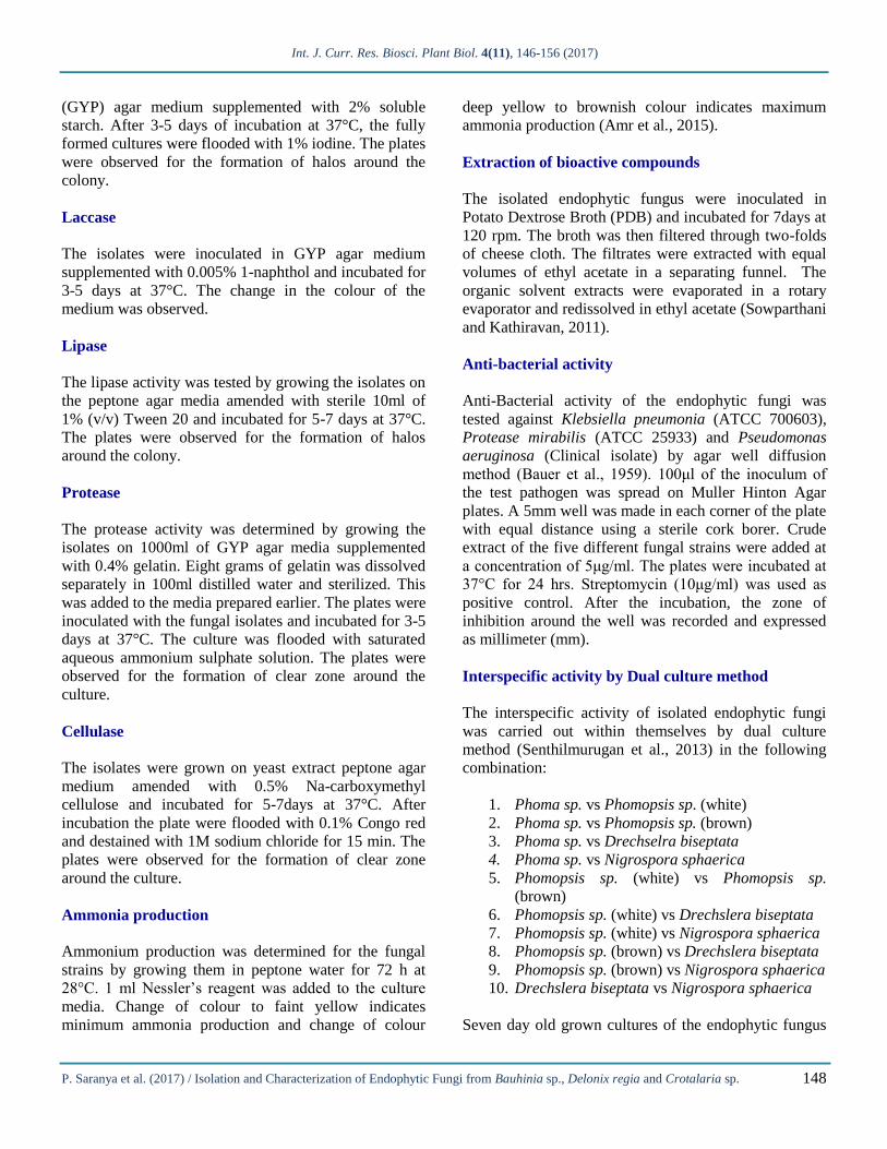
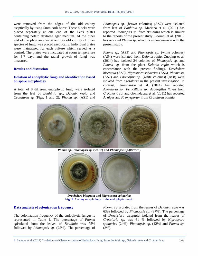
A total of 8 different endophytic fungi were isolated
from the leaf of Bauhinia sp., Delonix regia and
Crotalaria sp (Figs. 1 and 2). Phoma sp. (AS1) and
Phomopsis sp. (brown colonies) (AS2) were isolated
from leaf of Bauhinia sp. Mariana et al. (2011) has
reported Phomopsis sp. from Bauhinia which is similar
to the reports of the present study. Poorani et al. (2015)
has reported Phoma sp. which is in concurrence with the
present study.
Phoma sp. (AS3) and Phomopsis sp. (white colonies)
(AS4) were isolated from Delonix regia. Zuoping et al.
(2014) has isolated 24 colonies of Phomopsis sp. and
Phoma sp. from the plant Delonix regia which is
concordance with the present findings. Drechslera
biseptata (AS5), Nigrospora sphaerica (AS6), Phoma sp.
(AS7) and Phomopsis sp. (white colonies) (AS8) were
isolated from Crotalaria in the present investigation. In
contrast, Umashankar et al. (2014) has reported
Alternaria sp., Penicillium sp., Aspergillus flavus from
Crotalaria sp. and Govindappa et al. (2011) has reported
A. niger and F. oxysporum from Crotalaria pallida.
Phoma sp., Phomopsis sp. (white) and Phomopsis sp.(Brown)
Drechslera biseptata and Nigrospora sphaerica
Fig. 1: Colony morphology of the endophytic fungi.
Data analysis of colonization frequency
The colonization frequency of the endophytic fungus is
represented in Table 1. The percentage of Phoma
spisolated from the leaves of Bauhinia was 75%
followed by Phomopsis sp. (25%). The percentage of
Phoma sp. isolated from the leaves of Delonix regia was
63% followed by Phomopsis sp. (37%). The percentage
of Drechslera biseptata isolated from the leaves of
Crotalaria sp. was 61 % followed by Nigrospora
sphaerica (24%), Phomopsis sp. (12%) and Phoma sp.
(3%).
Int. J. Curr. Res. Biosci. Plant Biol. 4(11), 146-156 (2017)
P. Saranya et al. (2017) / Isolation and Characterization of Endophytic Fungi from Bauhinia sp., Delonix regia and Crotalaria sp.
150
Phoma sp., Phomopsis sp. (white) and Phomopsis sp. (Brown)
Drechslera biseptata and Nigrospora sphaerica
Fig. 2: Spore morphology of the endophytic fungi.
Table 1. Colonization frequency of endophytic fungi.
Plant source (Leaves) Fungal isolate Colonization frequency
Bauhinia sp. Phoma sp. (AS1)
Phomopsis sp. (Brown) (AS2)
75%
25%
Delonix regia Phoma sp. (AS3)
Phomopsis sp. (White) (AS4)
63%
37%
Crotalaria sp. Drechslera biseptata (AS5)
Nigrospora sphaerica (AS6)
Phoma sp. (AS7)
Phomopsis sp. (White) (AS8)
61%
24%
3%
12%
Extracellular enzyme assay
Amylase activities were shown by all the five
endophytic fungi as seen from the zone of clearance
(Fig. 3; Table 2). The results of the present study are in
concordance with that of Shubhpriya and Preeti (2015),
Amirita et al. (2012) who have reported amylase
production by Nigrospora sp. and Nigrospora sphaerica
respectively. Sunitha et al. (2013) has reported that
Drechslera sp. showed the positive activity of amylase
which is similar to the present study. In the present
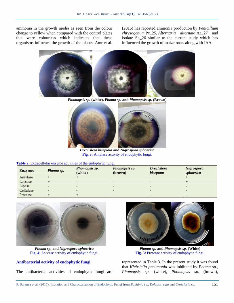
investigation, Laccase activities were shown by Phoma
sp. and Nigrospora sphaerica (Fig. 4). Sunitha et al.
(2013) has reported that Phoma sp. showed the positive
activity for the enzyme laccase whereas Shubhpriya and
Preeti (2015) has reported positive activity for laccase
by Nigrospora sp. in concordance with the present
findings. Protease activity was seen in Phoma sp. and
Phomopsis sp. (white) (Fig. 5). Orlandelli et al. (2015)
have reported protease activity in Phoma herbarum
JF766995 is similar to the current study. None of the
fungal isolates produced cellulase and lipase.
Ammonia production
All the five endophytic fungi had the ability to produce
Int. J. Curr. Res. Biosci. Plant Biol. 4(11), 146-156 (2017)
P. Saranya et al. (2017) / Isolation and Characterization of Endophytic Fungi from Bauhinia sp., Delonix regia and Crotalaria sp.
151
ammonia in the growth media as seen from the colour
change to yellow when compared with the control plates
that were colourless which indicates that these
organisms influence the growth of the plants. Amr et al.
(2015) has reported ammonia production by Penicillium
chrysogenum Pc_25, Alternaria alternata Aa_27 and
isolate Sh_26 similar to the current study which has
influenced the growth of maize roots along with IAA.
Phomopsis sp. (white), Phoma sp. and Phomopsis sp. (Brown)
Drechslera biseptata and Nigrospora sphaerica
Fig. 3: Amylase activity of endophytic fungi.
Table 2. Extracellular enzyme activities of the endophytic fungi.
Enzymes Phoma sp. Phomopsis sp.
(white)
Phomopsis sp.
(brown)
Drechslera
biseptata
Nigrospora
sphaerica
Amylase + + + + +
Laccase + - - - +
Lipase - - - - -
Cellulase - - - - -
Protease + + - - -
Phoma sp. and Nigrospora sphaerica
Fig. 4: Laccase activity of endophytic fungi.
Phoma sp. and Phomopsis sp. (White)
Fig. 5: Protease activity of endophytic fungi.
Antibacterial activity of endophytic fungi
The antibacterial activities of endophytic fungi are
represented in Table 3. In the present study it was found
that Klebsiella pneumonia was inhibited by Phoma sp.,
Phomopsis sp. (white), Phomopsis sp. (brown),

Int. J. Curr. Res. Biosci. Plant Biol. 4(11), 146-156 (2017)
P. Saranya et al. (2017) / Isolation and Characterization of Endophytic Fungi from Bauhinia sp., Delonix regia and Crotalaria sp.
152
Drechslera biseptata and Nigrospora sphaerica at a
concentration of 5mg/ml. Results similar to the present
study has been reported by Sandhu et al. (2014), where
Drechslera sp. and Phoma sp. showed inhibition against
Klebsiella pneumoniae. In the present study it was found
that Proteus mirabilis was inhibited by Phoma sp.,
Phomopsis sp. (white), Phomopsis sp. (brown) and
Nigrospora sphaerica at a concentration of 5mg/ml.
Liang et al. (2014) has reported that Phomopsis sp.
showed the best inhibitory responses against bacterial
pathogens. In the present study it was found that
Pseudomonas aeruginosa was inhibited by Phoma sp.,
Phomopsis sp. (white), Phomopsis sp. (brown),
Drechslera biseptata and Nigrospora sphaerica at a
concentration of 5mg/ml. Klebsiella pneumonia,
Pseudomonas aeruginosa and Proteus mirabilis are
Gram negative organisms that are resistant to antibiotics.
Antibacterial activity exhibited by the endophytic
fungus isolated in the current study suggests that potent
antibacterial compounds can be isolated from them.
Table 3. Antibacterial activity of endophytic fungi.
Fungal isolates
Klebsiella pneumonia Pseudomonas aeruginosa Protease mirabilis
Zone of inhibition (mm)
5mg Control
(10 mg) 5mg
Control
(10 mg) 5mg
Control
(10mg)
Phoma sp 15 35 12 32 5 28
Phomopsis sp (white) 10 30 15 34 4 25
Phomopsis sp (brown) 13 30 14 30 5 28
Drechslera biseptata 12 30 18 34 - 20
Nigrospora sphaerica 14 30 11 34 3 25
Control – Streptomycin.
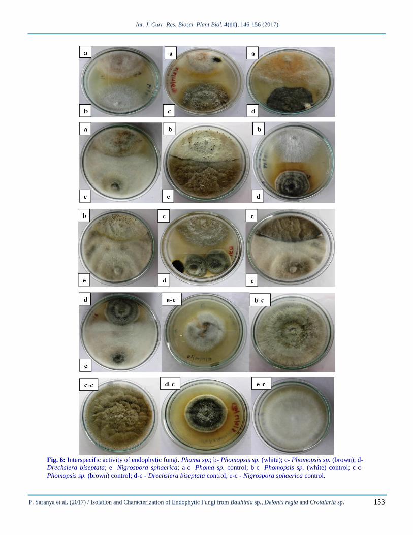
Interspecific activity
The interspecific activity of isolated endophytic fungi
is represented in Fig. 6. In the study of interspecific
activity of Phoma sp. vs Phomopsis sp. (white),
Phoma sp. had a growth of 2.5cm whereas Phomopsis
sp. (white) had 3cm. In the case of Phoma sp. vs
Phomopsis sp. (brown), Phoma sp. had a growth of
2.8cm whereas Phomopsis sp. (brown) had 3.8cm.
The control plates of Phoma sp., Phomopsis sp.
(white) and Phomopsis sp. (brown) had a growth of
5cm each. In the case of Phoma sp. vs Drechslera
biseptata, Phoma sp. had a growth of 3.5cm whereas,
Drechslera biseptata had 2cm. The control plates of
Phoma sp. had a growth of 5cm and Drechslera
biseptata had a growth of 4cm. In the study of
interspecific activity among Phoma sp. vs Nigrospora
sphaerica, Phoma sp. had a growth of 2.5cm whereas
Nigrospora sphaerica had 4cm. The control plates of
Phoma sp. and Nigrospora sphaerica had a growth of
5cm each.
In the study of interspecific activity of Phomopsis sp.
(white) vs Phomopsis sp. (brown), Phomopsis sp.
(white) had a growth of 2.9cm whereas Phomopsis sp.
(brown) had 3.1cm. The control plates of Phomopsis sp.
(white) and Phomopsis sp. (brown) had a growth of 5cm
each. In the case of Phomopsis sp. (white) vs Drechslera
biseptata it was found that Phomopsis sp. (white) had a
growth of 3.5cm whereas Drechslera biseptata had 2cm.
The control plates of Phomopsis sp. (white) had a
growth of 5cm while Drechslera biseptata had 4cm. In
the case of Phomopsis sp. (white) vs Nigrospora
sphaerica, Phomopsis sp. (white) had a growth of 2.3cm
whereas Nigrospora sphaerica had 4.2cm. The control
plates of Phomopsis sp. (white) and Nigrospora
sphaerica had a growth of 5cm each.
In the interspecific activity of Phomopsis sp. (brown) vs
Drechslera biseptata, Phomopsis sp. (brown) had a
growth of 3cm whereas Drechslera biseptata had 2cm.
The control plates of Phomopsis sp. (brown) had a
growth of 5cm and Drechslera biseptata had a growth of
4cm. In the case of Phomopsis sp. (brown) vs
Nigrospora sphaerica, Phomopsis sp (brown) had a
growth of 2.9cm whereas Nigrospora sphaerica had
4cm. The control plates of Phomopsis sp. (brown) and
Nigrospora sphaerica had a growth of 5cm each.
In the study of interspecific activity among the isolated
endophytic fungi Drechslera biseptata vs Nigrospora
sphaerica, Drechslera biseptata had a growth of 2.1cm
whereas Nigrospora sphaerica had 4.2cm. The control
plates of Drechslera biseptata had a growth of 4cm
whereas Nigrospora sphaerica had a growth of 5cm.The
results of the current study indicates that Nigrospora
sphaerica was the dominant organism in the
interspecific studies.
Int. J. Curr. Res. Biosci. Plant Biol. 4(11), 146-156 (2017)
P. Saranya et al. (2017) / Isolation and Characterization of Endophytic Fungi from Bauhinia sp., Delonix regia and Crotalaria sp.
153
Fig. 6: Interspecific activity of endophytic fungi. Phoma sp.; b- Phomopsis sp. (white); c- Phomopsis sp. (brown); d-
Drechslera biseptata; e- Nigrospora sphaerica; a-c- Phoma sp. control; b-c- Phomopsis sp. (white) control; c-c-
Phomopsis sp. (brown) control; d-c - Drechslera biseptata control; e-c - Nigrospora sphaerica control.
Int. J. Curr. Res. Biosci. Plant Biol. 4(11), 146-156 (2017)
P. Saranya et al. (2017) / Isolation and Characterization of Endophytic Fungi from Bauhinia sp., Delonix regia and Crotalaria sp.
154
Conclusion
In the present study Phoma sp was present in all the
leaves and were predominant in Bauhinia and Delonix
regia. Drechslera biseptata was predominant in
Crotalaria sp. Amylase was produced by all the
endophytic fungi. Emergence of resistance to antibiotics
require development of new antibiotics and in this view
the potent bioactive secondary metabolites from Phoma
sp. and Drechslera biseptata obtained in the current
study may be used for drug development paving way for
natural products.
Conflict of interest statement
Authors declare that they have no conflict of interest.
Acknowledgement
Authors thank the Principal and the Management of
Stella Maris College (Autonomous), Chennai,
Tamilnadu, India for the research facilities provided and
Dr. N.K. Udayaprakash, Assistant Professor,
Department of Biotechnology, Vel’s University,
Chennai for identification of the fungus.
References
Albrectsen, B.R., Björkén, L., Varad, A., Hagner, A.,
Wedin, M., Karlsson, J., Jansson, S., 2010.
Endophytic fungi in European aspen (Populus
tremula) leaves—diversity, detection, and a
suggested correlation with herbivory resistance.
Fungal Diversity. 41(1), 17-28.
Amirita, A., P. Sindhu, J. Swetha, N.S. Vasanthi K.P.
Kannan, 2012. Enumeration of endophytic fungi
from medicinal plants and screening of extracellular
enzymes. World J. Sci. Tech. 2(2): 13-19.
Amr, H. F., Hassan, S., Eid, A.M., Ewais, E.E. 2015.
Biotechnological applications of fungal endophyte
associated with medicinal plant Asclepias sinaica
(Bioss.). Ann. Agric. Sci. 60(1), 95–104.
Bal, H.B., Das, S., Dangar, T.K., Adhya, T.K., 2013.
ACC deaminase and IAA producing growth
promoting bacteria from the rhizosphere soil of
tropical rice plants. J. Basic Microbiol. 53(12), 972-
84.
Bauer, A.W., Kirby, W.M.M., Sherries, S.C., Tunk, M.
1966. Antibiotic susceptibility of testing by a
standard single disc method. Am J Clin Pathol.
36(4), 493-496.
Correa, R.C.G., Rhoden, S.A., Mota, T.R., Azevedo,
J.L., Pamphile, J.A., Marques de Souza, C.G.,
Polizeli, M.T.M., Bracht, A., Peralta, R,M. 2014.
Endophytic fungi: Expanding the arsenal of
industrial enzyme producers. J. Ind Microbiol
Biotechnol. 41, 1476-1478.
Das, A., Kamal, S., Shakil, N.A., Sherameti. I.,
Oelmuller, R., Dua, M., Tuteja, N., Johri, A.C.,
Varma, A. 2012. The root endophyte fungus
Piriformospora indica leads to early flowering,
higher biomass and altered secondary metabolites of
the medicinal plant, Coleus forskohlii. Plant Signal
Behav. 7,1–10.
Davey, M.L., Currah, R.S., 2006. Interactions between
mosses (Bryophyta) and fungi. Botany. 84(10),
1509-19.
Eaton, C.J., Cox, M.P., Scott, B. 2011. What triggers
grass endophytes to switch from mutualism to
pathogenism? Plant Sci. 180,190–195.
Elfita., Muharni., Munawar., Legasari, L., Darwati.
2011. Antimalaria compounds from endophytic
fungi of brotowali (Tinaspora crispa L). Indo J.
Chem. 11, 53-58.
Ellis, K. 1971. Dematiaceous Hyphomycetes.
Commonwealth Mycological Institute, Kew, Surrey,
England, p.608.
Govindappa, M., Bharath, N., Shruthi, H.B., Santoyo, G.
2011. In vitro Antioxidant Activity and
Phytochemical Screening of Endophytic Extracts of
Crotalaria pallida. Free Radicals and Antioxidants.
(3), 79-86.
Hassan, S.E., Liu, A., Bittman, S., Forge, T.A., Hunt,
D.E., Hijri, M., St-Arnaud, M., 2013. Impact of 12-
year field treatments with organic and inorganic
fertilizers on crop productivity and mycorrhizal
community structure. Biology and fertility of soils.
49(8), 1109-21.
Kannan, K.P., Madhan, K.D., Ramya, P.R., Madhu N.S.,
Meenatchi, G., Sowmya, A.N., Bhuvaneswari, S.
2014. Diversity of endophyticfFungi from salt
tolerant plants. Int. J. ChemTech Res. 6(9), 4084-
4088.
Krings, M., Taylor, T.N., Hass, H., Kerp, H., Dotzler,
N., Hermsen, E.J., 2007. Fungal endophytes in a
400‐million‐yr‐old land plant: Infection pathways,
spatial distribution, and host responses. New
Phytologist. 174(3), 648-657.
Krishnamurthy, Y.L., Shankar, N.B., Shashikala, J.,
2008. Fungal communities in herbaceous medicinal
plants, Malnad region, Southern India. Microbes.
Environ. 23, 24-28.
Int. J. Curr. Res. Biosci. Plant Biol. 4(11), 146-156 (2017)
P. Saranya et al. (2017) / Isolation and Characterization of Endophytic Fungi from Bauhinia sp., Delonix regia and Crotalaria sp.
155
Kumaresan, V., Suryanarayanan, T.S., 2002. Endophyte
assemblages in young, mature and senescent leaves
of Rhizophora apiculata: evidence for the role of
endophytes in mangrove litter degradation. Fungal
Diver. 1(9), 81-91.
Lee, J.M., Tan, W.S., Ting, A.S.Y. 2014. Revealing the
antimicrobial and enzymatic potentials of culturable
fungal endophytes from tropical pitcher plants
(Nepenthes spp.). Mycosphere. 5(2), 364–377.
Li, W.C., Guo, S.Y., Guo, L.D., 2007a. Endophytic
fungi associated with lichen Physcia stellaris using
different surface sterilization methods. J Fungal Res.
5, 202-6.
Li, W.C., Zhou, J., Guo, S.Y., Guo, L.D., 2007b.
Endophytic fungi associated with lichens in Baihua
mountain of Beijing, China. Fungal Divers. 30(25),
69-80.
Liang, Z.N., Zhu, H., Lai, K.P., Chen, L. 2014. Isolation
of endophytic fungi from medicinal plant Brucea
javanica and their microbial inhibition activity. J.
Chin. Medicinal Mat. 37(4), 564-568.
Mariana, R.P., Gustavo, M., Ana, P.D., Mario, R.M. Jr.,
Glaucia, M.P. 2011. The use of endophytes to obtain
bioactive compounds and their application in
biotransformation process. Chem. Rev. 111.
Mohamed, R., Jong, P.L., Zali, M.S., 2010. Fungal
diversity in wounded stems of Aquilaria
malaccensis. Fungal Divers. 43(1), 67-74.
Mohana, K.P., Sebastian, V., Vaidyanathan, P.,
Thimmappa, R.B., Singh, S., Gudasalamani, R.,
Ramesh, V., Retnabai, S.T., Michael, S., Uma, S.R.
2012. Fusarium proliferatum an endophytic fungus
from Dyxolum binetariferum Hook.f produces
rohitukine, a chromane alkaloid possessing anti-
cancer activity. Antonie van Leeuwenhoek. 101,
323-329.
Müller, C.B., Krauss, J., 2005. Symbiosis between
grasses and asexual fungal endophytes. Curr. Opin.
Plant Biol. 8, 450-456.
Onions, A.H.S., Allsopp, D., Eggins, H.O.W. 1981.
Smith’s Introduction to Industrial Mycology.
Edward Arnold Ltd. London, p.398.
Orlandelli, R.C., Almeida, T.T., Alberto, R.N., Polonio,
J.C., Azevedo, J.L., Pamphile, J.A. 2015.Antifungal
and proteolytic activities of endophytic fungi
isolated from Piper hispidum Sw. Braz. J.
Microbiol. 46(2), 359-66.
Petrini, O., Stone, J., Carroll, F.E., 1982. Endophytic
fungi in evergreen shrubs in western Oregon: a
preliminary study. Can. J. Bot. 60(6), 89-96.
Poorani, K., Manogaran, S., Dhakshinamoorthy, M.,
Kannan, K.P., 2015. Evaluation of antioxidant and
antibacterial activities of endophytic fungi isolated
from Bauhinia racemosa Lam and Phyllanthus
amarus Schum and Thonn. J. Chem. Pharm. Res.
7(9), 366-379.
Praptiwi., Jamal, Y., Fathoni, A., Nurkanto, A., Agusta,
A., 2013. 3-Acetyl-2,5,7-Trihydroxyl-1,4-
naphtalenedione, an antimicrobial metabolite from
the culture of endophytic fungus coelomycetes tcbp4
from Tinospora crispa. Media Litbangkes. 23(3),
95-101.
Purahong, W., Hyde, K.D., 2011. Effects of fungal
endophytes on grass and non-grass litter
decomposition rates. Fungal Divers. 47, 1–7.
Rodriguez, R.J., White, Jr J.F., Arnold, A.E., Redman,
R.S., 2009. Fungal endophytes: Diversity and
functional roles. New Phytologist. 182(2), 314-30.
Saikkonen, K., Wäli, P., Helander, M., Faeth, S.H.,
2004. Evolution of endophyte–plant symbioses.
Trends Plant Sci. 9(6), 275-80.
Sandhu, S.S., Suneel, K., Ravindra, P.A. 2014. Isolation
and identification of endophytic fungi from Ricinus
communis Linn. and their antibacterial activity. Int.
J. Res. Phar. Chem. 4(3), 611-618.
Senthilmurugan, G., Sekar, R., Suresh, K.,
Balamurugan, S., 2013. Phytochemical screening,
enzyme and antibacterial activity analysis of
endophytic fungi Botrytis sp. isolated from Ficus
benghalensis (L.). Int. J. Pharm. Res. BioSci. 2 (4),
264-273.
Shubhpriya, G., Preeti, C. 2015. Phytochemical
screening and extracellular enzymatic enumeration
of foliar endophytic fungal isolates of Centella
asiatica (L.) Urban. Int. J. Pharm. Sci. Rev. Res.
35(1), 21-24.
Sowparthani, K., Kathiravan, G. 2011. In vitro
antibacterial screening of ethyl acetate endophytic
fungi isolated from Phylum amarus (Schum and
Thonn) against pathogenic bacterial isolates. J.
Pharma Biomed. Sci. 10(10), 1-4.
Spaepen, S., Vanderleyden, J., 2011. Auxin and plant-
microbe interactions. Cold Spring Harbor
Perspectives in Biology. 3(4), a001438.
Su, Y. Y., Guo, L. D., Hyde, K.D., 2010. Response of
endophytic fungi of Stipa grandis to experimental
plant function group removal in Inner Mongolia
steppe, China. Fungal Divers. 43(1), 93-101
Sun, X., Guo, L.D., Hyde, K.D., 2011. Community
composition of endophytic fungi in Acer truncatum
and their role in decomposition. Fungal Divers.
47(1), 85-95.
Int. J. Curr. Res. Biosci. Plant Biol. 4(11), 146-156 (2017)
P. Saranya et al. (2017) / Isolation and Characterization of Endophytic Fungi from Bauhinia sp., Delonix regia and Crotalaria sp.
156
Sunitha, V.H., Devi, D.N., Srinivas, C., 2013.
Extracellular enzymatic activity of endophytic
fungal strains isolated from medicinal plants. World
J. Agric. Sci. 9(1), 1-9.
Suryanarayanan, T.S., Thirunavukkarasu, N., Hariharan,
G.N., Balaji, P., 2005. Occurrence of non-obligate
microfungi inside lichen thalli. Sydowia. 57(1), 120.
Sutton, B. C. 1980. The Coelomycetes. CMI, Kew,
Surrey, England. 696p.
Swatzell, L.J., Powell, M.J., Kiss, J.Z., 1996. The
relationship of endophytic fungi to the gametophyte
of the fern Schizaea pusilla. International journal of
plant sciences. 157(1), 53-62.
Tan, R.X., Zou, W.X., 2001. Endophytes: A rich source
of functional metabolites. Natural product reports.
18(4), 448-59.
Udayaprakash, N.K., 2004. Indoor Molds: Isolation and
Identification. Color Wings (P) Ltd., Chennai. 99p.
Umashankar, T., Govindappa, M., Ramachandra, Y.L.,
2014. In vitro antioxidant and antimicrobial activity
of partially purified coumarins from fungal
endophytes of Crotalaria pallida. Int. J. Curr.
Microbiol. Appl. Sci. 3(8), 58-72.
Zhang, D., Ge, H., Zou, J., Tao, X., Chen, R., Dai, J.
2014. Perioconianone A, a new 6/6/6 carbocyclic
sesquiterpenoid from endophytic fungus Periconia
sp. with neural anti-inflammatory activity. Organic
Lett. 16(5), 1410-1430.
Zhang, H.W., Song, Y.C., Tan, R.X., 2006. Biology and
chemistry of endophytes. Natural Prod. Rep. 23(5),
753-771.
Zuoping, Z., Changfei, Z., Wenna, Z., Wei, L., Long, C.,
Jinping, Y., Haiyan, L., 2014. Diversity and plant
growth-promoting ability of endophytic fungi from
the five flower plant species collected from Yunnan,
Southwest China. J. Plant Interact. 9(1), 585-591.
How to cite this article:
Saranya, P., Aruna Sharmili, S., Anbumalarmathi, J., 2017. Isolation and characterization of endophytic fungi from
Bauhinia sp., Delonix regia and Crotalaria sp. Int. J. Curr. Res. Biosci. Plant Biol. 4(11), 146-156. doi:
https://doi.org/10.20546/ijcrbp.2017.411.013