Biodeterioration of Construction Materials: State of the ...
Author's personal copy
A green biocide enhancer for the treatment of sulfate-reducing bacteria (SRB)biofilms on carbon steel surfaces using glutaraldehyde
Jie Wen a, Kaili Zhao a, Tingyue Gu a,*, Issam I. Raad b
a Department of Chemical and Biomolecular Engineering, Ohio University, Athens, OH 45701, USAb Department of Infectious Diseases, Infection Control, and Employee Health, The University of Texas M. D. Anderson Cancer Center, Houston, TX 77230, USA
a r t i c l e i n f o
Article history:Received 5 March 2009Received in revised form18 August 2009Accepted 14 September 2009Available online 4 October 2009
Keywords:Desulfovibrio desulfuricansBiofilm treatmentEDDSGlutaraldehydeMIC
a b s t r a c t
Generally speaking, a much higher concentration of biocide is needed to treat biofilms compared to thedosage used to for planktonic bacteria. With increasing restrictions of environmental regulations andsafety concerns on large-scale biocide uses such as oil field applications, it is highly desirable to make moreeffective use of biocides. In this paper a green biocide enhancer ethylenediaminedisuccinate (EDDS) that isa biodegradable chelator, was found to enhance the efficacy of glutaraldehyde in its treatment of sulfate-reducing bacteria (SRB) biofilms. Experiments were carried out in 100 ml anaerobic vials with carbon steelcoupons. The ATCC 14563 strain of Desulfovibrio desulfuricans was used. Biofilms on coupon surfaces werevisualized using scanning electron microscopy (SEM). Experimental results showed that EDDS reduced theglutaraldehyde dosages considerably in the inhibition of SRB biofilm establishment and the treatment ofestablished biofilms on carbon steel coupon surfaces.
� 2009 Elsevier Ltd. All rights reserved.
1. Introduction
Biofilms cause various problems such as medical infections, foulingof water cooling system, product contamination, and microbiologi-cally influenced corrosion (MIC) (Carpentier and Cerf,1993; Diosi et al.,2003; Raad et al., 2003; Kjellerup et al., 2006). MIC accounts for asmuch as 20% of all forms of corrosion, amounting to billons of dollarsin losses each year (Flemming, 1996). A major Alaska Prudhoe Bay oilfield pipeline had to be shut down due to a leak in 2006, which causeda turmoil in the global oil market. MIC was suspected to be one oftwo major factors (Jacobson, 2007) in the leak. MIC typically causeslocalized corrosion due to the patchy biofilms formed on a metalsurface. Among aerobic and anaerobic bacteria related to MIC, sulfate-reducing bacteria (SRB) are most often blamed (Kobrin, 1993; Feioet al., 2000). SRB reduce sulfate to sulfide and produce hydrogensulfide. Pitting may be initiated underneath biofilms. With furtherdevelopment of the biofilms, metabolic products may lower the localpH, thus deepening the pits (Videla and Herrera, 2005).
Based on their success in using ethylenediaminetetraacetic acid(EDTA) with antibiotics to eradicate biofilms on catheters, Raad andSherertz (2001) patented the idea of treating SRB biofilms usingchelators in combination with biocides. Initial experimental results
showed that EDTA enhanced the glutaraldehyde’s and THPS’ inhibi-tion of planktonic SRB growth (Zhao et al., 2005; Wen and Gu, 2007).EDTA’s slow biodegradability has sparked a call for its replacement invarious industrial applications with green chelators (EuropeanCommission, 2004). A popular biodegradable chelator is ethyl-enediaminedisuccinate (EDDS). The work below demonstrated thatEDDS enhanced glutaraldehyde’s treatment of SRB biofilms on carbonsteel surfaces.
2. Materials and methods
2.1. Bacteria and culture conditions
Desulfovibrio desulfuricans subsp. aestuarii ATCC 14563 was usedin this work. It is a marine strain SRB that favors a liquid mediumwith a salinity equivalent to 0.5–6 wt% NaCl (Ollivier et al., 1994).Enriched artificial seawater (ASW) and modified ATCC 1250 mediumwith 25 ppm Fe2þ were used for SRB growth. The composition ofenriched ASW medium included a salt mix (Instant Ocean�1 salt mixintended for marine aquariums) 36 g, Fe(NH4)2(SO4)2 125 mg,sodium lactate (60 wt% syrup) 4.5 ml and yeast extract 1 g, in 1 L ofwater. The comparison between ASW and the typical natural
* Corresponding author. Tel.: þ1 740 593 1499; fax: þ1 740 593 0873.E-mail address: [email protected] (T. Gu). 1 Instant Ocean is a registered trademark of Spectrum Brands, Atlanta, GA, USA.
Contents lists available at ScienceDirect
International Biodeterioration & Biodegradation
journal homepage: www.elsevier .com/locate/ ib iod
0964-8305/$ – see front matter � 2009 Elsevier Ltd. All rights reserved.doi:10.1016/j.ibiod.2009.09.007
International Biodeterioration & Biodegradation 63 (2009) 1102–1106
Author's personal copy
seawater was reported by Atkinson and Bingman (1996). Culturemedia were sterilized using an autoclave before use.
2.2. Substratum for biofilm growth
Disk shaped C1018 coupons with a top surface area of 1.12 cm2
were used as the substratum for biofilm growth in 100 ml vials.The bottom surface and the side surface were coated with Teflon toprevent exposure. Before immersion into the vials, the exposedsurface was polished using sand papers with 200, 400 and 600 gritssuccessively.
2.3. EDDS
Octaquest�2 E30, a trisodium salt of EDDS known as [S,S]-EDDS(Fig. 1), was used. It is a biodegradable chelator having similarchelation ability as EDTA, with no persistent metabolites formedduring biodegradation (Schowanek et al., 1997).
2.4. Efficacies of biocide enhancers for inhibitionof planktonic SRB growth
Planktonic SRB growth was carried out in 100 ml anaerobic vials.An anaerobic chamber with a nitrogen environment was used toprovide an anaerobic environment for inoculation. After distrib-uting 50 ml of the medium into each vial and adding an appropriateamount of a biocide and a biocide enhancer, it was inoculated witha two-day old SRB seed culture. The initial SRB cell concentrationswere 1.42 � 106 cells/ml right after inoculation. The vials weresealed and then placed in an incubator at 37 �C. SRB growth wasmonitored by counting the (viable) motile planktonic cells usinga hemocytometer under an optical microscope at 400� magnifi-cation in broth samples drawn from the vials using a syringe.
2.5. SEM observations
To study the SRB biofilm on a C1018 carbon steel coupon underSEM, the coupon’s exposed surface was exposed to 2.5 wt% glutar-aldehyde for 8 h and subsequently washed with a graded series(30%, 50%, 70%, 100% v/v) of ethanol for dehydration. The samplewas then critical point dried and coated with a gold alloy prior toSEM observations. The entire surface area of coupons was examinedunder SEM to locate sessile SRB.
2.6. EDDS and glutaraldehyde in inhibiting SRB biofilm formation
Laboratory experiments were carried out in 100 ml anaerobic vialsat 37 �C. Nitrogen sparging was used to remove oxygen in liquids. Insome cases, 0.5 g/L of cysteine as an oxygen scavenger was used toprevent contamination by trace amount of oxygen. Different concen-trations of glutaraldehyde and EDDS were added to vials before inoc-ulation (Table 1). 1 ml of two-day old SRB stock culture was used toinoculate each vial. The SRB cell concentration right after inoculationwas about 4.3 � 105–5.1 � 105 cells/ml. Initial pH was around 6.8 inmodified ATCC 1250 medium and around 7.8–8.1 in ASW. Couponswere taken out after around 8 days for SEM examination.
2.7. EDDS and glutaraldehyde in treating established SRB biofilms
Biofilms were pre-grown in ATCC 1250 medium. One coupon wastaken out for SEM analysis 4 days after inoculation to validate that the
coupon surface was indeed covered by a biofilm. Coupons coveredwith biofilms were washed with sterilized distilled water, and thentransferred into vials with fresh modified ATCC 1250 medium. EDDSand glutaraldehyde at different concentrations were then added(Table 1). All the procedures were conducted in a nitrogen-filledanaerobic chamber. Effects of EDDS and glutaraldehyde treatmentwere checked 8 days after they were introduced using SEM analysis.
3. Results and discussion
Glutaraldehyde is a widely used biocide in oil fields as well astetrakis hydroxymethyl phosphonium sulfate (THPS), quaternaryammonium compounds (QAC), bromo-nitropropanediol (BNPD),etc. Because of its broad-spectrum and biodegradability, glutaral-dehyde was selected in this study. Because of its interaction withthe SRB culture medium, glutaraldehyde exhibited inhibition (i.e.,suppression or delay) of SRB cell growth rather than killing them(Von Rege and Sand, 1998; Gardner and Stewart, 2002; De Savariaand de Mele, 2005; Cetin et al., 2007). Gardner and Stewart (2002)reported that 50 ppm of glutaraldehyde retarded the SRB planktonicgrowth to 143 h in Postgate C medium. Due to its ability to cross-linkproteins, glutaraldehyde is also a common fixative (Hayat, 2000).Since the experimental duration was around 8 days to examine theexistence of SRB, the surface morphology comparison would not beaffected by the addition of glutaraldehyde as a fixative (2.5 wt%) atthe end of experiments while the highest concentration used in theinhibition study was 500 ppm. The standard protocol using 2.5 wt%assures that all cells are immobilized by killing them.
Table 2 (data from Wen and Gu, 2007) shows the effects of EDDSon the enhancement of the inhibition of planktonic SRB growth.30 ppm of glutaraldehyde combined with 2000 ppm of EDDS iseffective to control the SRB growth, while biocide alone lost itsinhibition on SRB growth after 5 days of inoculation. Since a chelatoritself cannot inhibit planktonic SRB growth, it has to combine witha biocide to take effect. The treatment of 20 ppm of glutaraldehydecombined with 2000 ppm of EDDS is as good as or better than foundwith 30 ppm glutaraldehyde alone.
Biofilms protect sessile bacteria from biocide attacks (Denyer,1995; Morton et al., 1998). Stoodley et al. (1999) showed that densebiofilms with sessile cells glued together by extracellular polymericsubstances (EPS) increases mass transfer resistances. The limitednutrition supply decreases the bacteria metabolic activity andincreases the resistance to biocides. Others suggested that a biofilmmay change the physiology of sessile bacteria, which improve their
Fig. 1. Structure of [S,S]-EDDS.
Table 1Test matrix for EDDS enhancement of glutaraldehyde treatment of SRB biofilms oncarbon steel surfaces in ASW.
Inhibiting biofilmformation
Treating establishedbiofilms
Medium ASW Modified ATCC 1250 Modified ATCC 1250Glutaraldehyde (ppm) 25 0, 30 30, 500EDDS (ppm) 0, 2000 0, 2000 0, 2000
2 Octaquest is a registered trademark of Octel Performance Chemicals, Cheshire,United Kingdom.
J. Wen et al. / International Biodeterioration & Biodegradation 63 (2009) 1102–1106 1103
Author's personal copy
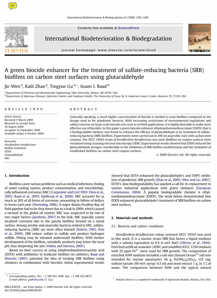
biocide resistance (Morton et al., 1998; Fux et al., 2005). A muchhigher concentration of biocide may be needed to remove thesessile bacteria, compared to the dosage for treating planktonicbacteria (Davies, 2003; Meyer, 2003). This is supported by theresults in Fig. 2 that indicates that even with a glutaraldehydeconcentration as high as 500 ppm for the treatment of a pre-grownSRB biofilm, sessile SRB cells were still visible. A more effectivetreatment is desired to reduce the dosage.
Trace metals such as manganese, zinc, iron are necessary forbacteria metabolism and biofilm growth (Dunne and Burd, 1992).Calcium was reported to be essential in the bonding of polymermolecules in biofilms (Carpentier and Cerf, 1993). Researchersfound that bacterial adhesion is sensitive to chelating agents, whichhave been introduced to treat biofilms during sanitization ofmedical instruments (Taweechaisupapong and Doyle, 2000; Baninet al., 2006; Chudzik et al., 2007). Raad et al. (2003) reportedthat EDTA combined with minocycline is effective in eradicatingbiofilms on catheter surfaces. They also found that the eradicationof Staphylococcus aureus and Candida parapsilosis biofilms wassped up greatly by using 25% (v/v) ethanol with minocycline–EDTA(Raad et al., 2007). Weinberg (2004) pointed out that biofilmformation was suppressed by iron chelators.
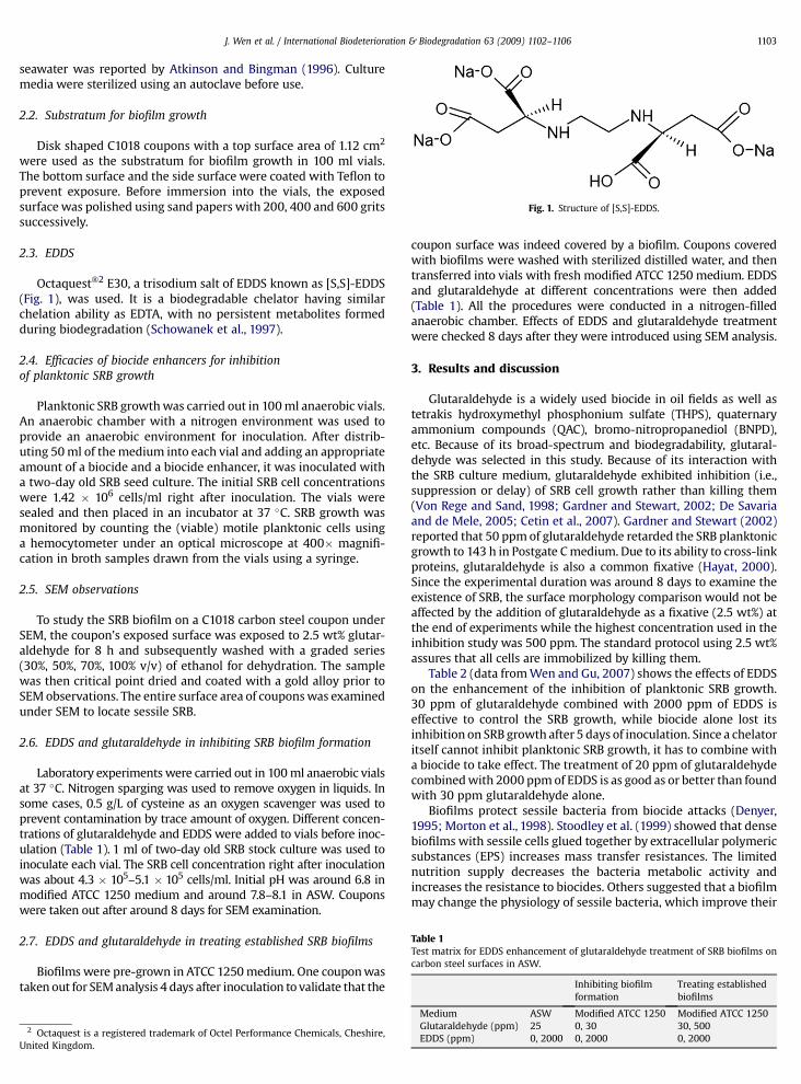
Fig. 3 shows the comparison of coupon surfaces with andwithout biocide enhancer. Surface morphology is different betweenFig. 3a (without EDDS) and Fig. 3b (with EDDS). In Fig. 3a sessile SRBcells are clearly visible, while in Fig. 3b they are absent. Figs. 4 and 5show the comparison of coupon surfaces with different EDDS andglutaraldehyde concentrations in modified ATCC 1250 medium.
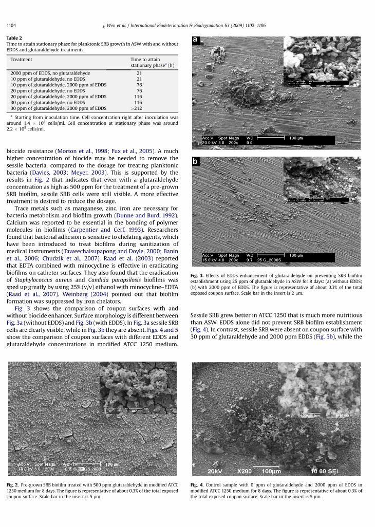
Sessile SRB grew better in ATCC 1250 that is much more nutritiousthan ASW. EDDS alone did not prevent SRB biofilm establishment(Fig. 4). In contrast, sessile SRB were absent on coupon surface with30 ppm of glutaraldehyde and 2000 ppm EDDS (Fig. 5b), while the
Table 2Time to attain stationary phase for planktonic SRB growth in ASW with and withoutEDDS and glutaraldehyde treatments.
Treatment Time to attainstationary phasea (h)
2000 ppm of EDDS, no glutaraldehyde 2110 ppm of glutaraldehyde, no EDDS 2110 ppm of glutaraldehyde, 2000 ppm of EDDS 7620 ppm of glutaraldehyde, no EDDS 7620 ppm of glutaraldehyde, 2000 ppm of EDDS 11630 ppm of glutaraldehyde, no EDDS 11630 ppm of glutaraldehyde, 2000 ppm of EDDS >212
a Starting from inoculation time. Cell concentration right after inoculation wasaround 1.4 � 106 cells/ml. Cell concentration at stationary phase was around2.2 � 108 cells/ml.
Fig. 2. Pre-grown SRB biofilm treated with 500 ppm glutaraldehyde in modified ATCC1250 medium for 8 days. The figure is representative of about 0.3% of the total exposedcoupon surface. Scale bar in the insert is 5 mm.
Fig. 3. Effects of EDDS enhancement of glutaraldehyde on preventing SRB biofilmestablishment using 25 ppm of glutaraldehyde in ASW for 8 days: (a) without EDDS;(b) with 2000 ppm of EDDS. The figure is representative of about 0.3% of the totalexposed coupon surface. Scale bar in the insert is 2 mm.
Fig. 4. Control sample with 0 ppm of glutaraldehyde and 2000 ppm of EDDS inmodified ATCC 1250 medium for 8 days. The figure is representative of about 0.3% ofthe total exposed coupon surface. Scale bar in the insert is 5 mm.
J. Wen et al. / International Biodeterioration & Biodegradation 63 (2009) 1102–11061104
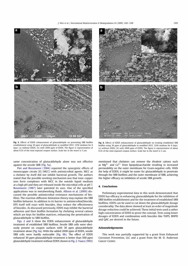
same concentration of glutaraldehyde alone was not effectiveagainst the sessile SRB (Fig. 5a).
Piet and Rossmoore (1984) reported the synergistic effects ofmonocopper citrate (II) (MCC) with antimicrobial agents. MCC asa chelator by itself did not inhibit bacterial growth. The authorsstated that the possible working mechanism was that toxic copperions form complexes with MCC in the outside liquid mediumat a high pH and they are released inside the microbial cells at pH 7.Rosemoore (1987) later patented its uses. One of the specifiedapplications was in metalworking fluids. Allison et al. (2000) dis-cussed the possible antimicrobial resistance mechanisms of bio-films. The reaction-diffusion-limitation theory may explain the SRBbiofilm behavior. In addition to its barrier to antimicrobial/biocide,EPS itself will react with biocides, thus reduce the effectivenessof biocides. As discussed previously, EDDS may inhibit the bacterialadhesion and then biofilm formation by chelating several cationswhich are keys for biofilm matrices, enhancing the penetration ofglutaraldehyde to SRB biofilm.
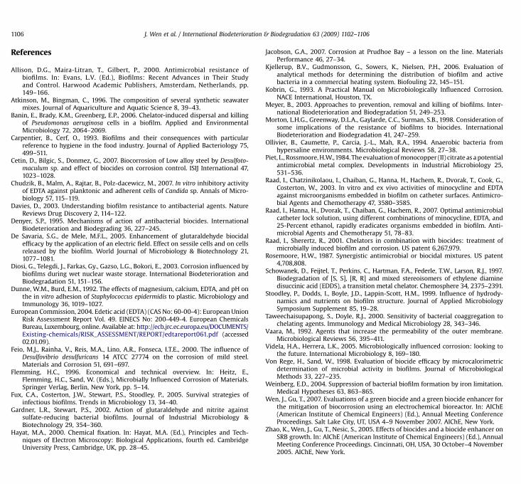
Figs. 2 and 6 show the EDDS enhancement of glutaraldehydetreatment of established SRB biofilms. Sessile SRB cells were obvi-ously present on coupon surfaces with 30 ppm glutaraldehydetreatment alone (Fig. 6a). With the added 2000 ppm of EDDS, sessileSRB cells were hardly noticeable (Fig. 6b). The effect of EDDSenhanced 30 ppm glutaraldehyde treatment is better than 500 ppmglutaraldehyde treatment without EDDS shown in Fig. 2. Vaara (1992)
mentioned that chelators can remove the divalent cations suchas Mg2þ and Ca2þ from lipopolysaccharide resulting in increasedpermeability on the outer membrane for Gram-negative cells. Withthe help of EDDS, it might be easier for glutaraldehyde to penetratethrough the SRB biofilms and the outer membrane of SRB, achievingthe higher efficacy on inhibition of sessile SRB growth.
4. Conclusions
Preliminary experimental data in this work demonstrated thatEDDS has efficacy in enhancing glutaraldehyde for the inhibition ofSRB biofilm establishment and for the treatment of established SRBbiofilms. EDDS can be used to cut down the glutaraldehyde dosageconsiderably. The data above showed at least an order of magnitudedosage reduction could be achieved. These initial tests used a ratherhigh concentration of EDDS to prove the concept. Tests using lowerdosages of EDDS and combination with biocides like THPS, BNPDand QAC are desired in the future.
Acknowledgements
This work was partially supported by a grant from EnhancedCorrosion Prevention, LLC and a grant from the M. D. AndersonCancer Center.
Fig. 5. Effects of EDDS enhancement of glutaraldehyde on preventing SRB biofilmestablishment using 30 ppm of glutaraldehyde in modified ATCC 1250 medium for 8days: (a) without EDDS; (b) with 2000 ppm of EDDS. The figure is representative ofabout 0.3% of the total exposed coupon surface. Scale bar in the insert is 5 mm.
Fig. 6. Effects of EDDS enhancement of glutaraldehyde on treating established SRBbiofilm using 30 ppm of glutaraldehyde in modified ATCC 1250 medium for 8 days:(a) without EDDS; (b) with 2000 ppm of EDDS. The figure is representative of about0.3% of the total exposed coupon surface. Scale bar in the insert is 5 mm.
J. Wen et al. / International Biodeterioration & Biodegradation 63 (2009) 1102–1106 1105
Author's personal copy
References
Allison, D.G., Maira-Litran, T., Gilbert, P., 2000. Antimicrobial resistance ofbiofilms. In: Evans, L.V. (Ed.), Biofilms: Recent Advances in Their Studyand Control. Harwood Academic Publishers, Amsterdam, Netherlands, pp.149–166.
Atkinson, M., Bingman, C., 1996. The composition of several synthetic seawatermixes. Journal of Aquariculture and Aquatic Science 8, 39–43.
Banin, E., Brady, K.M., Greenberg, E.P., 2006. Chelator-induced dispersal and killingof Pseudomonas aeruginosa cells in a biofilm. Applied and EnvironmentalMicrobiology 72, 2064–2069.
Carpentier, B., Cerf, O., 1993. Biofilms and their consequences with particularreference to hygiene in the food industry. Journal of Applied Bacteriology 75,499–511.
Cetin, D., Bilgic, S., Donmez, G., 2007. Biocorrosion of Low alloy steel by Desulfoto-maculum sp. and effect of biocides on corrosion control. ISIJ International 47,1023–1028.
Chudzik, B., Malm, A., Rajtar, B., Polz-dacewicz, M., 2007. In vitro inhibitory activityof EDTA against planktonic and adherent cells of Candida sp. Annals of Micro-biology 57, 115–119.
Davies, D., 2003. Understanding biofilm resistance to antibacterial agents. NatureReviews Drug Discovery 2, 114–122.
Denyer, S.P., 1995. Mechanisms of action of antibacterial biocides. InternationalBiodeterioration and Biodegrading 36, 227–245.
De Savaria, S.G., de Mele, M.F.L., 2005. Enhancement of glutaraldehyde biocidalefficacy by the application of an electric field. Effect on sessile cells and on cellsreleased by the biofilm. World Journal of Microbiology & Biotechnology 21,1077–1081.
Diosi, G., Telegdi, J., Farkas, Gy., Gazso, L.G., Bokori, E., 2003. Corrosion influenced bybiofilms during wet nuclear waste storage. International Biodeterioration andBiodegradation 51, 151–156.
Dunne, W.M., Burd, E.M., 1992. The effects of magnesium, calcium, EDTA, and pH onthe in vitro adhesion of Staphylococcus epidermidis to plastic. Microbiology andImmunology 36, 1019–1027.
European Commission, 2004. Edetic acid (EDTA) (CAS No: 60-00-4): European UnionRisk Assessment Report Vol. 49. EINECS No: 200-449-4. European ChemicalsBureau, Luxembourg, online. Available at: http://ecb.jrc.ec.europa.eu/DOCUMENTS/Existing-chemicals/RISK_ASSESSMENT/REPORT/edtareport061.pdf (accessed02.01.09).
Feio, M.J., Rainha, V., Reis, M.A., Lino, A.R., Fonseca, I.T.E., 2000. The influence ofDesulfovibrio desulfuricans 14 ATCC 27774 on the corrosion of mild steel.Materials and Corrosion 51, 691–697.
Flemming, H.C., 1996. Economical and technical overview. In: Heitz, E.,Flemming, H.C., Sand, W. (Eds.), Microbially Influenced Corrosion of Materials.Springer Verlag, Berlin, New York, pp. 5–14.
Fux, C.A., Costerton, J.W., Stewart, P.S., Stoodley, P., 2005. Survival strategies ofinfectious biofilms. Trends in Microbiology 13, 34–40.
Gardner, L.R., Stewart, P.S., 2002. Action of glutaraldehyde and nitrite againstsulfate-reducing bacterial biofilms. Journal of Industrial Microbiology &Biotechnology 29, 354–360.
Hayat, M.A., 2000. Chemical fixation. In: Hayat, M.A. (Ed.), Principles and Tech-niques of Electron Microscopy: Biological Applications, fourth ed. CambridgeUniversity Press, Cambridge, UK, pp. 28–45.
Jacobson, G.A., 2007. Corrosion at Prudhoe Bay – a lesson on the line. MaterialsPerformance 46, 27–34.
Kjellerup, B.V., Gudmonsson, G., Sowers, K., Nielsen, P.H., 2006. Evaluation ofanalytical methods for determining the distribution of biofilm and activebacteria in a commercial heating system. Biofouling 22, 145–151.
Kobrin, G., 1993. A Practical Manual on Microbiologically Influenced Corrosion.NACE International, Houston, TX.
Meyer, B., 2003. Approaches to prevention, removal and killing of biofilms. Inter-national Biodeterioration and Biodegradation 51, 249–253.
Morton, L.H.G., Greenway, D.L.A., Gaylarde, C.C., Surman, S.B., 1998. Consideration ofsome implications of the resistance of biofilms to biocides. InternationalBiodeterioration and Biodegradation 41, 247–259.
Ollivier, B., Caumette, P., Carcia, J.-L., Mah, R.A., 1994. Anaerobic bacteria fromhypersaline environments. Microbiological Reviews 58, 27–38.
Piet, L., Rossmoore, H.W.,1984. The evaluation of monocopper (II) citrate as a potentialantimicrobial metal complex. Developments in Industrial Microbiology 25,531–536.
Raad, I., Chatzinikolaou, I., Chaiban, G., Hanna, H., Hachem, R., Dvorak, T., Cook, G.,Costerton, W., 2003. In vitro and ex vivo activities of minocycline and EDTAagainst microorganisms embedded in biofilm on catheter surfaces. Antimicro-bial Agents and Chemotherapy 47, 3580–3585.
Raad, I., Hanna, H., Dvorak, T., Chaiban, G., Hachem, R., 2007. Optimal antimicrobialcatheter lock solution, using different combinations of minocycline, EDTA, and25-Percent ethanol, rapidly eradicates organisms embedded in biofilm. Anti-microbial Agents and Chemotherapy 51, 78–83.
Raad, I., Sherertz, R., 2001. Chelators in combination with biocides: treatment ofmicrobially induced biofilm and corrosion. US patent 6,267,979.
Rosemoore, H.W., 1987. Synergistic antimicrobial or biocidal mixtures. US patent4,708,808.
Schowanek, D., Feijtel, T., Perkins, C., Hartman, F.A., Federle, T.W., Larson, R.J., 1997.Biodegradation of [S, S], [R, R] and mixed stereoisomers of ethylene diaminedisuccinic acid (EDDS), a transition metal chelator. Chemosphere 34, 2375–2391.
Stoodley, P., Dodds, I., Boyle, J.D., Lappin-Scott, H.M., 1999. Influence of hydrody-namics and nutrients on biofilm structure. Journal of Applied MicrobiologySymposium Supplement 85, 19–28.
Taweechaisupapong, S., Doyle, R.J., 2000. Sensitivity of bacterial coaggregation tochelating agents. Immunology and Medical Microbiology 28, 343–346.
Vaara, M., 1992. Agents that increase the permeability of the outer membrane.Microbiological Reviews 56, 395–411.
Videla, H.A., Herrera, L.K., 2005. Microbiologically influenced corrosion: looking tothe future. International Microbiology 8, 169–180.
Von Rege, H., Sand, W., 1998. Evaluation of biocide efficacy by microcalorimetricdetermination of microbial activity in biofilms. Journal of MicrobiologicalMethods 33, 227–235.
Weinberg, E.D., 2004. Suppression of bacterial biofilm formation by iron limitation.Medical Hypotheses 63, 863–865.
Wen, J., Gu, T., 2007. Evaluations of a green biocide and a green biocide enhancer forthe mitigation of biocorrosion using an electrochemical bioreactor. In: AIChE(American Institute of Chemical Engineers) (Ed.), Annual Meeting ConferenceProceedings. Salt Lake City, UT, USA 4–9 November 2007. AIChE, New York.
Zhao, K., Wen, J., Gu, T., Nesic, S., 2005. Effects of biocides and a biocide enhancer onSRB growth. In: AIChE (American Institute of Chemical Engineers) (Ed.), AnnualMeeting Conference Proceedings. Cincinnati, OH, USA, 30 October–4 November2005. AIChE, New York.
J. Wen et al. / International Biodeterioration & Biodegradation 63 (2009) 1102–11061106