Internal diseases propedeutics (Part I). Diagnostics of...
94
1 Federal budgetary educational establishment of higher education Ulyanovsk State University The Institute of medicine, ecology and physical culture Smirnova A.Yu., Gnoevykh V.V. INTERNAL DISEASES PROPEDEUTICS PART I DIAGNOSTICS OF PULMONARY DISEASES Textbook of Medicine for medicine faculty students Ulyanovsk, 2016
Transcript of Internal diseases propedeutics (Part I). Diagnostics of...

1
Federal budgetary educational establishment of higher education
Ulyanovsk State University
The Institute of medicine, ecology and physical culture
Smirnova A.Yu., Gnoevykh V.V.
INTERNAL DISEASES PROPEDEUTICS
PART I
DIAGNOSTICS OF PULMONARY DISEASES
Textbook of Medicine for medicine faculty students
Ulyanovsk, 2016

2
УДК 811.11(075.8)
БКК 81.432.1-9я73
С50
Reviewers:
Savonenkova L.N. – MD, professor of Department of faculty therapy
Smirnova A.Yu., Gnoevykh V.V. Internal diseases propedeutics (Part I). Diagnostics of pulmonary diseases: Textbook of Medicine for medicine faculty students/Ulyanovsk: Ulyanovsk State University, 2016.-93
This publication is the first part of “Internal diseases propedeutics”, which main goal is the practical assistance for students in the development of the fundamentals of clinical diagnosis of diseases of the respiratory system. It contains a description of the main methods of laboratory and instrumental diagnostic tests of diseases of the respiratory system. The publication is illustrated with charts, drawings and tables. The textbook is intended for students of medical universities.
Smirnova A.Yu., Gnoevykh V.V., 2016
Ulyanovsk State University, 2016
3
THE CONTENTS OF A TEXT BOOK
Questioning and examination of patients with diseases of the lungs. 5
Main complains of patients with diseases of the lungs. 5
General inspection 7
Examination of the chest 8
Lungs percussion data in norm and pathology 13
Lungs auscultation data in norm and pathology 20
Pulmonary syndromes. 24
Pulmonary consolidation syndrome. 24
Inflammatory infiltration 25
Compressive atelectasis (pulmonary [lung] collapse) syndrome 28
Obturative atelectasis (segmental or lobar). 29
Pulmonary cavity syndrome 29
Pleural effusion syndrome 31
Syndrome of air in pleural cavity (pnemothorax) 35
Hyperinflated lung syndrome (emphysema) 37
A list of the main instrumental and laboratory methods of examination of respiratory system
39
Fiberoptic bronchoscopy 39
Blood gases 42
Pulmonary function testing 45
Airflow obstruction syndrome 54
Respiratory deficiency syndrome. 54
Blood tests 59

4
Sputum test 60
Questions for test control to engage the theme "questioning, general examination, inspection and palpation of the patient with respiratory diseases"
63
Questions for test control to engage the theme "comparative percussion of the lungs. pulmonary syndromes"
69
Application 80
References 94

5
Questioning and examination of patients with diseases of the lungs.
Main complains of patients with diseases of the lungs.
Shortness of breath, breathlessness (dyspnoe): patients subjective feeling of lack
of air; causes: stenosis of the larynx or (and) trachea, bronchial asthma, chronic
obstructive pulmonary disease, lung cancer (obstruction of the air passage in the
respiratory tract), pneumonia, tuberculosis, pulmonary infarction, pulmonary fibrosis,
obstructive pulmonary atelectasis, cardiac asthma (decreases the airiness of the
lungs), pneumothorax, hydrothorax (a decrease in respiratory surface of the lung due
to the accumulation of air or fluid in the pleural cavity), pulmonary and respiratory
failure. Types of dyspnea: inspiratory (inhale is difficult), expiratory (difficulty
exhaling), mixed, physiological and pathological.
Cough (tussis) - definition: a reflex act when a cluster of airway pathologic
discharge or ingress of foreign bodies; laryngitis, pleuritis, tracheitis, pneumonia;
causes: chronic obstructive pulmonary disease, bronchial asthma, multiple
bronchiectasis, lung abscess, tuberculosis, tumors.
Types of cough: cough without sputum, cough with sputum expectoration,
morning cough (bronchitis, bronchiectasis, lung abscess, and cavernous tuberculosis
of the lungs), night (tuberculosis, lymphogranulomatosis, malignant neoplasm),
evening (sometimes after pneumonia), persistent and periodic cough, barking
(whooping cough, compression of the trachea by a goiter or tumor, lesions of the
larynx with swelling of the vocal cords), cough with sputum "full mouth" (abscess of
the lungs), cough with release of large quantities of sputum (abscess and
bronchiectasis).
Sputum
Information should be obtained about its quantity, colour (white, grey, black,
pink, yellow or green), viscosity (serous or tacky), taste and odour (Table 1).
6
Table 1. Characteristics of sputum
Sputum Condition
Mucoid, excessive quantities Chronic bronchitis
Mucopurulent or purulent
(yellow or green)
Infection - acute or chronic bronchitis
Excessive in early mornings,
or at change of posture,
purulent
Bronchiectasia
Black Cigarette or atmospheric smoke, coal-miner's
sputum
Pink, frothy Acute pulmonary oedema
Rusty Lobar pneumonia
Blood-stained Acute bronchitis, tuberculosis, neoplasia
Viscous with plugs Asthmatic pulmonary eosinophilia
A change of colour from white to green or yellow suggests the onset of
infections in patients with chronic bronchitis. A pink and frothy sputum associated
with breathlessness is commonly encountered in pulmonary oedema.
Hemoptysis (haemoptoe) – definition: the allocation of blood with sputum
during cough; causes: viral pneumonia, abscess and gangrene of lungs,
bronchiectasis, tuberculosis, lung cancer, actinomycosis and ascariasis, mitral
stenosis, mitral insufficiency, cardiac asthma, pulmonary embolism, infarction of the
lungs. Features of hemoptysis in certain diseases: scarlet blood (tuberculosis, Central
bronchogenic cancer, bronchiectasis, ascariasis and actinomycosis of the lung,
pulmonary infarction in the first 2-3 days; "rusty" sputum (pneumonia in stage 2).
7
Chest pain characteristics: location, nature, intensity, duration, irradiation,
connection with the act of breathing, cough and body positions. Causes of pain in the
chest, and in certain diseases: superficial pain in the chest wall, worse on breathing,
coughing, sudden movements of the trunk, or in a position on the sick side (trauma,
erysipelas, herpes zoster, myalgia, myositis, thoracic osteochondrosis with radicular
syndrome, fractures and periostitis of the ribs, the tumor metastasis to the ribs and the
pleura); in lesions of the lung and pleura (dry pleurisy, pneumonia, abscess,
tuberculosis, infarction, metastasis of tumors to the pleura or the tumor of the pleura,
traumatic or spontaneous pneumothorax); other disorders (subphrenic abscess, acute
pancreatitis).
Risk factors of respiratory diseases: hypothermia, Smoking, atmospheric and
occupational pollutants, pathology of internal organs, burdened heredity (asthma,
Kartagener disease, cystic fibrosis, Oncology, primary emphysema, etc.), trauma,
aspiration, contact with tuberculosis patients, reducing the reactivity of the immune
system, the season of epidemics of viral or other infection.
General inspection
On general examination, there may be clues to the underlying disease:
Cachexia may occur in malignant disease, and in severe chronic lung disease,
including fibrosis, infection and emphysema. Cachexia may occur in a number of
severe disorders, including chronic lung disease such as pulmonary fibrosis,
tuberculosis and emphysema, malignant disease, including bronchial carcinoma, and
systemic infection, especially with HIV («slim disease»). Note the obvious signs
ofweight loss, fever, with widespread muscle and soft-tissues wasting.
Cyanosis - best seen in the lips, tongue, buccal mucosa and fingers - indicates
significant desatura-tion of circulating haemoglobin. Cyanosis is a fundamental sign
of cardiorespiratory disorders and suggests capillary oxygen desaturation of 85% or
lower.
A herpetic eruption on and around the lips is sometimes seen in a patient with a
respiratory infection.

8
Coal dust tattoos may be seen on the face, though these are more often seen on
the arms, as an occupational legacy in a patient with pulmonary fibrosis.
Nasal polyps frequently occur in patients with an atopic background and in those
with cystic fibrosis.
Eczema is often found in conjugation with hay fever and asthma.
The oscillations of the jugular venous pulse are difficult to interpret in patients
with chronic airways obstruction who generate a high intrathoracic pressure to drive
the air out through the narrowed bronchi. However, static engorgement of the neck
veins is an important sign of obstruction of the superior vena cava, usually caused by
mediastinal malignancy.
«Nicotine» stained fingers occur in heavy smokers, and typical pigmented scars
may occur in coal miners; in association with finger clubbing both signs have an
ominous significance, suggesting underlying bronchial carcinoma, pulmonary
fibrosis, bronchiectasia or chronic sepsis.
Finger clubbing is frequently present in a number of conditions, especially
bronchial carcino ma (occasionally with a pleural fibrinoma), and in those with
chronic purulent conditions such as bronchiectasia, lung abscess and empyema
(Application. Figure 1)
Examination of the chest.
Patients having respiratory system diseases may present the following problems:
chest pain, cough, dyspnea, asphyxiation. Pains caused by respiratory apparatus
lesion depend on pulmonary pleura involvement. If the process is confined to lungs
only, no pain can be registered since the lung tissue has no pain receptors. Thus, pain
can accompany any lung process provided it spread as far as pleura. Pleural pains are
characterized by the following features: they are of shooting character, not of
radiating nature, and are usually aggravated or detected only at the maximum of
inhale or while coughing and sneezing, that is, when pleural leaves overlap. It is
important to specially note some specific character of pains arising due to
diaphragmatic pleurisy. They are peculiar in having ability to spread to jugular region
via phrenic nerve. On the other hand, these pains radiate to abdominal cavity and can

9
be mistaken for abdominal diseases. Pleural pains should be distinguished from other
kinds that can arise in the thorax region: caused by thorax diseases: intercostal
muscles myosites, intercostal nerves pleurisy and nerve root compression
(osteochondrosis), rib injuries (fractures, fissures, etc.); pleural pains, cardiac and
vascular pains (angina pectoris, myocardial infarction, aortitis, etc.); reflex pains
(cholecystitis, diaphragmatic hernia, ulcer, appendicitis). The second characteristic
complaint is cough which categorizes according to pathogenesis: pulmonary, reflex,
central; according to duration: permanent, occasional; according to timbre: barking,
hoarse, noiseless, etc.; according to character: dry, productive (nature, smell, amount,
period of expectoration). The third characteristic patient complaint is dyspnea. It
categorizes: depending on breathing stages: inspiratory, expiratory, mixed;
accordingto pathogenesis: pulmonary, cardiac, anemic, etc. Asthma is an attack-like
abrupt dyspnea. It occurs not only with lung diseases (bronchial asthma), but also
with a number of other diseased states: bronchial, cardiac, mixed, cerebral, and
hysteric types.
On static examination, thorax shape characteristics are described. There exist
three normal types of thorax: asthenic chest, normosthenic (athletic) type, and
hypersthenic chest. Its pathologic shapes are paralytic chest, emphysematous (barrel)
chest, rickets breast, funnel breast, kyphoscoliotic chest. It is necessary to explain
here the notions of scoliosis, lordosis, and kyphosis. On static examination of thorax,
there can also be detected distortion in terms of restriction or enlargement of one side.
Examples can be given of the restriction of one side of the chest when having
pulmonary fibrosis, and enlargement with exudative pleurisy. Dynamic inspection
allows to evaluate the extent of thorax share in the breathing process, lagging of one
side, etc. It also allows to characterize breathing process. Palpation of chest
determines: chest elasticity or resistance, tenderness areas, vocal or tactile fremitus
(fremitus pectoralis).
Normal shape of the chest: normosthenic (truncated cone shape, transverse size
is larger than the Antero-posterior, supra - and infraclavicular fossa are moderately,
clearly defines the angle of Louis, the epigastric angle is sharp , the edges are

10
conisholme direction, the blades are tightly adjacent to the body, thoracic abdominal
equal; hypersthenic (cylinder in shape, the transverse dimension close to the Antero-
posterior, supra - and infraclavicular fossa are virtually absent, significantly
pronounced angle of Louis, the epigastric angle is obtuse , the ribs are horizontal, the
intercostal spaces are reduced, the vanes merge with the trunk, thoracic abdominal
less; asthenic (long and narrow in shape, reduced transverse and anteroposterior
dimensions, above and subclavial fossa pronounced, the angle of Louis is missing,
the epigastric angle is sharp, the edges are close to the vertical direction and "wing"
the scapula, thoracic and more abdominal.
Pathological shapes of the chest: emphysematous (with emphysema: barrel
shape, expansion of intercostal spaces, clearly defined the angle of Louis, the
epigastric angle is obtuse, the ribs are almost horizontal direction, the breath is
actively involved auxiliary respiratory muscles (sternokleidomastoid, trapezius, etc.
with indrawing of the intercostal spaces), lungs are in the phase of constant inhalation
and exhalation is much difficult), paralytic (for a total asthenia, disease Marfan's
tuberculosis: symptoms asthenic chest + atrophy of the muscles of the chest, an
asymmetric location of the clavicle, and the unequal retraction of the supraclavicular
fossae, shoulder blades at different levels and meshayutsya when breathing
asynchronously), rachitic (synonym-keel: increased anteroposterior size due to the
keeled sternum, "rachitic rosary" in the transition region from the rib cartilage into
bone) (Application pic. 2.), funnel (due to a congenital anomaly of the sternum: the
chest has a funnel-shaped indentation in the lower 1/3 of the sternum) (Application,
pic. 3), navicular (in syringomyelia: thoracic has indentation in the middle to upper
parts of the sternum).
Causes and types of spinal deformities: trauma, tuberculosis of the spine,
ankylosing spondylitis, etc. Scoliosis (curvature in the lateral direction), kyphosis
(backward curvature), lordosis (forward curvature), kyphoscoliosis – combination of
scoliosis and kyphosis. Kyphosis results in anterior concavity of thoracic spine and
thereby leads to shortening of the chest. Kyphosis is frequently seen in erdely people
with osteoporosis, chronic obstructive airways disease.
11
The cause of reduced 1/2 chest: pleural adhesions, pulmonary fibrosis, lung
carnification, pulmonary infarction,lung abscess, tuberculosis, pneumonectomy or
lobectomy, obstructive atelectasis.
The reasons for the increase of 1/2 chest: fluid in the pleural cavity, a
pneumothorax (the flattening and bulging of the intercostal spaces, asymmetry of the
clavicles and the shoulder blades, lag 1/2 of the chest during breathing).
Physiological types of breathing: thoracic (mostly in men), abdominal (more
common in women) and mixed. The breathing rate should be counted when the
patient is not conscious of it it can be done during the earlier part of the inspection/
The normal rate is between 14 and 18 breaths a minute. In opiate or barbiturate
poisoning this may fall to below eight breaths a minute (bradypnoe) whereas in acute
bronchopneumonia the rate may exceed 40 a minute (tahypnoe). The relationship
between inspiration and expiration should be determined. Normally, the inspiration is
active and longer whereas expiration is shorter and accomplished by the passive
recoil of the lungs. In small airways obstruction the expiration becomes active and
prolonged, due to a greater pressure gradient from small to major airways.
The deep inspiration and shorter expiration which follows immediately gives the
respiration its normal rhythm. Shallow breathing with short inspiration and
expiration occurs either when breathing is restricted (e.g. obesity, pulmonary fibrosis)
or is painful as in chest wall disease and pleurisy, or in anxiety states. Kussmaul
breathing with deep inspiration and expiration typically occurs in metabolic acidosis
(e.g. diabetic ketoac-idosis, renal failure, methyl alcohol poisoning, etc.). Cheyne-
Stokes breathing comprises periods of apnea alternating with a gradual resumption
of respiration with increasing depth which then declines to another period of
cessation of breathing. This pattern of breathing is also termed periodic or cyclical
breathing and occurs in advanced cardiac and respiratory failure, narcotic drug
poisoning and in cerebrovascular disease (Fig. 1). Pursed lip breathing is a sign of
severe small airways obstruction, as can be found in asthma and emphysema, but it
also occurs occasionally in left heart failure. It is an attempt by the patient to create
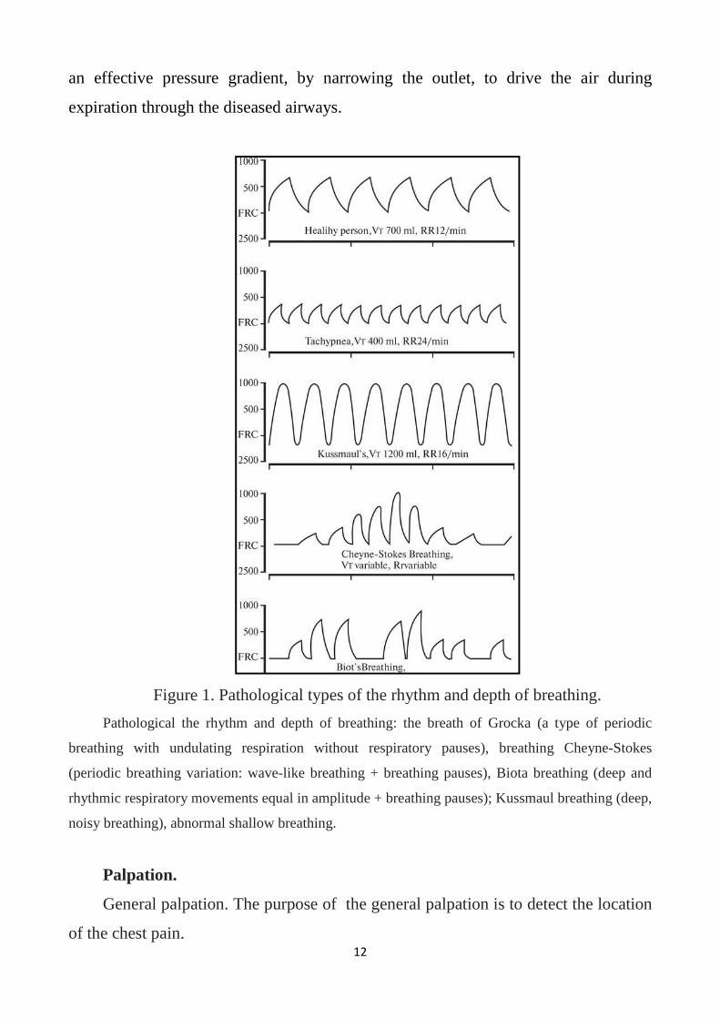
12
an effective pressure gradient, by narrowing the outlet, to drive the air during
expiration through the diseased airways.
Figure 1. Pathological types of the rhythm and depth of breathing.
Pathological the rhythm and depth of breathing: the breath of Grocka (a type of periodic
breathing with undulating respiration without respiratory pauses), breathing Cheyne-Stokes
(periodic breathing variation: wave-like breathing + breathing pauses), Biota breathing (deep and
rhythmic respiratory movements equal in amplitude + breathing pauses); Kussmaul breathing (deep,
noisy breathing), abnormal shallow breathing.
Palpation.
General palpation. The purpose of the general palpation is to detect the location
of the chest pain.

13
Resistance (expansion) of the chest - opposite the property of elasticity; causes:
emphysema of the lungs, ossification of ribs in the elderly, fluid in the pleural cavity,
tumors of the pleura
Vocal fremitus – carrying out oscillatory movements of the vocal cords in the bronchi
at the surface of the chest. Vocal fremitus is determined by palpation of the chest.
You can test the vocal fremitus by placing palms or more sensitive ulnar border of
your hand on the chest while the patient repeats «ninety nine» in a deep clear voice.
The corresponding areas on the chest must be tested simultaneously by both palms in
symmetrical areas. Vocal fremitus is increased through a consolidated lung (lobar
pneumonia) and decreased when the corre sponding bronchi are obstructed, or if there
is a pleural effusion. It is useful in distinguishing consolidation from pleural effusion,
both of which produce a dull note on percussion.
Causes physiological increase voice fremitus: over upper lobes of the lungs
compared to the lower, in men with a low voice, at astenikov with a thin rib cage;
Causes physiological weakening voice fremitus: increase of subcutaneous tissue in
women and children with high tone of voice, over the lower lung lobes than the
upper.
The causes for pathological voice fremitus increase: the inflammatory syndrome
seal lung tissue, compression atelectasis, pneumothorax communicating with a
bronchus, the air cavity in the lung communicating with the bronchus.
The causes of pathological weakening voice fremitus: obesity, hydrothorax,
pneumothorax is not communicating with a bronchus, obstructive atelectasis.
Lungs percussion data in norm and pathology
Percussion in its modern modification was proposed by Viennese physician
Leopold Auenbrugger in 1761. They distinguish the following peculiarities of
percussion note: loudness, i.e. amplitude of oscillation, duration, pitch, and tympanic
component. Loudness and duration of percussion note is relating to density of
underlying tissues. On comparative percussion the percussion note over the lung may
change towards either tympanic and flat note.
14
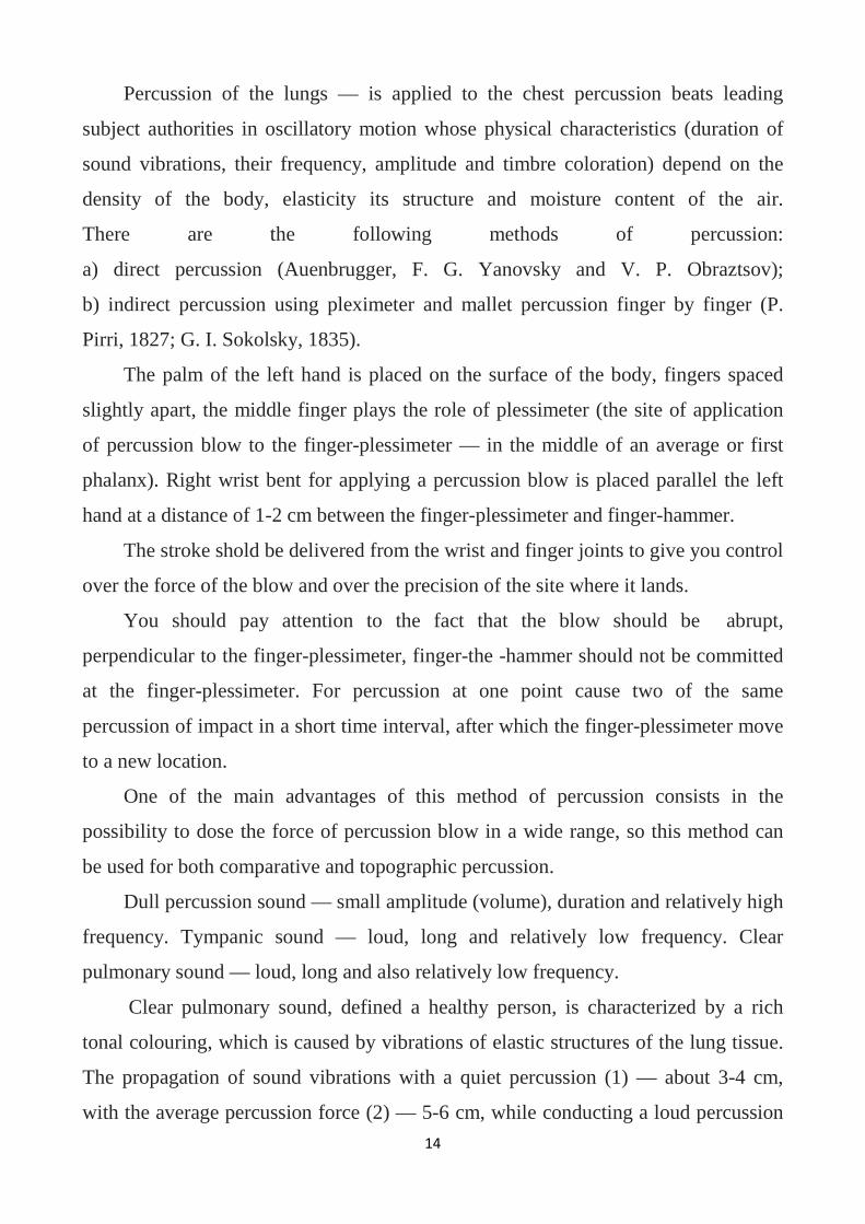
Percussion of the lungs — is applied to the chest percussion beats leading
subject authorities in oscillatory motion whose physical characteristics (duration of
sound vibrations, their frequency, amplitude and timbre coloration) depend on the
density of the body, elasticity its structure and moisture content of the air.
There are the following methods of percussion:
a) direct percussion (Auenbrugger, F. G. Yanovsky and V. P. Obraztsov);
b) indirect percussion using pleximeter and mallet percussion finger by finger (P.
Pirri, 1827; G. I. Sokolsky, 1835).
The palm of the left hand is placed on the surface of the body, fingers spaced
slightly apart, the middle finger plays the role of plessimeter (the site of application
of percussion blow to the finger-plessimeter — in the middle of an average or first
phalanx). Right wrist bent for applying a percussion blow is placed parallel the left
hand at a distance of 1-2 cm between the finger-plessimeter and finger-hammer.
The stroke shold be delivered from the wrist and finger joints to give you control
over the force of the blow and over the precision of the site where it lands.
You should pay attention to the fact that the blow should be abrupt,
perpendicular to the finger-plessimeter, finger-the -hammer should not be committed
at the finger-plessimeter. For percussion at one point cause two of the same
percussion of impact in a short time interval, after which the finger-plessimeter move
to a new location.
One of the main advantages of this method of percussion consists in the
possibility to dose the force of percussion blow in a wide range, so this method can
be used for both comparative and topographic percussion.
Dull percussion sound — small amplitude (volume), duration and relatively high
frequency. Tympanic sound — loud, long and relatively low frequency. Clear
pulmonary sound — loud, long and also relatively low frequency.
Clear pulmonary sound, defined a healthy person, is characterized by a rich
tonal colouring, which is caused by vibrations of elastic structures of the lung tissue.
The propagation of sound vibrations with a quiet percussion (1) — about 3-4 cm,
with the average percussion force (2) — 5-6 cm, while conducting a loud percussion
15
(3) — 7-8 cm. In quiet (threshold) percussion sound waves penetrate deep into
tissues 2-3 cm.
As the standard of the absolutely dull sound is the sound, which is determined
by percussion of the thigh muscles (femoral sound). The tympanic sound is a sound
that can only be detected by percussion of the abdominal cavity and space of Traube.
The standard of the clear pulmonary sound is the sound, which will be determined
during percussion of the axillary and subscapular areas in a healthy person. The
standard hyperresonant (tympanic) sound is the sound that appears when the
percussion cushion.
General rules of percussion of the lungs
1. Position of patient and physician should be comfortable to study.
2. Finger-plessimeter pressed tightly to the skin.
3. Finger-the hammer perpendicular to the finger-plessimeter.
4. Right-hand parallel the left (wrist joints placed one above the other).
5. 2 applied percussion blows are delivered through short time intervals.
6. Hand movements are carried out only in the wrist joint.
7. The doctor's hands should be warm.
Distinguish between comparative and topographic percussion of lungs.
Comparative percussion of the lungs is used to determine the nature of
pathological changes in the lungs and pleural cavity and used for the diagnosis of
bronchopulmonary syndromes.
Technique of comparative percussion is:
1. Conduct a comparison of the nature of percussion sounds obtained over
symmetrical areas of the chest.
2. Cause "rebounding" of percussion blows of medium strength. The volume of the
percussion sound can change depending on the thickness of the subcutaneous tissue,
the degree of development of muscles, the depth of location of the pathological
process and other reasons.
3. Percussion is carried out on the intercostal spaces.

16
To percuss the front of the chest, you should start by percussing over the
clavicle on one side, then on the other side, and then percuss on each ribspace and
compare the note elicited over the corresponding note on the other side.Percussion is
carried out 1 finger phalanx of plessimeter, because anatomically this is the most
convenient. Then put the direct percussion blows to the collarbone, using it as
plessimeter (Application Fig.4.).
Further percuss in the first, second and third right and left intercostal spaces at
the level of the midclavicular line. Below level III intercostal space on the left cardiac
dullness, so further research is carried out in the pits of Maranham. The patient stands
or sits, arms lowered along the torso, muscles tense, breathing smooth and shallow.
The doctor performs the percussion, usually standing to the right of the patient.
Finger-plessimeter is parallel to the ribs, but it is tightly pressed against the patient's
body.
For percussion axillary region finger-plessimeter put vertically in the upper part
of the right, and then left arm. The doctor is beside the patient, opposite the axillary
region. Then comparative percussion is carried out by comparing the percussion
blows in the third intercostal space of the axillary region on the right and left, and
then the percussion continue in the fourth intercostal space of the axillary region on
the right and left. The doctor is in front of the patient.
When performing comparative percussion on the posterior surface of the chest at
the beginning percuss suprascapular region, the finger-plessimeter set slightly above
the spine of the scapula and parallel to it, percussion is applied consistently blows
right and left with the patient standing with his hands at his sides, muscles tense.
Then percuss "alarm" zones and interscapular region. Finger-plessimeter is parallel to
the spine at the edge of the blades, sequentially from right to left. Hands patient is
asked to cross on his chest, putting hands on shoulders, with the blades of the
supplies are provided, expanding the interscapular space.
Further percuss subscapular area (Application Fig.5).. Finger-plessimeter is placed
horizontally below the angle of the scapula, alternately right and left. The arms of the
patient are lowered along the body, the muscles are relaxed. In detail, the technique
17
of comparative percussion of the lungs is represented on the website UlSU in the
form of a training video http://www.ulsu.ru/com/chairs/pii/stud/Uchebnie_filmi/), as
well as at the following address: http://youtu.be/Z9j26eDV5eA
The clinical significance of comparative percussion of lungs:
Percussion the clear pulmonary sound is heard in a healthy person over the lungs with
unchanged pulmonary tissue. Characteristic sound: loud, long and of low frequency
caused by fluctuations in the unmodified elastic structures of the lung tissue. The
standard is sound, as determined by percussion in the axillary and subscapular areas
in a healthy person.
Dull percussion sound – quiet, vague and high-frequency sound. Is formed over
the area of the lung containing less air than in norm or above the liquid.
Causes and anatomical localization of physiological shortening of percussion sound:
increase the thickness of the pulmonary layer over the right top of the shorter right
bronchus; in patient with muscled, in 2-3 intercostal space to the left due to the
proximity of the heart, over the upper lobes of both lungs, in the right axillary region
due to the proximity of the liver.
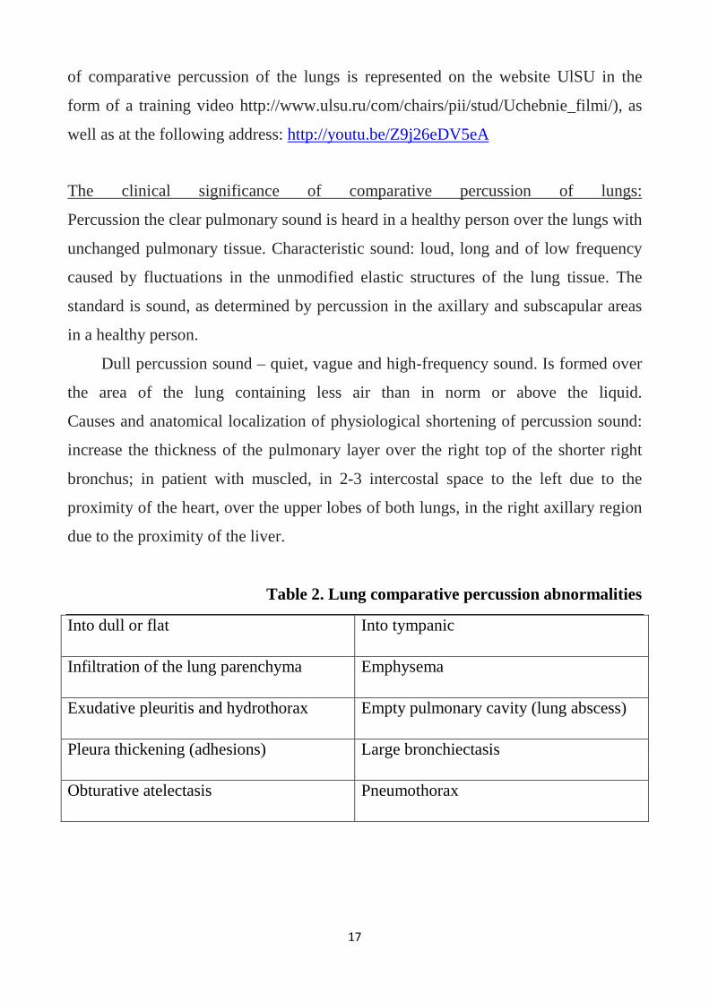
Table 2. Lung comparative percussion abnormalities
Into dull or flat Into tympanic
Infiltration of the lung parenchyma Emphysema
Exudative pleuritis and hydrothorax Empty pulmonary cavity (lung abscess)
Pleura thickening (adhesions) Large bronchiectasis
Obturative atelectasis Pneumothorax
18
Topographic percussion
In order to determine the exact size of the various organs or to differentiate the
borders of two organs that lie adjacent to one another, they must be of different
densities. Thus, by percussion it is easy to determine where the lung ends and the
heart begins because of the different densities of these organs. However, it is
extremely difficult to differentiate between heart and liver dullness, or between the
dullness of pleural effusion and liver dullness since the densities so closely
approximate one another.
The normal limits of pulmonary resonance correspond accurately to the
anatomic boundaries of the lung. With light percussion the inferior limits of the lung
are found at the level of the sixth rib in the mediclavicular line, the eighth rib in the
midaxillary line and the tenth rib in the scapular line (Application Fig 6-8.).
Table 3.
Lungs topographic percussion abnormalities (lower lung borders).
Elevation Depression
Shrinking of the lung Emphysema
Thickening of pleura Asthma
Pneumothorax (false depression) Chronic pulmonary congestion
Exudative pleuritis and hydrothorax High diaphragm
Flatulence(meteorism)
The lower limit of pulmonary resonance should in all instances be examined by
percussion during both forced inspiration and expiration; normally the difference in
space between these two extremes measures 3 to 4 cm. This space represents the
complemental pleural space, and by this means the degree of respiratory mobility is
attained. This respiratory mobility is diminished or absent in diseases of the lung such
as emphysema, pleural diaphragmatic adhesions and conditions that interfere with
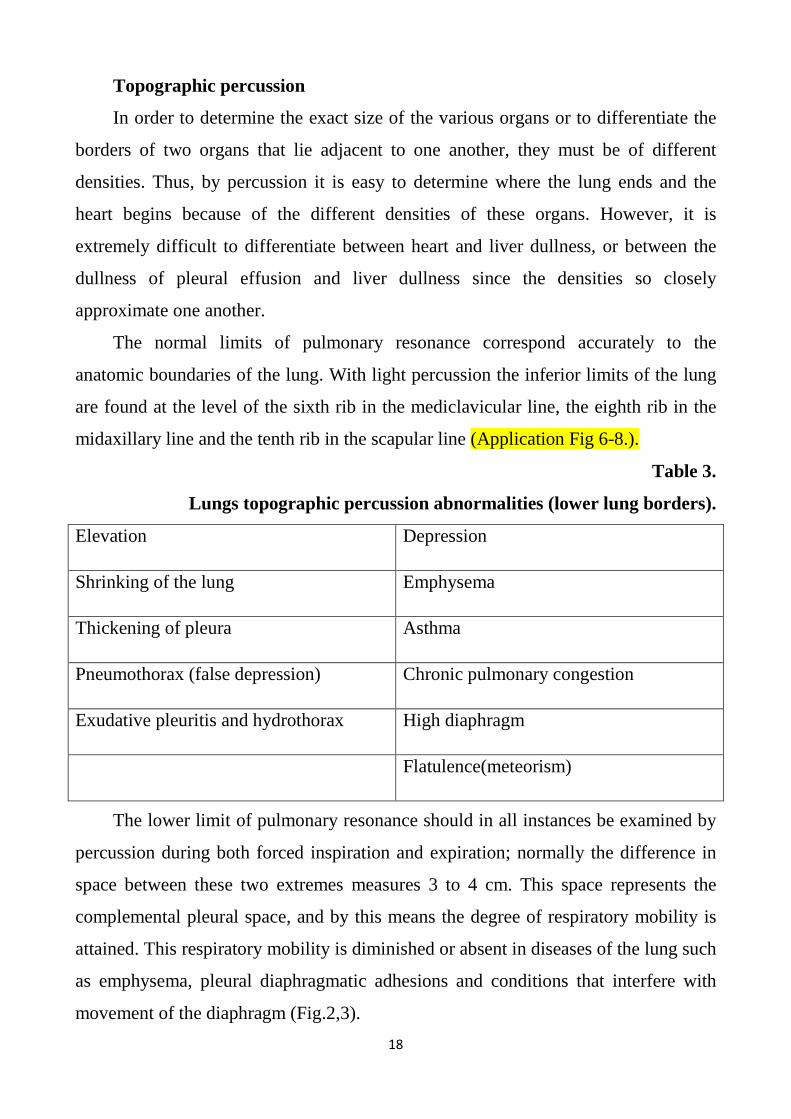
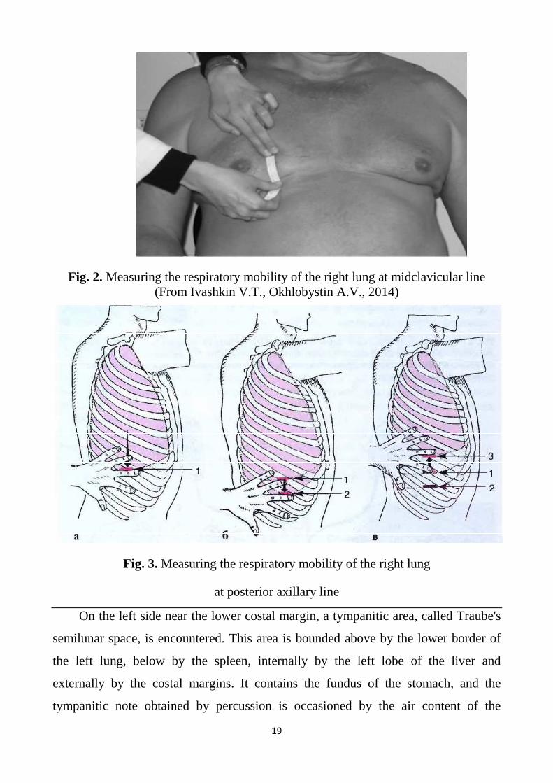
movement of the diaphragm (Fig.2,3).
19
Fig. 2. Measuring the respiratory mobility of the right lung at midclavicular line (From Ivashkin V.T., Okhlobystin A.V., 2014)
Fig. 3. Measuring the respiratory mobility of the right lung
at posterior axillary line
On the left side near the lower costal margin, a tympanitic area, called Traube's
semilunar space, is encountered. This area is bounded above by the lower border of
the left lung, below by the spleen, internally by the left lobe of the liver and
externally by the costal margins. It contains the fundus of the stomach, and the
tympanitic note obtained by percussion is occasioned by the air content of the

20
stomach. When the stomach is filled with food, the tympanitic note is decreased or
disappears, as it also does in cases of pericardial effusion and left pleural effusions.
Lungs auscultation data in norm and pathology
Auscultation is objective diagnostic method included listening to sound
phenomena arising in organs. This method was proposed by Laennec in 1819. The
sound of breathing originates somewhere between the pharynx and smaller bronchi,
although its exact source remains under study. Respiratory sounds are transmitted
through the lungs and chest wall. The tissues through which they pass, however, filter
out their higherpitched components. What you hear over most of the lungs are soft,
relatively low-pitched sounds that last through inspiration and fade out of your range
of hearing relatively early in expiration. Such sounds have been termed vesicular
breath sounds. According Laennec, a soft blowing murmur resembling the sound "f-
f" is caused by vibration of extending elastic alveolar walls, heard during the whole
inhalation. In the first third of exhalation the vibrations of collapsing alveoli walls are
still significant and may be heard, and during the last two thirds of exhalation
collapse of the alveoli is silent (Application Fig.10).
When you listen near the trachea − over the manubrium or between the scapulae
on the level of Th2-4, for example—your stethoscope is close enough to the source of
the breath sounds so that little filtration occurs. Here the breath sounds are louder and
higher in pitch. This difference is most noticeable during expiration, and you can hear
relatively high-pitched breath sounds throughout expiration. –laryngotracheal breath
sounds. Harsh and loud respiratory murmur of laryngotracheal respiration,
resembling the sound "H-H" is caused by turbulent air flow and associated vibrations
of adjacent dense tissues (Application Fig.11).
Quantitative changes of vesicular breath
sound:
enhancement:
Thin chest wall
Puerile breathing in children
diminishing:
Thick chest wall
Shallow breathing in weak
21
Vicarious hyperventilation patients
Narrowing of airways
Thickening of pleura
Emphysema
Presence of small
consolidation foci
Fluid or air in pleural cavity
Qualitative changes of vesicular breath sound: harsh breath sound occurs in
bronchitis due to airflow via unevenly narrow and thickened bronchi (Application
Fig. 12).
It is more rough and raspy; saccadic (interrupted) breath sound occurs in
nervous and shivering patients.
They distinguish the following variants of pathological bronchial breath sound:
infiltrative (inflammation, tumour, infarction etc.), compressive (in exudative
pleuritis above the fluid border), cavitary (in presence of large pulmonary cavities) –
amphoric (Application Fig. 13).
Mixed (bronchovesicular) breath sound appears in focal inflammatory
pulmonary consolidation (focal pneumonia). Weak bronchial respiration is
transmitted to the lung surface in the projection area of a small focus of consolidated
pulmonary tissue. The unchanged alveoli surrounding this focus induce vesicular
respiratory murmur (Application Fig. 14).
Breath sounds are diminished or absent over thickened pleura, pleural effusion,
pneumothorax or whenever there is fibrosis, collapse or infection in the underlying
lung (Application Fig. 15). Some people mistakenly state that air entry is diminished
into such an area where they do not hear any breath sounds. It should be appreciated
that breath sounds are not generated in the alveoli but in the major air passages and
conducted through the intervening lungs and pleura to the stethoscope.
22
Adventitious sounds.
Late inspiratory (fine) crackles. The opening up of multiple collapsed alveoli
produces discontinuous, non-musical crackling, clicking or bubbling sounds during
the middle or late phase of inspiration, sometimes spilling over the early part of
expiration. The cause of late inspiratory crackles is associated with the appearance in
the alveoli a small amount of viscous secretions (transudate, exudate, blood)
(Application Fig. 16).These sounds can be imitated by rubbing together a few hairs
between the thumb and finger in front of your ear.
Ispiratory crackles are characteristically heard over the lung bases in left heart
failure and fibrosing alveolitis, but they may be restricted to one area in lobar
pneumonia and localized fibrosis, and over the apex in tuberculosis.
Inspiratory and expiratory (coarse) crackles occur during inspiration and
expiration, when air passes through the above pathological liquid, forming bubbles
that, bursting, give a characteristic sound. Inspiratory and expiratory crackles depend
on the size of the lumen (caliber) of the bronchi: there are small, medium and major
“buble” crackles (Application Fig.17). As stated above, inspiratory crackles do not
occur at random but appear in the same sequence from breath to breath, suggesting
that pressure and volume changes determine their occurrence. However, in the late
stages of pulmonary oedema and in inflammatory conditions of bronchi, the larger
airways may be flooded with oedema fluid or bronchial secretions, and then the
crackles will be heard during both phases of respiration. In these cases the crackles
appear at random and are modified by coughing.
If crackles in the bronchi, lung tissue surrounded by a slightly changed, which
somewhat dampens the sounds, the crackles are heard muted or not euphonic.
Wheezes. Wheezes are high-pitched sounds which can be heard without the
stethoscope especially at the end of expiration. Polyphonic wheezes consist of a
cluster of continuous musical noises and are caused by high velocity of air flow
through narrowed small bronchi in asthma, emphysema and chronic bronchitis.
Healthy subjects can generate polyphonic wheezes towards the end of a forced
expiration as the bronchi are compressed and the velocity of air is increased. In
23
diffuse airways obstruction due to asthma and chronic bronchitis, wheezes occur at
submaximal respiration and may occur even at tidal breathing. In severe airways
obstruction, there may be a paradoxical absence of wheezes as the air flow through
the maximally narrowed bronchi is very slow. The absence of wheezes under such
circumstances is an ominous sign.
Bass wheezing – occurs in large and medium bronchi, the trachea. Auscultation
characteristics: low sounds "musical" character, like the humming or buzzing. Better
heard on inspiration and fickle (especially when coughing). Diagnostic value:
tracheitis, bronchitis (Application Fig. 18).
Whistling wheezing –occurs in the smaller bronchi and bronchioles due to
narrowing of the lumen due to the presence of parietal-phlegm, thickening of the
bronchial mucosa and or bronchospasm. Feature: a prolonged time of high-frequency,
high squealing sounds and "musical tone" similar to a whistle. Better are heard on
forced expiration, coughing little change. Observed in bronchitis, bronchoobstructive
syndrome (Application Fig. 19).
Pleural crackles (rub). These are coarse, non-musical sounds and are heard at
some point during both phases of respiration. Pleural crackles are localized to a small
area of the chest. They can be imitated by scratching the scalp while the
corresponding ear is blocked. The sound is caused by the two inflamed surfaces of
the pleura rubbing against each other during respiration and disappears when
sufficient fluid accumulates to separate the two layers of the pleura (Application Fig.
20). A pleural rub may be confused with a monophonic wheeze or lung crackles, but
it is usually confined to one area, has a fixed relation to both phases of respiration,
and is not changed by coughing. Sometimes a pleural rub is palpable.
Internet links:
1. Bronchial and vesicular breathing:
http://www.youtube.com/watch?v=nhUT5BfAFic&feature=BFa&list=PL9E04387C1
A0FBF82
2. Wheezes on the exhale:

24
http://www.youtube.com/watch?v=YG0-
ukhU1xE&feature=BFa&list=PL9E04387C1A0FBF82
3. Pleural crackles (rub):
http://www.youtube.com/watch?v=t2QE0O_exAQ&feature=BFa&list=PL9E04387C
1A0FBF82
4. Respiratory sounds:
http://www.youtube.com/watch?v=MzTcy6M3poM&feature=related
5. Auscultation of the lungs (1979):
http://www.youtube.com/watch?v=f0pSlN5j_v8
Pulmonary syndromes.
Pulmonary consolidation syndrome.
The essence of pulmonary consolidation syndrome is significant decrease or
complete absence of lung parenchyma airiness on more or less widespread area
(segment, lobe, few lobes simultaneously). This is one of the most frequent
syndromes in pulmonary pathology.
Causes. They distinguish the following causes of pulmonary consolidation:
1. Inflammatory infiltration (e.g. pneumonia focus, as well as specially defining
tuberculous infiltration with inclination to caseous abscess).
2. Pulmonary infarction due to thromboembolism or local vascular thrombosis.
3. Atelectasis and hypoventilation:
-obturative atelectasis (segmental or lobar);
-compressive atelectasis (pulmonary [lung] collapse);
-hypoventilation (e.g. middle lobe hypoventilation due to reduction of middle lobar
bronchus patency owing to bronchopulmonary lymph nodes, fibrous tissue; as is
well known, middle lobar bronchus incompletely ventilates middle lobe in norm).
4. Lung tumour.
5. Congestive heart failure (blood congestion in lower pulmonary parts).
Location. Consolidation focus might have different location (lower, upper parts,
middle lobe), that have differential diagnostic meaning. There is defined subpleural

25
location accompanied, as a rule, by visceral and then parietal pleura involvement.
Consolidation may sharply arise (acute pneumonia, lung infarction) or develop
gradually (tumour, atelectasis).
Inflammatory infiltration
Signs.
Cough
Sputum
Pains enhancing in deep inhalation particularly in subpleural location of
consolidation focus.
Asymmetric chest motions in respiration. In large consolidation focus and its
superficial location it may be discovered bulging and lag in motion of this part of
chest during respiration.
Increased vocal fremitus in consolidation area.
Dull or flat percussion note.
Vesicular breath sound changes into bronchial breath sound, bronchophony
increase (Application Fig.21).
At the initial and resolution stages of pneumonia when there is a little amount of
exudates in alveoli and they are stretched in air coming in, diminished vesicular
breath sound and fine (late inspiratory) crackles (crepitation) are listened above
infiltration area. At the height stage of pneumonia alveoli are filled up of exudates,
so, vesicular breath sound is replaced by bronchial.
Heterogeneous coarse (inspiratory and expiratory) crackles are heard because of
frequent involvement of bronchi in inflammatory process. Revealing of consonant
moist fine bubbling rales has particular diagnostic meaning because it witnesses
about presence of infiltration zones, increasing sound transmission, around small
bronchi.
X-ray allows to obtain a notion about focus shape and size. Consolidation focus
of lung parenchyma looks like local shading (Fig.4).

Fig.4. Consolidation focu
Pleural rub is defined in subpleural situation of infiltration or tumour and in
pulmonary infarction.
Lobar pneumonia: initial stage
Morphology. Congestion stage
engorgement, rapid bacterial proliferation.
Inspection. An increased respiratory rate is usually evident. Pain is a frequent
accompaniment, and with it the involved side shows a lag of respiratory motion.
Palpation. Palpation confirms the findings on inspection. Tactile
normal or even slightly decreased, and a pleural friction rub may be present.
Percussion. Impaired resonance may be elicited with light percussion. This
finding is extremely important.
Auscultation. Although the breath sounds may be diminished
prolonged and crepitation (crepitus indux)
pleural friction sound is determined.
Lobar pneumonia: stage of consolidation
26
Consolidation focus (inflammatory infiltration) of lung parenchyma
lower lobar)
Pleural rub is defined in subpleural situation of infiltration or tumour and in
Lobar pneumonia: initial stage
Morphology. Congestion stage - extensive serous exudation, vascular
apid bacterial proliferation.
Inspection. An increased respiratory rate is usually evident. Pain is a frequent
accompaniment, and with it the involved side shows a lag of respiratory motion.
Palpation. Palpation confirms the findings on inspection. Tactile
normal or even slightly decreased, and a pleural friction rub may be present.
Percussion. Impaired resonance may be elicited with light percussion. This
finding is extremely important.
Auscultation. Although the breath sounds may be diminished
(crepitus indux) is heard. With pleural involvement, a
pleural friction sound is determined.
Lobar pneumonia: stage of consolidation
of lung parenchyma (left
Pleural rub is defined in subpleural situation of infiltration or tumour and in
extensive serous exudation, vascular
Inspection. An increased respiratory rate is usually evident. Pain is a frequent
accompaniment, and with it the involved side shows a lag of respiratory motion.
Palpation. Palpation confirms the findings on inspection. Tactile fremitus is
normal or even slightly decreased, and a pleural friction rub may be present.
Percussion. Impaired resonance may be elicited with light percussion. This
Auscultation. Although the breath sounds may be diminished, expiration is
is heard. With pleural involvement, a

27
Morphology. Red hepatization stage - airspaces are filled with PMN cells,
vascular congestion, extravasation of RBC. Grey hepatization stage - accumulation of
fibrin, inflammatory WBCs and RBCs in various stages of disintegration, alveolar
spaces filled with inflammatory exudate.
Complaints. Coughing may be associated with a sharp pain in the affected side.
Mucoid sputum becomes rusty brown (prune juice color).
General inspection. Cyanosis of the lips and fingers. When the fever is high, the
face may be flushed. The patient's nostrils dilate on inspiration, and expiration is
often grunting.
Inspection. Dyspnea is invariably present. Respiratory movements are generally
decreased on the affected side.
Palpation. Diminished respiratory excursions, a pleural friction rub may be felt.
Tactile fremitus is increased.
Percussion. Dullness.
Auscultation. Bronchial breathing, bronchophony, pectoriloquy and whispered
bronchophony are evident with consolidation provided the bronchus to the involved
area is open. Rales are less numerous and distinct than in the stages of engorgement
or resolution.
Lobar pneumonia: stage of resolution
Morphology. Resolution stage - resorption of the exudate.
Inspection. The patient looks more comfortable and the cyanosis disappears. The
dyspnea disappears and the affected lung begins to expand again.
Palpation. The previously increased tactile fremi-tus becomes less marked and
gradually findings become normal.
Percussion. The dullness gradually disappears and normal resonance returns.
Auscultation. The bronchial breathing is gradually replaced by bronchovesicular
breathing and later by normal vesicular breathing. Crepitation reappears (crepitus
redux). Small and large moist rales are heard in increasing numbers.
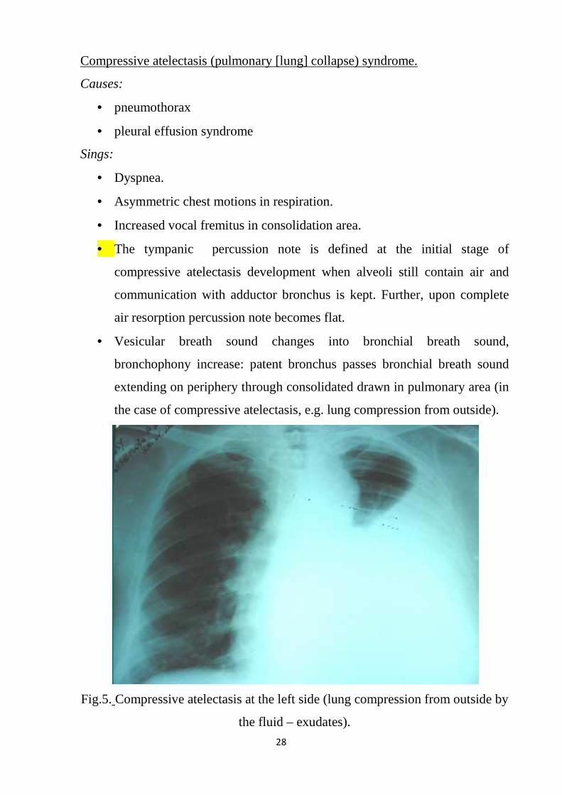
Compressive atelectasis (pulmonary [lung] collapse) syndrome.
Causes:
• pneumothorax
• pleural effusion syndrome
Sings:
• Dyspnea.
• Asymmetric chest motions in respiration.
• Increased vocal fremitus in consolidation area.
• The tympanic
compressive atelectasis development when alveoli still contain air and
communication with adductor bronchus is kept. Further, upon complete
air resorption percussion note becomes flat.
• Vesicular breath
bronchophony increase
extending on periphery through consolidated drawn in pulmonary area (in
the case of compressive atelectasis, e.g. lung compression from outside)
Fig.5. Compressive atelectasis at the left side (lung compression from outside by
28
Compressive atelectasis (pulmonary [lung] collapse) syndrome.
pleural effusion syndrome
Asymmetric chest motions in respiration.
fremitus in consolidation area.
percussion note is defined at the initial stage of
compressive atelectasis development when alveoli still contain air and
communication with adductor bronchus is kept. Further, upon complete
air resorption percussion note becomes flat.
Vesicular breath sound changes into bronchial breat
bronchophony increase: patent bronchus passes bronchial breath sound
extending on periphery through consolidated drawn in pulmonary area (in
the case of compressive atelectasis, e.g. lung compression from outside)
Compressive atelectasis at the left side (lung compression from outside by
the fluid – exudates).
Compressive atelectasis (pulmonary [lung] collapse) syndrome.
percussion note is defined at the initial stage of
compressive atelectasis development when alveoli still contain air and
communication with adductor bronchus is kept. Further, upon complete
bronchial breath sound,
: patent bronchus passes bronchial breath sound
extending on periphery through consolidated drawn in pulmonary area (in
the case of compressive atelectasis, e.g. lung compression from outside).
Compressive atelectasis at the left side (lung compression from outside by
29
Obturative atelectasis (segmental or lobar).
Causes:
• closure of airing bronchus lumen by endobronchial tumour, foreign body,
compression from outside
Sings:
Dyspnea.
Asymmetric chest motions in respiration.
Weakened or disappearance of vocal fremitus in consolidation area.
At the initial stage of atelectasis (hypoventilation stage) when a small amount of
aired alveoli in the collapsed area is still kept, diminished vesicular breath sound may
be defined. In obturative atelectasis at the complete bronchus closure stage no breath
sounds are heard above airless zone. No breath sounds are also heard above tumour.
Bronchophony reveals sound transmission increase above pulmonary consolidation
area. (Application Fig.22)
Pulmonary cavity syndrome
This syndrome is connected with presence of cavities with dense and smooth
walls, not rarely surrounded with infiltrate or fibrous tissue (cavern, abscess, cyst).
Symptomatology in every concrete case depends on many conditions:
• Cavity size
• Depth of its location
• Cavity contents: air only (empty cavity), air with some amount of fluid (e.g. air and
exudates).
• Cavity communication with respiratory tract (via drainage bronchus) or isolated
cavity.
Causes
1. Disintegrating (with emptying) lung infiltrate:
— pneumonia complicated by an abscess;
30
— pulmonary infarction complicated by an abscess;
— tuberculosis (cavern);
— granulomatous focus (necrotizing respiratory [Wegener's] granulomatosis).
2. Cysts (congenital and acquired).
Signs.
Decreased vocal fremitus is characreristic for large superficially located and
isolated cavities beyond dependence on their contents.
If cavity communicates with bronchus and even, if partially contains air, there is
increase vocal fremitus and tympanitic shade to percussion.
Above cavity filled with fluid there is dullness or flatness to percussion (much
as pulmonary consolidation syndrome).
Above empty cavity there is tympanic percussion sound.
Above isolated cavity no breath sounds are heard.
If a cavity communicates with drainage bronchus auscultated bronchial sounds
(breath sounds are easily transmitted from glottis along respiratory tract) due to the
sound resonance in cavity may acquire metallic shade (resemble a sound of blow on
metallic object). Metallic breath sounds should be distinguished from amphoric (also
appeared above cavities with very smooth walls) – the variant of bronchial sounds,
differed from usual with musical shade (appears due to resonance of smooth cavity
walls). Sounds resembling amphoric breath sounds may be simulated to wind over
neck of empty bottle.
Cavity partially containing a fluid not rarely may be issue of moist bubbling
rales which, as a rule, are consonant because their transmission is enhanced by
surrounding consolidated infiltrated tissues.
Independent stenotic noise which increases bronchial breath sounds, may be
heard above the place of cavity connection with drainage bronchus.
X-ray changes. More often pulmonary cavities are exactly discovered in the
course of X-ray examination (Fig. 6).
CAT allows to detect specific plural small cavities (cysts) forming at the late
stage of fibrosing alveolitis (―honeycomb lung).

It is necessary to point that all mentioned signs characterized the pulmonary
cavity syndrome are very dynamic as long as staging has place in cavity
development. Dynamics of signs is particularly demonstrative in the course of lung
abscess: fluid accumulation changes on complete or partial emptying and is
accompanied by appropriate symptomatic
Fig.6. Pulmonary cavity syndrome
Pleural effusion syndrome
Hydrothorax is the accumulation of increased amount of liquid in pleural cavity.
Liquid contents depends on pathologic process character, its stage and intensity. They
distinguish exudate and transudate due to liquid contents. Pus (in this case they say
pyothorax or empyema) and blood (
Effusion may have mixed character.
Causes.
1. Essential pleura lesion:
31
It is necessary to point that all mentioned signs characterized the pulmonary
me are very dynamic as long as staging has place in cavity
development. Dynamics of signs is particularly demonstrative in the course of lung
abscess: fluid accumulation changes on complete or partial emptying and is
accompanied by appropriate symptomatic changes.
Pulmonary cavity syndrome (lung abscess at the right side)
leural effusion syndrome
is the accumulation of increased amount of liquid in pleural cavity.
Liquid contents depends on pathologic process character, its stage and intensity. They
distinguish exudate and transudate due to liquid contents. Pus (in this case they say
) and blood (hemothorax) may also gather in pleural cavity.
Effusion may have mixed character.
Essential pleura lesion:
It is necessary to point that all mentioned signs characterized the pulmonary
me are very dynamic as long as staging has place in cavity
development. Dynamics of signs is particularly demonstrative in the course of lung
abscess: fluid accumulation changes on complete or partial emptying and is
at the right side).
is the accumulation of increased amount of liquid in pleural cavity.
Liquid contents depends on pathologic process character, its stage and intensity. They
distinguish exudate and transudate due to liquid contents. Pus (in this case they say
) may also gather in pleural cavity.
32
— Inflammation (pleuritis) with exudates production, that may be caused by
pathogens as well as immune mechanisms (nonspecific inflammation as
manifestation of rheumatic fever, systemic lupus erythematosus and others).
— Tuberculosis: more often para-tubercular nonspecific exudative pleural reaction
appears, rarely — proper tubercular pleura affection.
— Pleural tumour (e.g. mesothelioma) or pleural metastases.
2. Suppurative processes, including septicemia.
3. Pus (or blood) drain from adjacent foci in pulmonary tissue.
4. Trauma (wounds) of the thorax.
Signs
Fluid in pleural cavity squeezes the lung resulting in compressive atelectasis
formation and dyspnea appearance.
A large liquid amount is accompanied by smoothing of intercostal spaces,
protrusion of affected chest side, and its lag in motion on respiration.
Vocal fremitus over the liquid is sharply decreased down to absence.
On comparative percussion in fluid accumulation projection area dull or flat
percussion note is defined. Above the upper border of liquid badly ventilated
squeezed lung is situated near air containing bronchi, so according to the law of
compressive atelectasis it gives dull-tympanitic shade of percussion note.
On topographic percussion peculiarities of dullness upper boundary (which may
have various direction due to fluid contents) and also significant restriction on
diaphragm excursions on affected side are revealed.
— In case of inflammation (exudate) upper dullness border has the appearance of the
curve (Ellis-Damuazo-Sokolov's curve) with the apex along axillary lines, that is
characteristic of irregular fluid level elevation.
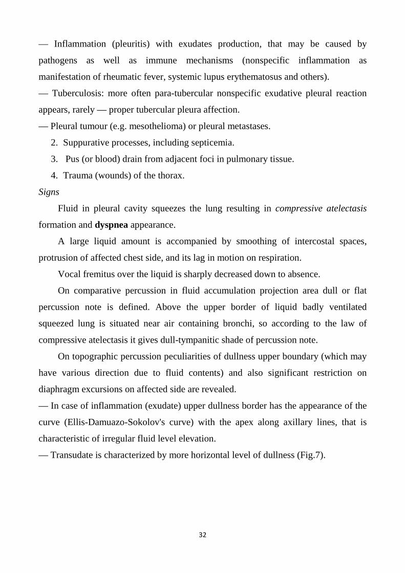
— Transudate is characterized by more horizontal level of dullness (Fig.7).
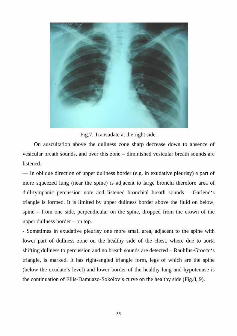
Fig.7
On auscultation above the dullness zone sharp decrease down to absence of
vesicular breath sounds, and over this zone
listened.
— In oblique direction of upper dullness border (e.g. in exudative pleurisy) a part of
more squeezed lung (near the spine) is adjacent to large bronchi
dull-tympanic percussion note and listened bronchial breath sounds
triangle is formed. It is limited by upper dullness border above the fluid on below,
spine – from one side, perpendicular on the spine, dropped from the crown of the
upper dullness border – on top.
- Sometimes in exudative pleurisy one more small area, adjacent to the spin
lower part of dullness zone on the healthy side of the chest, where due to aorta
shifting dullness to percussion and no breath sounds are detected
triangle, is marked. It has right
(below the exudate‘s level) and lower border of the healthy lung and hypotenuse is
the continuation of Ellis-Damuazo
33
Fig.7. Transudate at the right side.
On auscultation above the dullness zone sharp decrease down to absence of
and over this zone – diminished vesicular breath sounds are
In oblique direction of upper dullness border (e.g. in exudative pleurisy) a part of
more squeezed lung (near the spine) is adjacent to large bronchi
ercussion note and listened bronchial breath sounds
. It is limited by upper dullness border above the fluid on below,
from one side, perpendicular on the spine, dropped from the crown of the
on top.
Sometimes in exudative pleurisy one more small area, adjacent to the spin
lower part of dullness zone on the healthy side of the chest, where due to aorta
shifting dullness to percussion and no breath sounds are detected –
triangle, is marked. It has right-angled triangle form, legs of which are the spine
below the exudate‘s level) and lower border of the healthy lung and hypotenuse is
Damuazo-Sokolov‘s curve on the healthy side
On auscultation above the dullness zone sharp decrease down to absence of
diminished vesicular breath sounds are
In oblique direction of upper dullness border (e.g. in exudative pleurisy) a part of
more squeezed lung (near the spine) is adjacent to large bronchi therefore area of
ercussion note and listened bronchial breath sounds – Garlend‘s
. It is limited by upper dullness border above the fluid on below,
from one side, perpendicular on the spine, dropped from the crown of the
Sometimes in exudative pleurisy one more small area, adjacent to the spine with
lower part of dullness zone on the healthy side of the chest, where due to aorta
– Rauhfus-Grocco‘s
angled triangle form, legs of which are the spine
below the exudate‘s level) and lower border of the healthy lung and hypotenuse is
Sokolov‘s curve on the healthy side (Fig.8, 9).
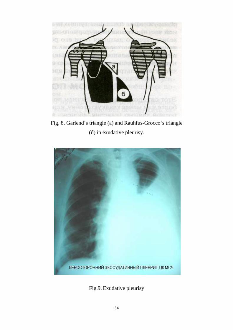
Fig. 8. Garlend‘s triangle (a) and Rauhfus
34
. Garlend‘s triangle (a) and Rauhfus-Grocco‘s triangle
(б) in exudative pleurisy.
Fig.9. Exudative pleurisy
Grocco‘s triangle
35
Syndrome of air in pleural cavity (pnemothorax)
The findings in pneumothorax depend on the size of the pleural airspace. Motion
may be normal or diminished, vocal fremitus may be decreased to absent, the
percussion note is usually normal but may be more resonant than over the
contralateral lung, and breath sounds and bronchophony are decreased to absent.
Pneumothorax
Spontaneous pneumothorax develops in the absence of any trauma to the chest.
When no obvious diseases of the lung are present, a spontaneous pneumothorax is
considered to be primary. In contrast, secondary spontaneous pneumothorax develops
as a complication of a wide variety of diseases of the airways and lungs.
Primary spontaneous pneumothorax is predominantly a disease of young males
and is six times more common in men than in women. It results from the rupture of
small apical sub- pleural emphysematous cysts that either are congenital or are
caused by bronchiolar inflammation and obstruction. Primary spontaneous
pneumothorax is more likely to occur in athenics. It has been suggested that in
athenics, the pleural pressure is more subatmospheric at the apex and as a result
apical alveoli are more greatly distended. This may play a role in cysts formation in
those who are congenitally predisposed. Cigarette smoking also increases the probability of primary spontaneous pneumothorax.
Primary spontaneous pneumothorax is not precipitated by exertion. It usually
occurs when the patient is at rest and only infrequently develops during exercise.
Chest pain and dyspnea are the most common symptoms, and only rarely are both
these symptoms absent. The chest pain is sudden in onset and pleuritic in nature;
shoulder pain reflects irritation of the diaphragmatic pleura. Compression and
collapse of the lung by a pneumothorax causes cough in over half the patients.
The characteristic findings on physical examination include impaired
expansion of the involved hemithorax, a tympanic percussion note, and diminished or
absent fremitus and breath sounds.
Marked respiratory distress with cyanosis, tachycardia, and hypotension signals
a tension pneumothorax.
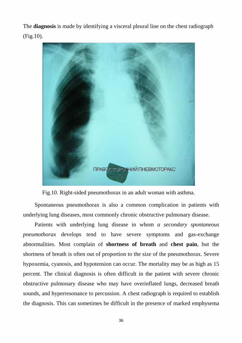
The diagnosis is made by identifying a visceral pleural line
(Fig.10).
Fig.10. Right-sided pneumothorax in an adult woman with asthma.
Spontaneous pneumothorax is also a common complication in patients with
underlying lung diseases, most commonly chronic obstructive pulmonary disease.
Patients with underlying lung disease in whom
pneumothorax develops tend to have severe symptoms and gas
abnormalities. Most complain of
shortness of breath is often out of propor
hypoxemia, cyanosis, and hypotension can occur. The mortality may be as high as 15
percent. The clinical diagnosis is often difficult in the patient with severe chronic
obstructive pulmonary disease who may have
sounds, and hyperresonance to percussion. A chest radiograph is required to establish
the diagnosis. This can sometimes be difficult in the presence of marked emphysema
36
is made by identifying a visceral pleural line on the c
sided pneumothorax in an adult woman with asthma.
Spontaneous pneumothorax is also a common complication in patients with
underlying lung diseases, most commonly chronic obstructive pulmonary disease.
Patients with underlying lung disease in whom a secondary spontaneous
develops tend to have severe symptoms and gas
abnormalities. Most complain of shortness of breath and chest pain
shortness of breath is often out of proportion to the size of the pneumothorax. Severe
hypoxemia, cyanosis, and hypotension can occur. The mortality may be as high as 15
percent. The clinical diagnosis is often difficult in the patient with severe chronic
obstructive pulmonary disease who may have overinflated lungs, decreased breath
sounds, and hyperresonance to percussion. A chest radiograph is required to establish
the diagnosis. This can sometimes be difficult in the presence of marked emphysema
on the chest radiograph
sided pneumothorax in an adult woman with asthma.
Spontaneous pneumothorax is also a common complication in patients with
underlying lung diseases, most commonly chronic obstructive pulmonary disease.
a secondary spontaneous
develops tend to have severe symptoms and gas-exchange
chest pain, but the
tion to the size of the pneumothorax. Severe
hypoxemia, cyanosis, and hypotension can occur. The mortality may be as high as 15
percent. The clinical diagnosis is often difficult in the patient with severe chronic
overinflated lungs, decreased breath
sounds, and hyperresonance to percussion. A chest radiograph is required to establish
the diagnosis. This can sometimes be difficult in the presence of marked emphysema

37
or bullous disease. Under these circumstances, the diagnosis of pneumothorax should
be made only if a visceral pleural line can be demonstrated.
Traumatic pneumothorax is most often due to penetrating chest trauma, but it
also can occur with closed chest trauma consequent to alveolar rupture from thoracic
compression, fracture of a bronchus, esophageal rupture, or rib fractures that lacerate
the pleura. Tube thoracostomy is required to evacuate air and blood from the pleural
space.
Jatrogenic Pneumothorax. Pneumothorax is also a common complication of
central venous line insertion, thoracentesis, pleural biopsy, percutaneous needle
aspiration of the lung, and transbronchial lung biopsy.
Tension Pneumothorax. When the pressure in a pneumothorax exceeds
atmospheric pressure, a tension pneumothorax is said to exist. It most commonly
occurs during mechanical ventilation or cardiopulmonary resuscitation, but it may
complicate any type of spontaneous or traumatic pneumothorax. Characteristic
findings on chest radiogram include a shift of the mediastmum away from the
pneumothorax and ipsilateral depression of the diaphragm. Since tension
pneumothorax is a medical emergency, the diagnosis must be made clinically.
Treatment cannot be delayed until a chest radiograph is obtained. Once the presence
of a tension pneumothorax is confirmed, a chest tube should be inserted immediately.
Hyperinflated lung syndrome (emphysema)
Emphysema is characterized by two features.
Anatomically, it is defined as an abnormal enlargement of the air spaces distal to
the terminal bronchioles, accompanied by destructive changes in the alveolar walls.
Physiologically, it is characterized by a loss of elastic recoil and thus an
increased lung compliance.
The degree of airways obstruction in patients with COPD correlates most
closely with the severity of emphysema, and patients who have significant functional
impairment usually have at least a moderate degree of emphysema.
The diagnosis of emphysema is usually inferred from the clinical and laboratory
findings.
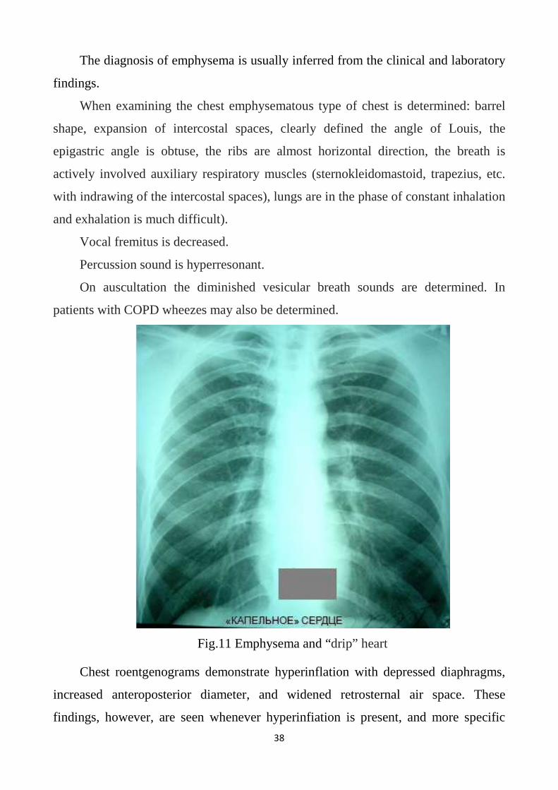
When examining the chest
shape, expansion of intercostal spaces, clearly defined the angle of Louis, the
epigastric angle is obtuse, the
actively involved auxiliary respiratory mu
with indrawing of the intercostal spaces)
and exhalation is much difficult)
Vocal fremitus is decreased.
Percussion sound is hyperresonant.
On auscultation the dimi
patients with COPD wheezes may also be determined.
Fig.11
Chest roentgenograms demonstrate hyperinflation with depressed diaphragms,
increased anteroposterior diameter, and
findings, however, are seen whenever h
38
The diagnosis of emphysema is usually inferred from the clinical and laboratory
When examining the chest emphysematous type of chest is determined: barrel
, expansion of intercostal spaces, clearly defined the angle of Louis, the
epigastric angle is obtuse, the ribs are almost horizontal direction, the breath is
d auxiliary respiratory muscles (sternokleidomastoid, trapezius, etc.
ng of the intercostal spaces), lungs are in the phase of constant inhalation
and exhalation is much difficult).
Vocal fremitus is decreased.
Percussion sound is hyperresonant.
On auscultation the diminished vesicular breath sounds are determined. In
patients with COPD wheezes may also be determined.
Fig.11 Emphysema and “drip” heart
Chest roentgenograms demonstrate hyperinflation with depressed diaphragms,
increased anteroposterior diameter, and widened retrosternal air space. These
gs, however, are seen whenever hyperinfiation is present, and more specific
The diagnosis of emphysema is usually inferred from the clinical and laboratory
ous type of chest is determined: barrel
, expansion of intercostal spaces, clearly defined the angle of Louis, the
are almost horizontal direction, the breath is
mastoid, trapezius, etc.
, lungs are in the phase of constant inhalation
vesicular breath sounds are determined. In
Chest roentgenograms demonstrate hyperinflation with depressed diaphragms,
widened retrosternal air space. These
yperinfiation is present, and more specific
39
features in emphysema include attenuation of the pulmonary vasculature. The one
finding that correlates well with the anatomic presence of emphysema is a reduction
in diffusing capacity because of the loss of alveolar capillaries.
A list of the main instrumental and laboratory methods of examination of
respiratory system
Chest x-ray
Computed tomography of chest organs
Bronchoscopy
Spirometry and peakflowmetry
The study of lung diffusion capacity (transfer factor)
Measurement of airway resistance (the method of "short-term interruption of the air
flow" or by the method of oscillations
Capnography
Bodyplethismography
Pulse oximetry, including monitoring of blood oxygenation
microtechnique Astrup (research arterializing gas composition of blood and acid-
base balance)
Blood tests
Sputum tests
sputum on flora and sensitivity to antibiotics
examination of sputum for Mycobacterium tuberculosis and atypical cells
blood test for tumor markers (Cyfra 21-1)
Fiberoptic bronchoscopy is used for diagnostic and therapeutic purposes (see
Table 4)
Diagnostic bronchoscopy allows visually to estimate respiratory tract
peculiarities from glottis to subsegmental bronchi, to obtain samples of content of
respiratory tract on different levels for bacteriological and cytological examination, to
perform bronchopulmonary lavage with subsequent sampling of received fluid. Using
bronchoscope it is possible to perform puncture biopsy of bronchial mucous and
tansbronchial biopsy of adjacent tissues (lymph node, lung parenchyma).

40
Bronchoscopy is used for therapeutic purposes, for example, for
bronchopulmonary lavage and local introduction of antibacterial drugs in
bronchiectasis (bronchi sanation), dilution and aspiration of mucus from corked
bronchi lumen in intractable asthmatic onset (particularly in presence of so-called
―dumb lung) and removal of foreign bodies.
Absolute contraindications: unstable angina, acute myocardial infarction, acute
stroke, pulmonary heart and cardiovascular failure article III, life-threatening
arrhythmias, severe hypoxemia, absence due to mental illness the patient contact the
doctor with the patient.
Table 4.
Indications for bronchoscopy
Indications for bronchoscopy Purposes
Hemoptysis Determination of source of bleeding and
hemostasis.
Chronic cough without visible
reason.
Detection of possible intrabronchial tumour
invisible on X-ray.
Delayed pneumonia resolution Exception of local bronchial obstruction
Atelectasis Determination of its cause.
Cancer of lung Biopsy, estimation of operable status
Abscess Exception of bronchial obstruction, receiving
of material for bacteriological examination and
cavity drainage improvement
Bronchiectasis Bronchial lavage, introduction of medications
(antibiotics, for example)
Dumb lung Dilution and aspiration of mucus
Foreign body Removal
Bronchoscopy plays a central role in the diagnosis of tumors of the
tracheobronchial localisation. central lung cancer when the tumor arises from the
mucosa of the bronchi of the 1st, 2nd and 3rd orders, which are radiographically the

41
root of the lung. On the nature of growth of central cancer is divided into
endobronchial and peribronchial, in turn, is divided into endobronchial exophytic
(nodular form) and endophytic (flat infiltrations and ulcers).
During the bronchoscopic studies the diagnosis of lung cancer is based on the direct
and indirect endoscopic signs. Direct signs include the presence of the tumor is
exophytic or endophytic (Application Fig. 23, 24). Exophytic tumour has a
hemispherical or semi-oval shape, rough surface, grayish-red, wide base. Partially or
completely occlusive the lumen of the bronchus.
It is difficult to make a differential diagnosis between a stenosis of bronchus
inflammatory and neoplastic etiologies in cases of infiltrative tumor growth.
Diagnostic difficulties help to resolve forceps and brush biopsy.
Ulcerative form of cancer is an ulcer of irregular shape, with fuzzy edges and
uneven bottom, covered with fibrinous coating (Application Fig. 25). Biopsies are
taken with the tongs from the edges of the ulceration.
When foreign bodies of the trachea the cough is paroxysmal in nature. On
bronchoscopic examination of patients is usually direct, as a rule, with other
diagnoses: pneumonia, lung cancer, hemoptysis of unclear etiology, bronchial asthma
(Application Fig. 26).
It is necessary to emphasize the bronchoscopy need for patients with hemoptysis
(especially repetitive) or bronchial [pulmonary] hemorrhage since it allows to
determine source of bleeding (trachea, bronchi, parenchyma) and its cause
(bronchiectasis, tumour, tuberculosis).
Also there are used thoracoscopy (inspection of the pleural sheets) and
mediastinoscopy (inspection of anterior mediastinum). One of the basic purposes of
these investigations is to receive biopsy material.
Blood gases The presence of respiratory failure may be suspected by the signs of central
cyanosis. It is important to define the type and extent of failure of oxygenation and
this is best done by measurement of arterial blood gas tensions (PaO2 and PaCO2),
oxygen saturation (Sa02) and pH.

42
The study of gas composition and blood acid-base status (Astrup
microtechnique).
Normally, in a healthy person the constancy of pH (7,4; 7,35-7,45) is maintained
by buffer systems of blood and lung and kidney (Shmidt R., Tevs G., 1996).
Equilibrium in acid-base status is determined by ratio of hydrogen (H+) and hydroxyl
(OH-) ions. With increasing concentration of H+ ions oxidizes the blood, while
increasing OH - ions oxalacetate. Bicarbonate buffer system regulates the buffering
capacity of the blood due to the change РаСО2 - voltage of carbon dioxide in the
blood (level РаСО2 depends on lung ventilation and at the same time affects it).
Adjusting the tension of CO2 in blood, respiratory system facilitates effective buffer
system in General.
Important buffer parameters:
1. BB - the sum of all the buffer bases of the blood ≈ 48 mmol/L. This value does not
change when the shifts РаСО2;
2. BE - the excess of bases (shows how the concentration of buffer bases is deviated
from the normal value (≈ 48 mmol/l). The normal value of VE varies from -2.5 to
+2.5 mmol/L. the Level BE > -5 mmol/l – metabolic acidosis.<5 mmol/l is
characteristic of metabolic alkalosis.
3. SB - standard bicarbonate. SB ≈ 24 mmol/l. SB corresponds to the content of
bicarbonate in the plasma of fully oxygenated and equilibrated with a gas mixture
(PCO2 = 40 mm Hg) at 37 degrees Celsius.
For the study of gas composition of blood and acid-base status is used
microtechnique Astrup. Analyzed blood obtained by puncture of the scarifier terminal
phalanx of one finger, pre-warmed for 10 minutes at a temperature of about 40 ° C
and treated with alcohol. Portion blood type in special capillaries that are on both
sides immediately after the blood sample are isolated from the air "plugs". Oxygen
tension is measured polarographically (Shmidt R., Tevs G., 1996).
Define:
1. indicators in acid-base status: pH, BE, BB, SB, etc.;

43
2. indicators of gas composition of blood arterialization: partial tension of oxygen
РаО2 (normal > 80 mm Hg) partial tension of carbon dioxide РаСО2 (normal 35-45
mm Hg), the saturation of hemoglobin by oxygen SaO2 (normal > 95%).
The main types of violations arterializing gas composition of blood and acid-
base balance:
1. Arterial hypoxemia (РаО2 <80 mm Hg). At persons of young age РаО2 is
about 95 mm Hg to 40 years it is reduced to 80 mm Hg and 70 years approximately
70 mm Hg. In the clinic chronic respiratory diseases are the most frequent cause of
arterial hypoxemia is the development of chronic respiratory insufficiency (IR) in
COPD patients
2. Arterial hypocapnia (РаО2 < 35 mm Hg). Characteristic of the early stages IR
when arterial hypoxemia is compensated by the increased pulmonary ventilation,
resulting in lower РаСО2 and development of respiratory alkalosis.
3. Arterial hypercapnia (РаСО2 > 45 mm Hg). A characteristic feature of the
later stages IR when the removal of carbon dioxide through the alveolo-capillary
membrane is disturbed. It is often noted respiratory acidosis is an important sign of
severity IR.
4. Respiratory alkalosis (РаСО2 < 35 mm Hg, BE = ± 2.5 mmol/l).
5. Respiratory acidosis (РаСО2 > 45 mm Hg, BE = ± 2.5 mmol/l)
6. Metabolic alkalosis (РаСО2 = 35-45 mm Hg BE > +5 mmol/l) is atypical for
a primary lesion of the lung.
7. Metabolic acidosis (РаСО2 = 35-45 mm Hg, BE < -5 mmol/l) - metabolic
acidosis atypical for a primary lesion of the lung.
The study of gas composition and blood acid-base balance is a powerful
technique for objectifying the severity of chronic respiratory failure. To determine
blood gas composition in COPD patients is recommended when the level of oxygen
saturation of hemoglobin (SpO2) by pulse oximetry data < 92% and FEV1 <50%.
The response to drugs and the therapeutic response to oxygen can then be
monitored easily. Haemoglobin saturation reflects oxygen carriage by the blood and
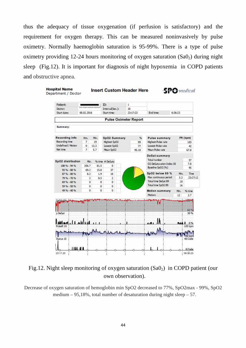
44
thus the adequacy of tissue oxygenation (if perfusion is satisfactory) and the
requirement for oxygen therapy. This can be measured noninvasively by pulse
oximetry. Normally haemoglobin saturation is 95-99%. There is a type of pulse
oximetry providing 12-24 hours monitoring of oxygen saturation (Sa02) during night
sleep (Fig.12). It is important for diagnosis of night hypoxemia in COPD patients
and obstructive apnea.
Fig.12. Night sleep monitoring of oxygen saturation (Sa02) in COPD patient (our own observation).
Decrease of oxygen saturation of hemoglobin min SpO2 decreased to 77%, SpO2max - 99%, SpO2 medium – 95,18%, total number of desaturation during night sleep – 57.
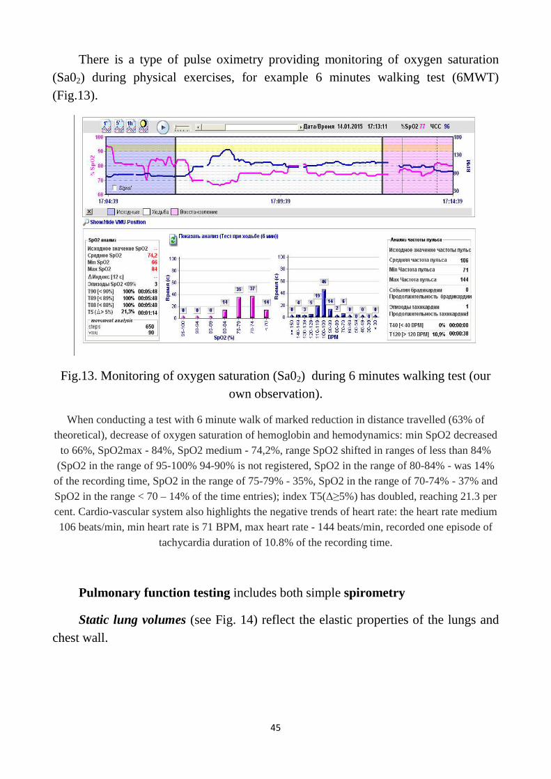
45
There is a type of pulse oximetry providing monitoring of oxygen saturation (Sa02) during physical exercises, for example 6 minutes walking test (6MWT) (Fig.13).
Fig.13. Monitoring of oxygen saturation (Sa02) during 6 minutes walking test (our own observation).
When conducting a test with 6 minute walk of marked reduction in distance travelled (63% of theoretical), decrease of oxygen saturation of hemoglobin and hemodynamics: min SpO2 decreased
to 66%, SpO2max - 84%, SpO2 medium - 74,2%, range SpO2 shifted in ranges of less than 84% (SpO2 in the range of 95-100% 94-90% is not registered, SpO2 in the range of 80-84% - was 14%
of the recording time, SpO2 in the range of 75-79% - 35%, SpO2 in the range of 70-74% - 37% and SpO2 in the range < 70 – 14% of the time entries); index T5(∆≥5%) has doubled, reaching 21.3 per cent. Cardio-vascular system also highlights the negative trends of heart rate: the heart rate medium 106 beats/min, min heart rate is 71 BPM, max heart rate - 144 beats/min, recorded one episode of
tachycardia duration of 10.8% of the recording time.
Pulmonary function testing includes both simple spirometry
Static lung volumes (see Fig. 14) reflect the elastic properties of the lungs and chest wall.

Fig. 14. Static lung volumes
Fig. 15. Static lung volumes ATS/ERS Task Force: Standartisation of
Vital capacity (VC or "slow VC") is the maximum volume of air that can be
expired slowly after a full inspiratory effort. Simple to perform, it is one of the most
valuable measurements of pulmonary function. Because VC decrease
46
. Static lung volumes ATS/ERS Task Force: Standartisation of lung
function testing, 2005
. Static lung volumes ATS/ERS Task Force: Standartisation of
function testing, 2005
(VC or "slow VC") is the maximum volume of air that can be
expired slowly after a full inspiratory effort. Simple to perform, it is one of the most
valuable measurements of pulmonary function. Because VC decrease
ATS/ERS Task Force: Standartisation of lung
. Static lung volumes ATS/ERS Task Force: Standartisation of lung
(VC or "slow VC") is the maximum volume of air that can be
expired slowly after a full inspiratory effort. Simple to perform, it is one of the most
valuable measurements of pulmonary function. Because VC decreases as a

47
restrictive lung disorder (eg, pulmonary edema, interstitial fibrosis) worsens, it can
be used along with the diffusing capacity to follow the course of such a disorder and
its response to therapy. The VC also reflects the strength of the respiratory muscles
and is often used to monitor the course of neuromuscular disorders.
Forced vital capacity (FVC), similar to VC, is the volume of air expired with
maximal force. It is usually measured along with expiratory flow rates in simple
spirometry (see Dynamic Lung Volumes and Flow Rates, below). The VC can be
considerably greater than the FVC in patients with airway obstruction. During the
FVC maneuver, terminal airways can close prematurely (ie, before the true residual
volume is reached), trapping gas distally and preventing its measurement by the
spirometer.
Total lung capacity (TLC) is the total volume of air within the chest after a
maximum inspiration.
Functional residual capacity (FRC) is the volume of air in the lungs at the end
of a normal expiration when all respiratory muscles are relaxed. Physiologically, it is
the most important lung volume because it approximates the normal tidal breathing
range. Outward elastic recoil forces of the chest wall tend to increase lung volume but
are balanced by the inward elastic recoil of the lungs, which tends to reduce it; these
forces are normally equal and opposite at about 40% of TLC. Loss of lung elastic
recoil in emphysema increases FRC. Conversely, the increased lung stiffness in
pulmonary edema, interstitial fibrosis, and other restrictive disorders decreases
FRC. Kyphoscoliosis leads to a decrease in FRC and in other lung volumes because a
stiff, noncompliant chest wall restricts lung expansion.
Inspiratory capacity is the difference between TLC and FRC.
The FRC has two components:
residual volume (RV), the volume of air remaining in the lungs at the end of a
maximal expiration, and
expiratory reserve volume (ERV); ERV = FRC - RV.
The RV normally accounts for about 25% of TLC (see Fig. 15).
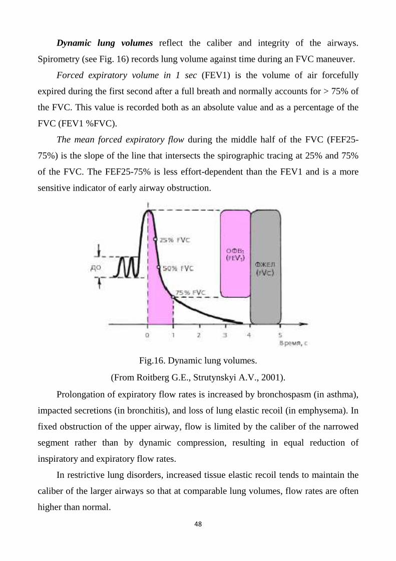
48
Dynamic lung volumes reflect the caliber and integrity of the airways.
Spirometry (see Fig. 16) records lung volume against time during an FVC maneuver.
Forced expiratory volume in 1 sec (FEV1) is the volume of air forcefully
expired during the first second after a full breath and normally accounts for > 75% of
the FVC. This value is recorded both as an absolute value and as a percentage of the
FVC (FEV1 %FVC).
The mean forced expiratory flow during the middle half of the FVC (FEF25-
75%) is the slope of the line that intersects the spirographic tracing at 25% and 75%
of the FVC. The FEF25-75% is less effort-dependent than the FEV1 and is a more
sensitive indicator of early airway obstruction.
Fig.16. Dynamic lung volumes.
(From Roitberg G.E., Strutynskyi A.V., 2001).
Prolongation of expiratory flow rates is increased by bronchospasm (in asthma),
impacted secretions (in bronchitis), and loss of lung elastic recoil (in emphysema). In
fixed obstruction of the upper airway, flow is limited by the caliber of the narrowed
segment rather than by dynamic compression, resulting in equal reduction of
inspiratory and expiratory flow rates.
In restrictive lung disorders, increased tissue elastic recoil tends to maintain the
caliber of the larger airways so that at comparable lung volumes, flow rates are often
higher than normal.

49
Retesting pulmonary function after the patient inhales a bronchodilator aerosol
(eg, albuterol, ipratropium) provides information about the reversibility of an
obstructive process (ie, the asthmatic component). Improvement in FVC or FEV1(L)
of > 15 to 20% is usually considered a significant response (Fig.17). In patients with
airway obstruction, absence of a response to a single exposure to a bronchodilator,
however, does not preclude a beneficial response to maintenance therapy. In
bronchoprovocation testing, a significant decrease in flow rates after inhaling
methacholine (a cholinergic drug) may indicate asthma.
Fig.17. FEV1 before and after bronchodilator aerosol inhale.
Maximal voluntary ventilation (MVV) is determined by encouraging the patient
to breathe at maximal tidal volume and respiratory rate for 12 sec; the volume of air
expired is expressed in L/min. The MVV generally parallels the FEV1 and can be
used to test internal consistency and estimate patient cooperation. The MVV can be
estimated from the spirogram by multiplying the FEV1(L)х40.
When the MVV is disproportionately low in a patient who seems to be
cooperating, neuromuscular weakness should be suspected. Except in advanced
neuromuscular disease, most patients can generate fairly good single-breath efforts
(eg, FVC). Because the MVV is much more demanding, it can reveal the diminished
reserves of weak respiratory muscles. The MVV decreases progressively with
Improvement in FEV1 > 15%
after bronchodilator aerosol inhale
before bronchodilator aerosol inhale
normal
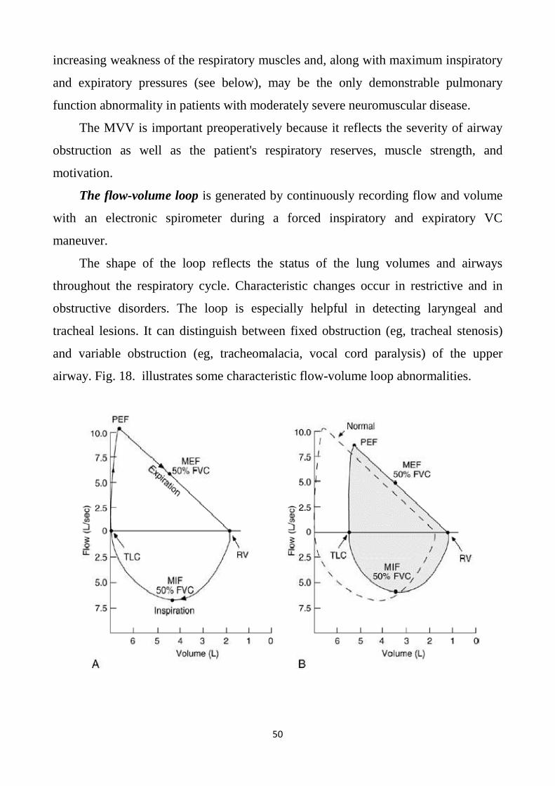
50
increasing weakness of the respiratory muscles and, along with maximum inspiratory
and expiratory pressures (see below), may be the only demonstrable pulmonary
function abnormality in patients with moderately severe neuromuscular disease.
The MVV is important preoperatively because it reflects the severity of airway
obstruction as well as the patient's respiratory reserves, muscle strength, and
motivation.
The flow-volume loop is generated by continuously recording flow and volume
with an electronic spirometer during a forced inspiratory and expiratory VC
maneuver.
The shape of the loop reflects the status of the lung volumes and airways
throughout the respiratory cycle. Characteristic changes occur in restrictive and in
obstructive disorders. The loop is especially helpful in detecting laryngeal and
tracheal lesions. It can distinguish between fixed obstruction (eg, tracheal stenosis)
and variable obstruction (eg, tracheomalacia, vocal cord paralysis) of the upper
airway. Fig. 18. illustrates some characteristic flow-volume loop abnormalities.
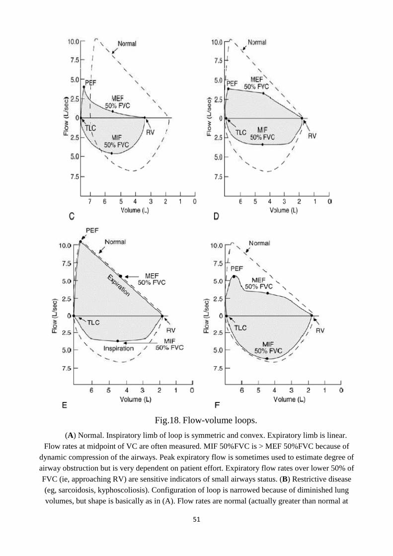
51
Fig.18. Flow-volume loops.
(A) Normal. Inspiratory limb of loop is symmetric and convex. Expiratory limb is linear. Flow rates at midpoint of VC are often measured. MIF 50%FVC is > MEF 50%FVC because of
dynamic compression of the airways. Peak expiratory flow is sometimes used to estimate degree of airway obstruction but is very dependent on patient effort. Expiratory flow rates over lower 50% of FVC (ie, approaching RV) are sensitive indicators of small airways status. (B) Restrictive disease (eg, sarcoidosis, kyphoscoliosis). Configuration of loop is narrowed because of diminished lung volumes, but shape is basically as in (A). Flow rates are normal (actually greater than normal at

52
comparable lung volumes because increased elastic recoil of lungs and/or chest wall holds airways open). (C) COPD, asthma. Though all flow rates are diminished, expiratory prolongation
predominates, and MEF is < MIF. (D) Fixed obstruction of upper airway (eg, tracheal stenosis, bilateral vocal cord paralysis, goiter). Top and bottom of loop are flattened so that the configuration
approaches that of a rectangle. The fixed obstruction limits flow equally during inspiration and expiration, and MEF = MIF. (E) Variable extrathoracic obstruction (eg, vocal cord paralysis). When a single vocal cord is paralyzed, it moves passively in accordance with pressure gradients across the
glottis. During a forced inspiration, it is drawn inward, resulting in a plateau of decreased inspiratory flow. During a forced expiration, it is passively blown aside and expiratory flow is unimpaired, ie, MIF 50%FVC is < MEF 50%FVC. (F) Variable intrathoracic obstruction (eg,
tracheomalacia). During a forced inspiration, negative pleural pressure holds the "floppy" trachea open. With forced expiration, the loss of structural support results in narrowing of the trachea and a
plateau of diminished flow (a brief period of maintained flow is seen before airway compression occurs). (From Oslopov B.N, Sadykova A.R., Karamysheva I.V. Introduction to internal diseases.
Manual. Part V.2nd edition., 2008).
Ordering Pulmonary Function Tests. As a general preoperative screen,
determination of the FVC, FEV1, FEV1 %FVC, and MVV usually suffices. Testing
should be performed before chest or abdominal surgery in smokers > 40 yr old and in
patients with respiratory symptoms. In patients with suspected laryngeal or tracheal
disorders, a flow-volume loop should be requested. If weakness of the respiratory
muscles is suspected, the MVV, MIP, MEP, and VC are the appropriate tests.
A complete set of pulmonary function tests should be requested when the
clinical picture does not coincide with the data obtained by simple spirometry or
when more complete characterization of an abnormal pulmonary process is desired.
A complete set includes determination of static and dynamic lung volumes, DLCO,
flow-volume loop, MVV, MIP, and MEP. However, extensive testing is tiring, time-
consuming, expensive, and unnecessary for adequate clinical assessment of most
patients. Periodic determinations of VC and DLCO usually suffice to monitor
patients with interstitial lung disease.
Table 5 is intended as general guidelines for interpreting pulmonary function
tests.

53
Table 5
Restrictive lung disease
Impairment None Mild Moderate Severe Very Severe
VC (%predicted) >80 60-80 50-60 35-50 <35
FEV1/FVC >75 >75 >75 >75 >75
MVV >80 >80 >80 60-80 <60
RV(%predicted) 80-120 80-120 70-80 60-70 <60
Obstructive lung diseases
Impairment None Mild Moderate Severe Very Severe
VC (%predicted) >80 >80 >80 ↓ ↓↓
FEV1/FVC >75 60-75 40-60 <40 <40
MVV >80 65-80 45-65 30-45 <30
RV(%predicted) 80-120 120-150 150-175 >200 >200
In the home for monitoring of lung function is recommended to determine the
peak expiratory flow (PEF) using the peak flow. This method belongs to an early
screening-diagnosis of bronchial obstruction – one of the most important biomarkers
of asthma and COPD. The below-mentioned patient after necessary training the
health worker is able to apply independently for objective control of their condition
and timely treatment to the doctor in case of its functional state. Note, however, that
the below-mentioned will never be able to replace spirometry, because peak flow can
be used to identify only the peak expiratory flow (PEF). Method of measurement of
PEF:
Measurement of PEF is once in the morning before bronchodilator. The
procedure is repeated three times and the highest peak expiratory flow (PEF) is
recorded. This can be compared with a nomogram that shows the patient‘s sex, age
and weight, and plotted on a chart to show the progress or response to treatment.
Measurements should be made 2-3 weeks (minimum 1 week). If the measurement
result is classified as proper or best for the period of measurement the indicator PEF
(the so-called "index PEF-variability or PSV
should change basic therapy
A. Initial level of PEF is low (55%), B. After change of basic therapy
54
variability or PSV-lability respiratory tract")
(Fig. 19). (GINA, 2002)
A.
B.
Fig.19. Monitoring of PEF.
A. Initial level of PEF is low (55%), index PEF-variability is very high (60%, Normally B. After change of basic therapy PEF is 110%, index PEF-variability is
spiratory tract") <80%, you
variability is very high (60%, Normally -20%). variability is 7%.
55
Airflow obstruction syndrome
Airflow obstruction syndrome — syndrome of bronchial patency alteration, in
chronic course of disease manifested by severe productive, rarely non-productive
cough and also emphysema development. In acute bronchial obstruction signs of
acute respiratory insufficiency occur, that is assessed as emergency situation.
Causes. In airflow obstruction syndrome the appearing changes are concerned
predominantly small bronchi. Inflammation and edema of bronchial mucous (chronic
bronchitis, allergic reactions), bronchospasm, usually with edema of bronchial
mucous (e.g. bronchial asthma) are the most frequent causes, rarely – diffuse
peribronchial fibrosis, compressing the bronchi from the outside, and also mechanical
bronchi compression from the outside in emphysema (on expiration intraalveolar
pressure increases resulting in small bronchi collapse).
Symptoms and signs. The main symptoms are cough, expiratory dyspnea. In
lingering course of airflow obstruction syndrome it is necessary to mark the particular
clinical meaning of cough not only as the symptom and sign of bronchi disorder but
as factor, which worsened pulmonary parenchyma lesion itself.
There are few outward manifestations of abnormality in patients with mild
airflow obstruction. However, as the process becomes more severe, the patient‘s
distress becomes evident from
- the labored breathing,
- the use of the accessory muscles on respiration,
- inspiratory retraction of the supraclavicular fossae and lower interspaces, and
- the positioning of the chest near total lung capacity.
When the patient is asked to empty the lungs forcibly and completely, it is
evident that expiration is prolonged and difficult, with pulmonary emptying
incomplete. The degree of expiratory slowing may be estimated by measuring the
forced expiratory time (which normally measures 4 seconds or less), with a watch
and a stethoscope. Auscultation over the larynx permits accurate determination of the
56
end of expiration. Sounds are audible at this site at the low airflows occurring near
residual volume, when breath sounds are no longer audible over the lungs.
On auscultation: harsh vesicular breath sounds with prolonged expiration,
wheezes and rhonchi, which presence gives an opportunity to define the level of
obstruction. Abnormality of inhalation and exhalation proportions and rough
prolonged exhalation appearance is the significant auscultative index of bronchial
obstruction. It must be remembered that when airway obstruction is very severe,
wheezes may completely disappear, usually with a marked decrease in the intensity
of breath sounds. The reappearance of wheezes indicates the response of the patient
to treatment, with diminution in the severity of airway obstruction.
In prominent acute bronchial obstruction the picture of ―silent lung appears,
when the bronchial patency is altered so much that breath sounds aren't listened at all.
Functional tests: FEV1 <80 % predicted, FEV1 / FVC<70% predicted and
FEF25-75% decrease.
Syndrome of hyperinflated lung closely linked with airflow obstruction
syndrome is discussed below.
Respiratory deficiency syndrome.
Diagnostics of respiratory insufficiency presence is an important and mandatory
moment in estimation of respiratory organs pathology. Respiratory insufficiency (RI)
is a condition when maintenance normal gas composition of arterial blood is not
supported or supported due to abnormal (heavy) work of external respiration
apparatus that leads to decrease of organism functional capacities.
Maintenance of normal gas exchange in the lungs is possible, as it was already
stated, only on condition of sharp interconnection of three components:
1. ventilation
2. gases diffusion through alveolocapillary membrane and
3. capillary blood perfusion in the lungs.
57
That is why any pathologic processes in the organism or unfavorable
environment factors (for example, decrease of oxygen partial pressure in atmospheric
air) that influence at least one of these components, may be the reasons of RI.
Two RI groups are distinguished:
group I with predominant lesion of extrapulmonary mechanisms;
group II with predominant lesion of pulmonary mechanisms: ventilation,
perfusion and alveolocapillary gases diffusion.
Main reasons and mechanisms of respiratory insufficiency, characteristic of
dyspnea. The following pathologic conditions may be included in group I of RI:
1. disturbance of central regulation of respiration (traumatic, metabolic,
circulatory, toxic, infectious and other brain lesions);
2. respiratory muscles lesion (trauma, intoxication, myalgia, myodystrophy, etc.)
or peripheral nerves lesion (poliomyelitis, polyradiculoneuritis, tetanus);
3. chest lesion (kyphoscoliosis, deformations, trauma, etc.). Group II of RI
includes the following pathologic conditions:
4. obstruction of large respiratory tracts (tumor, foreign body, dyskinesia of
membranous part of the trachea);
5. obstruction of small respiratory tracts (bronchial asthma, bronchiolitis);
6. disturbance of alveolar tissue restriction (interstitial edema, pleurisy,
pneumothorax, hydrothorax, etc.);
7. reduction of pulmonary tissue (massive inflammation, lung resection,
atelectases);
8. alveolocapillary membrane thickening (interstitial edema, pulmonary tissue
inflammation, pulmonary fibrosis, etc.);
9. pulmonary circulation lesions (blood congestion in the lesser circulation circle
in left ventricular cardiac failure, hypovolemia, etc.);
10. disturbance of ventilation - perfusion proportions (chronic obstructive
bronchitis, pneumonia, pulmonary artery branches thromboembolism, etc.).
Two forms of RI are distinguished depending on predominant lesion of three
respiratory system components (ventilation, perfusion and diffusion).

58
In ventilation form of RI external respiration lesion prevails which is
accompanied by development of hypoxemia as well as hypercapnia.
In the so-called parenchymatous form of RI disturbances of gases diffusion,
capillary blood perfusion or perfusion - ventilation proportions prevail. This form of
RI leads to development of hypoxemia whereas hypercapnia is not usually observed.
Attention should be paid to the fact that the majority of pulmonary pathologic
processes are accompanied by disturbance of several gas exchange mechanisms. For
example, in pneumonia restriction lesions are mainly observed, obstructive lesions
are somewhat less frequent, gases diffusion through alveolocapillary membrane
decreases, number of functioning alveoli lessens, etc.
In chronic obstructive bronchitis alongside with pronounced obstructive lesions
disturbances of ventilation-perfusion proportions are observed due to significant
unevenness of pulmonary ventilation, etc.
Dyspnea caused by respiratory center irritation and having a very variable
character is the most important symptom of RI. The type of dyspnea may be more
distinctly defined in small respiratory tracts obstruction (expiratory dyspnea) and in
restrictive disturbances (inspiratory dyspnea).
The traditional principle clinical classification RI is its division into degrees of
severity depending on the tolerance to physical activity and severity of dyspnea (by
A. G. Dembo, 1957; Schick, L. L., Kanaev N. N., 1980). Depending on the level of
physical activity in which there is shortness of breath, in our country it is customary
to distinguish three degrees of severity RI. When I grade dyspnea occurs with
increased physical activity; at II degree of dyspnea observed in moderate daily
physical activity; for III the severity of chronic pulmonary disease characterized by
shortness of breath at rest.
Almost all existing definitions of RI clearly indicate a gas composition of
arterial blood as a possible indicator of the degree days. The division of chronic
respiratory insufficiency in severity depends on the levels of the two most important
indicators of gas composition of arterial blood – РаО2 and SaO2 (saturation of
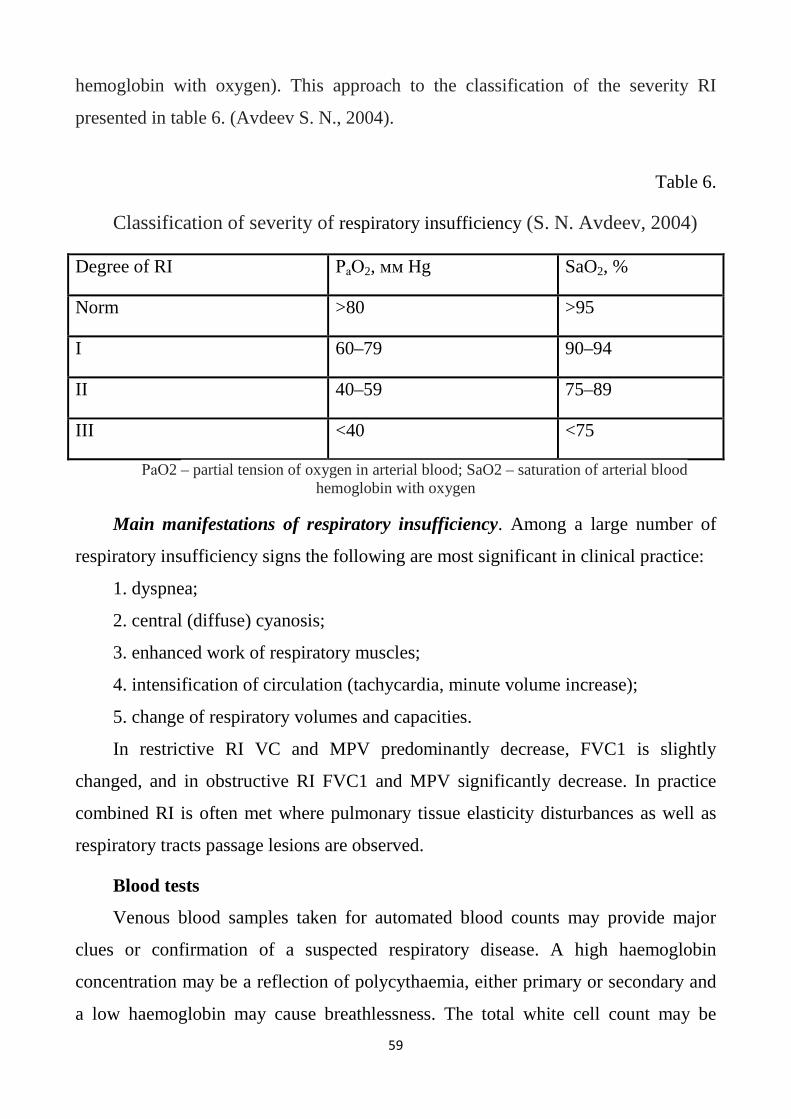
59
hemoglobin with oxygen). This approach to the classification of the severity RI
presented in table 6. (Avdeev S. N., 2004).
Table 6.
Classification of severity of respiratory insufficiency (S. N. Avdeev, 2004)
Degree of RI РаО2, мм Hg SaO2, %
Norm >80 >95
I 60–79 90–94
II 40–59 75–89
III <40 <75
РаО2 – partial tension of oxygen in arterial blood; SaO2 – saturation of arterial blood hemoglobin with oxygen
Main manifestations of respiratory insufficiency. Among a large number of
respiratory insufficiency signs the following are most significant in clinical practice:
1. dyspnea;
2. central (diffuse) cyanosis;
3. enhanced work of respiratory muscles;
4. intensification of circulation (tachycardia, minute volume increase);
5. change of respiratory volumes and capacities.
In restrictive RI VC and MPV predominantly decrease, FVC1 is slightly
changed, and in obstructive RI FVC1 and MPV significantly decrease. In practice
combined RI is often met where pulmonary tissue elasticity disturbances as well as
respiratory tracts passage lesions are observed.
Blood tests
Venous blood samples taken for automated blood counts may provide major
clues or confirmation of a suspected respiratory disease. A high haemoglobin
concentration may be a reflection of polycythaemia, either primary or secondary and
a low haemoglobin may cause breathlessness. The total white cell count may be

60
elevated in a range of acute bacterial infections and its subsequent fall is a reflection
of successful therapy. Normal or low white cell counts are found in mycoplasma or
viral infections. Eosinophilia suggests an allergic component or parasitic infection.
A range of serological tests that depend on agglutination, precipitation and
complement fixation provide evidence of the presence in the patient's serum of
specific antibodies against viral, bacterial, fungal, protozoal and helminthic
infections. Samples of blood should be tested on admission and repeated after 10- 14
days to detect a rising titer.
Radioallergoabsorbent tests (RASTs) on venous blood are an alternative to skin-
prick tests as a method of identifying specific IgE antibodies.
Sputum test
A sputum culture is a test to detect and identify bacteria or fungi that infect
the lungs or breathing passages. Sputum is a thick fluid produced in the lungs and in
the adjacent airways. A sample of sputum is placed in a sterile container and sent to
the laboratory for testing. Sampling may be performed by sputum being expectorated
(produced by coughing), induced (saline is sprayed in the lungs to induce sputum
production), or taken via an endotracheal tube with a protected specimen
brush (commonly used on patients on respirators) in an intensive care setting. For
selected organisms such as Cytomegalovirus or "Pneumocystis jiroveci" in specific
clinical settings (immunocompromised patients) a bronchoalveolar lavage might be
taken by an experienced pneumologist. If no bacteria or fungi grow, the culture is
negative. If organisms that can cause the infection (Pathogenicity organisms) grow,
the culture is positive. The type of bacterium or fungus is identified by microscopy,
colony morphology and biochemical tests of bacterial growth.
If bacteria or fungi that can cause infection grow in the culture, other tests can
determine which antimicrobial agent will most effectively treat the infection. This is
called susceptibility or sensitivity testing.
61
Bacterial culture
A portion of the sputum is smeared on a microscope slide for a Gram stain. Anot
her portion is spread over the surface ofseveral different types of culture plates, and p
laced in an incubator at body temperature for one to two days.
A Gram stain is done by staining the slide with purple and red stains, then exami
ning it under a microscope. Gramstaining checks that the specimen does not contain s
aliva or material from the mouth. If many epithelial (skin) cells andfew white blood c
ells are seen, the specimen is not pure sputum and is not adequate for culture. Depend
ing on laboratorypolicy, the specimen may be rejected and a new specimen requested.
If many white blood cells and bacteria of one typeare seen, this is an early confirmati
on of infection. The color of stain picked up by the bacteria (purple or red), their shap
e(such as round or rectangular), and their size provide valuable clues as to their identi
ty and helps the physician predictwhat antibiotics might work best before the entire te
st is completed. Bacteria that stain purple are called gram-positive;
those that stain red are called gram-negative.
During incubation, bacteria present in the sputum sample multiply and will appe
ar on the plates as visible colonies. Thebacteria are identified by the appearance of th
eir colonies, by the results of biochemical tests, and through a Gram stainof part of a
colony.
A sensitivity test, also called antibiotic susceptibility test, is also done. The bacte
ria are tested against differentantibiotics to determine which will treat the infection by
killing the bacteria.
The initial result of the Gram stain is available the same day, or in less than an h
our if requested by the physician. Anearly report, known as a preliminary report, is us
ually available after one day. This report will tell if any bacteria have beenfound yet,
and if so, their Gram stain appearance—for example, a gram-negative rod, or a gram-
positive cocci. The finalreport, usually available in one to three days, includes comple
te identification and an estimate of the quantity of thebacteria and a list of the antibiot
ics to which they are sensitive.
62
Fungal culture
To look for mold or yeast, a fungal culture is done. The sputum sample is spread
on special culture plates that willencourage the growth of mold and yeast. Different b
iochemical tests and stains are used to identify molds and yeast.Cultures for fungi ma
y take several weeks.
Viral culture
Viruses are a common cause of pneumonia. For a viral culture, sputum is mixed
with commercially
prepared animal cellsin a test tube. Characteristic changes to the cells caused by the g
rowing virus help identify the virus. The time tocomplete a viral culture varies with th
e type of virus. It may take from several days to several weeks.
Special procedures
Tuberculosis is caused by a slowgrowing bacteria called Mycobacterium tuberc
ulosis. Because it does not easily growusing routine culture methods, special procedu
res are used to grow and identify this bacteria. When a sputum sample fortuberculosis
first comes into the laboratory, a small portion of the sputum is smeared on a microsc
ope slide and stainedwith a special stain, called an acid
fast stain. The stained sputum is examined under a microscope for tuberculosisorgani
sms, which pickup the stain, making them visible. This smear is a rapid screen for the
organism, and allows thephysician to receive a preliminary report within 24 hours.
To culture for tuberculosis, portions of the sputum are spread on and placed into
special culture plates and tubes of broththat promote the growth of the organism. Gro
wth in broth is faster than growth on culture plates. Instruments are availablethat can
detect growth in broth, speeding the process even further. Growth and identification
may take two to four weeks.
Other microorganisms that cause various types of lower respiratory tract infectio
ns also require special cultureprocedures to grow and identify. Mycoplasma pneumon
ia causes a mild to moderate form of pneumonia, commonly calledwalking pneumoni
63
a; Bordetella pertussis causes whooping
cough; Legionella pneumophila, Legionnaire's disease;Chlamydia pneumoniae, an at
ypical pneumonia; and Chlamydia psittaci, parrot fever.
Pneumocystis carinii causes pneumonia in people with weakened immune syste
ms, such as people with AIDS. Thisorganism does not grow in culture. Special stains
are done on sputum when pneumonia caused by this organism issuspected. The diag-
osis is based on the results of these stains, the patient's symptoms, and medical histor
y.
Sputum culture is also called sputum culture and sensitivity.
It is possible that sputum cultures will eventually be replaced in the diagnosis of
tuberculosis by newer moleculartechniques. These advanced methods speed the diagn
ostic process as well as improve its accuracy. As of late 2002, fourmolecular techniqu
es are increasingly used in laboratories around the world to diagnose TB. They includ
e polymerasechain reaction to detect mycobacterial DNA in patient specimens; nuclei
c acid probes to identify mycobacteria in culture;restriction fragment length polymor
phism analysis to compare different strains of TB for epidemiological studies; andgen
etic-based susceptibility testing to identify drug-resistant strains of mycobacteria.
QUESTIONS FOR TEST CONTROL TO ENGAGE THE THEME "QUESTIONING, GENERAL EXAMINATION, INSPECTION AND PALPATION OF THE PATIENT WITH RESPIRATORY DISEASES" : 1. GIVE THE NAME OF THE NEXT FORM OF THE CHEST: Thorax elongated, narrow, flat. Over - and subclavian fossae are distinctly pronounced. The epigastric angle is acute. The ribs in the lateral parts directed more vertically. The muscles are poorly developed. a) paralytic b) emphysematous с) asthenic d) hypersthenic e) normasthenic 2. GIVE THE NAME OF THE NEXT FORM OF THE CHEST:
64
Increasing the transverse and anteroposterior size of the chest, short neck. Supraclavicular fossa smooth. The epigastric angle is obtuse. The direction of the horizontal ribs. Intercostal spaces are wide, breathing in zadanija departments indicated their retraction. The scapula firmly against the thorax. The breath is actively involved supporting musculature a) paralytic b) emphysematous c) asthenic d) hypersthenic e) normasthenic 3. GIVE THE NAME OF THE NEXT FORM OF THE CHEST: Wide but short ribcage. The ratio of anterior–posterior to lateral size of about 1.0. Supraclavicular fossa smooth. The epigastric angle is obtuse. The direction of the ribs are almost horizontal. Intercostal spaces narrow, their retractions when breathing is not marked. The scapula firmly against the thorax. a) paralytic b) emphysematous c) asthenic d) hypersthenic e) normasthenic 4. GIVE THE NAME OF THE NEXT FORM OF THE CHEST: Thorax elongated, narrow and flat. The ratio of anterior–posterior and lateral sizes close to 0.5. Over - and subclavian fossae are distinctly pronounced, but is located asymmetrically. The epigastric angle is acute. The ribs are placed obliquely. Intercostal spaces are wide Pronounced atrophy of the muscles of the chest. a) paralytic b) emphysematous c) asthenic d) hypersthenic e) normasthenic 5. GIVE THE NAME OF THE NEXT FORM OF THE CHEST: When inspecting the thorax, the ratio of anteroposterior to transverse size of about 0.7. Supraclavicular fossa is weakly developed. An epigastric angle of a straight line. The ribs in the lateral parts have moderately oblique direction. The scapula firmly

65
against the thorax. a) paralytic b) emphysematous c) asthenic d) hypersthenic e) normasthenic 6. WHAT IS THE NAME OF SPINAL CURVATURE FORWARD? a) lordosis b) scoliosis c) kyphosis d) kyphoscoliosis 7. WHAT IS THE NAME OF SPINAL CURVATURE IN A LATERAL DIRECTION? a) lordosis b) scoliosis c) kyphosis d) kyphoscoliosis 8. HOW IS CALLED THE CURVATURE OF THE SPINE AGO WITH THE FORMATION OF THE HUMP? a) lordosis b) scoliosis c) kyphosis d) kyphoscoliosis 9. WHAT IS THE NAME OF SPINAL CURVATURE IN A LATERAL DIRECTION AND BACK? a) lordosis b) scoliosis c) kyphosis d) kyphoscoliosis 10. SPECIFY THE MOST TYPICAL CHANGES OF THORAX IN PATIENTS WITH PNEUMOTHORAX: a) decrease half of thorax, its retraction and backlog in breath
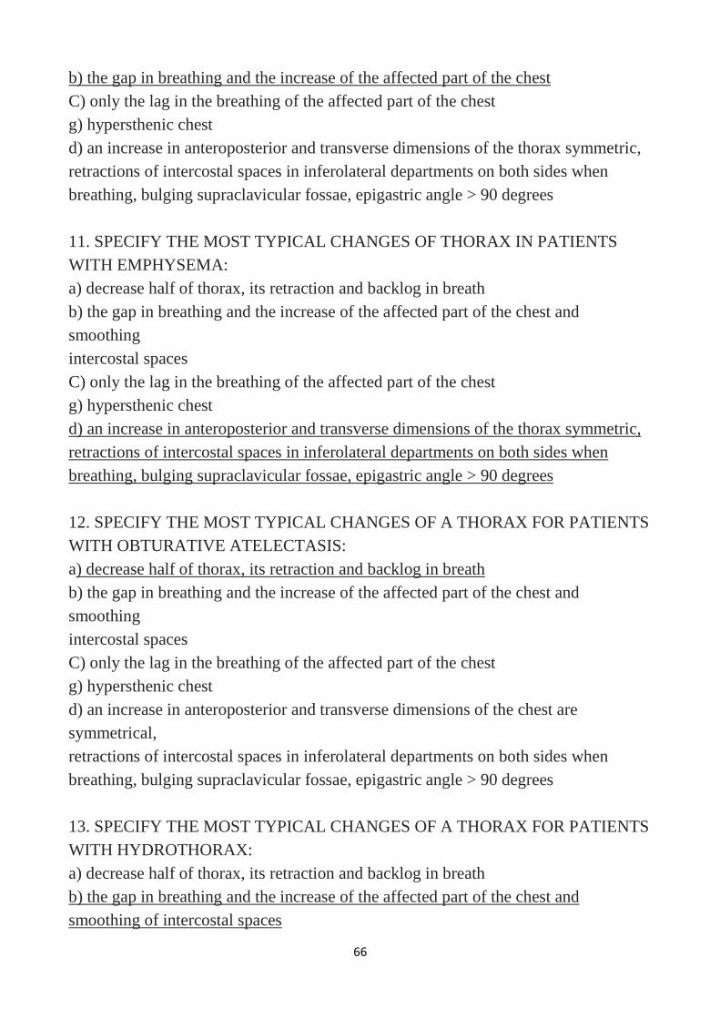
66
b) the gap in breathing and the increase of the affected part of the chest C) only the lag in the breathing of the affected part of the chest g) hypersthenic chest d) an increase in anteroposterior and transverse dimensions of the thorax symmetric, retractions of intercostal spaces in inferolateral departments on both sides when breathing, bulging supraclavicular fossae, epigastric angle > 90 degrees 11. SPECIFY THE MOST TYPICAL CHANGES OF THORAX IN PATIENTS WITH EMPHYSEMA: a) decrease half of thorax, its retraction and backlog in breath b) the gap in breathing and the increase of the affected part of the chest and smoothing intercostal spaces C) only the lag in the breathing of the affected part of the chest g) hypersthenic chest d) an increase in anteroposterior and transverse dimensions of the thorax symmetric, retractions of intercostal spaces in inferolateral departments on both sides when breathing, bulging supraclavicular fossae, epigastric angle > 90 degrees 12. SPECIFY THE MOST TYPICAL CHANGES OF A THORAX FOR PATIENTS WITH OBTURATIVE ATELECTASIS: a) decrease half of thorax, its retraction and backlog in breath b) the gap in breathing and the increase of the affected part of the chest and smoothing intercostal spaces C) only the lag in the breathing of the affected part of the chest g) hypersthenic chest d) an increase in anteroposterior and transverse dimensions of the chest are symmetrical, retractions of intercostal spaces in inferolateral departments on both sides when breathing, bulging supraclavicular fossae, epigastric angle > 90 degrees 13. SPECIFY THE MOST TYPICAL CHANGES OF A THORAX FOR PATIENTS WITH HYDROTHORAX: a) decrease half of thorax, its retraction and backlog in breath b) the gap in breathing and the increase of the affected part of the chest and smoothing of intercostal spaces

67
C) only the lag in the breathing of the affected part of the chest g) hypersthenic chest d) an increase in anteroposterior and transverse dimensions of the thorax symmetric, retractions of intercostal spaces in inferolateral departments on both sides when breathing, bulging supraclavicular fossae, epigastric angle > 90 degrees 14. DETERMINE the type of BREATHING: the patient is a feeling of incompleteness or obstruction of the exhalation; the exhalation with the participation of auxiliary muscles. a) respiration of Cheyne-Stokes b) expiratory dyspnea b) stridor d) inspiratory dyspnea d) Kussmaul breathing 15. DETERMINE the type of BREATHING: Periodic breathing with changing amplitude of the respiratory movements and long periods of apnea. a) respiration of Cheyne-Stokes b) expiratory dyspnea b) stridor d) inspiratory dyspnea d) Kussmaul breathing 16. DETERMINE the type of BREATHING: Deep noisy breath in patient in a coma a) respiration of Cheyne-Stokes b) expiratory dyspnea c) stridor d) inspiratory dyspnea e) Kussmaul breathing 17. DETERMINE THE TYPE OF BREATHING: THE patient dramatically difficult to inhale and exhale; jugular veins swollen. Inspiratory and expiratory breathing is noisy. a) respiration of Cheyne-Stokes b) expiratory dyspnea c) stridor

68
d) inspiratory dyspnea e) Kussmaul breathing 18. HOW TO CHANGE THE VOCAL FREMITUS IN SYNDROME OF AIR CAVITY? a) no change b) weakening of one side c) increase on both sides d) the weakening of both sides e) increase one side 19. HOW TO CHANGE VOCAL FREMITUS IN EMPHYSEMA? a) no change b) weakening of one side b) increase on both sides d) the weakening of both sides d) increase one side 20. HOW TO CHANGE VOCAL FREMITUS IN CANCER OF THE LARGE BRONCHUS WITH THE OBTURATIVE ATELECTASIS? a) no change b) weakening of one side b) increase on both sides d) the weakening of both sides d) increase one side 21. HOW TO CHANGE VOCAL FREMITUS IN THE PLEURAL EFFUSION SYNDROME? a) no change b) weakening of one side b) increase on both sides d) the weakening of both sides d) increase one side 22. HOW TO CHANGE VOCAL FREMITUS IN LOBAR PNEUMONIA? a) no change b) weakening of one side

69
b) increase on both sides d) the weakening of both sides d) increase one side 23. HOW TO CHANGE VOCAL FREMITUS WITH PNEUMOTHORAX, NOT COMMUNICATING WITH THE BRONCHUS? a) no change b) weakening of one side b) the gain on both sides d) the weakening of both sides d) increase one side
QUESTIONS FOR TEST CONTROL TO ENGAGE THE THEME
"COMPARATIVE PERCUSSION OF THE LUNGS. PULMONARY
SYNDROMES":
1. WHAT ARE THE 2 TYPES OF PERCUSSION SOUNDS APPEAR WHEN THE PLEURAL EFFUSE SYNDROME? a. dull sound b. clear lung sounds c. tympanic sound d. dullness with tympanic shade e. hyperresonant sound 2. WHAT PERCUSSION SOUND IS CHARACTERISTIC OF OBTURATIVE ATELECTASIS? a. dull sound b. clear lung sounds c. tympanic sound d. dulled-tympanic sound e. hyperresonant sound sound
3. WHAT PERCUSSION SOUND OCCURS OVER A LARGE CAVITY IN THE LUNG? a. femoral sound b. clear lung sounds

70
c. tympanic (metal) sound d. dullness with tympanic shade e. hyperresonant sound
4. WHAT PERCUSSION SOUND IS CHARACTERISTIC FOR INFLAMMATORY INFILTRATION OF LUNG TISSUE ( HEIGHT STAGE OF PNEUMONIA)? a. dull (femoral) sound b. clear lung sounds c. tympanic sound d. dullness with tympanic shade e. hyperresonant sound
5. WHAT PERCUSSION SOUND IS GENERATED AT THE INITIAL STAGE OF THE INFLAMMATORY INFILTRATION OF LUNG TISSUE (STAGE 1 OF PNEUMONIA)? a. absolutely dull (femoral) b. clear lung sounds c. tympanic sound d. dull sound e. hyperresonant sound
6. WHAT PERCUSSION SOUND ABOVE THE PLEURAL THICKENING? a. dull sound b. clear lung sounds c. tympanic sound d. dullness with tympanic shade e. hyperresonant sound
7. WHAT PERCUSSION SOUND IS CHARACTERISTIC FOR COMPRESSION ATELECTASIS? a. dull sound b. clear lung sounds c. tympanic sound d. dulled-tympanic sound e. hyperresonant sound

71
8. WHAT PERCUSSION SOUND IS CHARACTERISTIC FOR EMPHYSEMA? a. dull sound b. clear lung sounds c. tympanic sound d. dullness with tympanic shade e. hyperresonant sound
9. WHAT PERCUSSION SOUND IS CHARACTERISTIC FOR PNEUMOTHORAX? a. dull (femoral) or blunt sound b. clear lung sounds c. tympanic sound d. dullness with tympanic shade e. hyperresonant sound
10. WHAT PERCUSSION SOUND IS CHARACTERISTIC FOR THE FINAL STAGE OF THE INFLAMMATORY INFILTRATION OF LUNG (RESOLUTION STAGE OF PNEUMONIA)? a. dull (femoral) b. clear lung sounds c. tympanic sound d. blunt sound e. hyperresonant sound
11. WHAT SYNDROMES ARE CHARACTERIZED BY "SHORTENING" OF PERCUSSION SOUNDS? a. the syndrome of inflamantory of lung tissue b. the syndrome of pneumothorax not communicating with a bronchus c. syndrome of hydrothorax d. the syndrome of pneumothorax communicating with a bronchus e. syndrome of the air cavity in the lung f. syndrome of compression atelectasis g. the syndrome of obturative atelectasis h. the syndrome of pulmonary fibrosis i. the syndrome of emphysema 12. WHAT SYNDROMES ARE CHARACTERIZED BY TYMPANIC PERCUSSION SOUND?

72
a. the inflamantory infiltration syndrome b. the syndrome of pneumothorax not communicating with a bronchus c. syndrome of hydrothorax d. the syndrome of pneumothorax communicating with a bronchus e. syndrome of the air cavity in the lung f. the syndrome of obturative atelectasis g. the syndrome of pulmonary fibrosis h. the syndrome of emphysema 13. WHAT SYNDROME IS CHARACTERIZED BY DULL-TYMPANIC PERCUSSION SOUND? a. the inflamantory infiltration syndrome b. the syndrome of pneumothorax not communicating with a bronchus c. syndrome of hydrothorax d. the syndrome of pneumothorax communicating with a bronchus e. the syndrome of compressive atelectasis f. the syndrome of obturative atelectasis g. the syndrome of pulmonary fibrosis h. the syndrome of emphysema 14. FOR ANY OF THE SYNDROMES NOT CHARACTERISTIC METALLIC PERCUSSION SOUND? a. the inflamantory infiltration syndrome b. the syndrome of pneumothorax not communicating with a bronchus c. syndrome of hydrothorax d. the syndrome of pneumothorax communicating with a bronchus e. the syndrome of compressive atelectasis f. the syndrome of obturative atelectasis g. the large air cavity syndrome h. the syndrome of emphysema
15. IN CASE OF IDENTIFICATION THE INCREASE OF VOICE FREMITUS OVER THE AFFECTED SIDE OF THE CHEST WHAT PERCUSSION SOUND CAN I EXPECT?
a. dull percussion sound b. blunt percussion sound
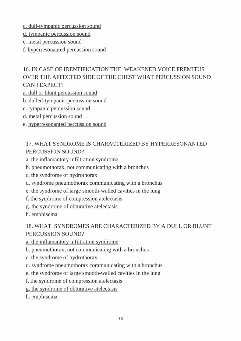
73
c. dull-tympanic percussion sound d. tympanic percussion sound e. metal percussion sound f. hyperresonanted percussion sound
16. IN CASE OF IDENTIFICATION THE WEAKENED VOICE FREMITUS OVER THE AFFECTED SIDE OF THE CHEST WHAT PERCUSSION SOUND CAN I EXPECT? a. dull or blunt percussion sound b. dulled-tympanic percussion sound c. tympanic percussion sound d. metal percussion sound e. hyperresonanted percussion sound
17. WHAT SYNDROME IS CHARACTERIZED BY HYPERRESONANTED PERCUSSION SOUND? a. the inflamantory infiltration syndrome b. pneumothorax, not communicating with a bronchus c. the syndrome of hydrothorax d. syndrome pneumothorax communicating with a bronchus e. the syndrome of large smooth-walled cavities in the lung f. the syndrome of compression atelectasis g. the syndrome of obturative atelectasis h. emphisema
18. WHAT SYNDROMES ARE CHARACTERIZED BY A DULL OR BLUNT PERCUSSION SOUND? a. the inflamantory infiltration syndrome b. pneumothorax, not communicating with a bronchus c. the syndrome of hydrothorax d. syndrome pneumothorax communicating with a bronchus e. the syndrome of large smooth-walled cavities in the lung f. the syndrome of compression atelectasis g. the syndrome of obturative atelectasis h. emphisema
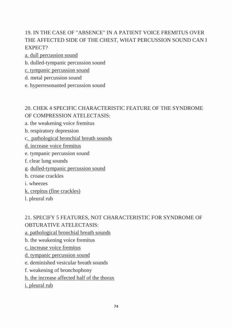
74
19. IN THE CASE OF "ABSENCE" IN A PATIENT VOICE FREMITUS OVER THE AFFECTED SIDE OF THE CHEST, WHAT PERCUSSION SOUND CAN I EXPECT? a. dull percussion sound b. dulled-tympanic percussion sound c. tympanic percussion sound d. metal percussion sound e. hyperresonanted percussion sound
20. CHEK 4 SPECIFIC CHARACTERISTIC FEATURE OF THE SYNDROME OF COMPRESSION ATELECTASIS: a. the weakening voice fremitus b. respiratory depression c. pathological bronchial breath sounds d. increase voice fremitus e. tympanic percussion sound f. clear lung sounds g. dulled-tympanic percussion sound h. croase crackles i. wheezes k. crepitus (fine crackles) l. pleural rub
21. SPECIFY 5 FEATURES, NOT CHARACTERISTIC FOR SYNDROME OF OBTURATIVE ATELECTASIS: a. pathological bronchial breath sounds b. the weakening voice fremitus c. increase voice fremitus d. tympanic percussion sound e. deminished vesicular breath sounds f. weakening of bronchophony h. the increase affected half of the thorax i. pleural rub

75
22. 5 SPECIFIC CHARACTERISTICS COMMON TO THE SYNDROME OF EMPHYSEMA: a. the weakening voice fremitus b. deminished vesicular breath sounds c. pathological bronchial breath sounds e. increase voice fremitus f. hyperresonant percussion sound g. funnel chest h. barrel chest i. pathological decrease in lung volume j. pathological increase in lung volume
23. SPECIFIC CHARACTERISTICS COMMON TO THE PLEURAL EFFUSION SYNDROME: a. the symmetry of participation in the breathing of both halves of the chest b. the lag in the breathing of the affected part of the chest c. the weakening voice fremitus d. the weakening or disappearance of breath e. pathological bronchial breath sounds f. tympanic percussion sound g. blunt or dull percussion sound h. pleural rub
24. 4 SPECIFIC SYMPTOMS, NOT CHARACTERISTIC FOR SYNDROME OF PNEUMOTHORAX THAT ARE NOT COMMUNICATING WITH A BRONCHUS: a. the appearance of pathological bronchial breathing b. the weakening voice fremitus c. tympanic percussion sound d. deminished vesicular breath sounds e. strengthen voice fremitus f. dull percussion sound g. the symmetry of participation in the breathing of both halves of the chest h. the lag in the breathing of the affected part of the chest
25. 4 SPECIFIC CHARACTERISTIC FEATURES OF THE INFLAMMATORY

76
INFILTRATION SYNDROME: a. the weakening voice fremitus b. pathological bronchial breathing c. increased voice fremitus d. tympanic percussion sound e. hyperresonanted percussion sound f. wheezes g. the symmetry of participation in the breathing of both halves of the chest h. the lag in the breathing of the affected part of the chest i. fine or croake crackles
26. 3 INDICATE SYMPTOM, NOT CHARACTERISTIC FOR SYNDROME OF THE AIR CAVITY connecting with bronchus: a. the weakening voice fremitus b. the appearance of pathological bronchial breathing c. increased voice fremitus d. tympanic percussion sound e. dull percussion sound f. crackles h. pleural rub
27. SPECIFY 5 FEATURES NOT CHARACTERISTIC FOR THE SYNDROME OF EMPHYSEMA: a. bronchial obstruction by tumor or foreign body b. pathological decrease in elastic properties of lungs with the development of "air traps" and pathological lung hyperinflation c. appearance in the pleural cavity fluid d. appearance in the pleural cavity air e. the appearance of air in the lung cavity in the case of the emptying of lung abscess or tuberculosis caverns f. inflammatory infiltration syndrome
28. WHAT ARE 5 SYNDROMES CHARACTERIZED BY PATHOLOGICAL BRONCHIAL BREATH SOUNDS?: a. syndrome of pleural effusion b. the syndrome of pneumothorax communicating with a bronchus c. pneumothorax, not communicating with a bronchus
77
d. the syndrome of obstructive atelectasis e. the syndrome of compression atelectasis f. the syndrome of the air cavity g. the syndrome of pulmonary fibrosis h. syndrome emphysema i. inflammatory infiltration syndrome
28. WHAT ARE 2 SYNDROMES CHARACTERIZED BY FINE CRACKLES?: a. syndrome of pleural effusion b. pneumothorax c. syndrome of compression atelectasis d. the syndrome of the air cavity e. the syndrome of emphysema f. the inflamatory infiltration syndrome
29. WHAT ARE THE CAUSE OF COMPRESSION ATELECTASIS: a. obturation (closing of the lumen) of the bronchus by tumor or foreign body b. pneumonia c. the pleural effusion syndrome d.decrease in the elastic properties of the small bronchi and bronchioles with the development of "expiratory closure of the small airways" e. infiltrative tuberculosis f. partial replacement of the alveoli by connective tissue
30. SPECIFY THE CAUSE OF OBTURATIVE ATELECTASIS: a. the pleural effusion syndrome b. pneumothorax c. the obstruction of the bronchus by tumor or foreign body d. pneumonia e. infiltrative tuberculosis f. "air trap" due to "expiratory closure of the small airways" g. partial replacement of the alveoli by connective tissue
31. SPECIFY 2 CAUSES OF INFLAMMATORY INFILTRATION SYNDROME: a. the pleural effusion syndrome
78
b. infiltrative tuberculosis c. pneumothorax d. the obturation (closing of the lumen) of the bronchus by tumor or foreign body e. pneumonia f. air trap due to the "expiratory closure of the small Airways"
32. SPECIFY 2 CAUSES OF THE SYNDROME OF THE AIR CAVITY: a. the pleural effusion syndrome b. pneumothorax c. obturation (closing of the lumen) of the bronchus by tumor or foreign body d. pneumonia e. infiltrative tuberculosis f. tuberculous cavity g. air trap due to the "expiratory closure of the small Airways" h. partial replacement of the alveoli by connective tissue i. lung abscess
33. 2 FEATURES OF BRONCHIAL OBSTRUCTION ACCORDING TO THE SPIROMETRY a. the decline of VC b. reduction FEV1/FVC<70% c. the decline of FEV1<80% from proper d. FEV1/FVC>70%
34. 2 FEATURES OF RESTRICTIVE DISORDERS OF RESPIRATORY FUNCTION ACCORDING TO SPIROMETRY a. the decline of VC b. reduction FEV1/FVC<70% c. the decline of FEV1<80% from proper d. FEV1/FVC>70%
35. CHOOSE THE CORRECT SYMPTOM OF RESPIRATORY ALKALOSIS a. РаСО2 > 35 mm Hg, BE = ± 2.5 mmol/l b. РаСО2 > 45 mm Hg, BE = ± 2.5 mmol/l

79
36. CHOOSE THE CORRECT SYMPTOM OF RESPIRATORY ACIDOSIS
a. РаСО2 < 35 mm Hg, BE = ± 2.5 mmol/l b. РаСО2 >45 mm Hg, BE = ± 2.5 mmol/l
37. SPECIFY 3 FEATURES OF RESPIRATORY INSUFFICIENCY (RI) STAGE I. a. signs of lung diseases that can cause RI b. shortness of breath during physical exertion exceeding the everyday c. shortness of breath with everyday physical activity d. shortness of breath at rest e. optional features of pulmonary hypertension and RI f. obligate signs of pulmonary hypertension and RI 38. A SIGN OF HYPOXEMIA IS a. РаО2<80 mm Hg b. PaCO2<45 mm Hg c. SaO2<95%
39. A SYMPTOM OF HYPERCAPNIA IS a. РаО2<35 mm Hg b. PaCO2>45 mm Hg
40. CURSHMANN'S SPIRALS IN SPUTUM ARE DETECTED IN: a) lung abscess; b) crupous pneumonia; c) chronic bronchitis; d) asthma; e) bronchiectasis. 41. ELASTIC FIBERS IN SPUTUM ARE DETECTED IN: a) dry pleuritis; b) crupous pneumonia; c) asthma; d) emphysema; e) lung abscess.
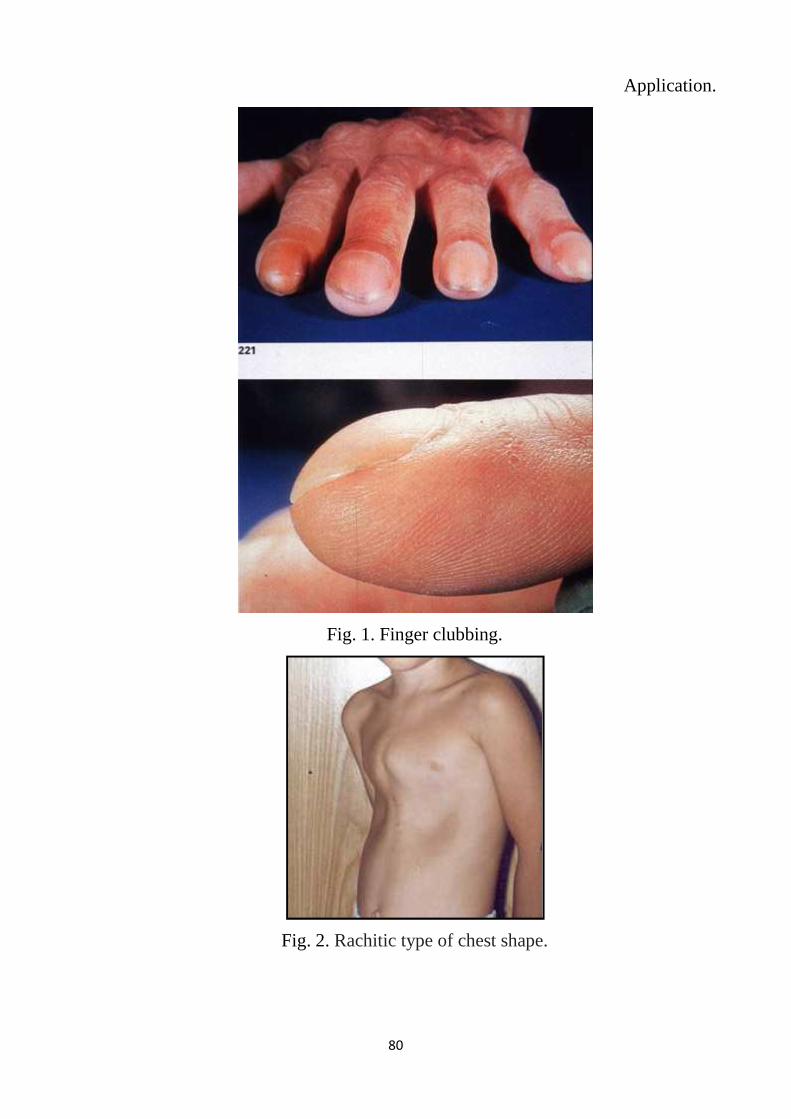
80
Application.
Fig. 1. Finger clubbing.
Fig. 2. Rachitic type of chest shape.

81
Fig. 3. Navicular type of chest shape.
Fig.4. Consequence of percuss of the anterior side of the chest.

82
Fig.5. Consequence of percuss of the posterior side of the chest.
a. suprascapular region; b. intrscapular region; в. subscapular region
Fig. 6. Percuss of lower border of the right lung (the anterior side of the chest).
a. parasternal line; b. mediaclavicular line
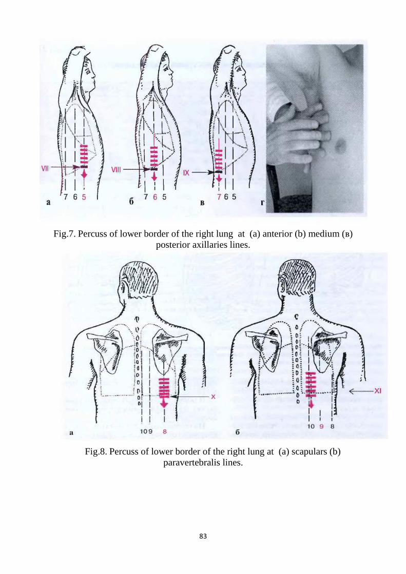
83
Fig.7. Percuss of lower border of the right lung at (a) anterior (b) medium (в) posterior axillaries lines.
Fig.8. Percuss of lower border of the right lung at (a) scapulars (b) paravertebralis lines.
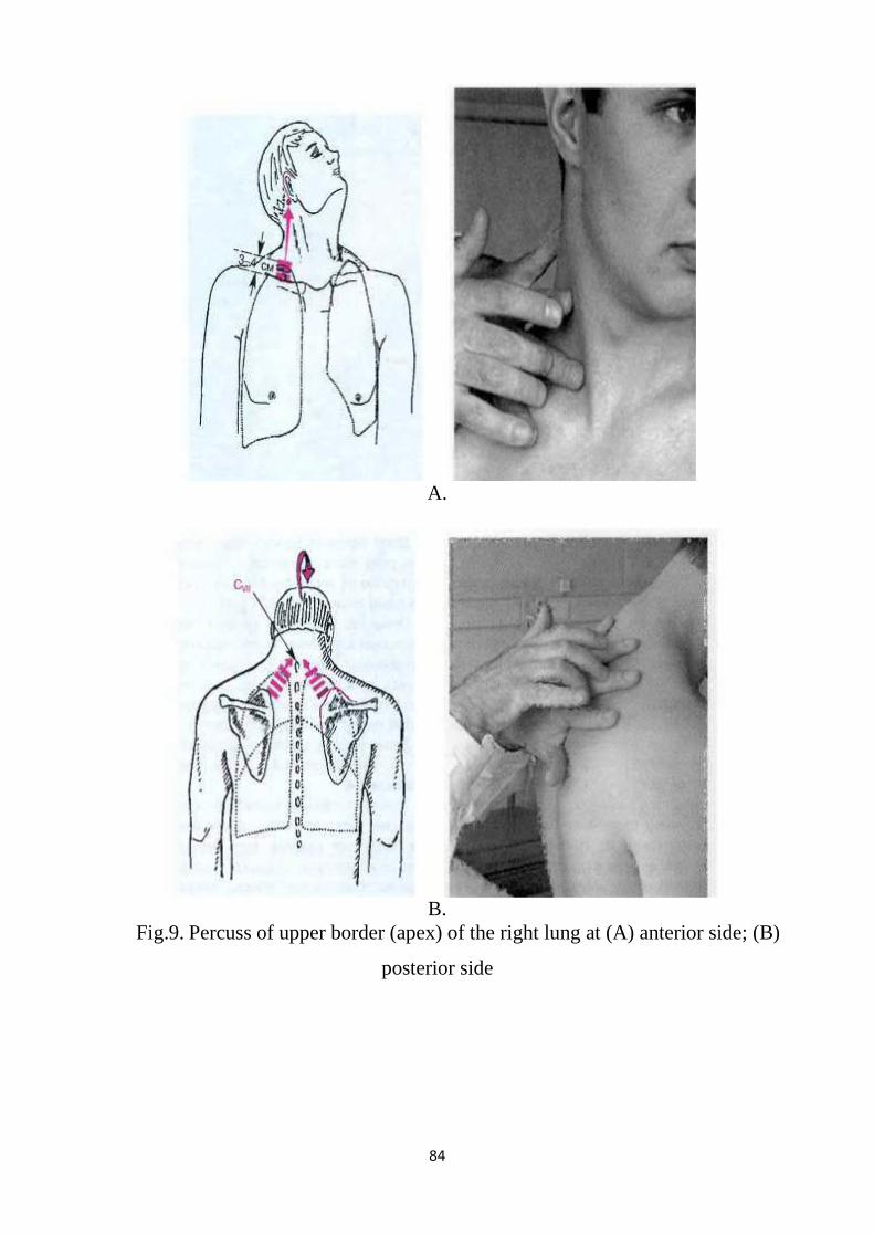
84
A.
B.
Fig.9. Percuss of upper border (apex) of the right lung at (A) anterior side; (B)
posterior side
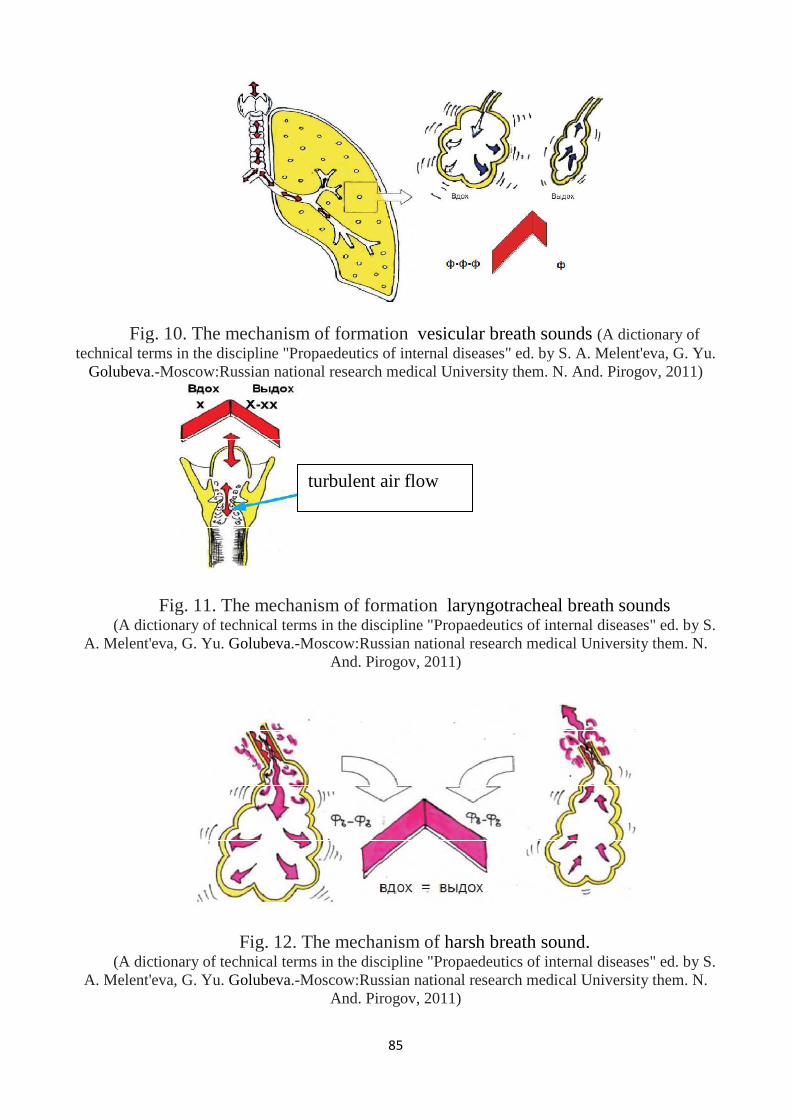
85
Fig. 10. The mechanism of formation vesicular breath sounds (A dictionary of technical terms in the discipline "Propaedeutics of internal diseases" ed. by S. A. Melent'eva, G. Yu.
Golubeva.-Moscow:Russian national research medical University them. N. And. Pirogov, 2011)
Fig. 11. The mechanism of formation laryngotracheal breath sounds (A dictionary of technical terms in the discipline "Propaedeutics of internal diseases" ed. by S.
A. Melent'eva, G. Yu. Golubeva.-Moscow:Russian national research medical University them. N. And. Pirogov, 2011)
Fig. 12. The mechanism of harsh breath sound. (A dictionary of technical terms in the discipline "Propaedeutics of internal diseases" ed. by S.
A. Melent'eva, G. Yu. Golubeva.-Moscow:Russian national research medical University them. N. And. Pirogov, 2011)
turbulent air flow
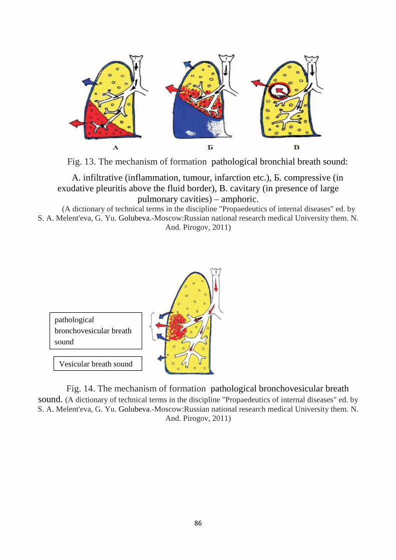
86
Fig. 13. The mechanism of formation pathological bronchial breath sound:
A. infiltrative (inflammation, tumour, infarction etc.), Б. compressive (in exudative pleuritis above the fluid border), B. cavitary (in presence of large
pulmonary cavities) – amphoric. (A dictionary of technical terms in the discipline "Propaedeutics of internal diseases" ed. by
S. A. Melent'eva, G. Yu. Golubeva.-Moscow:Russian national research medical University them. N. And. Pirogov, 2011)
Fig. 14. The mechanism of formation pathological bronchovesicular breath sound. (A dictionary of technical terms in the discipline "Propaedeutics of internal diseases" ed. by S. A. Melent'eva, G. Yu. Golubeva.-Moscow:Russian national research medical University them. N.
And. Pirogov, 2011)
pathological bronchovesicular breath sound
Vesicular breath sound
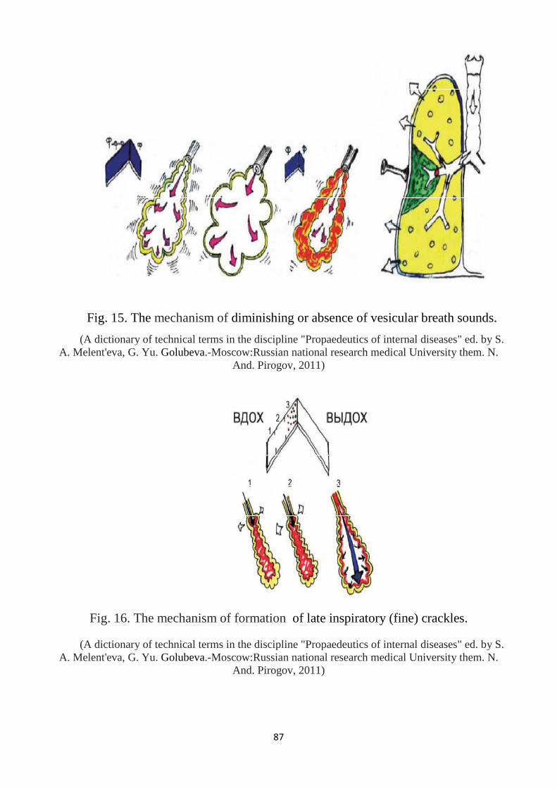
87
Fig. 15. The mechanism of diminishing or absence of vesicular breath sounds.
(A dictionary of technical terms in the discipline "Propaedeutics of internal diseases" ed. by S. A. Melent'eva, G. Yu. Golubeva.-Moscow:Russian national research medical University them. N.
And. Pirogov, 2011)
Fig. 16. The mechanism of formation of late inspiratory (fine) crackles.
(A dictionary of technical terms in the discipline "Propaedeutics of internal diseases" ed. by S. A. Melent'eva, G. Yu. Golubeva.-Moscow:Russian national research medical University them. N.
And. Pirogov, 2011)
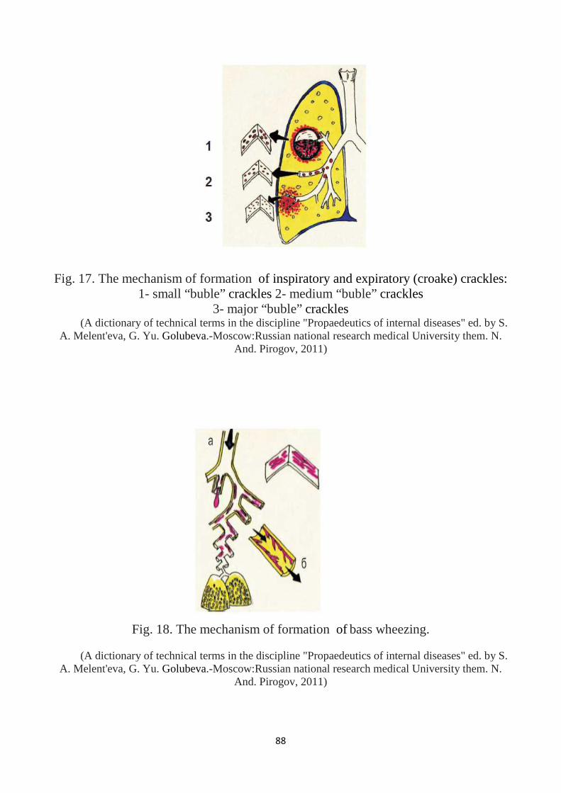
88
Fig. 17. The mechanism of formation of inspiratory and expiratory (croake) crackles: 1- small “buble” crackles 2- medium “buble” crackles
3- major “buble” crackles (A dictionary of technical terms in the discipline "Propaedeutics of internal diseases" ed. by S.
A. Melent'eva, G. Yu. Golubeva.-Moscow:Russian national research medical University them. N. And. Pirogov, 2011)
Fig. 18. The mechanism of formation of bass wheezing.
(A dictionary of technical terms in the discipline "Propaedeutics of internal diseases" ed. by S. A. Melent'eva, G. Yu. Golubeva.-Moscow:Russian national research medical University them. N.
And. Pirogov, 2011)

89
Fig. 19. The mechanism of formation of whistling wheezing: 1- thickening of the bronchial mucosa
2- narrowing of the lumen due to the presence of parietal-phlegm 3- bronchospasm
(A dictionary of technical terms in the discipline "Propaedeutics of internal diseases" ed. by S. A. Melent'eva, G. Yu. Golubeva.-Moscow:Russian national research medical University them. N.
And. Pirogov, 2011)
Fig. 20. The mechanism of formation of pleural crackles (rub).
(A dictionary of technical terms in the discipline "Propaedeutics of internal diseases" ed. by S. A. Melent'eva, G. Yu. Golubeva.-Moscow:Russian national research medical University them. N.
And. Pirogov, 2011)
two inflamed surfaces of the pleura rubbing against each other during respiration
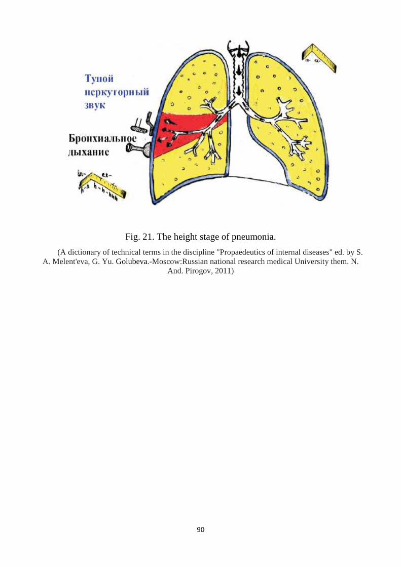
90
Fig. 21. The height stage of pneumonia.
(A dictionary of technical terms in the discipline "Propaedeutics of internal diseases" ed. by S. A. Melent'eva, G. Yu. Golubeva.-Moscow:Russian national research medical University them. N.
And. Pirogov, 2011)
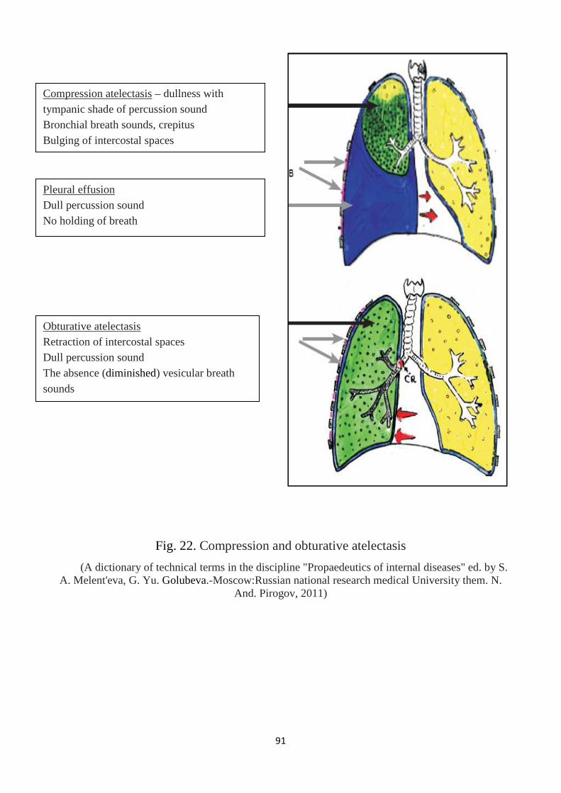
91
Fig. 22. Compression and obturative atelectasis
(A dictionary of technical terms in the discipline "Propaedeutics of internal diseases" ed. by S. A. Melent'eva, G. Yu. Golubeva.-Moscow:Russian national research medical University them. N.
And. Pirogov, 2011)
Compression atelectasis – dullness with tympanic shade of percussion sound Bronchial breath sounds, crepitus Bulging of intercostal spaces
Pleural effusion Dull percussion sound No holding of breath
Obturative atelectasis Retraction of intercostal spaces Dull percussion sound The absence (diminished) vesicular breath sounds

92
Fig.23. Exophytic cancer.
Fig.24. Endophytic cancer.

93
Fig. 25. Ulcerative form of cancer.
Fig. 26. Foreign body of the medium bronchus.
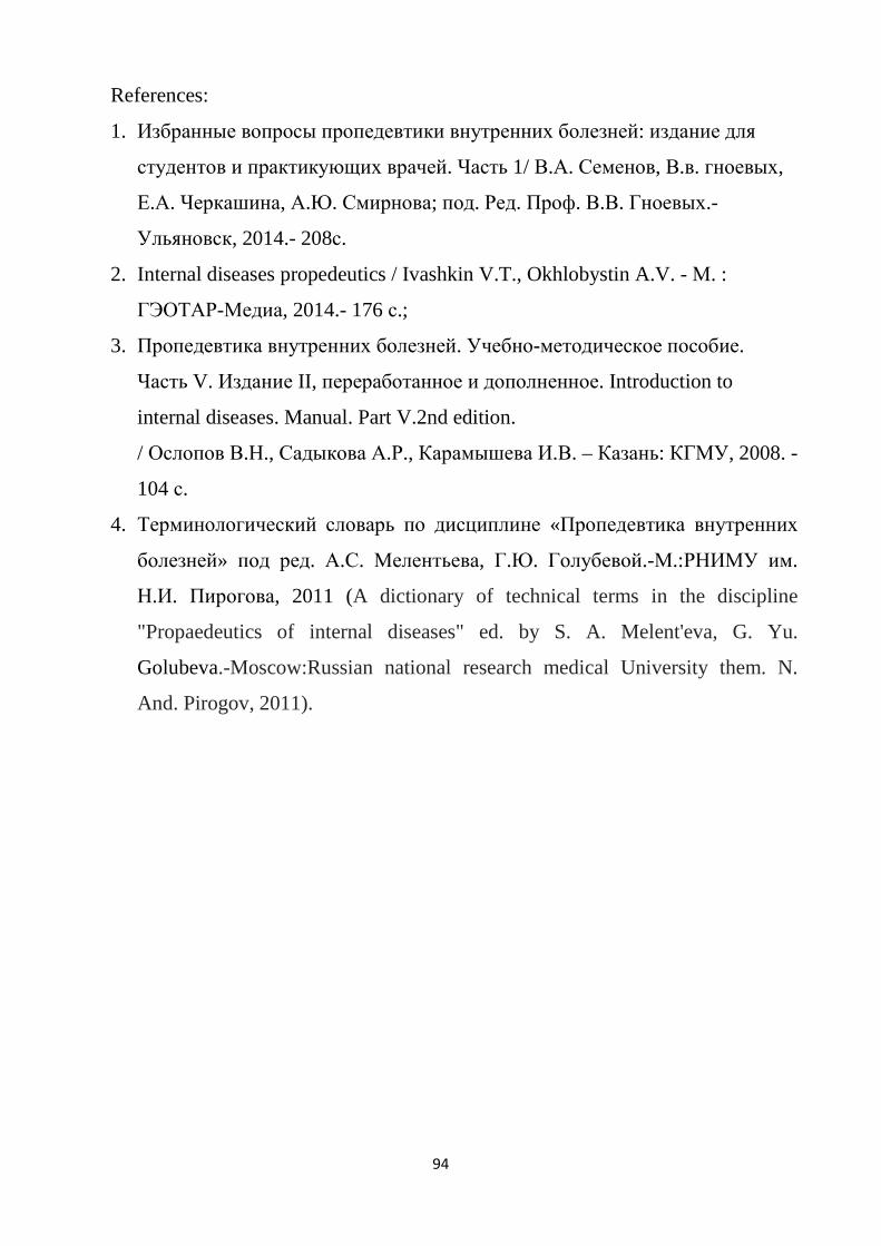
94
References:
1. Избранные вопросы пропедевтики внутренних болезней: издание для
студентов и практикующих врачей. Часть 1/ В.А. Семенов, В.в. гноевых,
Е.А. Черкашина, А.Ю. Смирнова; под. Ред. Проф. В.В. Гноевых.-
Ульяновск, 2014.- 208с.
2. Internal diseases propedeutics / Ivashkin V.T., Okhlobystin A.V. - М. :
ГЭОТАР-Медиа, 2014.- 176 с.;
3. Пропедевтика внутренних болезней. Учебно-методическое пособие.
Часть V. Издание II, переработанное и дополненное. Introduction to
internal diseases. Manual. Part V.2nd edition.
/ Ослопов В.Н., Садыкова А.Р., Карамышева И.В. – Казань: КГМУ, 2008. -
104 с.
4. Терминологический словарь по дисциплине «Пропедевтика внутренних
болезней» под ред. А.С. Мелентьева, Г.Ю. Голубевой.-М.:РНИМУ им.
Н.И. Пирогова, 2011 (A dictionary of technical terms in the discipline
"Propaedeutics of internal diseases" ed. by S. A. Melent'eva, G. Yu.
Golubeva.-Moscow:Russian national research medical University them. N.
And. Pirogov, 2011).