Agarose gel electrophoresis. Genomic DNA extraction PCR – Agarose gel electrophoresis.

Proc. Nati. Acad. Sci. USAVol. 84, pp. 6970-6974, October 1987Biochemistry
Internal amino acid sequence analysis of proteins separated by one-or two-dimensional gel electrophoresis after in situ proteasedigestion on nitrocellulose
(protein sequence analysis/electroblotting/peptide mapping/Edman degradation)
RUEDI H. AEBERSOLD*, JOHN LEAVITTt, RAUL A. SAAVEDRA*, LEROY E. HOOD*,AND STEPHEN B. H. KENT**Division of Biology, 147-75, California Institute of Technology, Pasadena, CA 91125; and tThe Linus Pauling Institute for Science and Medicine, Palo Alto,CA 94306
Contributed by Leroy E. Hood, May 20, 1987
ABSTRACT We have developed a general two-step meth-od for obtaining peptide fragments for sequence analysis frompicomole quantities of proteins separated by gel electrophore-sis. After separation by one- or two-dimensional polyacrylam-ide gel electrophoresis, proteins are electrophoretically trans-ferred (electroblotted) onto nitrocellulose, the protein-contain-ing regions are detected by reversible staining and are cut out,and each protein is digested in situ by proteolytic enzymes suchas trypsin or staphylococcal V-8 protease. The resulting peptidefragments are separated by narrow-bore reverse-phase HPLC,collected, and sequenced in a gas-phase sequenator. Excellentpeptide recoveries and the absence of extraneous contaminantsin the separation of the peptide fragment mixture allow thegeneration of extensive internal sequence information frompicomole amounts of protein. The method thus overcomes theproblem of obtaining amino acid sequence data from N-terminally blocked proteins and provides multiple, indepen-dent stretches of sequence that can be used to generateoligonucleotide probes for molecular cloning and/or used tosearch sequence data bases for related proteins. This methodhas been successfully applied to the routine amino acid se-quence analysis of a wide range of proteins isolated from one-and two-dimensional polyacrylamide gels.
Protein sequence analysis is central to modern biologicalresearch. Amino acid sequence data can aid in gene isolationeither through the use of synthetic oligonucleotides (1) orthrough the use of antisera of predetermined specificity (2).Sequence data can also be used to correlate the genestructure with the expressed polypeptide chain, to map activesites and domain boundaries, to define posttranslationalmodifications, or to establish evolutionary relationships be-tween proteins (3).
Currently, 20 pmol amounts of protein can give limitedN-terminal sequence information using optimized isolationmethods (4) and a commercially available gas-phase sequena-tor (5). It is desirable to develop a method of comparablesensitivity for internal amino acid sequence analysis ofproteins for the following reasons:
(i) Many proteins are not susceptible to the Edmandegradation. This is assumed to be due to artefactual orbiosynthetic blocking of the a-amino groups of the proteins.The nature of the modified N terminus is not easily deter-mined and only a few of the modifications can be chemicallyor enzymatically reversed (6). Therefore, the most generalway to obtain sequence information from a protein with ablocked a-amino group is to sequence peptide fragmentsgenerated by cleavage of the polypeptide chain.
(ii) In the molecular cloning of genes through the use ofsynthetic oligodeoxynucleotides reverse translated from theprotein sequence, it is advantageous to screen appropriatecDNA libraries with either a set of multiple probes or a singleprobe with minimum degeneracy to reduce the incidence of"false positives." Furthermore, the use of an oligodeoxynu-cleotide probe derived from the N terminus of the proteinrequires full-length cDNA clones or tedious and repetitivecloning from random-primed libraries (7). Multiple probescan be used to select long cDNA clones in a background ofshorter clones.
(iii) Amino acid sequences obtained at the low picomolelevel (10-20 pmol) are usually relatively short (10-25 aminoacid residues).'Homology searches of data bases for non-identical, but related, proteins using such short stretches ofamino acid sequence may give rise to numerous false match-es. Related proteins can more reliably be found by multiplescreening of the data ba'se with a set of peptides derived fromone protein.The highest resolving procedures for the analysis of mi-
crogram or submicrogram amounts of proteins are one-dimensional (iD) and two-dimensional (2D) polyacrylamidegel electrophoresis (PAGE) (8, 9). Although many proteinscan be identified through their behavior in analytical 2D gelelectrophoresis, sequence data are available for only a few ofthese proteins due to the difficulty of isolating sufficientquantities in a form suitable for sequence analysis (10). In thepast, proteins have been recovered from the gel matrix by(electro)elution (11) but they are frequently blocked at the Nterminus and are typically contaminated with large amountsof NaDodSO4 and salts, agents that hinder enzymatic cleav-age and separation of the resulting peptide fragments. Re-moval or exchange of the detergent and salts is tedious andis associated with significant losses of protein.
In this report we describe a method for the generation andisolation of peptide fragments for internal amino acid se-quence analysis after separation of <100 pmol of the intactpolypeptide chain by iD or 2D PAGE. The method consistsof the simultaneous electrophoretic transfer (electroblotting)of all separated proteins from the gel matrix onto nitrocellu-lose (12), detection of the proteins followed by in situenzymatic cleavage of individual proteins on the nitrocellu-lose matrix, and reverse-phase HPLC separation of releasedpeptide fragments. These peptide fragments are suitable foruse in the gas-phase microsequenator without further manip-ulation. The general utility of the method is illustrated here byextensive sequence data from a variety of proteins preparedfrom iD and 2D gel electrophoresis.
Abbreviations: MBP, myelin basic protein; ARP, actin-related pro-tein; iD, one-dimensional; 2D, two-dimensional; PVP-40, polyvinyl-pyrrolidone, average Mr = 40,000.
6970
The publication costs of this article were defrayed in part by page chargepayment. This article must therefore be hereby marked "advertisement"in accordance with 18 U.S.C. §1734 solely to indicate this fact.

Proc. Natl. Acad. Sci. USA 84 (1987) 6971
EXPERIMENTAL PROCEDURES
Materials. For gel electrophoresis, standard commercialsources of chemicals were used. Nitrocellulose (0.45-tumpore size) was obtained from Schleicher & Schuell. Theprotein stains employed were Ponceau S (Sigma) or amidoblack 10B (Sigma). Polyvinylpyrrolidone, average M, =40,000 (PVP-40), was purchased from Sigma. L-1-Tosyl-amido-2-phenylethyl chloromethyl ketone-treated trypsinand staphylococcal V-8 protease were obtained fromBoehringer Mannheim. Polybrene was obtained from Sigma.
Gel Electrophoresis. Proteins were separated in NaDod-S04/polyacrylamide gels with the dimensions 70 x 70 x 0.5mm according to Laemmli (8) or by 2D PAGE (9). Sharkmyelin proteins were separated in polyacrylamide gels ac-cording to Thomas and Kornberg (13). High-purity gelchemicals were used and gels were polymerized extensivelybefore use (11).
Electroblotting. Proteins were electroblotted in a Bio-Radtransblot system onto nitrocellulose as described (12) for 2 hrfor 0.5-mm-thick gels. For proteins that were difficult totransfer, up to 0.005% NaDodSO4 was added to the transferbuffer. After transfer, proteins were stained with amido black(14) or reversibly stained with Ponceau S using a modificationof the method described (15) as follows. Nitrocellulose filterswere immersed for 60 sec in a solution of 0.1% Ponceau S dyein 1% aqueous acetic acid. Excess stain was removed fromthe blot by gentle agitation for 1-2 min in 1% aqueous aceticacid. Protein-containing regions detected by either stain werecut out and transferred to Eppendorf tubes (1.5 ml), wherePonceau S-detected protein bands were destained by washingthe filter with 200 ,uM NaOH for 1-2 min. Finally, the filter(s)was washed with distilled water and stored wet at -20°C,without extensive drying which can inhibit the efficientrelease of peptides by protease treatment.
In Situ Enzymatic Cleavage of Electroblotted Proteins. Up tofive bands (NaDodSO4/PAGE) or 40 spots (2D gels) ofdestained nitrocellulose pieces containing the same proteinwere pooled in a single Eppendorf tube and incubated for 30min at 37°C in 1.2 ml of 0.5% PVP-40 dissolved in 100 mMacetic acid in order to prevent adsorption of the protease tothe nitrocellulose during digestion. Excess PVP-40 wasremoved by extensive washing with water (at least fivechanges). Because of the strong UV absorption of PVP-40,complete removal before HPLC analysis was essential.Nitrocellulose strips were then cut in small pieces of approx-imately 1 mm x 1 mm and put back into the same tube. Theprotein on the nitrocellulose pieces was digested as follows.(i) Trypsin: up to 300 ,l of 100 mM Tris-HCI, pH 8.2/acetonitrile, 95:5 (vol/vol), at 37°C overnight. (ii) V-8 prote-ase: 100 mM sodium phosphate, pH 7.8/acetonitrile, 95:5(vol/vol), at 37°C overnight. Only the minimum volume ofbuffer necessary to completely immerse the nitrocellulosepieces was used. Enzyme-to-substrate ratio was kept at-1:20 (wt/wt). After digestion, the whole reaction mixturewas frozen at -20°C or immediately loaded onto the HPLCcolumn, after acidification.
Reverse-Phase HPLC of the Cleavage Fragments. Enzymat-ic cleavage fragments were separated on a narrow-bore(2.1-mm i.d.) reverse-phase HPLC system using a dual-syringe Brownlee micropump (16). The system was equippedwith a Rheodyne model 7125 sample injector with a 500-,ulloop. The analytical column used was a Brownlee AquaporeBu-300, 2.1 x 100 mm, equipped with a guard cartridge filledwith the same material. The following buffer system wasused. Buffer A: 0.1% trifluoroacetic acid (Sequenal grade,Pierce) in water (double glass distilled). Buffer B: 0.08-0.095% trifluoroacetic acid in acetonitrile/H20, 70:30(vol/vol). The optical densities of buffers A and B werematched at 215 nm by titrating the trifluoroacetic acid
concentration in buffer B. Both buffers were degassed witha stream of helium. All experiments were carried out withcolumns at ambient temperature, at a flow rate of 100 ,l/min.Gradients were run as indicated in Figs. 1-3. Peptides weredetected by simultaneous monitoring at 215 nm, 260 nm, and280 nm with a Waters 490 detector.
Fresh or thawed peptide-containing supernatant was acid-ified with 30 ,ul of 10% trifluoroacetic acid, mixed quickly ina Vortex, and centrifuged for 1 min in a Microfuge at highspeed. The supernatant was removed and immediately in-jected into the HPLC unit. Buffer A was pumped through thecolumn for 5 min at a flow rate of 200 /l/min before the flowwas reduced to 100 Al/min and the gradient was started.
Peptide-containing fractions were collected manually intoEppendorf tubes based on the UV absorption at 215 nm.Collected fractions were frozen immediately. If this precau-tion was not taken the yield of recovered phenylthiohydan-toin amino acids dropped significantly at each serine residue.
Peptide Sequence Analysis. Amino acid sequence analysiswas performed on an automated Caltech gas-phase sequena-tor (5). Collected peptides were spotted onto glass fiber discsthat had been coated with Polybrene and pretreated accord-ing to the following method so that precycling in the gas-phase sequenator was not necessary. A sheet of GF/F glassfiber paper (13-cm diameter) (Whatman) was soaked in aglass Petri dish for 2 min in a solution of 2 mg of Polybreneper ml of water (17) and dried in an oven at 50'C for 1 hr.Excess Polybrene and other contaminants were electropho-retically removed by exposing the sheet to an electric field (50V) in a Transblot system (Bio-Rad) overnight at 40C. Thebuffer tank contained a solution of 2 M acetic acid/methanol(Baker, HPLC grade), 1:1 (vol/vol), and several grams ofmixed-bed ion-exchange resin (Amberlite MB-3, LKB). Af-ter electrophoretic purification, the glass fiber sheet wasrinsed thoroughly in water, dried in an oven at 50°C, andstored in a clean glass Petri dish. Phenylthiohydantoin aminoacids were identified essentially as described (18).
RESULTS AND DISCUSSIONGel Electrophoresis, Electrophoretic Transfer, and Detec-
tion. The separation of components of even complex proteinmixtures by 1D or 2D gel electrophoresis is perhaps the mostcommonly used protein analytical technique (8, 9). Similarly,the electrophoretic transfer of proteins from polyacrylamidegels onto nitrocellulose membranes is widely used for thedetection and analysis of proteins with specific reagents (12).Although electroblotting onto nitrocellulose is generally ef-ficient, the required presence of methanol in the transferbuffer tends to strip NaDodSO4 from proteins, with theconsequence that very basic and insoluble proteins transferless efficiently. We have overcome this problem by addingsmall amounts ofNaDodSO4 (0.005%, wt/vol) to the transferbuffer to provide additional negative charges and help keepproteins in solution in cases where transfers were otherwiseinefficient [e.g., myelin basic proteins (MBPs), pI > 9].
Transferred proteins were detected on the nitrocellulosemembrane with Ponceau S or amido black. Although thesensitivity of Ponceau S staining does not match the sensi-tivity of amido black, <500 ng of protein per typical band canbe detected. This sensitivity is sufficient to detect theamounts of proteins needed for internal sequence analysis.Staining with Ponceau S was preferred for the followingreasons: (i) The method is fast. Blots are stained anddestained in <5 min. (ii) Staining/destaining conditions arevery mild. Proteins are not exposed to high concentrations oforganic acids and solvents. (iii) Staining is reversible. Unlikeother dyes, Ponceau S can be efficiently removed from theblotted proteins under very mild conditions (200 ,M NaOH,3-5 min) without eluting the protein from the nitrocellulose.In some cases, because of the high charge density, very basic
Biochemistry: Aebersold et al.

6972 Biochemistry: Aebersold et al.
proteins such as MBPs cannot be completely destained.Neither residual Ponceau S nor amido black stain interfereswith reverse-phase HPLC analysis of the peptide fragmentsfrom in situ digestion, since the dyes either remain on thenitrocellulose (amido black) or dissociate completely fromthe peptide fragments in HPLC buffer A (Ponceau S). Thefree dyes do not bind to C4 columns and are washed throughin the void volume.
Prevention of Protease Adsorption to Nitroceflulose. It wasessential to pretreat the nitrocellulose before the proteasedigestion in order to prevent adsorption of the enzyme.Without such pretreatment, no cleavage was observed andthe enzyme disappeared from the supernatant. We evaluateda number of nonprotein compounds [polyoxyethylenesorbitan monolaurate (Tween-20), polyethylene glycol (av-erage Mr = 8000), PVP-40, Ficoll (approximate Mr =400,000), and salmon sperm DNA] for their ability to bind tonitrocellulose and prevent adsorption of the protease duringdigestion. Nitrocellulose pieces were incubated with eachreagent, reagents were washed away, and the saturatednitrocellulose pieces were incubated overnight at 37TC with1251I-radiolabeled bovine serum albumin and washed. Thenumber of radioactive cpm in the supernatant and on themembrane were determined. Bovine serum albumin waschosen as a model protein for these experiments because it isa relatively large and sticky protein. PVP-40 [0.5% (wt/vol)in 100 mM acetic acid, 30 min, 37°C] showed the highestsaturating capacity (>90%) of all reagents tested. Further-more, we found that the saturating capacity of PVP-40 was
a
31
9-.
C%
Cly
Po
MBP
U,cmn
not pH dependent (pH range, 2-8) and did not increase withincreasing concentration.
Efficiency of the Procedure. To quantitate the efficiency ofthe procedure, 10 ,g of 1251-radiolabeled a-lactalbumin wasapplied to 1-cm2 pieces of nitrocellulose. The protein wasstained with Ponceau S and destained, and the nitrocellulosepieces were saturated by treatment with 0.5% PVP-40 solu-tion. Digestion with trypsin was carried out for 12 hr at 37°Cand the resulting recovered peptide fragments were separatedby HPLC. The efficiency of each step of the procedure wasdetermined by y-radiation counting. Although a-lactalbuminis a relatively small and nonsticky protein and represents anunfavorable case in terms of washout losses, the overallrecovery of peptides amounted to =65% of the proteininitially present on the nitrocellulose.Two parameters proved to be critical for the high overall
efficiency of the procedure. (i) Proteins are more stronglyadsorbed to nitrocellulose at acidic than at neutral or basicpH. If the PVP-40 treatment was performed at acidic pH,very little protein was washed off during saturation. (ii)Hydrophilic interaction between peptide fragments and ni-trocellulose can generally be reduced by the addition oforganic solvent. Thus, 5% acetonitrile was included in thedigestion buffer and proved to be sufficient for the effectiverelease of the peptide fragments. No significant increase inthe release of the peptide fragments was observed with up to40% of acetonitrile in the digestion buffer, although this didnot inhibit the proteolytic activity of trypsin (data notshown).
100
30
'0
oWcLLL
0 m
RETENTION TIME (MIN)
W
LL~LL
nmo
RETENTION TIME ( MIN)FIG. 1. Proteins of a shark brain myelin preparation. (a) Ponceau S-stained nitrocellulose blot of the proteins separated by 18%
NADodSO4/PAGE (Kornberg gel) (13). Molecular weights are shown as Mr x 1O-3. (b) HPLC map of peptides released after in situ trypticdigestion of P0. (c) HPLC map of peptides released after in situ tryptic digestion of MBP. Asterisks (*) indicate peptides for which sequenceanalysis was carried out. AU, absorbance unit.
Proc. Natl. Acad. Sci. USA 84 (1987)

Proc. Natl. Acad. Sci. USA 84 (1987) 6973
Peptide Mapping. Enzymatic peptide maps of the sameprotein digested either in situ on nitrocellulose blots or insolution are somewhat different, although most of the majorpeptides can be found in both preparations. Repetition of theprocess with multiple, electroblotted samples of a-lactalbumin showed that the digestion pattern is reproducible(data not shown). The differences in the peptide maps afterenzymatic fragmentation in situ and in solution probablyreflect differences in accessibility ofenzymatic cleavage sitesrather than differential release of peptides. The overallrecovery, estimated from the relative peak areas in HPLCanalyses of in situ and solution digests, correlates with theoverall efficiency of 50-60% as calculated from radioactivelabeling experiments. For typical amounts of proteins, thepeptide cleavage fragments contained no contaminatingpeaks derived from the staining or PVP-40 saturation proce-dures or from the nitrocellulose itself. A protease blankreaction was always included to unambiguously identifysubstrate-derived peptides.
Application of the Method to Proteins Separated by NaDod-S04/PAGE. In the context of our ongoing effort to under-stand the structure and function of the myelin sheath in thevertebrate nervous system (19), myelin proteins from thecentral nervous system of a shark (Heterodontus) wereisolated and separated by NaDodSO4/PAGE (13). Afterelectrophoresis, the proteins were transferred onto nitrocel-lulose and stained with Ponceau S (Fig. la). The bandscorresponding to MBP (-5 Ag) and to the myelin protein P0(-12-14 pug) were cut out, destained, treated with PVP-40,and cleaved in situ with trypsin, and the resulting mixtures ofpeptide fragments were separated by reverse-phase HPLC.The peptide maps of shark PO and MBP are shown in Fig. 1b and c, respectively. Selected peptides were applied to agas-phase sequenator and their sequences were determinedat the 20-50 pmol level. For both proteins sufficient internalsequence information was obtained for the synthesis of threeindependent oligodeoxynucleotide probes to screen sharkbrain cDNA libraries. The PO protein has an N terminusaccessible to the Edman degradation, and therefore data forthe synthesis of an oligonucleotide probe could be generated
IEF*~ .4
a_ ~~~~~T
*
---s. ~SDSt *o w - -~~PAGE
d
0.05:
0 10 20 30 40 50 60
RETENTION TIME (MIN)
by conventional N-terminal sequence analysis (4). On theother hand, MBP is blocked at its N terminus, so thatsequence information could only be obtained from cleavagefragments of the protein. MBP is a very basic protein (pI >9). Although this protein could not be completely destainedafter Ponceau S treatment, no contaminating peaks interferedwith peptide separation or detection (Fig. 1c). The largenumber of basic residues in MBP led to the generation of anumber of very small tryptic cleavage fragments that did notbind to the C4 column, explaining the relatively small numberof peptides recovered after HPLC separation.
Application of the Method to Proteins Separated by 2DPAGE. We have applied the nitrocellulose blot/in situ diges-tion method to a number of proteins isolated from 2Dpolyacrylamide gels of whole-cell lysates. The sequence datawere used to confirm the identity of spots, to reveal homol-ogies to known proteins, or to synthesize oligodeoxynucleo-tide probes for isolation of the gene. The results obtained withtwo proteins are illustrated in Fig. 2. As 2D gels have a limitedloading capacity, multiple spots (20-40) (-10-15 ,ug) of thesame protein species were accumulated and simultaneouslyprocessed. To reduce the number of second-dimension andelectroblotting experiments, the relevant regions of multiple(up to five) first-dimension gels were aligned onto a singlesecond-dimension gel as described (21). Corresponding spotsfrom the repeated pattern on the nitrocellulose blots were cutout and simultaneously subjected to in situ digestion. Fig. 2shows peptide maps of two proteins from a human lympho-blastoid cell line: actin-related protein (ARP), a proteincorresponding to p24 in a 2D gel protein catalog (22) (Fig. 2b),and the p subunit of mitochondrial F1-ATPase (Fig. 2c). Atotal of 107 residues of sequence data were generated fromnine peptides from the F1-ATPase digest and aligned with theknown sequence of the bovine protein (23). The high degreeof homology (>90%) confirmed the identity of the humanlymphoblastoid protein in the 2D gel. Similar internal se-quence data were obtained for ARP, the other proteinexample in Fig. 2. Computer-assisted searches of sequencedata bases showed that ARP, which is known to be anabundant nucleus-encoded protein subsequently transported
rS i~ic ----100
< 0.051 '* 40ep ui
oL J ... ._o0 10 20> 30 40 50 60RETENTION TIME (MIN)
0.3.-
C 1 0O.21 * S 100
0. i Cc
400:~ ~ ~ ~ ¾ 2aw ~~~~~~~~~~~~~~~~~~~~~~~...... ......O 10.0 30 40 50 60RETENTION TIME (MIN)
FIG. 2. Proteins from a whole-cell lysate ofthe human lymphoblastoid cell line CCRF-CEM (ATCC no. CCL 119). (a) 2D [isoelectric focusing(IEF)/NaDodSO4/PAGE] separation of proteins metabolically labeled with [35S]methionine (20). Proteins used as examples are circled. (b)HPLC map of peptides released after in situ tryptic digestion of the protein ARP after electroblotting onto nitrocellulose. (c) HPLC map ofpeptides released after in situ tryptic digestion of the P-subunit of mitochondrial F1-ATPase after electroblotting onto nitrocellulose. (d) Blankdigest with trypsin. Asterisks (*) indicate peptides for which sequence analysis was carried out. AU, absorbance unit.
Biochemistry: Aebersold et al.
6974 Biochemistry: Aebersold et al.
into mitochondria, does not show significant homology to anyknown protein.
CONCLUSIONSWe have described a method for obtaining extensive internalsequence information from microgram amounts of protein. Thenitrocellulose blot/in situ digestion method offers a number ofadvantages over previous methods used to obtain internalsequence information from proteins. It employs the most highlyresolving and generally applicable protein separation methods,1D and 2D PAGE, and allows the parallel isolation, in a singleoperation, of multiple proteins in a form suitable for internalsequence analysis even if they are blocked at the N terminus.We estimate that, even with existing levels of protein sequenc-
ing sensitivity, on the order of 100 proteins from the 2D gelseparation of a whole-cell lysate (20) are present in sufficientamounts to yield extensive amino acid sequence informationusing the strategies outlined in this report.The method is compatible with proteolytic enzymes of
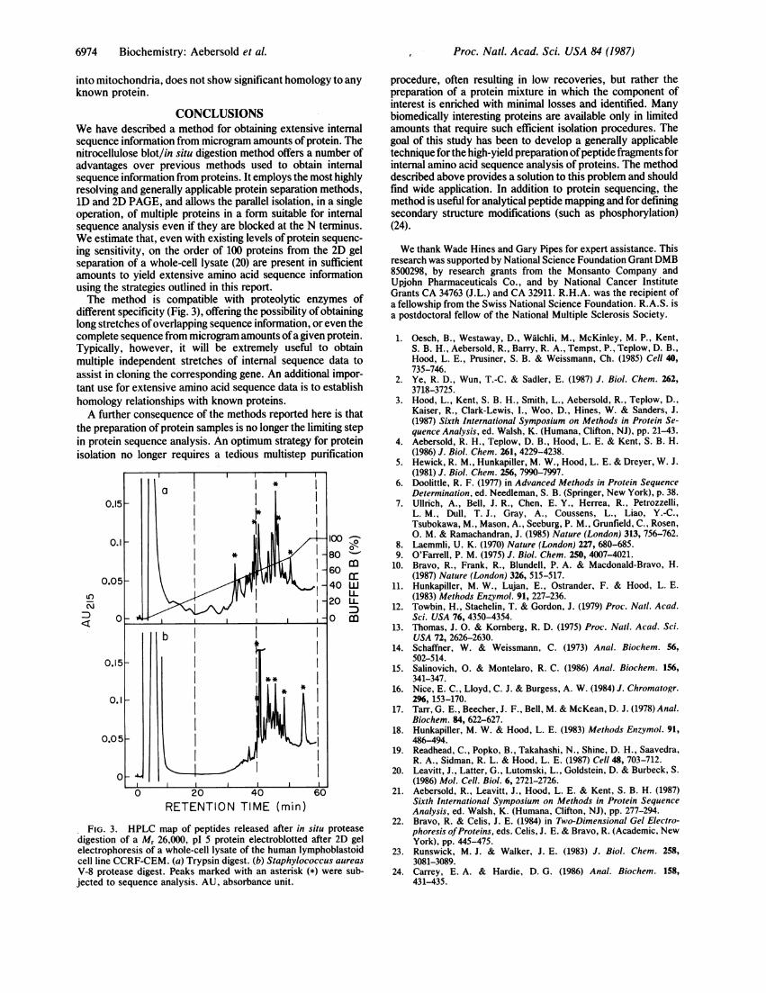
different specificity (Fig. 3), offering the possibility of obtaininglong stretches ofoverlapping sequence information, or even thecomplete sequence from microgram amounts ofa given protein.Typically, however, it will be extremely useful to obtainmultiple independent stretches of internal sequence data toassist in cloning the corresponding gene. An additional impor-tant use for extensive amino acid sequence data is to establishhomology relationships with known proteins.A further consequence of the methods reported here is that
the preparation of protein samples is no longer the limiting stepin protein sequence analysis. An optimum strategy for proteinisolation no longer requires a tedious multistep purification
LC
:D
0o<
m
wLLmLmD
RETENTION TIME (min)
FIG. 3. HPLC map of peptides released after in situ proteasedigestion of a M, 26,000, pl 5 protein electroblotted after 2D gelelectrophoresis of a whole-cell lysate of the human lymphoblastoidcell line CCRF-CEM. (a) Trypsin digest. (b) Staphylococcus aureas
V-8 protease digest. Peaks marked with an asterisk (*) were sub-jected to sequence analysis. AU, absorbance unit.
procedure, often resulting in low recoveries, but rather thepreparation of a protein mixture in which the component ofinterest is enriched with minimal losses and identified. Manybiomedically interesting proteins are available only in limitedamounts that require such efficient isolation procedures. Thegoal of this study has been to develop a generally applicabletechnique for the high-yield preparation ofpeptide fragments forinternal amino acid sequence analysis of proteins. The methoddescribed above provides a solution to this problem and shouldfind wide application. In addition to protein sequencing, themethod is useful for analytical peptide mapping and for definingsecondary structure modifications (such as phosphorylation)(24).
We thank Wade Hines and Gary Pipes for expert assistance. Thisresearch was supported by National Science Foundation Grant DMB8500298, by research grants from the Monsanto Company andUpjohn Pharmaceuticals Co., and by National Cancer InstituteGrants CA 34763 (J.L.) and CA 32911. R.H.A. was the recipient ofa fellowship from the Swiss National Science Foundation. R.A.S. isa postdoctoral fellow of the National Multiple Sclerosis Society.
1. Oesch, B., Westaway, D., Walchli, M., McKinley, M. P., Kent,S. B. H., Aebersold, R., Barry, R. A., Tempst, P., Teplow, D. B.,Hood, L. E., Prusiner, S. B. & Weissmann, Ch. (1985) Cell 40,735-746.
2. Ye, R. D., Wun, T.-C. & Sadler, E. (1987) J. Biol. Chem. 262,3718-3725.
3. Hood, L., Kent, S. B. H., Smith, L., Aebersold, R., Teplow, D.,Kaiser, R., Clark-Lewis, I., Woo, D., Hines, W. & Sanders, J.(1987) Sixth International Symposium on Methods in Protein Se-quence Analysis, ed. Walsh, K. (Humana, Clifton, NJ), pp. 21-43.
4. Aebersold, R. H., Teplow, D. B., Hood, L. E. & Kent, S. B. H.(1986) J. Biol. Chem. 261, 4229-4238.
5. Hewick, R. M., Hunkapiller, M. W., Hood, L. E. & Dreyer, W. J.(1981) J. Biol. Chem. 256, 7990-7997.
6. Doolittle, R. F. (1977) in Advanced Methods in Protein SequenceDetermination, ed. Needleman, S. B. (Springer, New York), p. 38.
7. Ullrich, A., Bell, J. R., Chen, E. Y., Herrea, R., Petrozzelli,L. M., Dull, T. J., Gray, A., Coussens, L., Liao, Y.-C.,Tsubokawa, M., Mason, A., Seeburg, P. M., Grunfield, C., Rosen,0. M. & Ramachandran, J. (1985) Nature (London) 313, 756-762.
8. Laemmli, U. K. (1970) Nature (London) 227, 680-685.9. O'Farrell, P. M. (1975) J. Biol. Chem. 250, 4007-4021.
10. Bravo, R., Frank, R., Blundell, P. A. & Macdonald-Bravo, H.(1987) Nature (London) 326, 515-517.
11. Hunkapiller, M. W., Lujan, E., Ostrander, F. & Hood, L. E.(1983) Methods Enzymol. 91, 227-236.
12. Towbin, H., Staehelin, T. & Gordon, J. (1979) Proc. NatI. Acad.Sci. USA 76, 4350-4354.
13. Thomas, J. 0. & Kornberg, R. D. (1975) Proc. NatI. Acad. Sci.USA 72, 2626-2630.
14. Schaffner, W. & Weissmann, C. (1973) Anal. Biochem. 56,502-514.
15. Salinovich, 0. & Montelaro, R. C. (1986) Anal. Biochem. 156,341-347.
16. Nice, E. C., Lloyd, C. J. & Burgess, A. W. (1984) J. Chromatogr.296, 153-170.
17. Tarr, G. E., Beecher, J. F., Bell, M. & McKean, D. J. (1978) Anal.Biochem. 84, 622-627.
18. Hunkapiller, M. W. & Hood, L. E. (1983) Methods Enzymol. 91,486-494.
19. Readhead, C., Popko, B., Takahashi, N., Shine, D. H., Saavedra,R. A., Sidman, R. L. & Hood, L. E. (1987) Cell 48, 703-712.
20. Leavitt, J., Latter, G., Lutomski, L., Goldstein, D. & Burbeck, S.(1986) Mol. Cell. Biol. 6, 2721-2726.
21. Aebersold, R., Leavitt, J., Hood, L. E. & Kent, S. B. H. (1987)Sixth International Symposium on Methods in Protein SequenceAnalysis, ed. Walsh, K. (Humana, Clifton, NJ), pp. 277-294.
22. Bravo, R. & Celis, J. E. (1984) in Two-Dimensional Gel Electro-phoresis ofProteins, eds. Celis, J. E. & Bravo, R. (Academic, NewYork), pp. 445-475.
23. Runswick, M. J. & Walker, J. E. (1983) J. Biol. Chem. 258,3081-3089.
24. Carrey, E. A. & Hardie, D. G. (1986) Anal. Biochem. 158,431-435.
Proc. Natl. Acad. Sci. USA 84 (1987)