Interleukin-6 as a Photoreceptor Neuroprotectant in an … Interleukin-6 as a Photoreceptor...
8
Interleukin-6 as a Photoreceptor Neuroprotectant in an Experimental Model of Retinal Detachment Deborah Y. Chong, Christopher S. Boehlke, Qiong-Duan Zheng, Linda Zhang, Ying Han, and David N. Zacks PURPOSE. To test the hypothesis that interleukin (IL)-6 prevents photoreceptor cell death during periods of retinal separation from the retinal pigment epithelium (RPE). METHODS. Retinal–RPE separation was created in wild-type C57BL mice, IL-6 / mice, and Brown Norway rats by subreti- nal injection of 1% hyaluronic acid. In some animals, anti–IL-6 neutralizing antibody (NAB) or exogenous IL-6 was adminis- tered into the subretinal space at the time of separation or at specified times afterward. Terminal deoxynucleotidyl trans- ferase dUTP nick-end labeling (TUNEL) was performed 3 days after separation to detect apoptotic photoreceptors. Photore- ceptor cell counts were performed after 1 and 2 months. RESULTS. Loss of IL-6 function through genetic ablation (IL-6 / mice) or injection of anti–IL-6 NAB resulted in significantly increased levels of TUNEL-positive staining 3 days after retinal– RPE separation. One month after separation, outer nuclear layer (ONL) cell counts were significantly lower in IL-6 / mice or in animals injected with anti–IL-6 NAB than in controls. Gain of IL-6 function through the addition of exogenous IL-6 resulted in significantly increased ONL counts at 1 month but not at 2 months. Reinjection of IL-6 at 1 month led to contin- ued preservation of ONL counts compared with controls. A window of opportunity for treatment was detected because delaying injection of exogenous IL-6 to 2 weeks after retinal– RPE separation still resulted in significantly greater ONL cell counts compared with controls. CONCLUSIONS. IL-6 may serve as a photoreceptor neuropro- tectant in the setting of retinal–RPE separation. (Invest Oph- thalmol Vis Sci. 2008;49:3193–3200) DOI:10.1167/iovs.07-1641 R etinal detachment (RD), defined as the separation of the neurosensory retina from subjacent retinal pigment epithe- lium (RPE), results in the apoptotic death of photoreceptor cells. 1–4 Rodent and feline models of RD have demonstrated the activation of proapoptotic pathways nearly immediately after the retina becomes separated from the RPE. 1–4 Histologic markers of apoptosis, such as terminal deoxynucleotidyl trans- ferase nick-end label (TUNEL) staining, reach a peak at approx- imately 3 days after RD, with apoptotic activity and progressive cell death persisting for the duration of the detachment period. Clinical experience in the repair of retinal detachments, how- ever, has demonstrated that there is a window of opportunity for repair with preservation of good visual acuity. Retrospec- tive case series have demonstrated that significant numbers of patients with macula-off RDs repaired within 5 to 10 days after onset of detachment can retain relatively good visual function but that visual acuity drops significantly as the time between detachment and repair extends. 5–7 The delayed time between the activation of proapoptosis pathways and the clinical onset of visual loss suggests that intrinsic neuroprotective factors may become activated within the neural retina and may serve to counterbalance the effects of the proapoptotic pathways activated by retinal–RPE separation. Previous work in our laboratory using gene microarray analysis of experimental detachment in rats revealed the in- creased expression of genes involved in stress–response path- ways. 8 Of particular interest was the increased transcription and translation of interleukin (IL)-6 and downstream com- ponents of its associated signal transduction pathway. IL-6 is a pleiotropic cytokine with a role in inflammation, hemato- poiesis, angiogenesis, cell differentiation, and neuronal sur- vival. 9–11 In the central nervous system, IL-6 is synthesized by microglia, astrocytes, and neurons. 12,13 In the retina, IL-6 is synthesized by Mu ¨ller cells and the RPE. 14,15 A neuroprotective role for IL-6 has been previously suggested in different models of ocular injury. In rat models of retinal ischemia–reperfusion injury, IL-6 protein levels are upregulated within 8 hours after injury. 16 Furthermore, intravitreal injection of exogenous IL-6 immediately after ischemia–reperfusion injury or before N- methyl-D-aspartate (NMDA)-induced toxicity increases survival of retinal ganglion cells. 16,17 In vitro studies have shown that IL-6 increases the duration of rat retinal ganglion cell survival in primary tissue culture. 18,19 Collectively, these data suggest that IL-6 upregulation after injury may serve to function as a neu- ronal survival factor. The goal of this study is to test the hypothesis that IL-6 func- tions as an inhibitor of photoreceptor apoptosis after RD. Under- standing the effect of IL-6 on photoreceptor survival may provide insight into potential therapeutic strategies for protecting photo- receptors during periods of photoreceptor–RPE separation. METHODS Experimental Model of Retinal Detachment All experiments were performed in accordance with the ARVO State- ment for the Use of Animals in Ophthalmic and Vision Research and the guidelines established by the University Committee on Use and Care of Animals of the University of Michigan. Detachments were created in adult male Brown-Norway rats (300 – 400 g; Charles River Laboratories, Wilmington, MA), wild-type C57BL mice (age 3– 6 weeks; Jackson Laboratory, Bar Harbor, ME), and IL-6 / mice on a C57BL background (age 3– 6 weeks; Jackson Laboratory), as previously de- scribed. 20 Briefly, rodents were anesthetized with a 50:50 mix of ketamine (100 mg/mL) and xylazine (20 mg/mL), and pupils were dilated with topical phenylephrine (2.5%) and tropicamide (1%). A From the Department of Ophthalmology and Visual Sciences, W. K. Kellogg Eye Center, University of Michigan, Ann Arbor, Michigan. Supported by National Eye Institute Grant K08-124-14705 (DNZ), the International Retinal Research Foundation, Inc. (DNZ), Research to Prevent Blindness, Inc. (DNZ), and Departmental Core Center for Vision Research Grant EY-07003. Submitted for publication December 20, 2007; revised March 7, 2008; accepted May 14, 2008. Disclosure: D.Y. Chong, None; C.S. Boehlke, None; Q.-D. Zheng, None; L. Zhang, None; Y. Han, None; D.N. Zacks, None The publication costs of this article were defrayed in part by page charge payment. This article must therefore be marked “advertise- ment” in accordance with 18 U.S.C. §1734 solely to indicate this fact. Corresponding author: David N. Zacks, Department of Ophthal- mology and Visual Sciences, W. K. Kellogg Eye Center, 1000 Wall Street, Ann Arbor, MI 48105-0714; [email protected]. Investigative Ophthalmology & Visual Science, July 2008, Vol. 49, No. 7 Copyright © Association for Research in Vision and Ophthalmology 3193 Downloaded from iovs.arvojournals.org on 07/23/2019
Transcript of Interleukin-6 as a Photoreceptor Neuroprotectant in an … Interleukin-6 as a Photoreceptor...
Interleukin-6 as a Photoreceptor Neuroprotectant in anExperimental Model of Retinal Detachment
Deborah Y. Chong, Christopher S. Boehlke, Qiong-Duan Zheng, Linda Zhang,Ying Han, and David N. Zacks
PURPOSE. To test the hypothesis that interleukin (IL)-6 preventsphotoreceptor cell death during periods of retinal separationfrom the retinal pigment epithelium (RPE).
METHODS. Retinal–RPE separation was created in wild-typeC57BL mice, IL-6�/� mice, and Brown Norway rats by subreti-nal injection of 1% hyaluronic acid. In some animals, anti–IL-6neutralizing antibody (NAB) or exogenous IL-6 was adminis-tered into the subretinal space at the time of separation or atspecified times afterward. Terminal deoxynucleotidyl trans-ferase dUTP nick-end labeling (TUNEL) was performed 3 daysafter separation to detect apoptotic photoreceptors. Photore-ceptor cell counts were performed after 1 and 2 months.
RESULTS. Loss of IL-6 function through genetic ablation (IL-6�/�
mice) or injection of anti–IL-6 NAB resulted in significantlyincreased levels of TUNEL-positive staining 3 days after retinal–RPE separation. One month after separation, outer nuclearlayer (ONL) cell counts were significantly lower in IL-6�/�
mice or in animals injected with anti–IL-6 NAB than in controls.Gain of IL-6 function through the addition of exogenous IL-6resulted in significantly increased ONL counts at 1 month butnot at 2 months. Reinjection of IL-6 at 1 month led to contin-ued preservation of ONL counts compared with controls. Awindow of opportunity for treatment was detected becausedelaying injection of exogenous IL-6 to 2 weeks after retinal–RPE separation still resulted in significantly greater ONL cellcounts compared with controls.
CONCLUSIONS. IL-6 may serve as a photoreceptor neuropro-tectant in the setting of retinal–RPE separation. (Invest Oph-thalmol Vis Sci. 2008;49:3193–3200) DOI:10.1167/iovs.07-1641
Retinal detachment (RD), defined as the separation of theneurosensory retina from subjacent retinal pigment epithe-
lium (RPE), results in the apoptotic death of photoreceptorcells.1–4 Rodent and feline models of RD have demonstratedthe activation of proapoptotic pathways nearly immediatelyafter the retina becomes separated from the RPE.1–4 Histologicmarkers of apoptosis, such as terminal deoxynucleotidyl trans-ferase nick-end label (TUNEL) staining, reach a peak at approx-imately 3 days after RD, with apoptotic activity and progressive
cell death persisting for the duration of the detachment period.Clinical experience in the repair of retinal detachments, how-ever, has demonstrated that there is a window of opportunityfor repair with preservation of good visual acuity. Retrospec-tive case series have demonstrated that significant numbers ofpatients with macula-off RDs repaired within 5 to 10 days afteronset of detachment can retain relatively good visual functionbut that visual acuity drops significantly as the time betweendetachment and repair extends.5–7 The delayed time betweenthe activation of proapoptosis pathways and the clinical onsetof visual loss suggests that intrinsic neuroprotective factorsmay become activated within the neural retina and may serveto counterbalance the effects of the proapoptotic pathwaysactivated by retinal–RPE separation.
Previous work in our laboratory using gene microarrayanalysis of experimental detachment in rats revealed the in-creased expression of genes involved in stress–response path-ways.8 Of particular interest was the increased transcriptionand translation of interleukin (IL)-6 and downstream com-ponents of its associated signal transduction pathway. IL-6 isa pleiotropic cytokine with a role in inflammation, hemato-poiesis, angiogenesis, cell differentiation, and neuronal sur-vival.9 –11 In the central nervous system, IL-6 is synthesized bymicroglia, astrocytes, and neurons.12,13 In the retina, IL-6 issynthesized by Muller cells and the RPE.14,15 A neuroprotectiverole for IL-6 has been previously suggested in different modelsof ocular injury. In rat models of retinal ischemia–reperfusioninjury, IL-6 protein levels are upregulated within 8 hours afterinjury.16 Furthermore, intravitreal injection of exogenous IL-6immediately after ischemia–reperfusion injury or before N-methyl-D-aspartate (NMDA)-induced toxicity increases survivalof retinal ganglion cells.16,17 In vitro studies have shown thatIL-6 increases the duration of rat retinal ganglion cell survival inprimary tissue culture.18,19 Collectively, these data suggest thatIL-6 upregulation after injury may serve to function as a neu-ronal survival factor.
The goal of this study is to test the hypothesis that IL-6 func-tions as an inhibitor of photoreceptor apoptosis after RD. Under-standing the effect of IL-6 on photoreceptor survival may provideinsight into potential therapeutic strategies for protecting photo-receptors during periods of photoreceptor–RPE separation.
METHODS
Experimental Model of Retinal Detachment
All experiments were performed in accordance with the ARVO State-ment for the Use of Animals in Ophthalmic and Vision Research andthe guidelines established by the University Committee on Use andCare of Animals of the University of Michigan. Detachments werecreated in adult male Brown-Norway rats (300–400 g; Charles RiverLaboratories, Wilmington, MA), wild-type C57BL mice (age 3–6 weeks;Jackson Laboratory, Bar Harbor, ME), and IL-6�/� mice on a C57BLbackground (age 3–6 weeks; Jackson Laboratory), as previously de-scribed.20 Briefly, rodents were anesthetized with a 50:50 mix ofketamine (100 mg/mL) and xylazine (20 mg/mL), and pupils weredilated with topical phenylephrine (2.5%) and tropicamide (1%). A
From the Department of Ophthalmology and Visual Sciences,W. K. Kellogg Eye Center, University of Michigan, Ann Arbor,Michigan.
Supported by National Eye Institute Grant K08-124-14705 (DNZ),the International Retinal Research Foundation, Inc. (DNZ), Research toPrevent Blindness, Inc. (DNZ), and Departmental Core Center forVision Research Grant EY-07003.
Submitted for publication December 20, 2007; revised March 7,2008; accepted May 14, 2008.
Disclosure: D.Y. Chong, None; C.S. Boehlke, None; Q.-D.Zheng, None; L. Zhang, None; Y. Han, None; D.N. Zacks, None
The publication costs of this article were defrayed in part by pagecharge payment. This article must therefore be marked “advertise-ment” in accordance with 18 U.S.C. §1734 solely to indicate this fact.
Corresponding author: David N. Zacks, Department of Ophthal-mology and Visual Sciences, W. K. Kellogg Eye Center, 1000 WallStreet, Ann Arbor, MI 48105-0714; [email protected].
Investigative Ophthalmology & Visual Science, July 2008, Vol. 49, No. 7Copyright © Association for Research in Vision and Ophthalmology 3193
Downloaded from iovs.arvojournals.org on 07/23/2019
20-gauge microvitreoretinal blade (Walcott Scientific, Marmora, NJ)was used to create a sclerotomy 2 mm posterior to the limbus, carefullyavoiding lens damage. A Glaser subretinal injector (32-gauge tip; BDOphthalmic Systems, Sarasota, FL) was introduced through the scle-rotomy into the vitreous cavity and then through a peripheral retino-tomy into the subretinal space. Sodium hyaluronate (10 mg/mL; Phar-macia and Upjohn Co., Kalamazoo, MI) was slowly injected to detachthe neurosensory retina from the underlying RPE. In all experiments,approximately one third to one half of the neurosensory retina wasdetached. Detachments were created in the left eye, leaving the righteye as the control. In some eyes, either 0.15 �g anti–human IL-6neutralizing antibody (NAB; R&D Systems, Minneapolis, MN) or 15 ngexogenous human IL-6 (R&D Systems) was injected into the subretinalspace of the detachment in a 10-�L volume at the time of creation ofthe detachment or at a subsequent time point. Doses were based onmanufacturers’ recommendations based on in vitro activity assays.
Western Blot Analysis
Retinas from experimental eyes with detachments and control eyeswithout detachments were dissected from the RPE–choroid at 3 daysafter retinal detachment, homogenized, and lysed in buffer containing10 mM HEPES (pH 7.6), 0.5% IgEPal, 42 mM KCl, 1 mM phenylmeth-ylsulfonyl fluoride (PMSF), 1 mM EDTA, 1 mM EGTA, 1 mM dithiothre-itol (DTT), 5 mM MgCl2, and 1 tablet of protease inhibitors per 10 mLbuffer (Complete Mini; Roche Diagnostics GmbH, Mannheim, Ger-many). The homogenates were incubated on ice and centrifuged at22,000g at 4°C for 60 minutes. The protein concentration of thesupernatant was then determined (DC Protein Assay kit; Bio-Rad Lab-oratories, Hercules CA). Protein samples were loaded and run onSDS-polyacrylamide gels (Tris-HCl Ready Gels; Bio-Rad Laboratories).After electrophoretic separation, the proteins were transferred ontopolyvinylidene fluoride (PVDF) membranes (Immobilon-P; AmershamPharmacia Biotech, Piscataway, NJ). Protein bands were visualizedwith Ponceau S staining, and the lanes were assessed for equal loadingby densitometry of a nonspecific band present across all lanes. Mem-branes were then immunoblotted for phospho-STAT1 (Tyr701) orphospho-STAT3 (Tyr705) using an immunoblotting kit (PhosphoPlusStat1 [Tyr701] Antibody Kit 9170 or PhosphoPlus Stat3 [Tyr705] An-tibody Kit 9130, respectively; Cell Signaling Technology, Danvers, MA)according to the manufacturer’s instructions using a 1:1000 dilution ofthe primary antibody.
TUNEL Staining and Histology
At varying intervals after creation of the detachment, the animals werehumanely killed, and the eyes were enucleated. For TUNEL staining,whole eyes were fixed overnight at 4°C in phosphate-buffered salinewith 4% paraformaldehyde (pH 7.4). The specimens were embeddedin paraffin and sectioned at a thickness of 5 to 6 �m. TUNEL stainingwas performed on the sections with a fluorescein in situ apoptosisdetection kit (ApopTag; Millipore, Billerica, MA) according to themanufacturer’s instructions. For light microscopic analysis, paraffinsections were stained with 0.5% toluidine blue in 0.1% borate buffer.
Data Analysis
Photoreceptor cell apoptosis was quantified as the percentage of totalcells in the outer nuclear layer (ONL) that was TUNEL positive. Threenonoverlapping high-power fields (40�) at the maximal height of theRD were selected per section and were averaged unless there werefewer than three nonoverlapping high-power fields, in which casefewer fields were used. One representative section was used per eye.The total number of cells in the ONL was measured in a similar fashion.The total thickness of the retina (measured from the outer edge of theONL to the inner limiting membrane) was measured in three places ineach of three nonoverlapping high-power fields (40�) at the maximalheight of the RD per section and was averaged for each eye. Photore-ceptor inner and outer segments were not included in the total retinalthickness measurement given the variable retraction of these elements
after detachment of the neurosensory retina, which does not neces-sarily correlate with post-reattachment viability of the photorecep-tors.21,22 For toluidine blue–stained specimens, normalization of ONLcell count to the total retinal thickness of each section (i.e., ONL cellcount divided by total retinal thickness) was performed to account forpossible differences in angles of sectioning and to allow for inter-sample comparison. ONL cell counts and total retinal thicknesses ineach group of the rat experiments were also normalized to correspond-ing values of attached retinas in that group to allow intersamplecomparison. For each experimental group, measurements were per-formed on three sections from four to 11 eyes, each eye from aseparate animal.
Statistical analysis comparing percentages of TUNEL-positive cellsin the ONL between groups and comparing the ONL cell count/totalretinal thickness ratio between groups was performed using two-tailedStudent’s t-tests without assuming equal variance. Differences wereconsidered significant at P � 0.05.
RESULTS
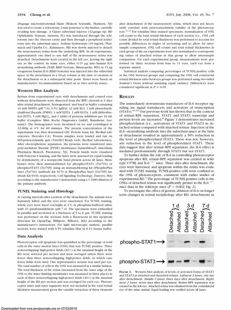
The immediately downstream transducers of IL-6 receptor sig-naling are signal transducers and activators of transcription(STATs).10,23 Our previous work has shown that in the contextof retinal–RPE separation, STAT1 and STAT3 transcript andprotein levels are increased.8 Figure 1 demonstrates increasedphosphorylation (i.e., activation) of STAT1 and STAT3 in de-tached retinas compared with attached retinas. Injection of theIL-6–neutralizing antibody into the subretinal space at the timeof detachment resulted in approximately a 50% reduction inthe level of phosphorylated STAT3. There was not, however,any reduction in the level of phosphorylated STAT1. Thesedata suggest that after retinal–RPE separation, the IL-6 effect ismediated predominantly through STAT3 but not STAT1.
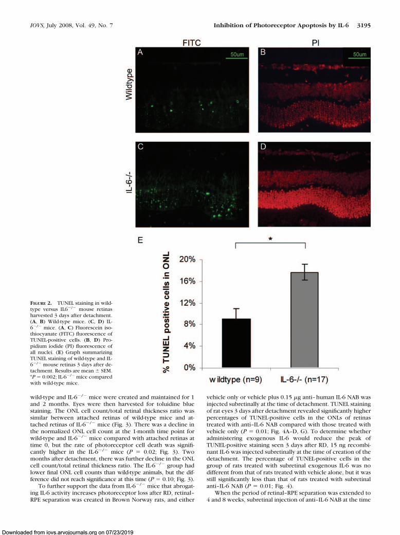
To further define the role of IL-6 in controlling photoreceptorapoptosis after RD, retinal–RPE separation was created in wild-type C57BL and IL-6�/� mice. Three days after detachment, theeyes were harvested, and apoptosis within the retina was evalu-ated with TUNEL staining. TUNEL-positive cells were confined tothe ONL of photoreceptors, consistent with earlier studies ofexperimental RD.3 The percentage of TUNEL-positive cells in theONLs of detached retinas was significantly greater in the IL-6�/�
mice than in the wild-type mice (P � 0.002; Fig. 2).To investigate the effect of genetic ablation of IL-6 on longer
term changes in retinal morphology after RD, detachments in
FIGURE 1. Western blot analysis of levels of activated forms of STAT1and STAT3 in attached and detached retinas. Leftmost 2 lanes: one dayafter detachment. Middle 2 lanes: three days after detachment. Right-most 2 lanes: seven days after detachment. Retina–RPE separation wascreated in the left eye. Attached retina was obtained from the contralateraleye of the same animal. Equal loading was verified across all lanes.
3194 Chong et al. IOVS, July 2008, Vol. 49, No. 7
Downloaded from iovs.arvojournals.org on 07/23/2019
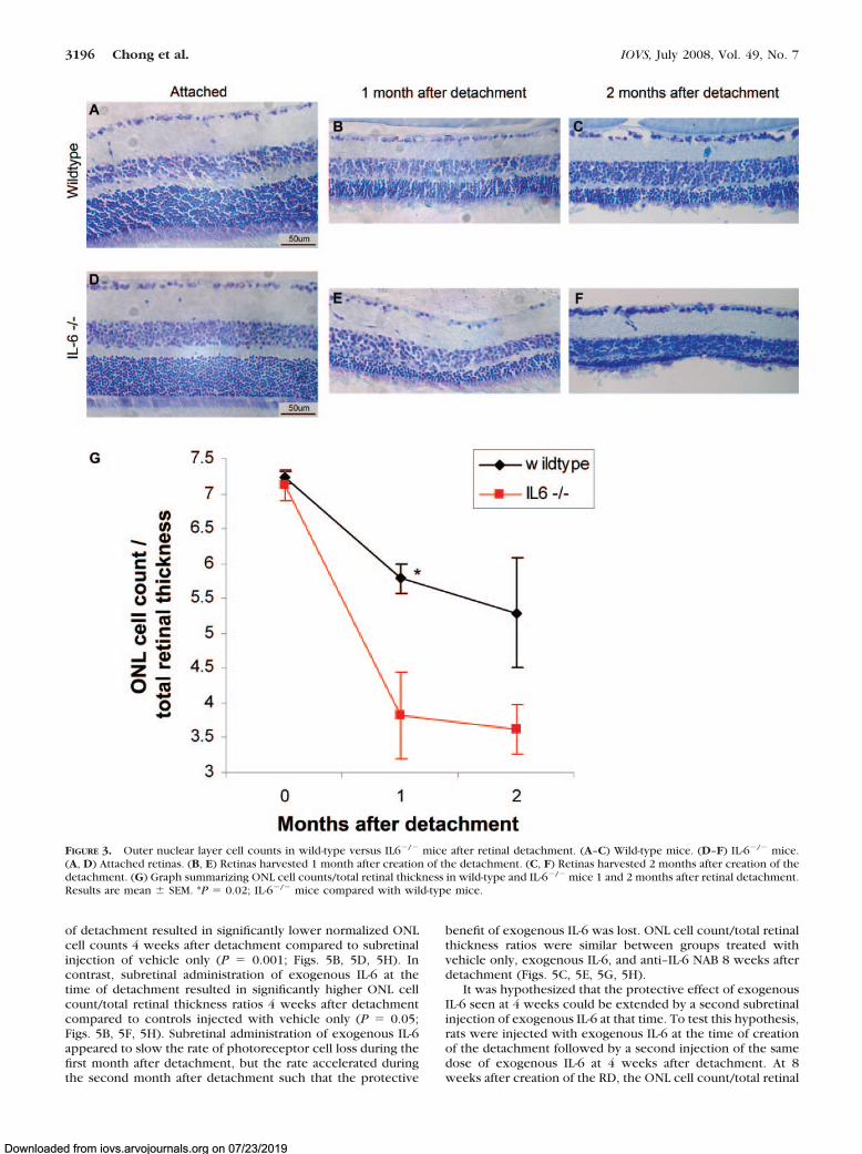
wild-type and IL-6�/� mice were created and maintained for 1and 2 months. Eyes were then harvested for toluidine bluestaining. The ONL cell count/total retinal thickness ratio wassimilar between attached retinas of wild-type mice and at-tached retinas of IL-6�/� mice (Fig. 3). There was a decline inthe normalized ONL cell count at the 1-month time point forwild-type and IL-6�/� mice compared with attached retinas attime 0, but the rate of photoreceptor cell death was signifi-cantly higher in the IL-6�/� mice (P � 0.02; Fig. 3). Twomonths after detachment, there was further decline in the ONLcell count/total retinal thickness ratio. The IL-6�/� group hadlower final ONL cell counts than wild-type animals, but the dif-ference did not reach significance at this time (P � 0.10; Fig. 3).
To further support the data from IL-6�/� mice that abrogat-ing IL-6 activity increases photoreceptor loss after RD, retinal–RPE separation was created in Brown Norway rats, and either
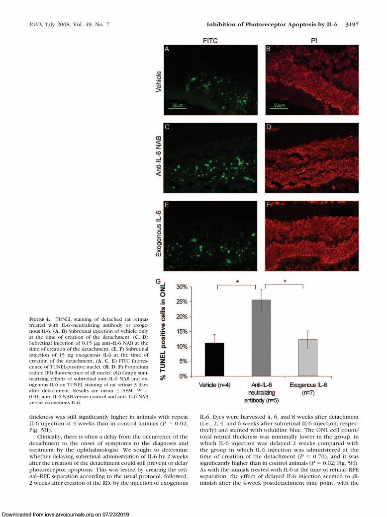
vehicle only or vehicle plus 0.15 �g anti–human IL-6 NAB wasinjected subretinally at the time of detachment. TUNEL stainingof rat eyes 3 days after detachment revealed significantly higherpercentages of TUNEL-positive cells in the ONLs of retinastreated with anti–IL-6 NAB compared with those treated withvehicle only (P � 0.01; Fig. 4A–D, G). To determine whetheradministering exogenous IL-6 would reduce the peak ofTUNEL-positive staining seen 3 days after RD, 15 ng recombi-nant IL-6 was injected subretinally at the time of creation of thedetachment. The percentage of TUNEL-positive cells in thegroup of rats treated with subretinal exogenous IL-6 was nodifferent from that of rats treated with vehicle alone, but it wasstill significantly less than that of rats treated with subretinalanti–IL-6 NAB (P � 0.01; Fig. 4).
When the period of retinal–RPE separation was extended to4 and 8 weeks, subretinal injection of anti–IL-6 NAB at the time
FIGURE 2. TUNEL staining in wild-type versus IL6�/� mouse retinasharvested 3 days after detachment.(A, B) Wild-type mice. (C, D) IL-6�/� mice. (A, C) Fluorescein iso-thiocyanate (FITC) fluorescence ofTUNEL-positive cells. (B, D) Pro-pidium iodide (PI) fluorescence ofall nuclei. (E) Graph summarizingTUNEL staining of wild-type and IL-6�/� mouse retinas 3 days after de-tachment. Results are mean � SEM.*P � 0.002; IL-6�/� mice comparedwith wild-type mice.
IOVS, July 2008, Vol. 49, No. 7 Inhibition of Photoreceptor Apoptosis by IL-6 3195
Downloaded from iovs.arvojournals.org on 07/23/2019
of detachment resulted in significantly lower normalized ONLcell counts 4 weeks after detachment compared to subretinalinjection of vehicle only (P � 0.001; Figs. 5B, 5D, 5H). Incontrast, subretinal administration of exogenous IL-6 at thetime of detachment resulted in significantly higher ONL cellcount/total retinal thickness ratios 4 weeks after detachmentcompared to controls injected with vehicle only (P � 0.05;Figs. 5B, 5F, 5H). Subretinal administration of exogenous IL-6appeared to slow the rate of photoreceptor cell loss during thefirst month after detachment, but the rate accelerated duringthe second month after detachment such that the protective
benefit of exogenous IL-6 was lost. ONL cell count/total retinalthickness ratios were similar between groups treated withvehicle only, exogenous IL-6, and anti–IL-6 NAB 8 weeks afterdetachment (Figs. 5C, 5E, 5G, 5H).
It was hypothesized that the protective effect of exogenousIL-6 seen at 4 weeks could be extended by a second subretinalinjection of exogenous IL-6 at that time. To test this hypothesis,rats were injected with exogenous IL-6 at the time of creationof the detachment followed by a second injection of the samedose of exogenous IL-6 at 4 weeks after detachment. At 8weeks after creation of the RD, the ONL cell count/total retinal
FIGURE 3. Outer nuclear layer cell counts in wild-type versus IL6�/� mice after retinal detachment. (A–C) Wild-type mice. (D–F) IL-6�/� mice.(A, D) Attached retinas. (B, E) Retinas harvested 1 month after creation of the detachment. (C, F) Retinas harvested 2 months after creation of thedetachment. (G) Graph summarizing ONL cell counts/total retinal thickness in wild-type and IL-6�/� mice 1 and 2 months after retinal detachment.Results are mean � SEM. *P � 0.02; IL-6�/� mice compared with wild-type mice.
3196 Chong et al. IOVS, July 2008, Vol. 49, No. 7
Downloaded from iovs.arvojournals.org on 07/23/2019
thickness was still significantly higher in animals with repeatIL-6 injection at 4 weeks than in control animals (P � 0.02;Fig. 5H).
Clinically, there is often a delay from the occurrence of thedetachment to the onset of symptoms to the diagnosis andtreatment by the ophthalmologist. We sought to determinewhether delaying subretinal administration of IL-6 by 2 weeksafter the creation of the detachment could still prevent or delayphotoreceptor apoptosis. This was tested by creating the reti-nal–RPE separation according to the usual protocol, followed,2 weeks after creation of the RD, by the injection of exogenous
IL-6. Eyes were harvested 4, 6, and 8 weeks after detachment(i.e., 2, 4, and 6 weeks after subretinal IL-6 injection, respec-tively) and stained with toluidine blue. The ONL cell count/total retinal thickness was minimally lower in the group, inwhich IL-6 injection was delayed 2 weeks compared withthe group in which IL-6 injection was administered at thetime of creation of the detachment (P � 0.70), and it wassignificantly higher than in control animals (P � 0.02; Fig. 5H).As with the animals treated with IL-6 at the time of retinal–RPEseparation, the effect of delayed IL-6 injection seemed to di-minish after the 4-week postdetachment time point, with the
FIGURE 4. TUNEL staining of detached rat retinastreated with IL-6–neutralizing antibody or exoge-nous IL-6. (A, B) Subretinal injection of vehicle onlyat the time of creation of the detachment. (C, D)Subretinal injection of 0.15 �g anti–IL-6 NAB at thetime of creation of the detachment. (E, F) Subretinalinjection of 15 ng exogenous IL-6 at the time ofcreation of the detachment. (A, C, E) FITC fluores-cence of TUNEL-positive nuclei. (B, D, F) Propidiumiodide (PI) fluorescence of all nuclei. (G) Graph sum-marizing effects of subretinal anti–IL-6 NAB and ex-ogenous IL-6 on TUNEL staining of rat retinas 3 daysafter detachment. Results are mean � SEM. *P �0.01; anti–IL-6 NAB versus control and anti–IL-6 NABversus exogenous IL-6.
IOVS, July 2008, Vol. 49, No. 7 Inhibition of Photoreceptor Apoptosis by IL-6 3197
Downloaded from iovs.arvojournals.org on 07/23/2019
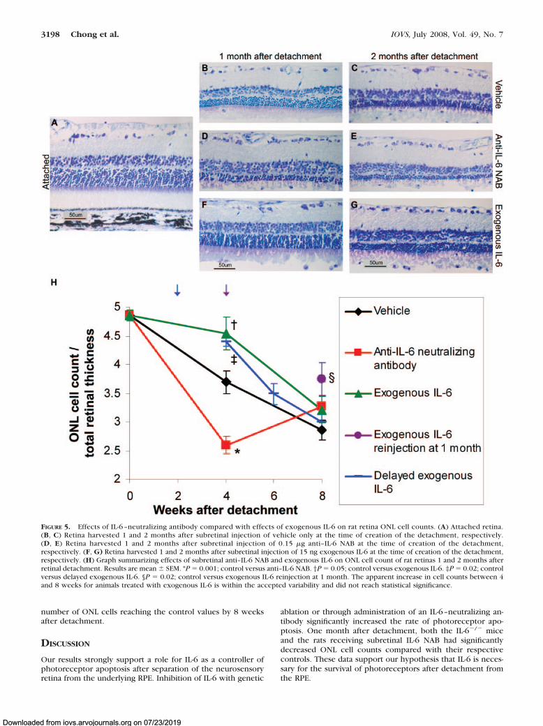
number of ONL cells reaching the control values by 8 weeksafter detachment.
DISCUSSION
Our results strongly support a role for IL-6 as a controller ofphotoreceptor apoptosis after separation of the neurosensoryretina from the underlying RPE. Inhibition of IL-6 with genetic
ablation or through administration of an IL-6–neutralizing an-tibody significantly increased the rate of photoreceptor apo-ptosis. One month after detachment, both the IL-6�/� miceand the rats receiving subretinal IL-6 NAB had significantlydecreased ONL cell counts compared with their respectivecontrols. These data support our hypothesis that IL-6 is neces-sary for the survival of photoreceptors after detachment fromthe RPE.
FIGURE 5. Effects of IL-6 –neutralizing antibody compared with effects of exogenous IL-6 on rat retina ONL cell counts. (A) Attached retina.(B, C) Retina harvested 1 and 2 months after subretinal injection of vehicle only at the time of creation of the detachment, respectively.(D, E) Retina harvested 1 and 2 months after subretinal injection of 0.15 �g anti–IL-6 NAB at the time of creation of the detachment,respectively. (F, G) Retina harvested 1 and 2 months after subretinal injection of 15 ng exogenous IL-6 at the time of creation of the detachment,respectively. (H) Graph summarizing effects of subretinal anti–IL-6 NAB and exogenous IL-6 on ONL cell count of rat retinas 1 and 2 months afterretinal detachment. Results are mean � SEM. *P � 0.001; control versus anti–IL-6 NAB. †P � 0.05; control versus exogenous IL-6. ‡P � 0.02; controlversus delayed exogenous IL-6. §P � 0.02; control versus exogenous IL-6 reinjection at 1 month. The apparent increase in cell counts between 4and 8 weeks for animals treated with exogenous IL-6 is within the accepted variability and did not reach statistical significance.
3198 Chong et al. IOVS, July 2008, Vol. 49, No. 7
Downloaded from iovs.arvojournals.org on 07/23/2019
Experiments looking at the effect of increasing IL-6 levelshad mixed effects, depending on the time point examined andthe assay measured. When compared with injection of vehicleonly, subretinal injection of exogenous IL-6 did not signifi-cantly affect the percentage of TUNEL-positive cells 3 daysafter RD but did significantly increase the number of photore-ceptors that survived longer term detachments. A possibleexplanation for this difference is that the dose of exogenousIL-6 might have been insufficient to prevent apoptosis 3 daysafter creation of the detachment, but RD may cause surround-ing cells, such as Muller cells and RPE cells, to express IL-6 ina delayed fashion, which, in addition to the exogenous IL-6, isenough to inhibit apoptosis in the longer term. It has beenshown that subretinal IL-6 levels in patients with RD are higherthan in the vitreous of controls,24 and the amount of subretinalIL-6 in patients with RD is highest 5 to 8 weeks after RD.25
Another possible explanation is that a cofactor may need timeto be upregulated to maximally transduce IL-6 signaling. IL-6signals through a combination of its ligand-binding subunit(gp80, also known as IL-6R) and a common signal-transducingsubunit (gp130).26 Although we administered very high dosesof exogenous IL-6, perhaps there was not enough gp80 orgp130 to transduce the signal, and there might have been adelay in upregulation such that maximal IL-6 signal transduc-tion might not have been seen 3 days after RD but was manifest1 month after RD. Some studies have shown that the adminis-tration of exogenous IL-6 alone does not have as much anti-apoptotic activity as the administration of IL-6 in conjunctionwith a soluble form of IL6-R.17,27 The time to upregulation ofgp80 and gp130 after retinal detachment is unknown.
Despite the activation of endogenous IL-6, there was still arelatively linear rate of cell loss. Our results demonstrate thatthe rate of this death can be significantly decreased by theaddition of exogenous IL-6. Correlating this to clinical out-comes of retinal detachment repair described in the literature,in which a significant portion of patients with prolonged de-tachment do not achieve visual acuity greater than 20/40,5–7
our data suggest that adding exogenous IL-6 might serve toincrease that percentage. We do not know whether treatmentwith exogenous IL-6 is sufficient to preserve visual acuity, butcertainly the presence of an increased number of photorecep-tor cells appears to be a requisite starting point for improvedoutcomes.
Although there was significantly greater preservation ofphotoreceptors in the group of rats treated with exogenousIL-6 at the time of detachment than in the control group 1month after RD, the difference was lost 2 months after RDbecause of accelerated photoreceptor loss in the exogenousIL-6 group in the second month. Reinjection of exogenous IL-6at 1 month suggested that the duration of the effect of IL-6could be extended, perhaps allowing for a greater therapeutic“window of opportunity” to achieve retinal–RPE reattachment.The half-life of IL-6 in the subretinal space was uncertain in ourexperiments. Further work is necessary to clarify the optimaldosing amount and frequency to achieve continued preserva-tion of photoreceptors. This could be most useful when reti-nal–RPE separation might continue for extended periods oftime, such as in neovascular age-related macular degenerationor tractional retinal detachment caused by diabetes or prolif-erative vitreoretinopathy.
Patients often have retinal detachments for an unknownduration of time before they seek care from an ophthalmolo-gist. In our model, delay of the initial subretinal injection ofexogenous IL-6 by 2 weeks from the creation of the detach-ment still allowed significantly greater preservation of photo-receptors 1 month after detachment compared with controls,suggesting that IL-6 may still be useful in preserving photore-ceptors despite the delayed presentation of patients. Additional
studies to evaluate the time window of effectiveness of sub-retinal IL-6 administration are needed. Interestingly, the effectof a single IL-6 injection 2 weeks after retinal–RPE separationonly lasted for 2 weeks, not 4 weeks as might have beenexpected from the results seen with IL-6 injection at the timeof creation of the detachment. This suggests that other con-trollers of photoreceptor apoptosis may override the effect ofa single IL-6 injection as the duration of detachment lengthens.
With regard to the mechanism by which IL-6 acts as anantiapoptotic factor, IL-6 is known to be a strong activator ofthe Janus kinase (JAK)/STAT pathway.10,23 In our model, reti-nal–RPE separation potently activates STAT1 and STAT3. Ingeneral, STAT1 is associated with tumor suppression and pro-apoptotic activity, whereas STAT3 is predominantly associatedwith cellular proliferation and is considered to be antiapop-totic.23,28–31 Our data suggest that the IL-6 effect is mediatedpredominantly through STAT3 and not STAT1. Modulation ofapoptotic pathways by STATs may be through the downstreamtranscriptional regulation of factors that trigger cell death, suchas FAS and caspases, or factors that promote cell survival, suchas Bcl-xL and FLICE (FADD (Fas-associated death domain)-likeinterleukin-1�-converting enzyme) inhibitory protein (FLIP).31,32
FLIP is an enzymatically inactive homologue of caspase-8 thatcan compete with caspase-8 for recruitment to death-inducingsignaling complexes (DISCs) and thus acts as a dominant-negative inhibitor of apoptosis.33 IL-6 may stabilize proteinlevels of FLIP because FLIP is more rapidly degraded in IL-6�/�
mice.34 We cannot conclusively state, based on our data,whether IL-6 acted directly on the photoreceptors or second-arily through an effect on a second cell type. Further work isrequired to define the IL-6 source and target cells.
Studies have shown that IL-6 may protect against retinaldegeneration induced by a range of insults, including ischemi-a–reperfusion injury,16 NMDA toxicity,17 pressure-induceddeath,19 and, in our study, retinal detachment. The therapeuticpotential of IL-6 is tempered by evidence suggesting that ele-vated levels of IL-6 in the vitreous have been documented inproliferative vitreoretinopathy,35 inflammation,36 diabetic andbranch retinal vein occlusion-related macular edema,37,38 andproliferative diabetic eye disease.39 IL-6 has also been impli-cated in ocular neovascularization.11,40 It is unclear, however,whether elevated IL-6 in these conditions is the cause of pa-thology or whether IL-6 is upregulated in response to theretinal disease. Our data provide compelling proof-of-principlefor the photoreceptor-protective role of IL-6 in the context ofretinal–RPE separation and suggest that this may be a valuablepoint of therapeutic intervention for improving the visual out-come in patients with this type of retinal injury.
References
1. Cook B, Lewis GP, Fisher SK, Adler R. Apoptotic photoreceptordegeneration in experimental retinal detachment. Invest Ophthal-mol Vis Sci. 1995;36(6):990–996.
2. Hisatomi T, Sakamoto T, Goto Y, et al. Critical role of photorecep-tor apoptosis in functional damage after retinal detachment. CurrEye Res. 2002;24(3):161–172.
3. Zacks DN, Hanninen V, Pantcheva M, Ezra E, Grosskreutz C, MillerJW. Caspase activation in an experimental model of retinal detach-ment. Invest Ophthalmol Vis Sci. 2003;44(3):1262–1267.
4. Yang L, Bula D, Arroyo JG, Chen DF. Preventing retinal detach-ment-associated photoreceptor cell loss in Bax-deficient mice. In-vest Ophthalmol Vis Sci. 2004;45(2):648–654.
5. Burton TC. Recovery of visual acuity after retinal detachmentinvolving the macula. Trans Am Ophthalmol Soc. 1982;80:475–497.
6. Ross WH, Kozy HW. Visual recovery in macula-off rhegmatog-enous retinal detachments. Ophthalmology. 1998;105(11):2149–2153.
IOVS, July 2008, Vol. 49, No. 7 Inhibition of Photoreceptor Apoptosis by IL-6 3199
Downloaded from iovs.arvojournals.org on 07/23/2019
7. Hassan TS, Sarrafizadeh R, Ruby AJ, Garretson BR, Kuczynski B,Williams GA. The effect of duration of macular detachment onresults after the scleral buckle repair of primary, macula-off retinaldetachments. Ophthalmology. 2002;109(1):146–152.
8. Zacks DN, Han Y, Zeng Y, Swaroop A. Activation of signalingpathways and stress-response genes in an experimental model ofretinal detachment. Invest Ophthalmol Vis Sci. 2006;47(4):1691–1695.
9. John GR, Lee SC, Brosnan CF. Cytokines: powerful regulators ofglial cell activation. Neuroscientist. 2003;9(1):10–22.
10. Heinrich PC, Behrmann I, Haan S, Hermanns HM, Muller-Newen G,Schaper F. Principles of interleukin (IL)-6-type cytokine signallingand its regulation. Biochem J. 2003;374(pt 1):1–20.
11. Ebrahem Q, Minamoto A, Hoppe G, Anand-Apte B, Sears JE. Tri-amcinolone acetonide inhibits IL-6- and VEGF-induced angiogene-sis downstream of the IL-6 and VEGF receptors. Invest Ophthal-mol Vis Sci. 2006;47(11):4935–4941.
12. Gadient RA, Otten UH. Interleukin-6 (IL-6)–a molecule with bothbeneficial and destructive potentials. Prog Neurobiol. 1997;52(5):379–390.
13. Schobitz B, de Kloet ER, Sutanto W, Holsboer F. Cellular localiza-tion of interleukin 6 mRNA and interleukin 6 receptor mRNA in ratbrain. Eur J Neurosci. 1993;5(11):1426–1435.
14. Yoshida S, Sotozono C, Ikeda T, Kinoshita S. Interleukin-6 (IL-6)production by cytokine-stimulated human Muller cells. Curr EyeRes. 2001;22(5):341–347.
15. Benson MT, Shepherd L, Rees RC, Rennie IG. Production of inter-leukin-6 by human retinal pigment epithelium in vitro and itsregulation by other cytokines. Curr Eye Res. 1992;11(suppl):173–179.
16. Sanchez RN, Chan CK, Garg S, et al. Interleukin-6 in retinal isch-emia reperfusion injury in rats. Invest Ophthalmol Vis Sci. 2003;44(9):4006–4011.
17. Inomata Y, Hirata A, Yonemura N, Koga T, Kido N, Tanihara H.Neuroprotective effects of interleukin-6 on NMDA-induced ratretinal damage. Biochem Biophys Res Commun. 2003;302(2):226–232.
18. Mendonca Torres PM, de Araujo EG. Interleukin-6 increases thesurvival of retinal ganglion cells in vitro. J Neuroimmunol. 2001;117(1–2):43–50.
19. Sappington RM, Chan M, Calkins DJ. Interleukin-6 protects retinalganglion cells from pressure-induced death. Invest OphthalmolVis Sci. 2006;47(7):2932–2942.
20. Zacks DN, Boehlke C, Richards AL, Zheng QD. Role of the fas-signaling pathway in photoreceptor neuroprotection. Arch Oph-thalmol. 2007;125(10):1389–1395.
21. Guerin CJ, Lewis GP, Fisher SK, Anderson DH. Recovery of pho-toreceptor outer segment length and analysis of membrane assem-bly rates in regenerating primate photoreceptor outer segments.Invest Ophthalmol Vis Sci. 1993;34(1):175–183.
22. Lewis GP, Matsumoto B, Fisher SK. Changes in the organizationand expression of cytoskeletal proteins during retinal degenera-tion induced by retinal detachment. Invest Ophthalmol Vis Sci.1995;36(12):2404–2416.
23. Samardzija M, Wenzel A, Aufenberg S, Thiersch M, Reme C, GrimmC. Differential role of Jak-STAT signaling in retinal degenerations.FASEB J. 2006;20(13):2411–2413.
24. La Heij EC, Van De Waarenburg MP, Blaauwgeers HG, et al. Levelsof basic fibroblast growth factor, glutamine synthetase, and inter-leukin-6 in subretinal fluid from patients with retinal detachment.Am J Ophthalmol. 2001;132(4):544–550.
25. Bakunowicz-Lazarczyk A, Sulkowski S, Moniuszko T. Comparativestudies of morphological changes and interleukin concentration insubretinal fluid of patients with retinal detachment. Ophthalmo-logica. 1999;213(1):25–29.
26. Heinrich PC, Graeve L, Rose-John S, et al. Membrane-bound andsoluble interleukin-6 receptor: studies on structure, regulation ofexpression, and signal transduction. Ann N Y Acad Sci. 1995;762:222–236.
27. Curnow SJ, Scheel-Toellner D, Jenkinson W, et al. Inhibition ofT cell apoptosis in the aqueous humor of patients with uveitisby IL-6/soluble IL-6 receptor trans-signaling. J Immunol. 2004;173(8):5290 –5297.
28. Aaronson DS, Horvarth CM. A roadmap for those who don’t knowJAK-STAT. Science. 2002;296(5573):1653–1655.
29. Stephanou A, Latchman DS. STAT-1: a novel regulator of apoptosis.Int J Exp Pathol. 2003;84(6):239–244.
30. Stephanou A, Latchman DS. Opposing actions of STAT-1 andSTAT-3. Growth Factors. 2005;23(3):177–182.
31. Battle TE, Frank DA. The role of STATs in apoptosis. Curr MolMed. 2002;2(4):381–392.
32. Haga S, Terui K, Zhang HQ, et al. Stat3 protects against Fas-inducedliver injury by redox-dependent and -independent mechanisms.J Clin Invest. 2003;112(7):989–998.
33. Budd RC, Yeh WC, Tschopp J. cFLIP regulation of lymphocyteactivation and development. Nat Rev Immunol. 2006;6(3):196–204.
34. Kovalovich K, Li W, DeAngelis R, Greenbaum LE, Ciliberto G, TaubR. Interleukin-6 protects against Fas-mediated death by establish-ing a critical level of antiapoptotic hepatic proteins FLIP, Bcl-2, andBcl-xL. J Biol Chem. 2001;276(28):26605–26613.
35. Yamamoto H, Hayashi H, Uchida H, Kato H, Oshima K. Increasedsoluble interleukin-6 receptor in vitreous fluid of proliferativevitreoretinopathy. Curr Eye Res. 2003;26(1):9–14.
36. Wakefield D, Lloyd A. The role of cytokines in the pathogenesis ofinflammatory eye disease. Cytokine. 1992;4(1):1–5.
37. Funatsu H, Yamashita H, Noma H, Mimura T, Yamashita T, Hori S.Increased levels of vascular endothelial growth factor and inter-leukin-6 in the aqueous humor of diabetics with macular edema.Am J Ophthalmol. 2002;133(1):70–77.
38. Noma H, Minamoto A, Funatsu H, et al. Intravitreal levels ofvascular endothelial growth factor and interleukin-6 are correlatedwith macular edema in branch retinal vein occlusion. Graefes ArchClin Exp Ophthalmol. 2006;244(3):309–315.
39. Mocan MC, Kadayifcilar S, Eldem B. Elevated intravitreal interleu-kin-6 levels in patients with proliferative diabetic retinopathy. CanJ Ophthalmol. 2006;41(6):747–752.
40. Izumi-Nagai K, Nagai N, Ozawa Y, et al. Interleukin-6 receptor-mediated activation of signal transducer and activator of transcrip-tion-3 (STAT3) promotes choroidal neovascularization. Am JPathol. 2007;170(6):2149–2158.
3200 Chong et al. IOVS, July 2008, Vol. 49, No. 7
Downloaded from iovs.arvojournals.org on 07/23/2019