INTERFERENCE REFLECTION MICROSCOPY IN CELL BIOLOGY ...
24
J. Cell Sci. 75, 279-301 (1985) 279 Printed in Great Britain © The Company ofBiobgists Limited 1985 INTERFERENCE REFLECTION MICROSCOPY IN CELL BIOLOGY: METHODOLOGY AND APPLICATIONS HENDRIK VERSCHUEREN Pasteur Instituut van Brabant, Engelandstraat, 642, 1180 Brussels, Belgium SUMMARY Since its introduction into cell biology by Curtis in 1964, interference reflection microscopy (IRM) has been used by an increasing number of researchers to study cell-substrate interactions in living cells in culture. With the use of antiflex objectives, high-contrast IRM images can now be readily obtained. From the different theories on image formation in IRM that have been put forward, it can be seen that a zero-order interference pattern is generated at high illuminating numerical aperture. This yields information on the closeness of contact between cell and substrate, with only minor perturbation by reflections from the dorsal cell surface. Therefore, the proper use of illuminating apertures is crucial. Nevertheless, IRM images have to be interpreted with caution, especially under thin cytoplasmic sheets. Quantitative IRM is possible only with a mathematical model for finite illuminating aperture interferometry and with an independent measurement of cell thickness for values up to 1 fim. IRM has been applied qualitatively to a large number of cell types, and it seems that there are two universal types of adhesion. Focal contacts are small regions of closest cell-substrate apposition, possibly of immediate contact, that are associated with the distal end of actin filament bundles. They are firm attachment structures that hold the cell in place and in its spread shape. Close contacts are broad areas of reduced cell-to-substrate distance. They are weaker but highly dynamic adhesions that sustain rapid movements of cells or cell parts over the substrate. Although a number of independent observations suggest that adhesion patterns of malignantly transformed cells differ from those of their normal counterparts, there is no simple correlation between malignancy in vivo and altered contact formation in vitro. The adhesion pattern seems to be determined by the locomotory state of the cells rather than by their tissue of origin. Finally, IRM can also be used to enhance contrast in images of fixed preparations. INTRODUCTION After the famous work of Ernst Abbe at the Zeiss works, the optical microscope seemed to have reached its peak of achievement in the last decades of the nineteenth century, with only minor technical improvements left to be added. A few decades later, the development of the electron microscope opened the way to resolutions that are inaccessible to optical microscopy. However, far from being superseded com- pletely by electron microscopy, light microscopy has successfully continued its evolution as an advanced technique in cell biology thanks to the introduction of several new methods. Phase-contrast, developed by Zernike in the 1930s (see Zernike, 1955), has become standard equipment in all cell biological laboratories, because of its simplicity and its excellent performance in detailed observations on living cells. Nomarski differential interference contrast (Nomarski, 1957) and Hoffman Key words: microscopy, cell adhesion.
Transcript of INTERFERENCE REFLECTION MICROSCOPY IN CELL BIOLOGY ...

J. Cell Sci. 75, 279-301 (1985) 279Printed in Great Britain © The Company ofBiobgists Limited 1985
INTERFERENCE REFLECTION MICROSCOPY IN CELL
BIOLOGY: METHODOLOGY AND APPLICATIONS
HENDRIK VERSCHUEREN
Pasteur Instituut van Brabant, Engelandstraat, 642, 1180 Brussels, Belgium
SUMMARY
Since its introduction into cell biology by Curtis in 1964, interference reflection microscopy(IRM) has been used by an increasing number of researchers to study cell-substrate interactions inliving cells in culture. With the use of antiflex objectives, high-contrast IRM images can now bereadily obtained. From the different theories on image formation in IRM that have been putforward, it can be seen that a zero-order interference pattern is generated at high illuminatingnumerical aperture. This yields information on the closeness of contact between cell and substrate,with only minor perturbation by reflections from the dorsal cell surface. Therefore, the proper use ofilluminating apertures is crucial. Nevertheless, IRM images have to be interpreted with caution,especially under thin cytoplasmic sheets. Quantitative IRM is possible only with a mathematicalmodel for finite illuminating aperture interferometry and with an independent measurement of cellthickness for values up to 1 fim.
IRM has been applied qualitatively to a large number of cell types, and it seems that there are twouniversal types of adhesion. Focal contacts are small regions of closest cell-substrate apposition,possibly of immediate contact, that are associated with the distal end of actin filament bundles. Theyare firm attachment structures that hold the cell in place and in its spread shape. Close contacts arebroad areas of reduced cell-to-substrate distance. They are weaker but highly dynamic adhesionsthat sustain rapid movements of cells or cell parts over the substrate. Although a number ofindependent observations suggest that adhesion patterns of malignantly transformed cells differfrom those of their normal counterparts, there is no simple correlation between malignancy in vivoand altered contact formation in vitro. The adhesion pattern seems to be determined by thelocomotory state of the cells rather than by their tissue of origin. Finally, IRM can also be used toenhance contrast in images of fixed preparations.
INTRODUCTION
After the famous work of Ernst Abbe at the Zeiss works, the optical microscopeseemed to have reached its peak of achievement in the last decades of the nineteenthcentury, with only minor technical improvements left to be added. A few decadeslater, the development of the electron microscope opened the way to resolutions thatare inaccessible to optical microscopy. However, far from being superseded com-pletely by electron microscopy, light microscopy has successfully continued itsevolution as an advanced technique in cell biology thanks to the introduction ofseveral new methods. Phase-contrast, developed by Zernike in the 1930s (see Zernike,1955), has become standard equipment in all cell biological laboratories, becauseof its simplicity and its excellent performance in detailed observations on livingcells. Nomarski differential interference contrast (Nomarski, 1957) and Hoffman
Key words: microscopy, cell adhesion.

280 H. Venchueren
modulation contrast (Hoffman & Gross, 1975) are two other modern techniques thatmarkedly increase contrast in images of unstained cells, but these are still of lesswidespread use. In addition to these contrast-enhancing methods for the observationof living cells, fluorescence microscopy, initiated by Kohler at the beginning of thiscentury, has developed into an extremely powerful tool with the use of fluorochrome-labelled antibodies, yielding high-contrast images of specific cellular constituents.Besides these major trends in modern light microscopy, interference reflectionmicroscopy (IRM) is a recent innovation for more specific applications. Since thesurface-contact microscope, devised by Ambrose (1956, 1961) to study cell-substratecontact has not been used further by others, the importance of IRM lies in its uniquesuitability for studying cell—substrate adhesion patterns in living, moving cells.
The development of IRM started when Curtis (1964) introduced into cell biologythe results of the work of Vasicek (1960) and Van den Tempel (1958) on thin-layeroptics. After Curtis's pioneering work, IRM was abandoned for a decade, but once ithad been taken up again by Izzard & Lochner (Lochner & Izzard, 1973; Izzard &Lochner, 1976), Abercrombie and his collaborators (Abercrombie & Dunn, 1975;Abercrombie, Dunn & Heath, 1976; Abercrombie, Dunn & Heath, 1977) and Ploem(1975a,6), numerous authors have applied IRM to the study of cell adhesion to glass.Despite this increasing number of users, much confusion remains about the principlesunderlying image formation in IRM and their practical consequences. This preventsthe technique from becoming more popular among cell biologists.
It is the aim of this review to contribute to a better understanding of this interestingmethod, and to indicate some of its current applications in advanced cell biology.
The method that is the subject of the present article has been given different names:interference reflection microscopy (Curtis, 1964), reflection contrast microscopy(Ploem, 1975a) and reflection interference contrast microscopy (Beck & Bereiter-Hahn, 1981). Because of its primacy, I will use the term interference reflectionmicroscopy (IRM) throughout this paper.
BASIC PRINCIPLES OF IMAGE FORMATION IN IRM
Under epi-illumination the image of a non-opaque object results from light re-flected at interfaces between media of different refractive indices (n), and the intensityof the refracted beam increases with increasing differences between n values; theFresnel reflection coefficient (r) at the boundary between media with refractiveindices ri\ and n-i is:
The reflectivity R, or the ratio between the intensity of the reflected beam Ir and theintensity of the incident beam /o, is given by:

IRM in cell biology 281
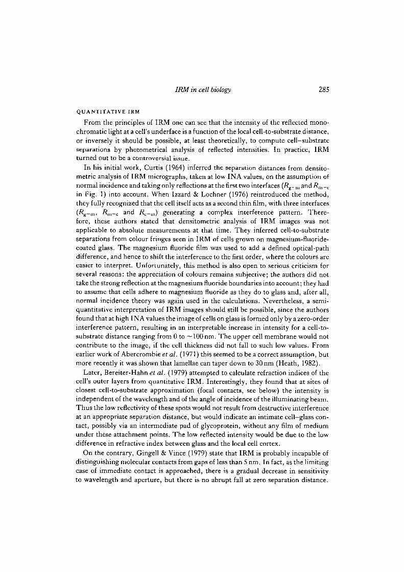
When cells are grown on a coverglass and observed with epi-illumination through anoil-immersion objective, the first reflection will occur at the transition from glass toculture medium (Rg-m in Fig. 1). From the n values one can see that this will be arelatively strong reflection; in other words, the background will be relatively clear.The presence of a cell on the coverglass can then modify this background intensity.
At sites where the cell surface is in immediate contact with the coverglass the re-flectivity will be determined by the glass-cell transition (Rg-C), which more pre-cisely is a glass-membrane or glass-cell coat transition. The higher refractive index ofthe membrane (n 2s 1 -4: Gingell & Todd, 1979; Bereiter-Hahn, Fox & Thorell, 1979)results in a low reflectivity, in other words a site of direct apposition of the cellmembrane to the glass will appear darker than the cell-free background. As I willdiscuss later, there might be some debate about whether this situation actually occurs.
Alternatively, when the cell is separated from the substrate by a film of culturemedium, reflection will occur at the glass-medium (Rg-m) and medium-cell (Rm-C)interfaces. If the thickness of the medium film is of the order of magnitude of thewavelength used, then both reflected beams can interfere. The optical pathdifference, A, between the two reflections is given by the cosine law (Tolansky, 1973,p. 53): A = 2nmd cos 8, where nm is the refractive index of the medium, d is thedistance between the interfaces, and 0is the angle of refraction in the medium. A darkfringe will be produced when A = N A, a bright one when A = (N+k) A. The integralnumber N is the order of interference. With monochromatic incident light theseinterferences result in bright and dark zones compared to the background; white light
Medium n = 1-34
Glass n = 1-515
Fig. 1. Schematic representation of a vertical section through a cell on a coverglass, andthe reflections occurring at different interfaces. Two situations are illustrated: atRg^ thecell is in direct apposition to the glass; in the other case the cell is separated from thesubstrate by a thin film of medium, generating two wavefronts, Rg-m and Rm-c, that caninterfere with one another. Reflections at the dorsal cell surface i?c_m (illustrated only inthe latter case here) can interfere with the earlier reflections.

282 H. Verschueren
yields a coloured pattern. In other words, the intensity of monochromatic light or thecolour of white light, reflected at the cell's underface contains information about thecloseness of contact between the cell and the substrate.
However, the image will be complicated by reflections at any other interface that isnear these first two transitions, e.g. the nuclear membrane or the dorsal cell mem-brane. In fact, beneath the thin cytoplasmic sheets or lamellae, the contribution fromthe beam reflected at the upper cell-medium transition (/?c_m) can completelyinvalidate conclusions about cell-substrate distances (Gingell, 1981).
At this point, the illuminating numerical aperture (INA) deserves special attention.Although Curtis (1964) had already reported on the influence of opening the apertureiris on the image, Izzard & Lochner (1976) were the first to analyse and interpret thephenomenon. Since the optical path length within the medium layer between cell andglass is a function of the angle of refraction in the medium, the broad range of angles ofincidence within the illuminating cone produced at high INA results in a continuousseries of overlapping interference patterns, which cancel out each other almostentirely. Only for the zero-order fringe (optical path difference, A, smaller than onewavelength) do these overlapping patterns converge; there is an increase in intensityfor a gap increasing from 0 to ~100nm. Thus, at high INA (>1) the higher-orderinterference fringes are lost, and the image is constituted solely from zero-orderinterferences, giving information on cell-substrate separations within that range. Forlarger distances, the image appears as a uniform grey as for the cell-free areas.Secondly, on the assumption that cell thickness does not fall to 100 nm (Aber-crombie, Heaysman & Pegrum, 1971; see, however, Heath, 1982), Izzard & Lochner(1976) concluded that the upper cell membrane does not contribute to the image athigh INA values. For these reasons, in order to observe adhesion patterns, it isnecessary to open the iris aperture in the illuminator fully.
Beck & Bereiter-Hahn (1981) proposed a different explanation, from results ofwork with ring-shaped apertures of increasing diameter. In their view, the contri-butions from the far side of the cell are decreased at high INA values because of thedecreased depth of focus; at the more distant reflecting surfaces the incident light isdistributed over a larger surface, and so the reflection per unit area is lowered.
The conclusion from either point of view is that the disturbing contributions fromthe dorsal cell surface are diminished at high INA values. However, the explanationdue to loss of focal depth is valid only for ring-shaped apertures, in which case thecentral beam stop concomitantly reduces the actual range of angles of incidence, andthen the phenomenon of overlapping interference patterns loses much of its signi-ficance.
PRACTICAL IRM
Although a conventional microscope can be used, it is desirable in many instances toobserve cells in normal orientation with an inverted microscope. Obviously the micro-scope needs to be equipped with an epi-illuminator. Since monochromatic light(usually 546 nm) is selected through a narrow band filter, and since the intensity of the

IRM in cell biology 283
reflected light remains below 1 % of the incident intensity, a powerful light source, forexample a mercury arc, is commonly used. Another important part of the instrumentis the field diaphragm since it is advantageous to cut off stray light from areas outsidethe field of interest as much as possible by encircling a relatively small field. This iswhy the field iris is often visible in IR micrographs (Izzard&Lochner, 1976). Becauseof the strong influence of the INA on the IRM image, the aperture diaphragm is ofdecisive importance; at higher INA the contribution of deeper structures is muchweaker than in the case of normal incidence. Therefore, small apertures should by nomeans be used to study adhesion. Interpretable images can be produced with either afully opened iris diaphragm, or large ring-shaped diaphragms. The commercial, smallring-shaped gratings do not suit the purpose (Bereiter-Hahn et al. 1979; Beck &Bereiter-Hahn, 1981). Moreover, ring-shaped apertures do not offer the advantage ofsuch a broad range of angles of incidence. In fact, since the function of the centralbeam stop is to cut off some of the stray light coming from reflections within theobjective lens (Patzelt, 1979), there is no reason to prefer, in general, ring-shapedapertures to fully opened iris apertures when using the antiflex device (see below). Inthe Leitz system, however, the central beam stop serves primarily to shade off thephase plate of the phase-contrast objectives.
A third, simple alternative is to use no aperture diaphragm at all. The author uses aconventional epi-fluorescence illuminator from Zeiss, where only a field diaphragmand filter housing are provided (Fig. 2). The actual INA is approximately 1-06 withthe 63/1-25 Antiflex Neofluar oil-immersion objective. A complete Kb'hler illumi-nator does not yield better images, but the variable INA offers possibilities forstudying the optical contributions from deeper regions in the specimen. Thus, Izzard& Lochner (1980) used an INA = 0-8 to visualize fringes under leading lamellae thatare inclined away from the substrate, and Bereiter-Hahn et al. (1981) studied rufflingmembranes at the dorsal cell surface at INA = 0-62.
Finally, the avoidance of stray light coming from reflections outside the object is ofdecisive importance. Obviously, oil-immersion is a prerequisite, avoiding reflection atthe glass—air interfaces between object and objective, but even the weaker reflectionsinside the objective produce enough stray light to destroy the whole image, unless thefield stop is narrowed to impracticable low sizes. As the disturbing reflectionsoriginate largely from the central portions of the lens (Beck & Bereiter-Hahn, 1981),ring-shaped apertures avoid much of the stray light, but for reasons explained abovethe use of a central beam stop in the illuminator is not particularly recommended.
The antiflex device, introduced into IRM by Ploem (1975a) is an ingenious, verysatisfactory solution to the problem (Fig. 2). The incoming light is plane-polarized(protect polaroid filter from ultraviolet light and heatl) before being reflected into theobjective, and the analyser behind the objective is crossed to cut off all reflectionsoccurring within the microscope body and the objective. As the antiflex objective has arotatable plane-parallel quartz plate (A/4) mounted on the front lens, the light beamthat forms the image passes through this quartz plate on its way to and back from theobject. Provided the plate is properly oriented, the reflected light becomes circularlypolarized and passes through the analyser. Thus only the light reflected by the object
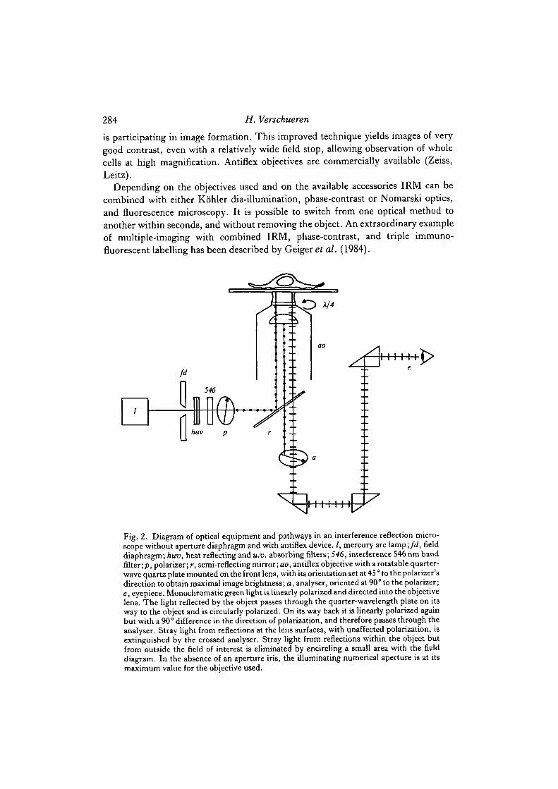
284 H. Verschueren
is participating in image formation. This improved technique yields images of verygood contrast, even with a relatively wide field stop, allowing observation of wholecells at high magnification. Antiflex objectives are commercially available (Zeiss,Leitz).
Depending on the objectives used and on the available accessories IRM can becombined with either Kbhler dia-illumination, phase-contrast or Nomarski optics,and fluorescence microscopy. It is possible to switch from one optical method toanother within seconds, and without removing the object. An extraordinary exampleof multiple-imaging with combined IRM, phase-contrast, and triple immuno-fluorescent labelling has been described by Geiger et al. (1984).
Fig. 2. Diagram of optical equipment and pathways in an interference reflection micro-scope without aperture diaphragm and with antiflex device. /, mercury arc lamp;/<i, fielddiaphragm; huv, heat reflecting and u.v. absorbing filters; 546, interference 546nm bandfilter;/), polarizer; r, semi-reflecting mirror; ao, antiflex objective with a rotatable quarter-wave quartz plate mounted on the front lens, with its orientation set at 45 ° to the polarizer'sdirection to obtain maximal image brightness; a, analyser, oriented at 90° to the polarizer;e, eyepiece. Monochromatic green light is linearly polarized and directed into the objectivelens. The light reflected by the object passes through the quarter-wavelength plate on itsway to the object and is circularly polarized. On its way back it is linearly polarized againbut with a 90° difference in the direction of polarization, and therefore passes through theanalyser. Stray light from reflections at the lens surfaces, with unaffected polarization, isextinguished by the crossed analyser. Stray light from reflections within the object butfrom outside the field of interest is eliminated by encircling a small area with the fielddiagram. In the absence of an aperture iris, the illuminating numerical aperture is at itsmaximum value for the objective used.
IRM in cell biology 285
QUANTITATIVE IRM
From the principles of IRM one can see that the intensity of the reflected mono-chromatic light at a cell's underface is a function of the local cell-to-substrate distance,or inversely it should be possible, at least theoretically, to compute cell-substrateseparations by photometrical analysis of reflected intensities. In practice, IRMturned out to be a controversial issue.
In his initial work, Curtis (1964) inferred the separation distances from densito-metric analysis of IRM micrographs, taken at low INA values, on the assumption ofnormal incidence and taking only reflections at the first two interfaces (.Rg_m andi?m_c
in Fig. 1) into account. When Izzard & Lochner (1976) reintroduced the method,they fully recognized that the cell itself acts as a second thin film, with three interfaces(Rg-m, Rm-C and Rc-m) generating a complex interference pattern. There-fore, these authors stated that densitometric analysis of IRM images was notapplicable to absolute measurements at that time. They inferred cell-to-substrateseparations from colour fringes seen in IRM of cells grown on magnesium-fluoride-coated glass. The magnesium fluoride film was used to add a defined optical-pathdifference, and hence to shift the interference to the first order, where the colours areeasier to interpret. Unfortunately, this method is also open to serious criticism forseveral reasons: the appreciation of colours remains subjective; the authors did nottake the strong reflection at the magnesium fluoride boundaries into account; they hadto assume that cells adhere to magnesium fluoride as they do to glass and, after all,normal incidence theory was again used in the calculations. Nevertheless, a semi-quantitative interpretation of IRM images should still be possible, since the authorsfound that at high INA values the image of cells on glass is formed only by a zero-orderinterference pattern, resulting in an interpretable increase in intensity for a cell-to-substrate distance ranging from 0 to ~100nm. The upper cell membrane would notcontribute to the image, if the cell thickness did not fall to such low values. Fromearlier work of Abercrombie et al. (1971) this seemed to be a correct assumption, butmore recently it was shown that lamellae can taper down to 30 nm (Heath, 1982).
Later, Bereiter-Hahn et al. (1979) attempted to calculate refraction indices of thecell's outer layers from quantitative IRM. Interestingly, they found that at sites ofclosest cell-to-substrate approximation (focal contacts, see below) the intensity isindependent of the wavelength and of the angle of incidence of the illuminating beam.Thus the low reflectivity of these spots would not result from destructive interferenceat an appropriate separation distance, but would indicate an intimate cell-glass con-tact, possibly via an intermediate pad of glycoprotein, without any film of mediumunder these attachment points. The low reflected intensity would be due to the lowdifference in refractive index between glass and the local cell cortex.
On the contrary, Gingell & Vince (1979) state that IRM is probably incapable ofdistinguishing molecular contacts from gaps of less than 5 nm. In fact, as the limitingcase of immediate contact is approached, there is a gradual decrease in sensitivityto wavelength and aperture, but there is no abrupt fall at zero separation distance.

286 H. Verschueren
Moreover, Bereiter-Hahnef at. (1979) did not take cell thickness and reflections fromthe dorsal membrane into account (see Gingell, 1981 for a critique).
The final breakthrough in reflected light interferometry of living cells came whenGingell & Todd (1979) proposed their finite aperture theory. Preliminary work hadbeen done by Heavens & Yuan (1979), who described a limited theory for calculatingreflected irradiances in the case of large INA values, and applied it to a simplified cellmodel to predict interference fringes generated by combined reflections from ventraland dorsal membranes. Gingell & Todd (1979) developed this approach into a com-prehensive theory, applicable to non-coherent Kohler illumination, predictingreflectance in a multilayer as a function of INA and the thickness of the layers. Theychecked the validity of their model by comparing computed irradiances with thosemeasured in a water wedge. For this simple situation, there is an almost perfect fitbetween theory and experimental values. In a later paper (Gingell, Todd & Heavens,1982) the theory was further validated with magnesium fluoride films, and again withwater wedges for up to 30 interference fringes. Applying their finite aperture theory toan idealized, multilayered model cell, composed of glass, water, membrane, cyto-plasm, membrane and water, Gingell & Todd (1979, figs 10-15) constructed anumber of curves that indicate the relative irradiance as a function of glass-membraneseparation, for different INA values and different cytoplasmic thicknesses. The moststriking feature of these curves is a strong dampening of the higher-order fringes at alarge INA value (I118). Concomitantly, the curves at high INA become independentof cytoplasmic thickness and, interestingly, also of the assumed value for the refrac-tion index of the membrane. Thus, in a general way, the view of Izzard and Lochnerwas confirmed: at high INA the interference pattern is determined merely by zero-order interference, characterized by a continuous increase in intensity for separationsfrom 10 to ~150nm; in addition, the contributions from the far side of the cell areweakened at high INA. However, it is only at cytoplasmic thicknesses exceeding 1 fimthat these contributions can be neglected for quantitative purposes. In a further paper(Gingell, 1981) the effect of thin cytoplasm on image brightness was calculated inmore detail for fixed medium gap values of 0 and 50 nm. It emerged that dark fringeswill occur under cytoplasmic lamellae of 50— 150nm thick, the exact value dependingon the medium gap underneath.
In conclusion, IRM at high INA allows semi-quantitative appreciation of cell-substrate distances under a cytoplasmic layer of at least 1 ^m. At low INA, or underthe thin cytoplasm often occurring at the cell periphery, even a qualitativeinterpretation of the image is hazardous. Quantitative IRM is now possible with thefinite aperture interferometry model of Gingell & Todd (1979), provided that for thecell periphery cytoplasmic thickness, independently measured, is taken into account.
Cell thickness can be determined by transmission electron microscopy of verticalsections through the cell studied, or by transmitted light interference microscopy.Both methods further complicate quantitative IRM considerably, and there are only afew limited attempts described in the literature. Heath (1982) combined IRM andtransmission electron microscopy in a semi-quantitative analysis of the cell—glassseparation under a cultured cell. In accordance with Gingell's (1981) theoretical

IRM in cell biology 287
calculations, he found a very dark fringe at the cell margin, which could not beexplained by low cell-substrate distance, but was determined by the thin cytoplasmiclamella (70 nm) with a gap of 30-50 nm underneath it. For detailed quantitativeanalysis, however, the method may not be completely reliable, as there is no firm proofthat fixation and embedding procedures leave cell thickness and medium gap strictlyunaltered (Gingell, 1981; Heath, 1982). As for transmitted light interferometry,Gingell, Todd & Heavens (1982) showed a fair correlation between IRM and multiplebeam interferometry of magnesium fluoride films, but in cells the situation is consider-ably more complicated. Izzard & Lochner (1976) compared data from IRM andJamin interferometry with a Zeiss-Lebedeff microscope, to evaluate the thickness offibroblasts. Unfortunately, only crude estimations, with an error of tens or hundredsof nanometres could be made. It therefore seems unlikely that interferometricmethods will overcome the difficulties inherent in IRM of thin cytoplasmic sheets.
Clearly, quantitative IRM remains extremely complex, beyond the scope of mostcell biologists. However, the insights that came from these mathematical approachesare also important for qualitative work.
QUALITATIVE IRM: FOCAL AND CLOSE CONTACTS
Despite the difficulties in developing an adequate theoretical model for quantitativeIRM, many useful qualitative observations on cell adhesion have been made since theoriginal publication of Izzard & Lochner (1976). The typical features these authorsfirst described, the focal and close contacts, have been reported now by many obser-vers, working with different cell types.
Focal contacts (FCs)
These are also termed focal adhesions, adhesion plaques, feet or vinculin plaques;for a concise review, see Birchmeier (1981).
FCs appear as very dark streaks (Figs 3, 4, 5) 0*2—1 ^m in width and 2— lO/im inlength. Although it is generally accepted that FCs are the sites of minimal cell-to-substrate distance, there has been some debate as to whether or not there is an inter-mediate layer of culture medium left between the cell and the culture substrate.Bereiter-Hahn et al. (1979) provided some evidence in favour of a direct cell-to-glasscontact, possibly via a pad of glycoprotein, but for reasons discussed above IRMmight not be an appropriate method for distinguishing zero gaps from extremely lowseparations (Gingell & Vince, 1979). The value of 10-15 nm proposed by Izzard &Lochner (1976) for the gap under FCs has also been criticized, insofar as it had beenderived from interferometric methods (Gingell, 1981), but it is in agreement with theseparation distances found by electron microscopy (Heath, 1982; Chen & Singer,1982). Nevertheless, even if there is a small gap of ~ 15 nm between the membranelipid bilayer and the substrate, this gap might well be filled with cell-depositedmaterial, and then the FCs could yet be considered as real intimate cell—substratecontacts.

H. Verschueren
Figs 3-5

IRM in cell biology 289
In combined IRM and differential interference contrast microscopy, FCs are seento coincide with the termination site of cytoplasmic stress fibres (Lochner & Izzard,1973; Izzard & Lochner, 1976; Abercrombie & Dunn, 1975). In a correlated IRMand high-voltage electron-microscopic study, Heath & Dunn (1978) decisively con-firmed this association. FCs as described from IRM can therefore be considered of thesame nature as the adhesion plaques described earlier from electron microscopy(Abercrombie et al. 1971; Brunk, Ericsson, Ponten & Westermark, 1971). Theassociation of FCs with the distal end of microfilament bundles was further confirmedin an independent way by combined IRM and immunofluorescence microscopy. Infact, immuno-staining methods, both at the optical and the electron-microscopic levelhave allowed some insight into the molecular architecture of cellular adhesions. FCsare associated with the different components of the actomyosin system: actin, a-actinin, myosin and tropomyosin (Badley et al. 1978; Wehland, Osborn & Weber,1979). Vinculin has been found to be localized within the FCs (Geiger, 1979) andseems to play a role, together with tf-actinin, in linking the actin filaments to the innerside of the plasma membrane (Chen & Singer, 1982; Singer, 1982; Avnur, Small &Geiger, 1983; Couchman, Badley & Rees, 1983). Avnur & Geiger (19816) proposed amethod for the isolation of substrate-attached ventral membranes. They showed thatmicrotubules and intermediate filaments are easily removed by shear, but micro-filament-associated proteins such as actin, vinculin, ar-actinin, filamin andtropomyosin were retained in FCs.
There have been contradictory reports on the presence of fibronectin at the outerside of the cell membrane in FCs. Birchmeier et al. (1980) and Chen & S. J. Singer
Fig. 3. First-passage mouse embryo cell, 24h after seeding, cc, close contacts;/c, focalcontacts; n, nucleus; fr, higher-order fringes. This fibroblastic cell is in a motileconfiguration. Behind the edge of the large extending lamellae at the right the CCs form acontinuous band, while under the cell centre the CC is intersected by white zones withlarger separation distance (100-150 nm). FCs are seen within the CC of the lamellae and atthe trailing end of the cell. Despite an I N A > 1, the nucleus is faintly visible, as well as afew successive fringes within the CC and at concave parts of the cell contour. In the formercase the fringes are due to a progressive thickening of the cytoplasm towards the cell centre,in the latter to the inclination of the cell's underface away from the substrate. (Micrographreproduced with permission, from Verschueren et al. 1983.) X1050.
Fig. 4. First-passage mouse embryo cell, 72 h after seeding. Is, sites of large cell-to-substrate distance;//!, presumable fibronectin fibres or extracellular matrix contacts. Thiscell has reached a stationary configuration 3 days after seeding. It is anchored firmly to thesubstrate by FCs, but some CC can be seen under the small lamella extending at the lowerright. At different locations, the cell-substrate separation is wide enough (> 150 nm) forthe white band to merge into a few successive higher-order fringes and background inten-sity. The black streaks under the nucleus probably correspond to extracellular matrixcontacts, i.e. fibronectin-containing deposits forming a cell-to-substrate bridge within anarea of large separation. X750.
Fig. 5. Lamellar cytoplasm of a C3H/lOTi/C18 mouse embryo cell. The extreme edge ofthe protruding cytoplasm shows a white fringe (arrowheads) where the lamellipodiumextends ahead of the CCs at a certain distance from the substrate. The strong influence ofreflections from the dorsal cell surface in the case of thin lamellar cytoplasm is illustrated bythe black fringes (arrows) resulting from destructive interference between the differentreflected beams. X750.
290 H. Verschueren
(1980) found no fibronectin in FCs, Rees et al. (1982) and Woods et al. (1983)illustrated the progressive disappearance of fibronectin from FCs during theirformation, and Grinnell (1980) and Avnur & Geiger (1981a) reported the removal offibronectin from pre-coated substrates at the FC sites. Later, however, I . I . Singer(1982) found fibronectin to be associated with vinculin plaques in the perinuclear areaof quiescent cells, and proposed that the FC should be considered as a structureundergoing a maturation process from fibronectin-negative in motile cells, to fibro-nectin-positive in the case of more stable attachment. On the other hand, Chen & S. J.Singer (1982) proposed differentiating between focal adhesions (fibronectin lackingFCs) and what they called extracellular matrix contacts, where fibronectin is foundtogether with other matrix proteins, forming a bridge between the cell and thesubstrate at sites of separation distances exceeding 100 nm. Thus it is clear now thatchemically and functionally different structures may have been called FCs in earlierpapers, and that more extensive characterization of the contacts seen in IRM isdesirable. In this context, the recent discovery of new proteins within FCs (Burridge,Kelly & Connell, 1982; Oesch & Birchmeier, 1982; Burridge & Connell, 1982;Burridge & Mangeat, 1984; Mangeat & Burridge, 1984) is promising.
FCs were reported to be stationary relative to the substrate as the cell moves(Lochner & Izzard, 1973; Izzard & Lochner, 1980; Abercrombie & Dunn, 1975). Itwas suggested that FCs are important in cell translocation by providing attachmentpoints for the stress fibres that in- turn would draw the bulk cytoplasm forward(Abercrombie et al. 1971; Dunn & Heath, 1976; Izzard & Lochner, 1980).
FCs are, however, most conspicuous under stationary cells: the number of FCsunder embryonic chick heart fibroblasts and mesoderm cells increases with time inculture, together with a decrease in translocation rate (Couchman & Rees, 1979a,6;Sanders, 1984). Numerous FCs are seen under confluent epithelial cells (Cottier-Fox,Sparring, Zetterberg & Fox, 1979; Kolega, Shure, Chen & Young, 1982), while theymay be lacking under highly motile cell types such as rabbit neutrophil granulocytes(Armstrong & Lackie, 1975), amphibian leucocytes (Bereiter-Hahn, 1977; Kolega etal. 1982) and leukaemia cells (Haemmerli & Ploem, 1979).
Thus,<FCs are firm attachment structures, anchoring cultured cells to their solidsubstrate through a membrane specialization that forms a bridge from the cell coat tothe cytoplasmic actin fibres. Although they may have a role in cell translocation, thereis circumstantial evidence that the primary role of FCs is to hold the cell in place ratherthan to move it. Practically, FCs are easily recognized in IRM thanks to their typicalstreaky shape and low intensity. Nevertheless, some caution is recommended ininterpreting the very dark fringe often seen at the margin of spread, flattened cells (seeFig. 5; and Heath, 1982): these can well be caused by interference with reflections atthe upper membrane (Gingell, 1981) and should not necessarily be regarded asdensely packed or contiguous FCs (Segel, Volk & Geiger, 1983). Secondly, as pointedout before, the dark stripes often seen under the cell centre (Fig. 4) might well befibronectin-containing extracellular matrix bridges between cell and substrate and notFCs.

IRM in cell biology 291
Close contacts (CCs)
CCs appear as broad areas of uniform low intensity (grey) reflections under movingcells or cell parts (Figs 3,5). Their shape and dimensions are undefined, and theircontours are continually changing. Moreover, reflections at the dorsal cell surface caninterfere to alter significantly the intensity of reflection when the cell thickness fallsbelow 1 nm (Gingell, 1981), a situation occurring frequently under the lamellar cyto-plasm of different cell types (Heath, 1982). Thus it is not always possible to ascertainthe identity of grey areas in any point of the image. The dark fringes under the wedge-shaped cell periphery, especially, should not be called CCs without due consideration.Nevertheless, the extent of CC formation can be appreciated qualitatively in mostIRM images.
For the same reasons as for FCs, measurement of the cell-to-substrate distanceunder the CC cannot come from IRM alone, but again the value of ~ 30 nm, found byIzzard & Lochner (1976) is in fair agreement with findings from electron microscopy(Heath, 1982; Chen & Singer, 1982) and is now widely accepted. The evidence for theadhesive nature of CCs has been discussed in detail by Izzard & Lochner (1980) andHeath (1982). To date, CCs have received less attention that FCs and their associatedmicrofilament bundles. There is some evidence, nevertheless, that CCs also representcytoskeleton-associated membrane specializations: Heath & Dunn (1978) demon-strated a frequent association with a meshwork of microfilaments, and Chen & Singer(1982), showing the presence of tf-actinin but not vinculin in CC regions, proposedthat there the microfilaments are connected laterally to the membrane.
CCs are seen mostly beneath the entire underface of highly motile cells (Armstrong& Lackie, 1975; Bereiter-Hahn, 1977; Kolega et al. 1982) and behind the margin ofextending lamellae of slower moving cell types such as epithelial cells (Heath, 1982)and fibroblasts (Izzard & Lochner, 1980). In the latter case they are broken up underthe cell centre, and stationary cells hardly display any CC at all. Thus, in contrast tothe strong, stationary FCs, CCs are weaker but highly dynamic adhesions. Whilebeing strong enough to withstand the tractions involved in cell translocation, they aremore appropriate than FCs for sustaining rapid movement because they readily breakup. Further study of the structure and regulation of CCs should certainly providevaluable information about the mechanisms of cell movement.
Besides the typical FCs and CCs, IRM images show zones of larger cell-substrateseparations (100 nm and more). These appear as bright reflections due to constructiveinterference, or as background intensity zones due to overlapping first and higher-order interference patterns (see below). Sometimes one can also recognize a fewsucceeding fringes at sites where the cell surface is inclined away from the substrate atan appropriate angle.
APPLICATIONS OF IRM IN CELL BIOLOGY
It is beyond the scope of this article to cover in depth the results that have beenobtained so far by the increasing number of cell biologists who started to use IRM in

292 H. Verschueren
the last decade. I will only indicate the different fields where IRM has been applied,and point out some less well-known possibilities. The reader is referred to the originalarticles for full discussion.
Many different cell types have by now been studied with IRM, although fibro-blastic cells have received most attention. Embryonic chick heart fibroblasts wereused in the pioneering work of Curtis (1964) and Izzard & Lochner (Lochner &Izzard, 1973; Izzard & Lochner, 1976), who defined the basic features of IRM imagesof living cells. IRM of chick heart fibroblasts has been used further to study therelationship between adhesion patterns and locomotion (Dunn & Heath, 1976; Dunn,1979; Izzard & Lochner, 1980; Chen, 1981), contact inhibition of movement(Abercrombie & Dunn, 1975), the association of adhesions with the microfilamentsystem (Heath & Dunn, 1978; Heath, 1983) and the interrelationship of adhesion,spreading, growth and movement (Badley, 1980; Couchman & Rees, 1979a,b; Rees,Badley& Woods, 1979; Badley, Woods, Carruthers & Rees, 1980). In their importantarticle defining the 'grip and stick' model for cell adhesion, Rees, Lloyd & Thorn(1977) discussed IRM images of rat dermal fibroblasts and baby hamster kidney(BHK) cells during detachment from glass. The role of fibronectin in locomotion andanchorage of fibroblasts has also been studied using IRM (Couchman, Rees, Green &Smith, 1982; Rees et al. 1982; Chandrasekhar, Norton, Millis & Izzard, 1983;Sanders, 1984), while Couchman, Hook, Rees & Timpl (1983) described the forma-tion of FCs on laminin substrates in the absence of fibronectin. In their recent work,Izzard's group (Yates, Norton & Izzard, 1979; Yates & Izzard, 1981; Norton &Izzard, 1982) have described Balb/c3T3 cells and an adhesion-defective mutant ofthis line. Verschueren, Wildemauwe & Van Larebeke (1983) described the formationof extremely large CCs beneath mouse embryo fibroblasts after treatment withdipyridamole.
Several authors have published results in IRM of epithelial cell types. Cottier-Foxet al. (1979) first described FCs and CCs during attachment of primary mouse kidneyepithelial cells. An interesting model for the study of cell locomotion isXenopus laevisepidermis, because it offers an exceptional opportunity for comparing in vitro resultswith a situation in situ. IRM was possible only in vitro, where CCs were predominant,but some FCs were also present. In situ however, there seem to be no microfilamentbundles or adhesion plaques (Radice, 1978; Radice, 1980a,6; Bereiter-Hahn et al.1981). Heath (1982) and Billig et al. (1982), working with chick embryo cornealepithelial cells, and Turksen, Opas, Aubin & Kalnins (1983) with embryonic chickretinal pigment epithelial cells, came to the conclusion that epithelial cell sheets invitro tend to adhere to the substrate mainly by their marginal cells through FCs andCCs, while the central cells adhere more loosely. For a comprehensive discussion onadhesion and locomotion of fibroblastic and epithelial cells in culture, the reader isreferred to Heath (1982).
Concerning malignant cells, Haemmerli and associates have observed by IRM aseries of human and rabbit carcinoma lines (Haemmerli, Strauli & Ploem, 1980;Haemmerli, 1981; Haemmerli & Strauli, 1981; Haemmerli, Strub, Jockusch &Strauli, 1982; Jockusch, Haemmerli & In Albon, 1983), and rat leukaemia cells

IRM in cell biology
(Haemmerli & Ploem, 1979). Other authors used different murine carcinoma celllines (Cottier-Fox, Ryd, Hagmar & Fox, 1980; Couchman, Yates, King & Badley,1981) and Chinese hamster ovary (CHO) cells (Leader, Stopak & Harris, 1983).Other cell types studied with IRM are neutrophil granulocytes (Armstrong & Lackie,1975; Keller, Barandun, Kistler & Ploem, 1979; Keller, Zimmermann & Cottier,1983), macrophages (Ploem, 1975a) and lymphoblastoid cells (Ploem, 19756), Frienderythroleukaemia cells and human glia and glioma cells (Bereiter-Hahn et al. 1979),red blood cells (Gingell & Vince, 1979; Gingell &Todd, 1980; Wolf & Gingell, 1983;Donath& Gingell, 1983; Pera, 1984), cultured rat liver cells (Pentz & Schulle, 1981;see also Fig. 6), growth cones of chick embryo neurones (Letourneau, 1979), platelets(Alexandrova & Vasiliev, 1984) and amoebae (Opas, 1978; Preston & King, 1978;Gingell & Vince, 1979, 1982; Vince & Gingell, 1980; King, Preston & Miller, 1983).
The general impression emerging from all these results is that FCs with theirassociated microfilament bundles and CCs can occur in most cell types of extremelydiverse species and the relative abundance of these two features is related primarily tothe locomotory behaviour of the cells. The conclusion from a comparison of 10 celltypes by Trinkaus' group (Shure, Young, Kolega & Chen, 1979; Kolega et al. 1982)will probably hold for a broad range of cell types: CCs and not FCs are associated withrapid cellular translocation, while the presence of FCs is indicative of reducedtranslocation and of stabilization of the spread cell shape.
An exciting finding with IRM is that Rous sarcoma virus-transformed fibroblastshave fewer FCs than their untransformed counterparts (David-Pfeuty & Singer,
Fig. 6. Rat hepatocyte in primary culture, 24 h after isolation. A. Differential interferencecontrast; B, IRM. The IRM image of this liver parenchyma cell, isolated by collagenaseperfusion, is characterized by numerous, contiguous black patches that do not resembleFCs in shape. These patches disappear within —48 h and then typical FCs can be recog-nized under newly formed lamellae. The patches of low reflectivity could be due tointerfering reflections from the upper membrane, but could alternatively be interpreted asdeposits of extracellular material that is formed only by freshly isolated hepatocytes, andnot by cells in long-term cultures. X710.

294 H. Verschueren
Fig. 7. Unstained chromosomes. IRM of chromosomes from human lymphocytes, pre-pared for sister chromatid exchange analysis, but before Giemsa staining. X750.
Fig. 8. Coomassie-Blue-stained cytoskeleton. A. Kohlerdia-illumination; B, IRM. A low-passage mouse embryo cell, extracted with detergent, fixed with glutaraldehyde andstained with Coomassie-Blue (Pena, 1980; Opas & Kalnins, 1982). In IRM themicrofilament bundles appear with enhanced contrast, and are visible even under thenucleus and the cell centre, where granules that are trapped in the cytoskeleton obscure theimage in transmitted light. X620.

IRM in cell biology 295
1980). At the same time, Rohrschneider (1980) found that the src gene product ispresent in focal contacts of RSV-transformed cells. It seems that phosphorylation oftyrosine of vinculin by the src gene product might be an important event in theacquisition of anchorage independence and malignancy (Rohrschneider, Rosok &Shriver, 1982; Rohrschneider & Rosok, 1983). The issue has been discussed byAlitalo&Vaheri(1982)andHynes(1982). In addition, Couchmaneial. (1981) foundthat androgen-responsive fibroblastic cells derived from a murine mammary carci-noma have predominantly CCs and lack FCs, while the androgen-unresponsiveepithelioid cells do have FCs and a few CCs. Furthermore, there is some recentevidence that reverse transformation might be accompanied by an increase in thenumber of FCs. In chick embryo chondrocytes, infected with transformation-defective temperature-sensitive mutants of Rous sarcoma virus, adhesion plaquesreappeared when the cells were shifted from the permissive to the restrictive tem-perature (Marchisio et al. 1984). Further, Leader et al. (1983) showed in Chinesehamster ovary cells that broad CCs are characteristic of the transformed state and aregradually replaced by numerous FCs following the addition of cyclic AMP andtestosterone, while Lehtonen, Lehto, Badley & Virtanen (1983) described theappearance of vinculin plaques during retinoic-acid-induced differentiation of terato-carcinoma cells. Another putative correlation between altered adhesion pattern invitro and malignancy in vivo was suggested by Cottier-Fox et al. (1980), who foundthat for a tumour pair originating from one murine carcinoma, the non-metastasizingmember formed CCs and FCs, while the highly metastatic ascitic form has onlylimited areas of CC and generally fails to form FCs. A similar correlation betweenincreased metastatic potential and a decreased number of FCs in cells from murinemelanoma and fibrosarcoma has been noted by Raz & Geiger (1982).
From the work of different groups, however, it is clear that there is no simplecorrelation between deficient FC formation and malignancy. Cells from six humancarcinoma lines are able to form numerous FCs when they are stationary (Haemmerli& Strauli, 1981), while the highly motile rabbit V2 carcinoma (Haemmerli et al. 1980)and L5222 rat leukaemia cells (Haemmerli & Ploem, 1979) show CCs predominantly.These results from Haemmerli's group confirm that for malignant cells also the type ofcontacts formed depends on cellular behaviour rather than on the cell type. In thesame context it is noteworthy that the adhesion-defective, FC-lacking mutant of 3T3cells studied by Izzard's group (Yates et al. 1979; Yates & Izzard, 1981; Norton &Izzard, 1982) is a non-malignant, anchorage-dependent cell line.
A methodological improvement in the quantitative study of adhesion patterns andparticularly the extent of FC formation in different cell types and different situationsmight come from the use of automatic image analysis in IRM (Bereiter-Hahn, 1977).Since automatic contour finding in low-contrast images is a major problem, IRMimages might well prove easier to analyse than, for example, phase-contrast ordifferential interference contrast images.
In addition to the description of adhesion patterns in different cell types and theelucidation of the molecular events involved in the formation of specialized adhesivestructures, IRM has also been used to study the role of physical long-range forces,

296 H. Verschueren
electrostatic repulsion and London—van der Waals' attraction, in cell—substrateinteraction by a number of authors (King, Heaysman & Preston, 1979; Gingell &Todd, 1977, 1980; Gingell & Vince, 1979, 1982; Vince & Gingell, 1982).
A particular application of IRM is found in a recent paper by Pera (1984), whoreconstructed the three-dimensional shape of erythrocytes by analysing interferencefringes generated at low-aperture operation.
So far this review has been concerned only with observations of living cells. IRMcan, however, be used to increase contrast in fixed, stained or unstained preparations,such as histological sections, blood smears and chromosomes (Pera, 1979a,b\ see alsoFig. 7). A recently developed method to visualize cytoskeletal structures is that ofIRM of detergent-extracted, Coomassie-Blue-stained cells (Pena, 1980; Opas &Kalnins, 1982). This rapid technique shows, at high resolution and high contrast,microfilament bundles at the ventral surface of the cell (Fig. 8). In a similar technique(Opas & Kalnins, 1984), glutaraldehyde-fixed and NBD-phallacidin-stained cells areobserved alternatively in IRM or fluorescence microscopy. These latter examplessuggest that IRM is likely to be further developed in the next years, and to become ofinterest to an increasing number of microscopists.
The author thanks L. Schoofs for helping with the literature, D. Dekegel for finishing the micro-graphs, A. De Roose and R. Nyssen for the drawings, J. de Gerlacheand M. Lans for providing thehepatocyte shown in Fig. 6, G. Grutman for preparing the chromosomes shown in Fig. 7, G.Hibbert for correcting the typescript, and N. Van Larebeke in whose laboratory the author isemployed.
REFERENCES
ABERCROMBIE, M. & DUNN, G. A. (197S). Adhesion of fibroblasts to substratum during contactinhibition observed by interference reflection microscopy. Expl Cell Res. 92, 57-62.
ABERCROMBIE, M., DUNN, G. A. & HEATH, J. P. (1976). Locomotion and contraction in non-muscle cells. In Contractile Systems in Non-muscle Tissues (ed. J. V. Perry et al.), pp. 3—11.Amsterdam: Elsevier North-Holland Biomedical Press.
ABERCROMBIE, M., DUNN, G. A. & HEATH, J. P. (1977). The shape and movement of fibroblasts inculture. In Cell and Tissue Interaction (ed. J. W. Lash&M. M. Burger), pp. 57-70. New York:Raven Press.
ABERCROMBIE, M., HEAYSMAN, J. E. M. &PEGRUM, S. M. (1971). The locomotion of fibroblasts inculture. IV. Electron microscopy of the leading lamella. Expl Cell Res. 67, 359-367.
ALEXANDROVA, A. Y. &. VASILIEV, J. M. (1984). Focal contacts of spreading platelets with thesubstratum. Expl Cell Res. 153, 254-258.
ALITALO, K. &. VAHERI, A. (1982). Pericellular matrix in malignant transformation. Adv. CancerRes. 37, 111-158.
AMBROSE, E. J. (1956). A surface contact microscope for the study of cell movements. Nature, Lond.178, 1194.
AMBROSE, E. J. (1961). The movements of fibrocytes. Expl Cell Res. (suppl.) 8, 54-73.ARMSTRONG, P. B. & LACKIE, J. M. (1975). Studies on intercellular invasion in vitro using rabbit
peritoneal neutrophil granulocytes (PMNS). I. Role of contact inhibition of locomotion. J. CellBiol. 65, 439-462.
AVNUR, Z. & GEIGER, B. (1981a). The removal of extracellular fibronectin from areas of cell-substrate contact. Cell 25, 121-132.
AVNUR, Z. & GEIGER, B. (19816). Substrate-attached membranes of cultured cells: isolation andcharacterization of ventral membranes and the associated cytoskeleton. J. molec. Biol. 153,361-379.

IRM in cell biology 297
AVNUR, Z., SMALL, J. V. & GEIGER, B. (1983). Actin-dependent association of vinculin with thecytoplasmic aspect of the plasma membrane in cell-contact areas. J. Cell Biol. 96, 1622-1630.
BADLEY, R. A. (1980). Cytoskeletons, membranes and fibroblast adhesion. Cell Biol. Int. Rep. 4,792.
BADLEY, R. A., LLOYD, C. W., WOODS, A., CARRUTHERS, L., ALCOCK, C. & REES, D. A. (1978).
Mechanisms of cellular adhesion. III. Preparation and preliminary characterization of adhesions.Expl Cell Res. 117, 231-244.
BADLEY, R. A., WOODS, A., CARRUTHERS, L. & REES, D. A. (1980). Cytoskeleton changes infibroblast adhesion and detachment. J . Cell Sri. 43, 379-390.
BECK, K. & BEREITER-HAHN, J. (1981). Evaluation of reflection interference contrast microscopeimages of living cells. Microsc. Ada 84, 153-178.
BEREITER-HAHN, J. (1977). MikroskopischeSpezialverfahrenzurselektivenKontraststeigerungfurdie automatische Bildanalyse. Microsc. Ada (suppl.) 1, 165-180.
BEREITER-HAHN, J., Fox, C. H . & T H O R E L L , B. (1979). Quantitative reflection contrast microscopyof living cells. J. Cell Biol. 82, 767-779.
BEREITER-HAHN, J., STROHMEIER, R., KUNZENBACHER, I., BECK, K. & VOTH, M. (1981).
Locomotion of Xenopus epidermal cells in primary culture../. Cell Sri. 52, 289-311.BILLIG, D., NICOL, A., MCGINTY, R., COWIN, P., MORGAN, J. & GARROD, D. (1982). The
cytoskeleton and substratum adhesion in chick embryonic corneal epithelial cells. J. Cell Sri. 57,51-71.
BIRCHMEIER, C , KREIS, T. E., EPPENBERGER, H. M., WINTERHALTER, K. H. & BIRCHMEIER, W.
(1980). Corrugated attachment membrane in WI-38 fibroblasts: alternating fibronectin fibersand actin-containing focal contacts. Proc. natn. Acad. Sri., U.SA. 77, 4108-4112.
BIRCHMEIER, W. (1981). Fibroblast's focal contacts. Trends Biochem. Sri. 6, 234-237.BRUNK, U., ERICSSON, J. L E., PONTEN, J. & WESTERMARK, B. (1971). Specialization of cell
surfaces in contact-inhibited human gha-like cells in vitro. Expl Cell Res. 67, 407-415.BURRIDGE, K., KELLY, T. & CONNELL, L. (1983). A new protein of adhesion plaques and ruffling
membranes. J. Cell Biol. 97, 359-367.BURRIDGE, K. & CONNELL, L. (1982). Proteins involved in the attachment of actin to the plasma
membrane. Phil. Trans. R. Soc. Lond. B, 299, 291-299.BURRIDGE, K. & MANGEAT, P. (1984). An interaction between vinculin and talin. Nature, Lond.
308, 744-745.CHANDRASEKHAR, S., NORTON, E., MILLIS, A. J. T. &IZZARD, C. S. (1983). Functional changes in
cellular fibronectin from late passage fibroblasts in vitro. Cell Biol. Int. Rep. 7, 11-21.CHEN, W.-T. (1981). Mechanism of retraction of the trailing edge during fibroblast movement.
J. Cell Biol. 90, 187-200.CHEN, W-T. & SINGER, S. J. (1980). Fibronectin is not present in the focal adhesions formed
between normal cultured fibroblasts and their substrata. Proc. natn. Acad. Sri. U.SA. 77,7318-7322.
CHEN, W.-T. & SINGER, S. J. (1982). Immunoelectron microscopic studies of the sites of cell-substratum and cell-cell contacts in cultured fibroblasts. J. Cell Biol. 95, 205-222.
COTTLER-FOX, M., RYD, W., HAGMAR, B. & Fox, C. H. (1980). Adhesion of metastatic and non-metastatic carcinoma cells to glass surfaces. Int.jf. Cancer 26, 689-694.
COTTLER-FOX, M., SPARRING, K. M., ZETTERBERG, A. & Fox, C. H. (1979). The process ofepithelial cell attachment to glass surfaces studied by reflexion contrast microscopy. Expl Cell Res.118,414-418.
COUCHMAN, J. R., BADLEY, R. A. &REES, D. A. (1983). Redistribution of microfilament-associatedproteins during the formation of focal contacts and adhesions in chick fibroblasts. J. Muscle Res.CellMotil. 4, 647-661.
COUCHMAN, J. R., HOOK, M., REES, D. A. & TIMPL, R. (1983). Adhesion, growth, and matrixproduction by fibroblasts on laminin substrates. J. Cell Biol. 96, 177-183.
COUCHMAN, J. R. & REES, D. A. (1979a). Actomyosin organization for adhesion, spreading, growthand movement in chick fibroblasts. Cell Biol. Int. Rep. 3, 431-439.
COUCHMAN, J. R. & REES, D. A. (19796). The behaviour of fibroblasts migrating from chick heartexplants: changes in adhesion, locomotion and growth, and in the distribution of actomyosin andfibronectin. J. Cell Sri. 39, 149-165.

298 H. Verschueren
COUCHMAN, J. R., REES, D. A., GREEN, M. R. & SMITH, C. G. (1982). Fibronectin has a dual rolein locomotion and anchorage of primary chick fibroblasts and can promote entry into the divisioncycle. J. CellBiol. 93, 402-410.
COUCHMAN, J. R., YATES, J . , K I N G , R . J. B.&BADLEY, R. A. (1981). Changes in microfilament andfocal adhesion distribution with loss of androgen responsiveness in cultured mammary tumorcells. Cancer Res. 41, 263-269.
CURTIS, A. S. G. (1964). The mechanism of adhesion of cells to glass. A study by interferencereflection microscopy. J . CellBiol. 20, 199-215.
DAVID-PFEUTY, T. & SINGER, S. J. (1980). Altered distributions of the cytoskeletal proteinsvinculin and or-actinin and cultured fibroblasts transformed by Rous sarcoma virus. Proc. natn.Acad. Sci. U.SA. 77, 6687-6691.
DONATH, E. & GINGELL, D. (1983). A sharp cell surface conformational transition at low ionicstrength changes the nature of the adhesion of enzyme-treated red blood cells to a hydrocarboninterface. J . Cell Set. 63, 113-124.
DUNN, G. A. (1979). Mechanisms of fibroblast locomotion. In Cell Adhesion and Motility (ed.A. S. G. Curtis & J. D. Pitts), pp. 409-423. Cambridge University Press.
DUNN, G. A. & HEATH, J. P. (1976). A new hypothesis of contact guidance in tissue cells. ExplCellRes. 101, 1-14.
GEIGER, B. (1979). A 130K protein from chicken gizzard: its localization at the termini of micro-filament bundles in cultured chicken cells. Cell 18, 193-205.
GEIGER, B., AVNUR, Z., RINNERTHALER, G., HINSSEN, H. & SMALL, V. J. (1984). Microfilament-
organizing centers in areas of cell contact: cytoskeletal interactions during cell attachment andlocomotion. J. CellBiol. 99, 83s-91s.
GINGELL, D. (1981). The interpretation of interference reflection images of spread cells: significantcontributions from thin peripheral cytoplasm. J. Cell Sci. 49, 237-247.
GlNGELL, D. & TODD, I. (1977). Long range attraction between red cells and a hydrocarbonsurface. Nature, Land. 268, 767-768.
GrNGELL, D. & TODD, I. (1979). Interference reflection microscopy: a quantitative theory forimage interpretation and its application to cell-substratum separation .measurement. Biophys.J.26, 507-526.
GINGELL, D. & TODD, I. (1980). Red blood cell adhesion. II. Interferometric examination of theinteraction with hydrocarbon oil and glass. J. Cell Sci. 41, 135-149.
GINGELL, D., TODD, I. & HEAVENS, O. S. (1982). Quantitative interference microscopy: effect ofmicroscope aperture. OpticaActa 29, 901-908.
GINGELL, D. & VINCE, S. (1979). Long-range forces and adhesion: an analysis of cell-substratumstudies. In Cell Adhesion and Motility (ed. A. S. G. Curtis & J. D. Pitts), pp. 1-37. CambridgeUniversity Press.
GINGELL, D. & VINCE, S. (1982). Cell-glass separation depends on salt concentration and valency:measurements on Dictyostelium amoebae by finite aperture interferometry. J. Cell Sci. 54,299-310.
GRINNELL, F. (1980). Visualization of cell-substratum adhesion plaques by antibody exclusion.CellBiol. Int. Rep. 4, 1031-1036.
HAEMMERLI, G. (1981). Use of reflection contrast microscopy in the analysis of cellular motility.Virchows Arch. Zellpath 36, 35-40.
HAEMMERLI, G. & PLOEM, J. S. (1979). Adhesion patterns of cell interactions revealed by reflectioncontrast microscopy. Expl Cell Res. 118, 438-442.
HAEMMERLI, G. & STRAULI, P. (1981). In vitro motility of cells from human epidermoid carci-nomas. A study by phase-contrast and reflection-contrast cinematography. Int. J. Cancer 27,603-610.
HAEMMERLI, G., STRAULI, P. & PLOEM, J. S. (1980). Cell-to-substrate adhesions during spreadingand locomotion of carcinoma cells. A study by microcinematography and reflection contrastmicroscopy. Expl Cell Res. 128, 249-256.
HAEMMERLI, G., STRUB, A. M., JOCKUSCH, B. M. & STRAUU, P. (1982). Filament patternsassociated with in vitro motility of human carcinoma cells. CellBiol. Int. Rep. 6, 471-487.
HEATH, J. P. (1982). Adhesions to substratum and locomotory behaviour of fibroblastic andepithelial cells in culture. In Cell Behaviour (ed. R. Bellairs, A. Curtis & G. Dunn), pp. 77-108.Cambridge University Press.

IRM in cell biology 299
HEATH, J. P. (1983). Behaviour and structure of the leading lamella in moving fibroblasts. I.Occurrence and centripetal movement of arc-shaped microfilament bundles beneath the dorsalcell surface. J . Cell Set. 60, 331-354.
HEATH, J. P. & DUNN, G. A. (1978). Cell to substratum contacts of chick fibroblasts and theirrelation to the microfilament system. A correlated interference-reflexion and high-voltageelectron-microscope study. J. Cell Set. 29, 197-212.
HEAVENS, 0 . S. & YUAN, Y. F. (1979). Interference effects in observation of cells. Phys med. Biol.24, 810-814.
HOFFMAN, R. & GROSS, L. (1975). Modulation contrast microscope. Appl. Optics 14, 1169-1176.HYNES, R. (1982). Phosphorylation of vinculin by pp60*re: what might it mean ? Cell 28, 437-438.IZZARD, C. S. & LOCHNER, L. R. (1976). Cell-to-substrate contacts in living fibroblasts: an
interference reflexion study with an evaluation of the technique..?. Cell Set. 21, 129-159.IZZARD, C. S. & LOCHNER, L. R. (1980). Formation of cell-to-substrate contacts during fibroblast
motility: an interference-reflexion study. J'. Cell Set. 42, 81-116.JOKUSCH, B. M., HAEMMERLI, G. & IN ALBON, A. (1983). Cytoskeletal organization in locomoting
cells of the V2 rabbit carcinoma. Expl Cell Res. 144, 251-263.KELLER, H. U., BARANDUN, S., KISTLER, P. & PLOEM, J. S. (1979). Locomotion and adhesion of
neutrophil granulocytes. Effects of albumin, fibrinogen and gamma globulins studied byreflection contrast microscopy. Expl Cell Res. 122, 351-362.
KELLER, H. U., ZIMMERMANN, A. & COTTIER, H. (1983). Crawling-like movements, adhesion tosolid substrata and chemokinesis of neutrophil granulocytes. J. Cell Set. 64, 89-106.
KING, C. A., HEAYSMAN, J. E. M. & PRESTON, T. M. (1979). Experimental evidence for the role oflong range forces in fibroblast-substrate interaction. Expl Cell Res. 119, 406—410.
KING, C. A., PRESTON, T. M. & MILLER, R. H. (1983). Cell-substrate interactions in amoeboidlocomotion - a matched reflexion interference and transmission electron microscopy study. CellBiol. Int. Rep. 7, 641-649.
KOLEGA, J., SHURE, M. S., CHEN, W.-T. & YOUNG, N. D. (1982). Rapid cellular t rans la t ion isrelated to close contacts formed between various cultured cells and their substrata. J. Cell Set. 54,23-34.
LEADER, W. M., STOPAK, D. & HARRIS, A. K. (1983). Increased contractile strength and tightenedadhesions to the substratum result from reverse transformation of CHO cells by dibutyryl cyclicadenosine monophosphate. J. Cell Sci. 64, 1—11.
LEHTONEN, E., LEHTO, V.-P., BADLEY, R. A. & VIRTANEN, I. (1983). Formation of vinculinplaques precedes other cytoskeletal changes during retinoic acid-induced teratocarcinoma celldifferentiation. Expl Cell Res. 144, 191-197.
LETOURNEAU, P. C. (1979). Cell-substratum adhesion of neurite growth cones, and its role inneurite elongation. Expl Cell Res. 124, 127-138.
LOCHNER, L. & IZZARD, C. S. (1973). Dynamic aspects of cell-substrate contact in fibroblastmotility. jf. Cell Biol. 59, 199a.
MANGEAT, P. & BURRIDGE, K. (1984). Actin-membrane interaction in fibroblasts: what proteinsare involved in this association 1 J. Cell Biol. 99, 95s-103s.
MARCHISIO, P. C , CAPASSO, O., NITSCH, L., CANCEDDA, R. & GIONTI, E. (1984). Cytoskeleton
and adhesion patterns of cultured chick embryo chondrocytes during cell spreading and Roussarcoma virus transformation. Expl Cell Res. 151, 332-343.
NOMARSKI, G. (1957). Du contraste de phase au contraste par interf6rences. Revue d'hematologie12, 439-442.
NORTON, E. K. & IZZARD, C. S. (1982). Fibronectin promotes formation of the close cell-to-substrate contact in cultured cells. Expl Cell Res. 139, 463-468.
OESCH, B. & BIRCHMEIER, W. (1982). New surface component of fibroblast's focal contacts identi-fied by a monoclonal antibody. Cell 31, 671-679.
O P A S . M . (1978). Interference reflection microscopy of adhesion of Amoeba pmteus.J.Microsc. 112,215-221.
OPAS, M. &KALNINS, V. I. (1982). Reflection interference contrast microscopy of microfilaments incultured cells. Cell Biol. Int. Rep. 6, 1041-1046.
OPAS, M. & KALNINS, V. I. (1984). Microfilament distribution and adhesion patterns in culturedcells after glutaraldehyde-formaldehyde fixation. Eur.J. Cell Biol. 33, 60-65.

300 H. Verschueren
PATZELT, W. J. (1979). Reflection contrast, a new optical microscopic method. Leitz Sci. tech.Inform. 7, 141-143.
PENA, S. D. J. (1980). A new technique for the visualization of the cytoskeleton in culturedfibroblasts with Coomassie blue R250. Cell Biol. Int. Rep. 4, 149-153.
PENTZ, S. &SCHULLE, H. (1981). Revealing the adhesion mechanisms of cultured liver cells to glasssurfaces during mitosis by reflection-contrast microscopy. Zeiss Inform. 25, 41-43.
PERA, F. (1979a). Effects of reflection-contrast microscopy in stained histological, hematologicaland chromosome preparations. Mikroskopie (Wien) 35, 93-100.
PERA, F. (19796). Uses of the Leitz reflection contrast device in histology and cytology. Leitz Sci.tech. Inform. 7, 147-151.
PERA, F. (1984). Three-dimensional image analysis of cells in reflexion contrast. I. Methods fordetermination of surface, volume and angle of deflectivity of human erythrocytes. MicmnMicrosc.Acta 15, 7-16.
PLOEM, J. S. (1975a). Reflection-contrast microscopy as a tool for investigation of the attachment ofliving cells to a glass surface. In Mononuclear Phagocytes in Immunity, Infection and Pathology(ed. R. Van Furth), pp. 405-421. London: Blackwell Scientific Publications.
PLOEM, J. S. (19756). General introduction. Ann. N Y. Acad. Sci. 254, 4-20.PRESTON, T. M. & KING, C. A. (1978). Cell-substrate associations during the amoeboid loco-
motion of Naegleria. J. gen. Microbiol. 104, 347-351.RADICE, G. P. (1978). Analysis of the migration of epithelial cells invivo and invitro.J. CellBiol. 79,
270a.RADICE, G. P. (1980a). The spreading of epithelial cells during wound closure mXenopus larvae.
Devi Biol. 76,26-46.RADICE, G. P. (19806). Locomotion and cell-substratum contacts oiXenopus epidermal cells in
vitrv and in situ. J. Cell Sci. 44, 201-223.RAZ, A. & GEIGER, B. (1982). Altered organization of cell-substrate contacts and membrane-
associated cytoskeleton in tumor cell variants exhibiting different metastatic capabilities. CancerRes. 42, 5183-5190.
REES, D. A., BADLEY, R. A. & WOODS, A. (1979). Relationship between actomyosin stress fibresand some cell surface receptors in fibroblast adhesion. In Cell Adhesion and Motility (ed. A. S. G.Curtis & J. D. Pitts), pp. 389-408. Cambridge University Press.
REES, D. A., COUCHMAN, J. R., SMITH, C. G., WOODS, A. & WILSON, G. (1982). Cell-substratum
interactions in the adhesion and locomotion of fibroblasts. Phil. Trans. R. Soc. Land. 299,169-176.
REES, D. A., LLOYD, C. W. & T H O M , D. (1977). Control of grip and stick in cell adhesion throughlateral relationships of membrane glycoproteins. Nature, Land. 267, 124-128.
ROHRSCHNEIDER, L. R. (1980). Adhesion plaques of Rous sarcoma virus-transformed cells containthe src gene product. Proc. natn. Acad. Sci. U.SA. 77, 3514-3518.
ROHRSCHNEIDER, L. &ROSOK, M. J. (1983). Transformation parameters and pp60"rc localization incells infected with partial transformation mutants of Rous sarcoma virus. Mol. cell. Biol. 3,731-746.
ROHRSCHNEIDER, L., ROSOK, M. & SHRIVER, K. (1982). Mechanism of transformation by Roussarcoma virus: events within adhesion plaques. Cold Spring Harbor Symp. Quant. Biol. 46,953-965.
SANDERS, E. J. (1984). Substratum attachment of embryonic mesoderm cells in culture. In Vitro 20,521-527.
SEGEL, L. A., VOLK, T. & GEIGER, B. (1983). On spatial periodicity in the formation of celladhesions to a substrate. CellBiophys. 5, 95-104.
SHURE, M. S., YOUNG, N. D., KOLEGA, J. & CHEN, W.-T. (1979). Relationship between cell tosubstratum contact pattern and rate of cellular translocation in culture. J'. Cell Biol. 83, 491a.
SINGER, I. I. (1982). Association of fibronectin and vinculin with focal contacts and stress fibers instationary hamster fibroblasts. J. CellBiol. 92, 398-408.
TOLANSKY, S. (1973). Aw Introduction to Interferometry. 2nd edn. London: Longman.TURKSEN, K., OPAS, M., AUBIN, J. E. &KALNINS, V. I. (1983). Microtubules, microfilaments and
adhesion patterns in differentiating chick retinal pigment epithelial cells in vitro. Expl Cell Res.147, 379-391.

IRM in cell biology 301
VAN DENTEMPEL, M. (1958). Distance between emulsified oil globules upon coalescence. J. ColloidSet. 13, 125-133.
VASICEK, A. (1960). Optics of Thin Films. Amsterdam: North Holland Publishing Company.VERSCHUEREN, H., WILDEMAUWE, C. & VAN LAREBEKE, N. (1983). Effects of dipyridamole
(Persantin) on morphology and motility of mouse embryo cells. CellBiol. Int. Rep. 7, 263-270.VINCE, S. & GINGELL, D. (1960). Cationic modulation of the interaction of Dictyostelium
discoideum amoebae with glass. Evidence from quantitative interference microscopy. Expl CellRes. 126,462-465.
WEHLAND, J., OSBORN, M. &WEBER, K. (1979). Cell-to-substratum contacts in living cells: a directcorrelation between interference-reflexion and indirect-immunofluorescence microscopy usingantibodies against actin and ar-actinin..?. Cell Sci. 37, 257-273.
WOLF, H. & GINGELL, D. (1983). Conformational response of the glycocalyx to ionic strength andinteraction with modified glass surfaces: a study of live red cells by interferometry. j ' . Cell Sci. 63,101-112.
WOODS, A., SMITH, C. G., REES, D. A. & WILSON, G. (1983). Stages in specialization of fibroblastadhesion and deposition of extracellular matrix. Eur.jf. Cell Biol. 32, 108-116.
YATES, I. R. & IZZARD, C. S. (1981). Cell-to-substrate contacts in an adhesion-defective mutant ofBalb/c3T3 cells. 7. Cell Sci. 52, 183-196.
YATES, J. R., NORTON, E. &IZZARD, C. S. (1979). Cell-to-substrate contacts in Balb/c3T3 cells andtheir adhesion-defective mutant AD6. J. Cell Biol. 83, 63a.
ZERNIKE, F. (1955). How I discovered phase contrast. Science 121, 345-349.
(Received 30 May 1984 -Accepted, in revised form, 19 November 1984)