Intercostal Spaces - Avinash.ppt lectures/Anatomy/Intercostal Spaces - Avinash.pdf · Intercostal...
24
Thoracic Wall Coverings • Skin – Thin anteriorly & thick posteriorly, variable hair distribution • Superficial Fascia – More dense posteriorly • Deep Fascia – Thin , ill defined for free movement of chest for breathing • Extrinsic muscles –Upper limb , Back, Abdomen & Head & Neck
Transcript of Intercostal Spaces - Avinash.ppt lectures/Anatomy/Intercostal Spaces - Avinash.pdf · Intercostal...
Thoracic Wall
Coverings• Skin – Thin anteriorly & thick posteriorly,
variable hair distribution• Superficial Fascia – More dense posteriorly• Deep Fascia – Thin , ill defined for free
movement of chest for breathing• Extrinsic muscles –Upper limb , Back,
Abdomen & Head & Neck
Intercostal Spaces• Eleven (11) intercostal spaces on each side• Last two spaces are open in front
Features of Space• Each directed downward & forward• Narrow towards vertebral column & broad towards
sternum, widest at costo-chondral junction• Posterior part is inter-osseous while ant part is inter-
cartilaginous
Contents – Intercostal muscles , vessels & nerves
Intercostal SpacesTypical I/C space
Spaces b/w typical ribs & transversed by nerves & vessels & confined to thoracic wall
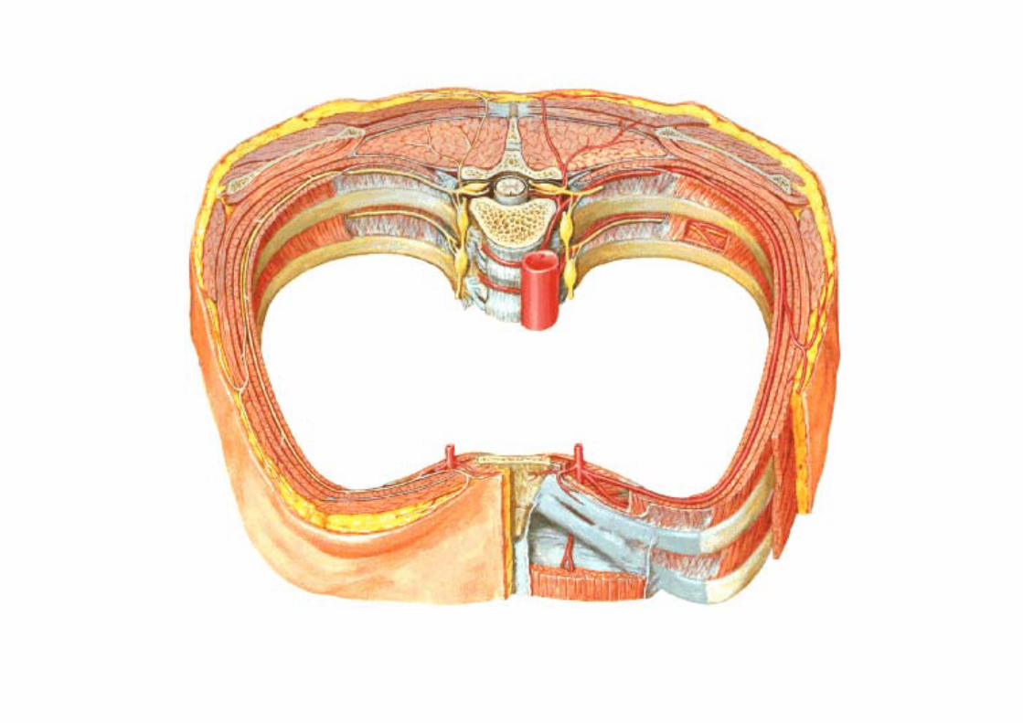
Boundaries of a typical I/c space – 3rd to 6th• Above – Sharp lower margin of upper rib & its cartilage• Below – Blunt upper margin of lower rib & its cartilage• In front – Lateral border of sternum b/w costal notches• Behind – Body of corresponding thoracic vertebra

Intercostal muscles
Arranged in three sheets from outside inward• External Intercostal• Internal Intercostal• Transverses thoracis – intercostalis intimi
Subcostalissternocostalis
Main actionPrevent retraction during inspiration & bulgingduring expiration of the intercostal spaces
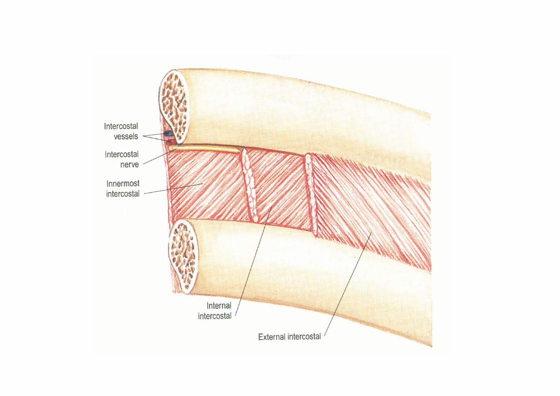
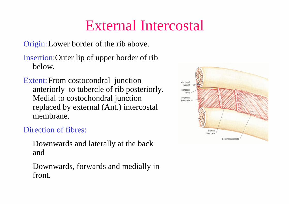
External IntercostalOrigin:Lower border of the rib above.
Insertion:Outer lip of upper border of rib below.
Extent:From costocondral junction anteriorly to tubercle of rib posteriorly. Medial to costochondral junction replaced by external (Ant.) intercostal membrane.
Direction of fibres:
Downwards and laterally at the back and
Downwards, forwards and medially in front.
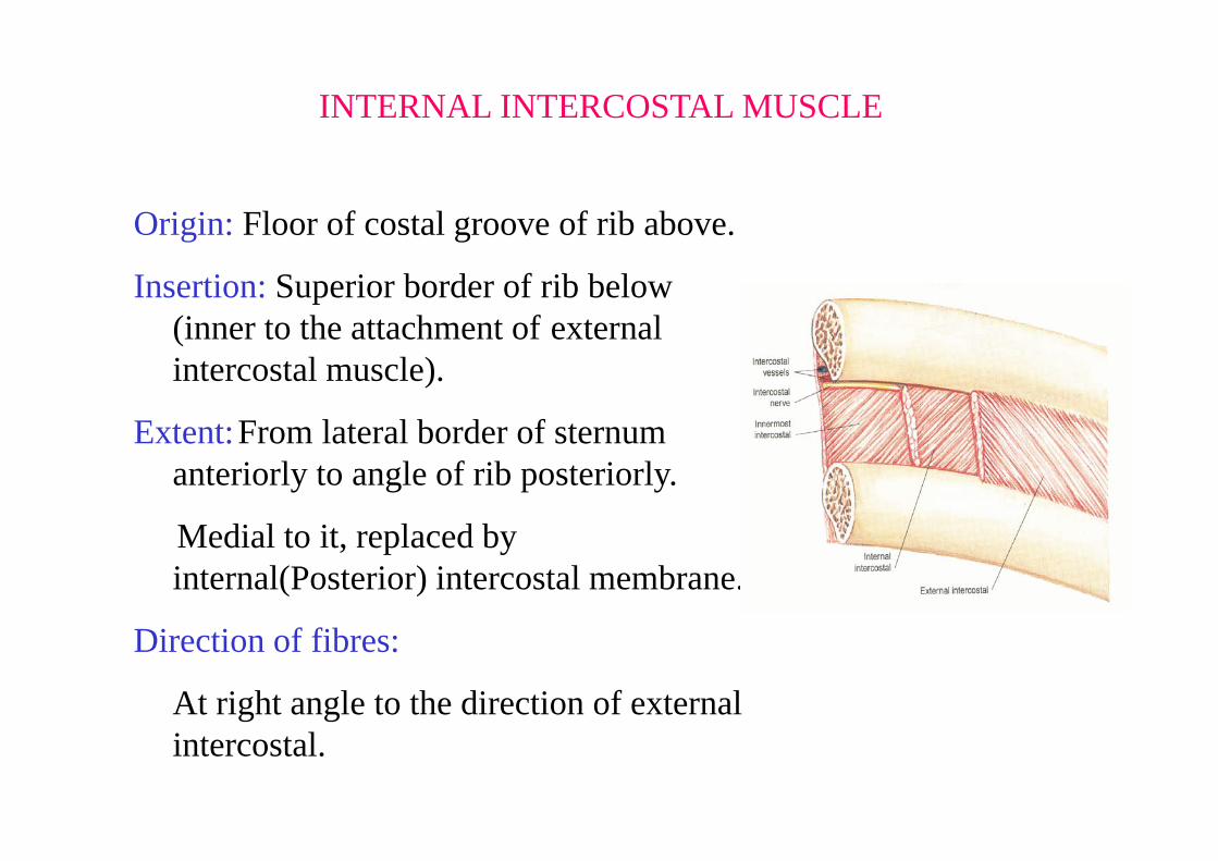
INTERNAL INTERCOSTAL MUSCLE
Origin: Floor of costal groove of rib above.
Insertion: Superior border of rib below (inner to the attachment of external intercostal muscle).
Extent:From lateral border of sternum anteriorly to angle of rib posteriorly.
Medial to it, replaced by internal(Posterior) intercostal membrane.
Direction of fibres:
At right angle to the direction of external intercostal.
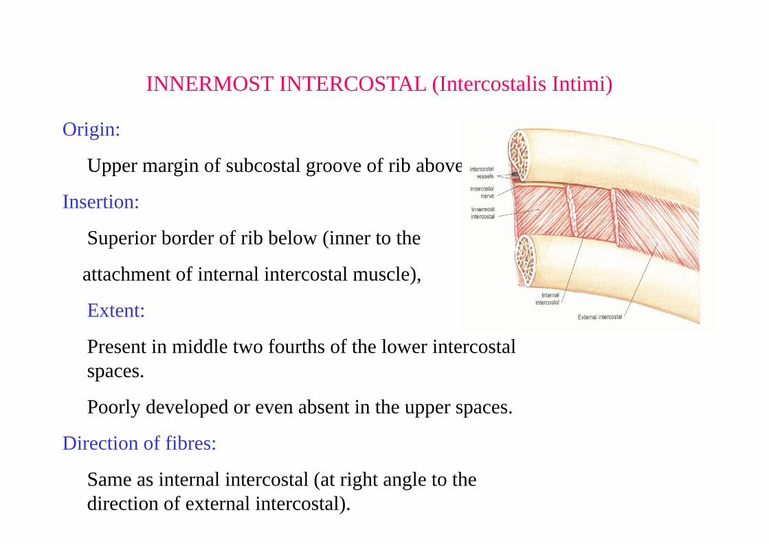
INNERMOST INTERCOSTAL (Intercostalis Intimi)
Origin:
Upper margin of subcostal groove of rib above.
Insertion:
Superior border of rib below (inner to the
attachment of internal intercostal muscle),
Extent:
Present in middle two fourths of the lower intercostal spaces.
Poorly developed or even absent in the upper spaces.
Direction of fibres:
Same as internal intercostal (at right angle to the direction of external intercostal).
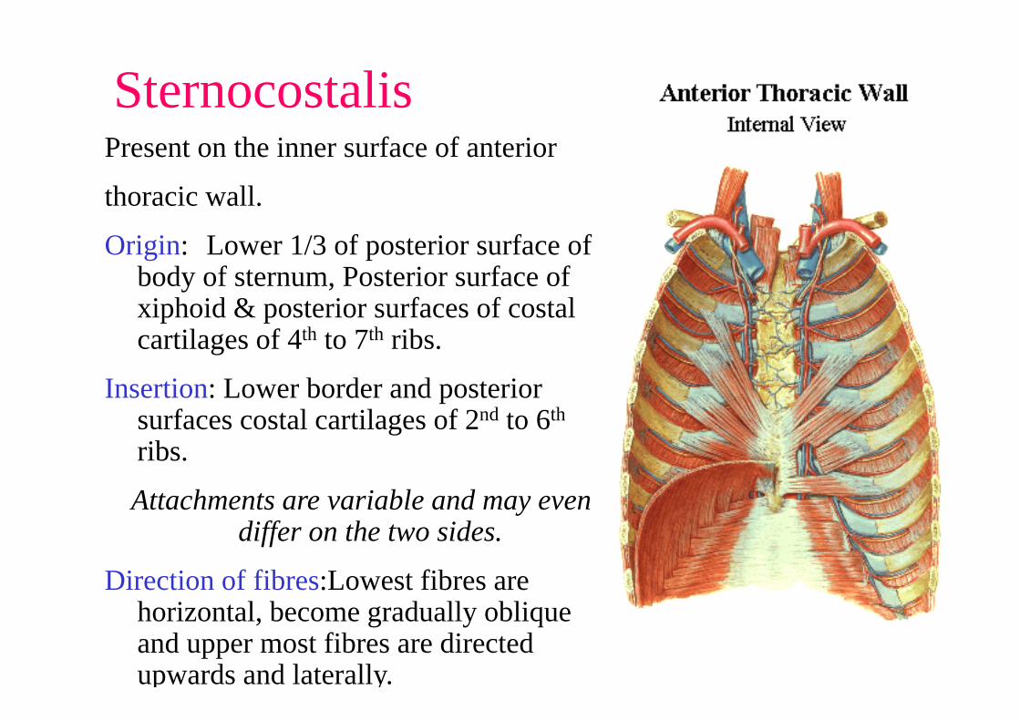
SternocostalisPresent on the inner surface of anterior
thoracic wall.
Origin: Lower 1/3 of posterior surface of body of sternum, Posterior surface of xiphoid & posterior surfaces of costal cartilages of 4th to 7th ribs.
Insertion: Lower border and posterior surfaces costal cartilages of 2nd to 6th
ribs.
Attachments are variable and may even differ on the two sides.
Direction of fibres:Lowest fibres are horizontal, become gradually oblique and upper most fibres are directed upwards and laterally.
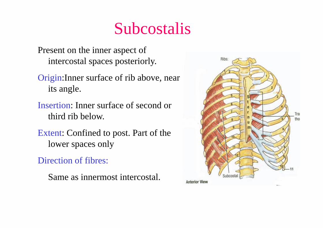
SubcostalisPresent on the inner aspect of
intercostal spaces posteriorly.
Origin:Inner surface of rib above, near its angle.
Insertion: Inner surface of second or third rib below.
Extent: Confined to post. Part of the lower spaces only
Direction of fibres:
Same as innermost intercostal.
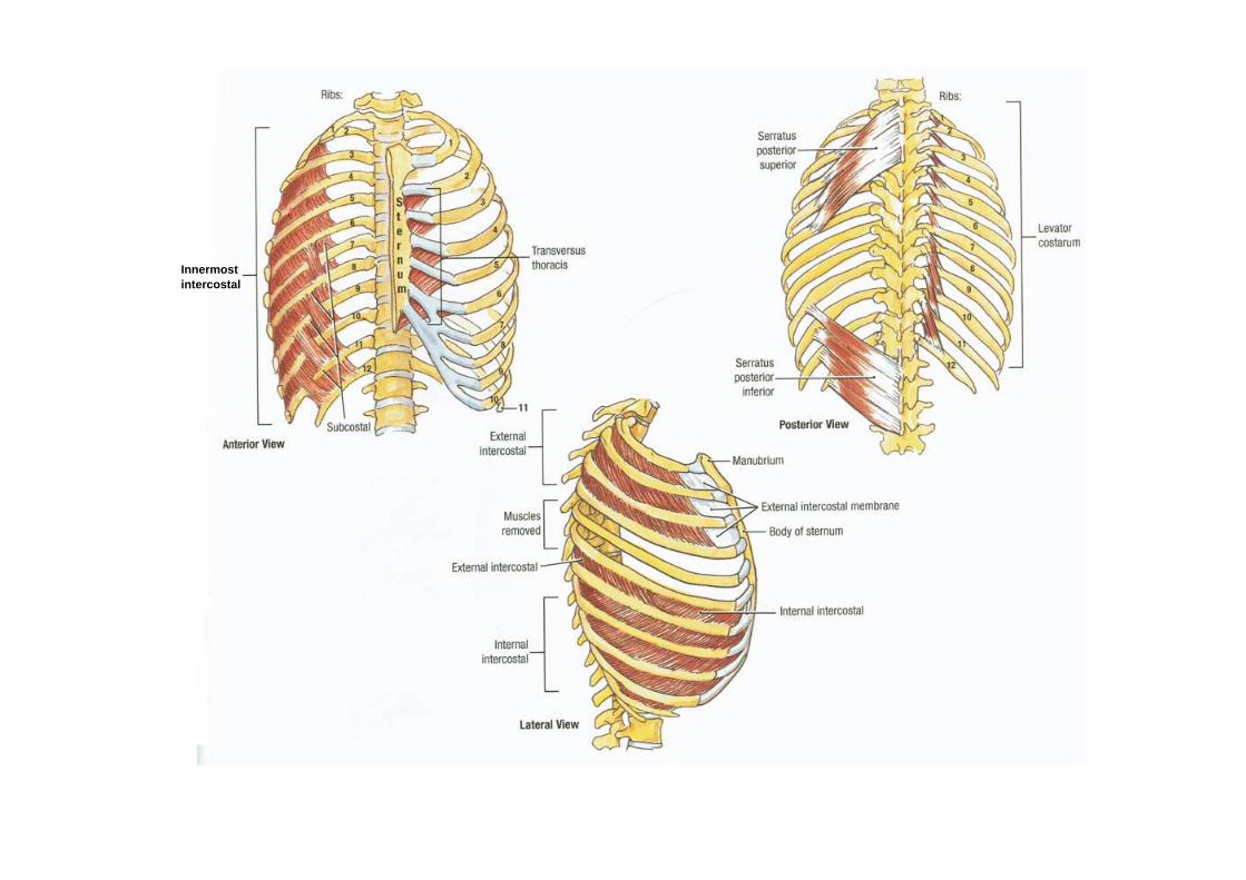
Innermostintercostal
Actions• Ext. intercostal-Inspiration, moves ribs superiorly• Int. intercostal- Expiration, moves ribs inferiorly• Innermost intercostal-Expiration• Subcostales – depress ribs• S.P.S elevates sup. 4 ribs, raising the sternum
and AP diameter• S.P.I. depresses the inf. Ribs, so prevents then
to be picked sup. By dia.• Transverse throcis• Lev. Costarum unimportant
Intercostal vessels
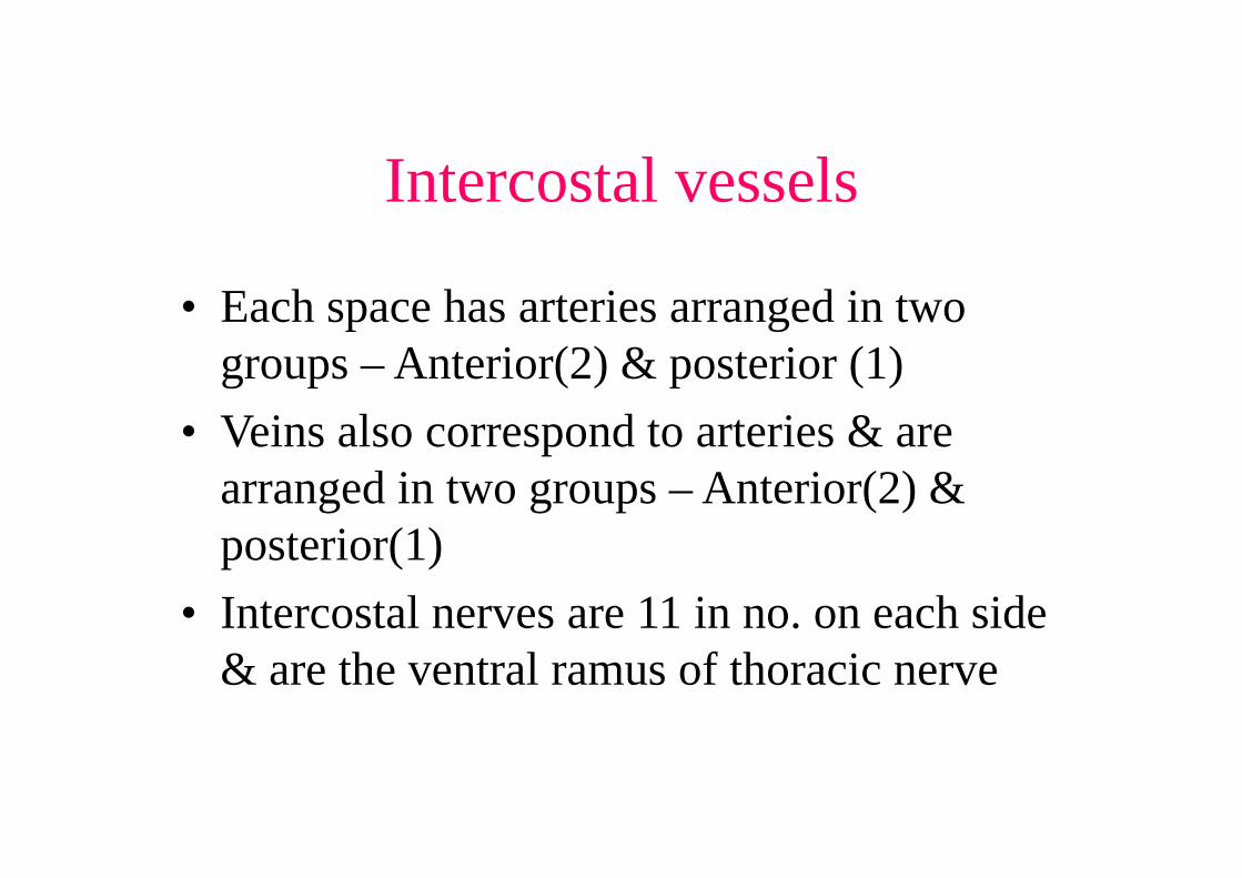
• Each space has arteries arranged in two groups – Anterior(2) & posterior (1)
• Veins also correspond to arteries & are arranged in two groups – Anterior(2) & posterior(1)
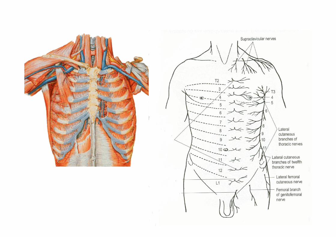
• Intercostal nerves are 11 in no. on each side & are the ventral ramus of thoracic nerve
Intercostal Arteries
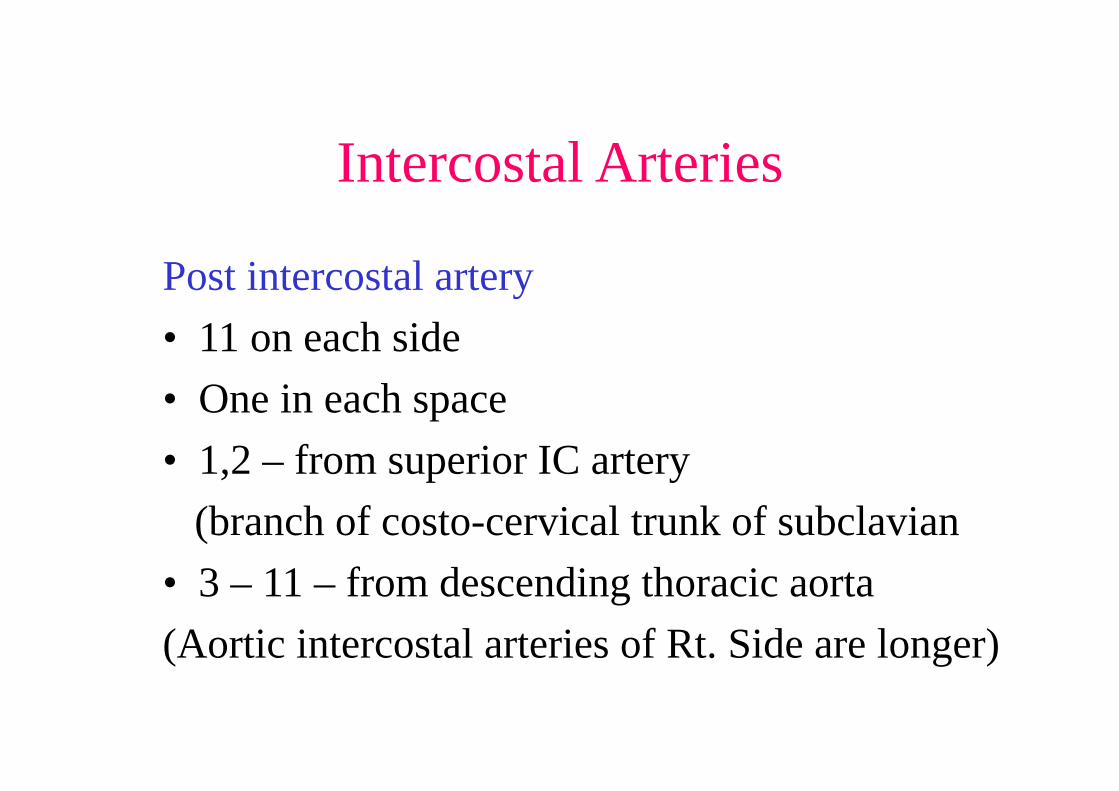
Post intercostal artery• 11 on each side • One in each space• 1,2 – from superior IC artery
(branch of costo-cervical trunk of subclavian• 3 – 11 – from descending thoracic aorta(Aortic intercostal arteries of Rt. Side are longer)


Post. Intercostal artery
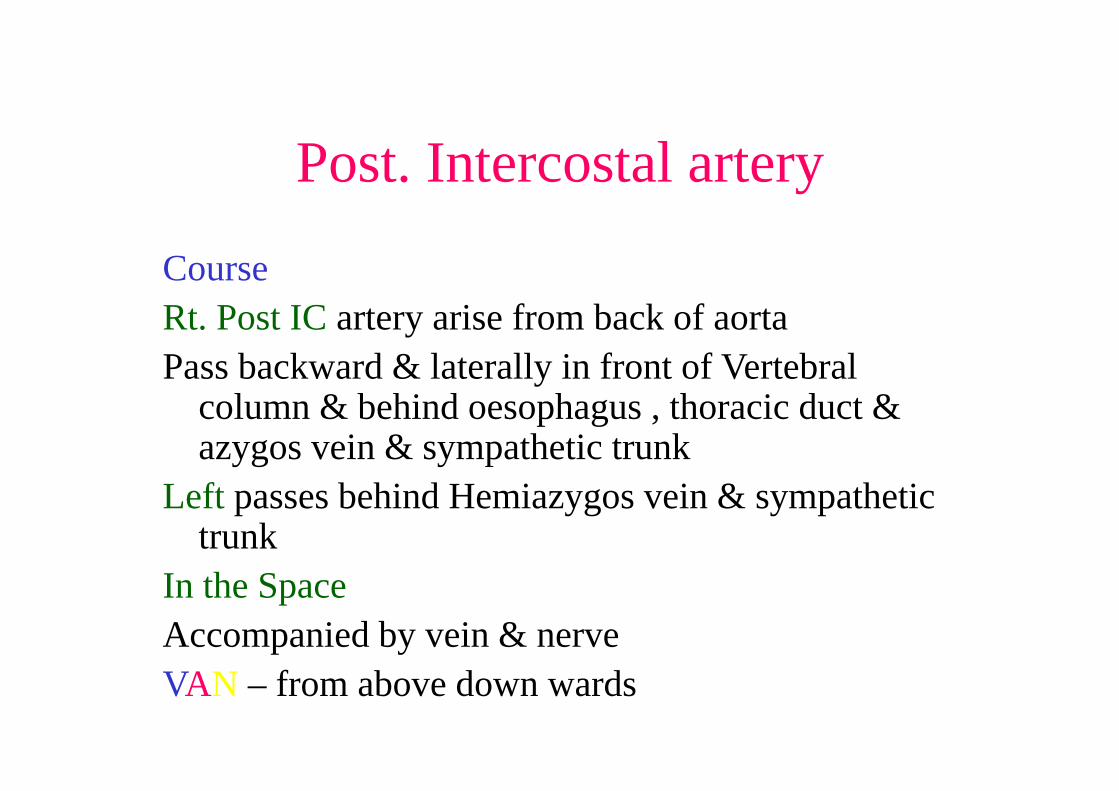
CourseRt. Post IC artery arise from back of aortaPass backward & laterally in front of Vertebral
column & behind oesophagus , thoracic duct & azygos vein & sympathetic trunk
Left passes behind Hemiazygos vein & sympathetic trunk
In the SpaceAccompanied by vein & nerveVAN – from above down wards

Post. Intercostal artery
• Each artery passes upward & laterally toward angle of upper rib
• Run along the costal groove between 2nd & 3rd
layer• Give a collateral branch at angle of rib &main
branch continue & anastomose with upper anterior intercostal artery & collateral with lower ant. IC artery at costochondral junction

Anterior Intercostal artery
• Present in all spaces except last two which are open in front
1-6 arise from internal thoracic artery7-9 from musculophrenic arteryEach space has two (upper & lower)
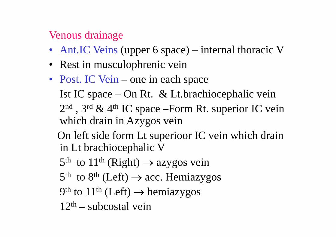
Venous drainage• Ant.IC Veins (upper 6 space) – internal thoracic V• Rest in musculophrenic vein• Post. IC Vein – one in each space
Ist IC space – On Rt. & Lt.brachiocephalic vein 2nd , 3rd & 4th IC space –Form Rt. superior IC vein which drain in Azygos vein On left side form Lt superioor IC vein which drain in Lt brachiocephalic V5th to 11th (Right) azygos vein5th to 8th (Left) acc. Hemiazygos 9th to 11th (Left) hemiazygos12th – subcostal vein
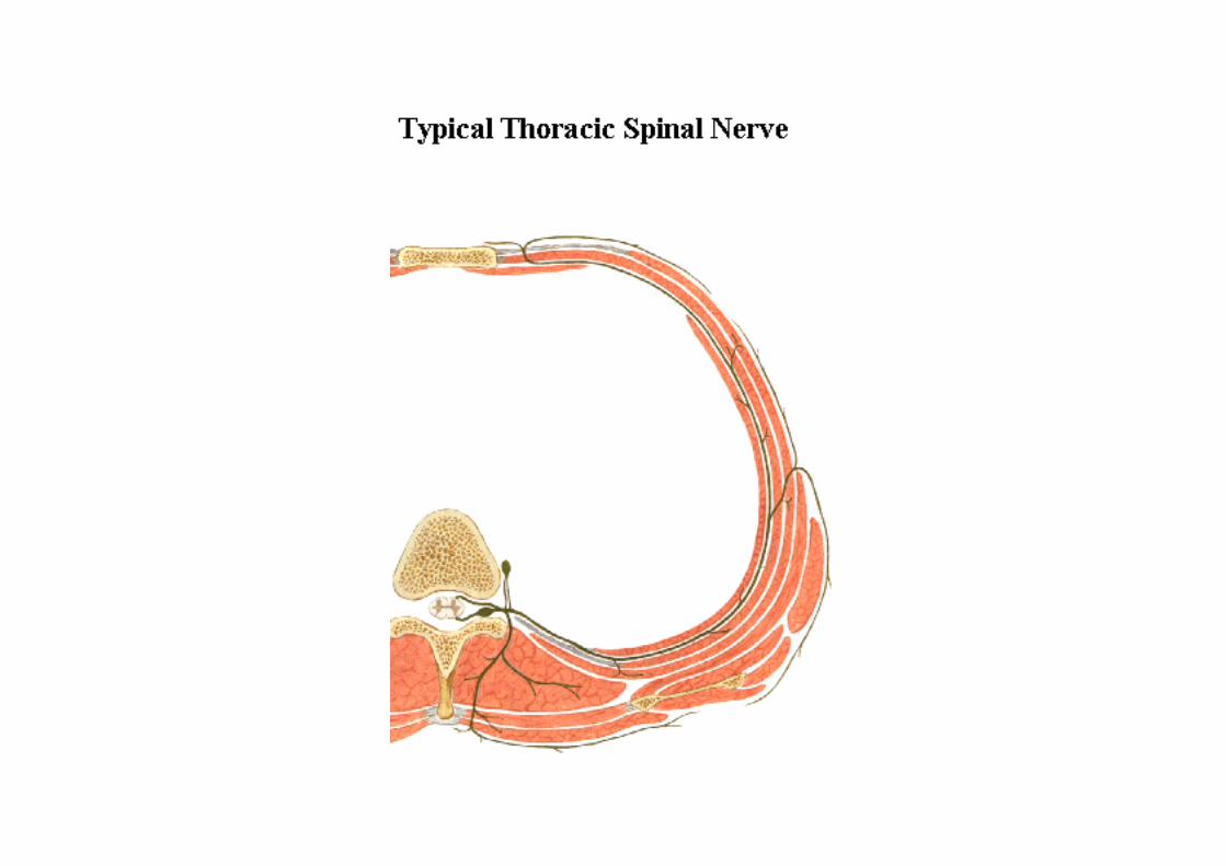
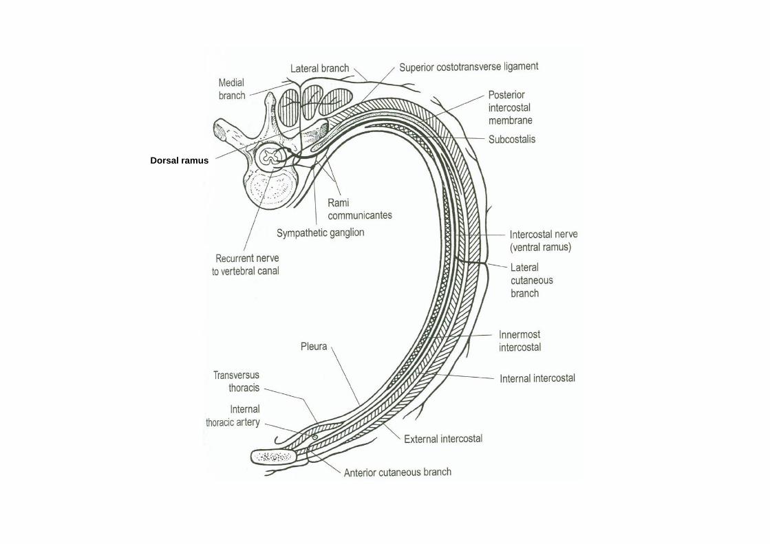
Dorsal ramus