INTEGUMENTARY SYSTEM - Folsom Cordova Unified School … 4... · INTEGUMENTARY SYSTEM. Body...
32
INTEGUMENTARY SYSTEM
Transcript of INTEGUMENTARY SYSTEM - Folsom Cordova Unified School … 4... · INTEGUMENTARY SYSTEM. Body...
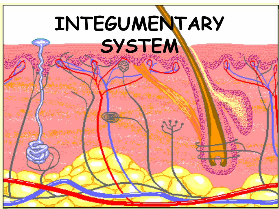
INTEGUMENTARY SYSTEM
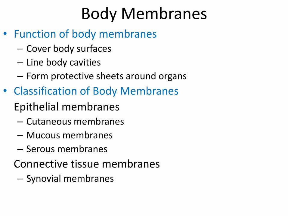
Body Membranes• Function of body membranes
– Cover body surfaces
– Line body cavities
– Form protective sheets around organs
• Classification of Body Membranes
Epithelial membranes– Cutaneous membranes
– Mucous membranes
– Serous membranes
Connective tissue membranes– Synovial membranes
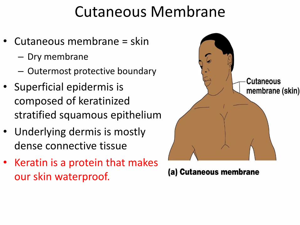
Cutaneous Membrane
• Cutaneous membrane = skin
– Dry membrane
– Outermost protective boundary
• Superficial epidermis is composed of keratinized stratified squamous epithelium
• Underlying dermis is mostly dense connective tissue
• Keratin is a protein that makes our skin waterproof.

Mucous Membranes• Surface epithelium type
depends on site
– Stratified squamous epithelium (mouth, esophagus)
– Simple columnar epithelium (rest of digestive tract)
• Lines all body cavities that open to the exterior body surface
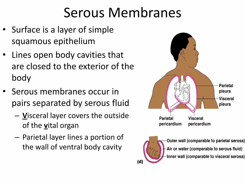
Serous Membranes• Surface is a layer of simple
squamous epithelium
• Lines open body cavities that are closed to the exterior of the body
• Serous membranes occur in pairs separated by serous fluid
– Visceral layer covers the outside of the vital organ
– Parietal layer lines a portion of the wall of ventral body cavity

• Specific serous membranes
– Peritoneum
• Abdominal cavity
– Pleura
• Around the lungs
– Pericardium
• Around the heart

Connective Tissue Membrane• Synovial membrane
– Connective tissue only
– Lines fibrous capsules surrounding joints
– Secretes a lubricating fluid
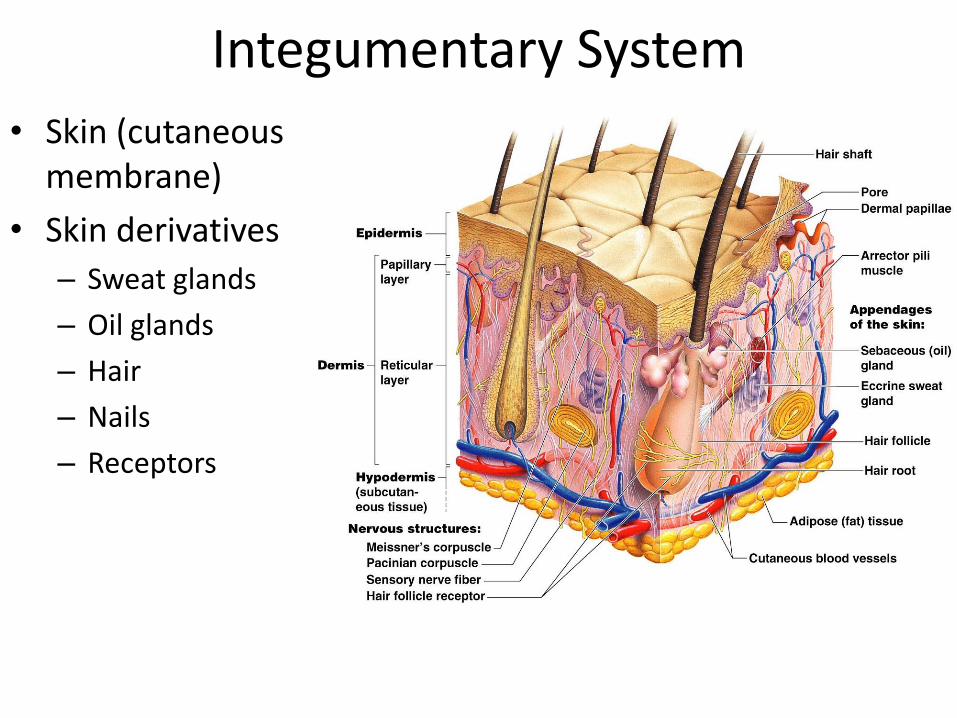
Integumentary System
• Skin (cutaneous membrane)
• Skin derivatives
– Sweat glands
– Oil glands
– Hair
– Nails
– Receptors
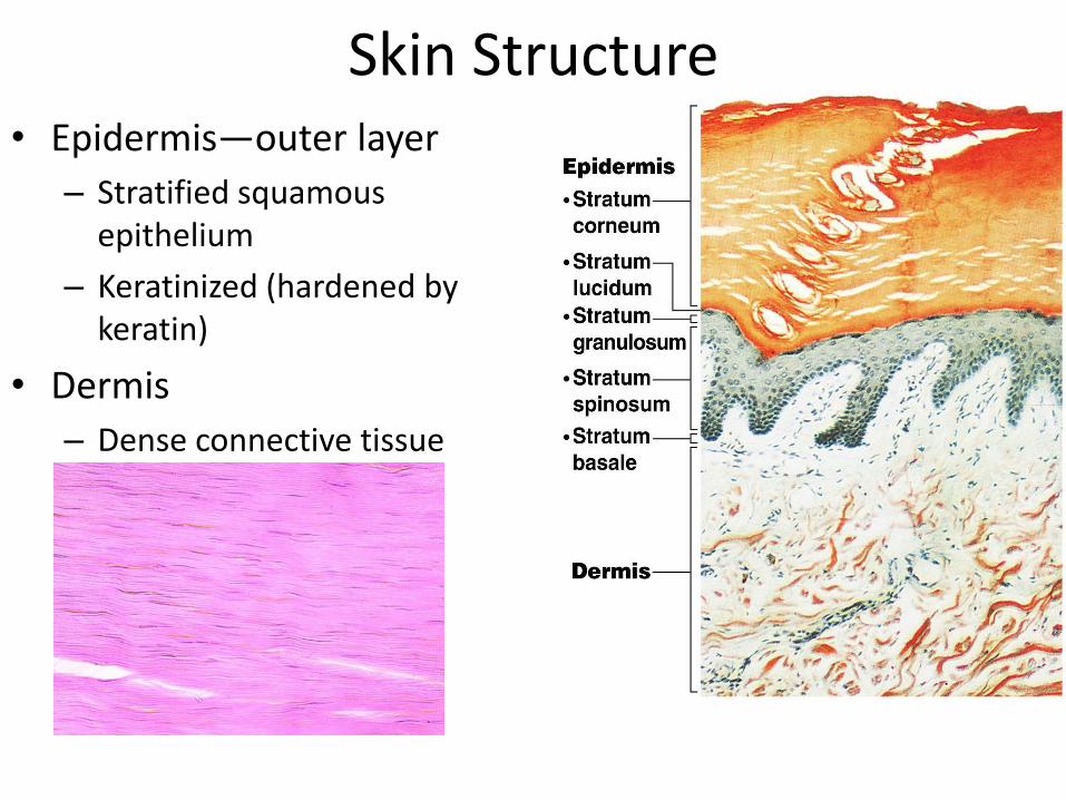
Skin Structure• Epidermis—outer layer
– Stratified squamous epithelium
– Keratinized (hardened by keratin)
• Dermis
– Dense connective tissue
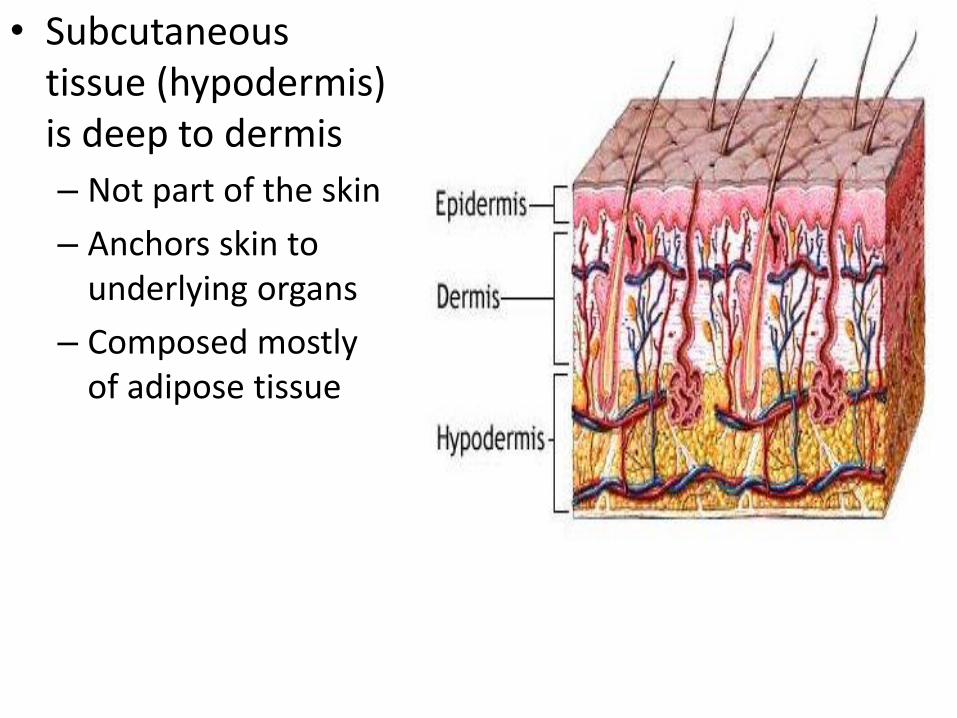
• Subcutaneous tissue (hypodermis) is deep to dermis
– Not part of the skin
– Anchors skin to underlying organs
– Composed mostly of adipose tissue

The Skin*Primary organ of Integumentary System & largest body organ*
Structure:→Epidermis→ Dermis→ Subcutaneous (hypodermis)
Appendages:→ Hair→ Receptors→ Nails→ Skin Glands
-Sudoriferous gland (sweat)-Sebaceous gland (oil)
Function: → Protection→ Temperature Regulation→ Sense organ activity
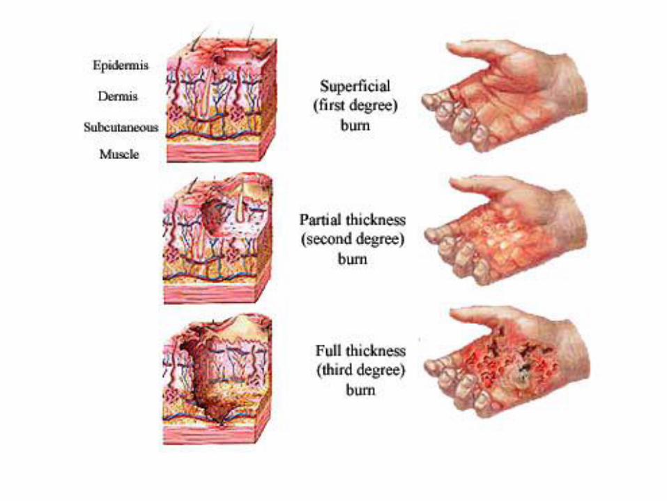
Burns:→Surface area estimation→Classification
Layers of the Epidermis• Stratum basale (stratum germinativum)
– Deepest layer of epidermis
– Lies next to dermis
– Cells undergoing mitosis
– Daughter cells are pushed upward to become the more superficial layers
• Stratum spinosum
• Stratum granulosum
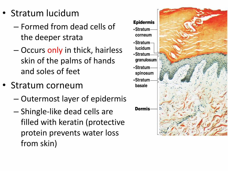
• Stratum lucidum
– Formed from dead cells of the deeper strata
– Occurs only in thick, hairless skin of the palms of hands and soles of feet
• Stratum corneum
– Outermost layer of epidermis
– Shingle-like dead cells are filled with keratin (protective protein prevents water loss from skin)
• Come up with a pneumonic device to help you remember the layers of the epidermis
• (either superficial to deep, as listed, or deep to superficial)
• Corneum
• Lucidum
• Granulosum
• Spinosum
• Basale
Melanin• Pigment (melanin) produced by melanocytes
• Melanocytes are mostly in the stratum basale
• Color is yellow to brown to black
• Amount of melanin produced depends upon genetics and exposure to sunlight

Dermis• Two layers
– Papillary layer (upper dermal region)• Projections called dermal
papillae (form fingerprints)– Some contain capillary loops
– Other house pain receptors and touch receptors
– Reticular layer (deepest skin layer)• Blood vessels
• Sweat and oil glands
• Deep pressure receptors
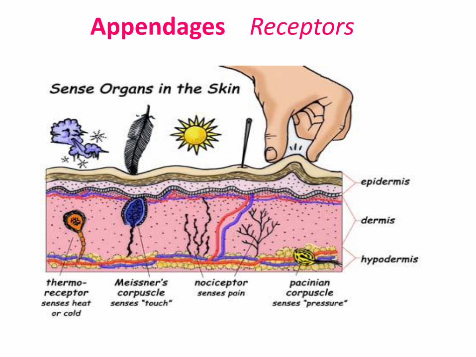
Appendages Receptors
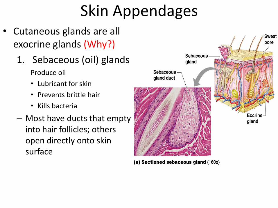
Skin Appendages• Cutaneous glands are all
exocrine glands (Why?)
1. Sebaceous (oil) glandsProduce oil
• Lubricant for skin
• Prevents brittle hair
• Kills bacteria
– Most have ducts that empty into hair follicles; others open directly onto skin surface
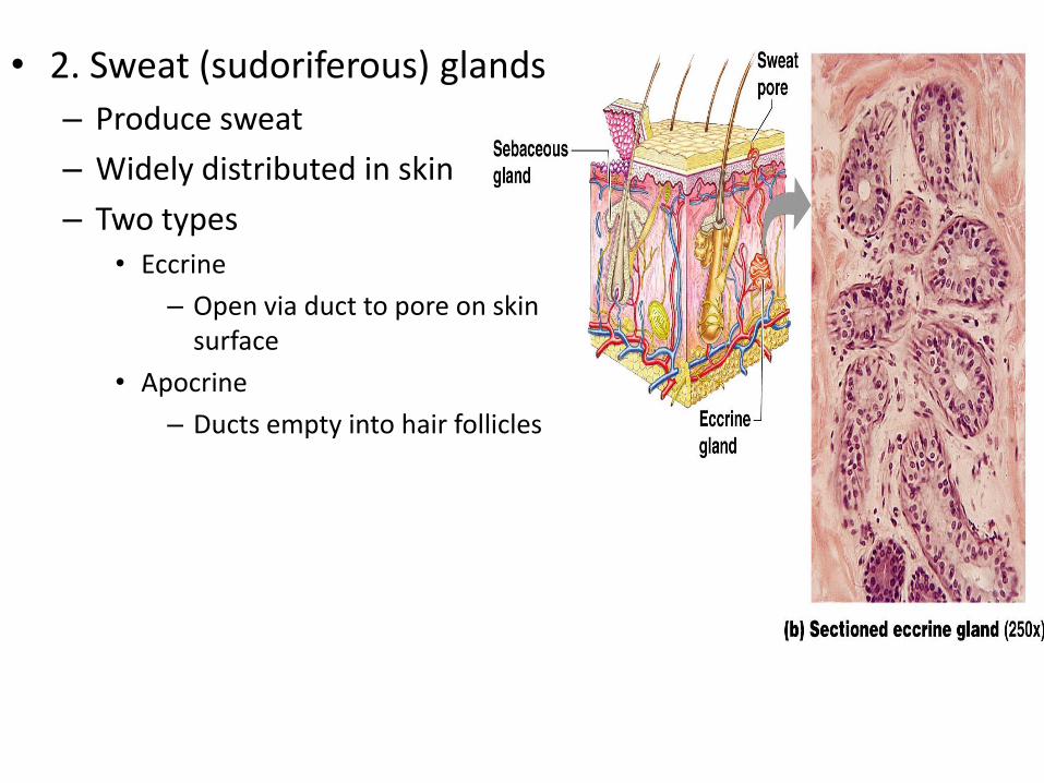
• 2. Sweat (sudoriferous) glands
– Produce sweat
– Widely distributed in skin
– Two types• Eccrine
– Open via duct to pore on skin surface
• Apocrine
– Ducts empty into hair follicles
• Composition of sweat– Mostly water
– Salts and vitamin C
– Some metabolic waste
– Fatty acids and proteins (apocrine only)
• Function– Helps dissipate excess heat
– Excretes waste products
– Acidic nature inhibits bacteria growth
• Odor is from associated bacteria

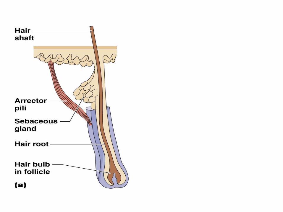
• Hair
– Produced by hair follicle
– Consists of hard keratinized epithelial cells
– Melanocytes provide pigment for hair color

• Nails
– Scale-like modifications of the epidermis
• Heavily keratinized
– Stratum basale extends beneath the nail bed
• Responsible for growth
– Lack of pigment makes them colorless
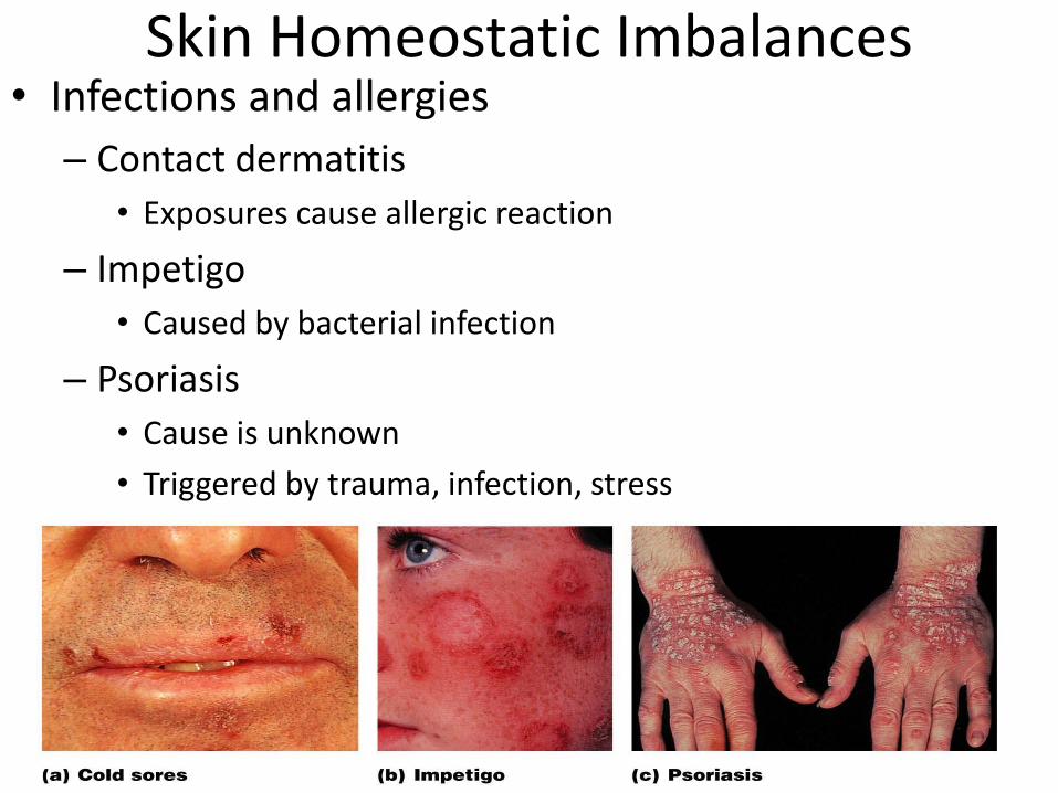
Skin Homeostatic Imbalances• Infections and allergies
– Contact dermatitis
• Exposures cause allergic reaction
– Impetigo
• Caused by bacterial infection
– Psoriasis
• Cause is unknown
• Triggered by trauma, infection, stress
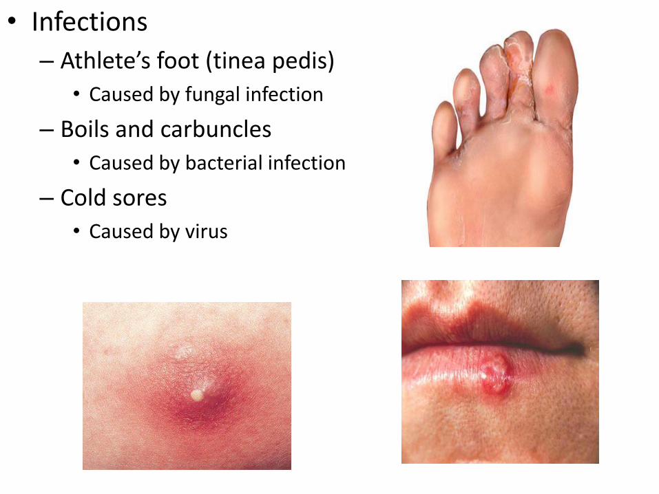
• Infections
– Athlete’s foot (tinea pedis)
• Caused by fungal infection
– Boils and carbuncles
• Caused by bacterial infection
– Cold sores
• Caused by virus
Skin Cancer
• Cancer—abnormal cell mass
• Classified two ways
– Benign
• Does not spread (encapsulated)
– Malignant
• Metastasized (moves) to other parts of the body
• Skin cancer is the most common type of cancer
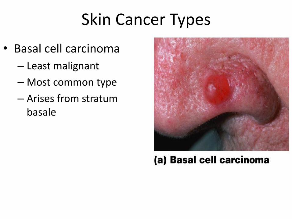
Skin Cancer Types
• Basal cell carcinoma
– Least malignant
– Most common type
– Arises from stratum basale
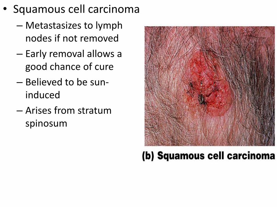
• Squamous cell carcinoma
– Metastasizes to lymph nodes if not removed
– Early removal allows a good chance of cure
– Believed to be sun-induced
– Arises from stratum spinosum
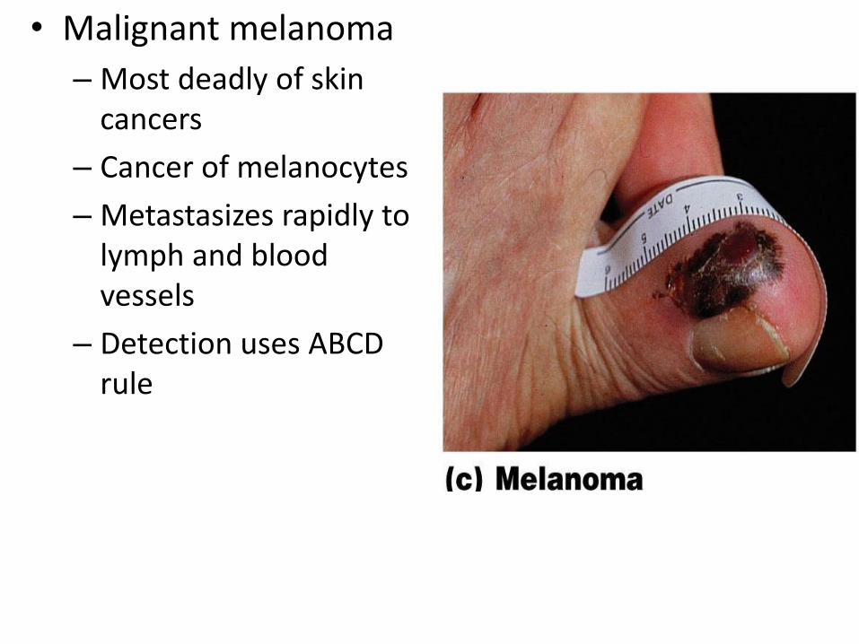
• Malignant melanoma
– Most deadly of skin cancers
– Cancer of melanocytes
– Metastasizes rapidly to lymph and blood vessels
– Detection uses ABCD rule
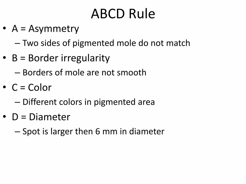
ABCD Rule• A = Asymmetry
– Two sides of pigmented mole do not match
• B = Border irregularity
– Borders of mole are not smooth
• C = Color
– Different colors in pigmented area
• D = Diameter
– Spot is larger then 6 mm in diameter
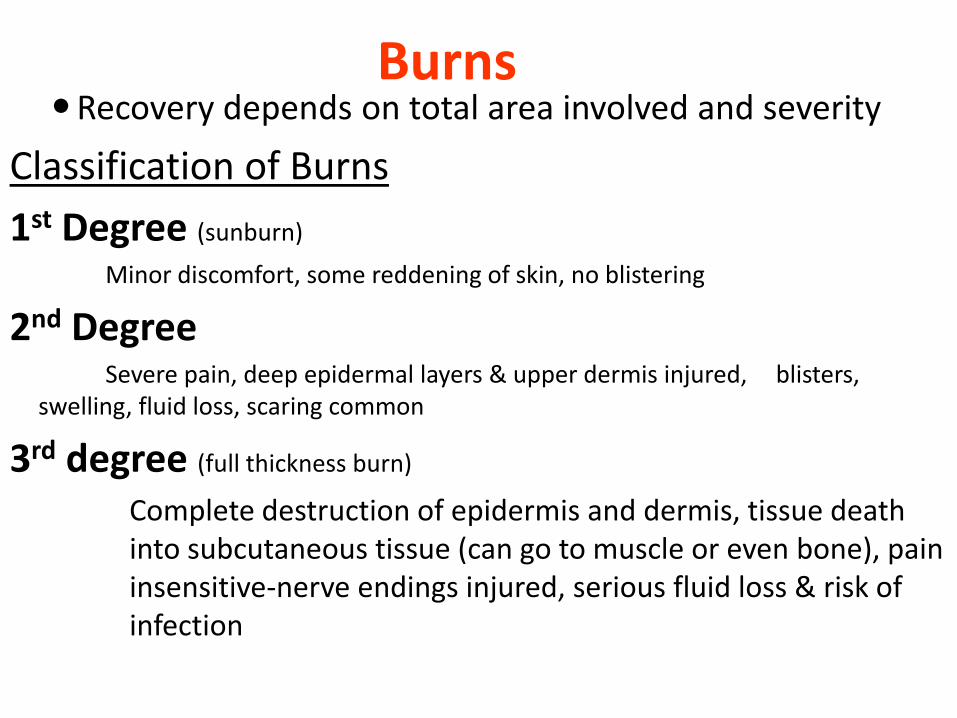
BurnsRecovery depends on total area involved and severity
Classification of Burns
1st Degree (sunburn)
Minor discomfort, some reddening of skin, no blistering
2nd Degree Severe pain, deep epidermal layers & upper dermis injured, blisters,
swelling, fluid loss, scaring common
3rd degree (full thickness burn)
Complete destruction of epidermis and dermis, tissue death into subcutaneous tissue (can go to muscle or even bone), pain insensitive-nerve endings injured, serious fluid loss & risk of infection