Insulin-loaded alginate microspheres for oral delivery – Effect of polysaccharide reinforcement on...
7
Insulin-loaded alginate microspheres for oral delivery – Effect of polysaccharide reinforcement on physicochemical properties and release profile Susana Martins, Bruno Sarmento * , Eliana B. Souto, Domingos C. Ferreira Department of Pharmaceutical Technology, Faculty of Pharmacy, University of Porto, Porto, Portugal Received 27 April 2006; received in revised form 11 January 2007; accepted 12 February 2007 Available online 20 February 2007 Abstract Oral administration of insulin requires protein protection from degradation in the gastric environment and its absorption improve- ment in the intestinal tract. To achieve this objective several types of microspheres composed of alginate, chitosan and dextran sulphate have been prepared by ionotropic gelation. Parameters such as the mean particle size, swelling behaviour, insulin encapsulation effi- ciency, loading capacity and release profiles in simulated gastric and intestinal fluids have been compared for the systems developed. In this study, attempts have been made to increase insulin protection and to improve its release from microspheres by reinforcing the alginate matrix with chitosan and/or dextran sulphate. Dextran sulphate was able to avoid insulin release at pH 1.2, protecting the pro- tein from the acidic environment and reducing the total insulin released at pH 6.8. This effect was explained by an interaction between the permanent negatively charged groups of dextran sulphate and insulin molecules. Ó 2007 Elsevier Ltd. All rights reserved. Keywords: Alginate; Chitosan; Dextran sulphate; Insulin; Microspheres; Gastric retention 1. Introduction The development of improved delivery systems for oral administration of insulin is a field of great interest in the treatment of diabetes mellitus to overcome the problem of daily subcutaneous injections. Oral administration is a more convenient approach considering the pain and possi- ble infections caused by injections, thus leading to a higher patient compliance. This administration route prevents the occasional hyperinsulinaemia observed by subcutaneous administration, since the principal organ in glucose homeo- stasis is the liver and this should obviously be the prime tar- get for intervention (Arbit, 2004). A successful oral drug delivery system needs to be developed on the basis of increased resistance of the drug carrier against gastrointes- tinal enzymes and pH gradients (i.e. from 1to 3 in the stom- ach to 6 to7 in the intestine). In addition, time-controlled release is also a pre-requisite for such oral drug delivery systems (Janes & Alonso, 2003). It is known that microencapsulation of labile proteins improves their protection against gastric pH and enzymatic attack, providing a controlled release profile of the entrapped molecules (Onal & Zihnioglu, 2002), and further enhancement of their intestinal absorption. Microencapsu- lation of proteins can be performed using alginate, chitosan and dextran sulphate. Being biodegradable, biocompatible, non-toxic and mucoadhesive macromolecules these natural polysaccharides have been widely used in the formulation of several drug delivery systems (Lin, Yu, & Chien, 2005; Onal & Zihnioglu, 2002; Yu et al., 2004). Alginate is an anionic polysaccharide consisting of var- ious ratios of guluronic and mannuronic acid units linked by glycosidic bonds. Gelation can be induced by cross-link- ing the guluronic acid units with di- or polyvalent cations such as calcium. Major disadvantages of alginate beads 0144-8617/$ - see front matter Ó 2007 Elsevier Ltd. All rights reserved. doi:10.1016/j.carbpol.2007.02.012 * Corresponding author. Tel.: +351 222078949; fax: +351 222003977. E-mail address: bruno.sarmento@ff.up.pt (B. Sarmento). www.elsevier.com/locate/carbpol Carbohydrate Polymers 69 (2007) 725–731
-
Upload
susana-martins -
Category
Documents
-
view
218 -
download
4
Transcript of Insulin-loaded alginate microspheres for oral delivery – Effect of polysaccharide reinforcement on...

www.elsevier.com/locate/carbpol
Carbohydrate Polymers 69 (2007) 725–731
Insulin-loaded alginate microspheres for oral delivery – Effectof polysaccharide reinforcement on physicochemical
properties and release profile
Susana Martins, Bruno Sarmento *, Eliana B. Souto, Domingos C. Ferreira
Department of Pharmaceutical Technology, Faculty of Pharmacy, University of Porto, Porto, Portugal
Received 27 April 2006; received in revised form 11 January 2007; accepted 12 February 2007Available online 20 February 2007
Abstract
Oral administration of insulin requires protein protection from degradation in the gastric environment and its absorption improve-ment in the intestinal tract. To achieve this objective several types of microspheres composed of alginate, chitosan and dextran sulphatehave been prepared by ionotropic gelation. Parameters such as the mean particle size, swelling behaviour, insulin encapsulation effi-ciency, loading capacity and release profiles in simulated gastric and intestinal fluids have been compared for the systems developed.In this study, attempts have been made to increase insulin protection and to improve its release from microspheres by reinforcing thealginate matrix with chitosan and/or dextran sulphate. Dextran sulphate was able to avoid insulin release at pH 1.2, protecting the pro-tein from the acidic environment and reducing the total insulin released at pH 6.8. This effect was explained by an interaction between thepermanent negatively charged groups of dextran sulphate and insulin molecules.� 2007 Elsevier Ltd. All rights reserved.
Keywords: Alginate; Chitosan; Dextran sulphate; Insulin; Microspheres; Gastric retention
1. Introduction
The development of improved delivery systems for oraladministration of insulin is a field of great interest in thetreatment of diabetes mellitus to overcome the problemof daily subcutaneous injections. Oral administration is amore convenient approach considering the pain and possi-ble infections caused by injections, thus leading to a higherpatient compliance. This administration route prevents theoccasional hyperinsulinaemia observed by subcutaneousadministration, since the principal organ in glucose homeo-stasis is the liver and this should obviously be the prime tar-get for intervention (Arbit, 2004). A successful oral drugdelivery system needs to be developed on the basis ofincreased resistance of the drug carrier against gastrointes-tinal enzymes and pH gradients (i.e. from 1to 3 in the stom-
0144-8617/$ - see front matter � 2007 Elsevier Ltd. All rights reserved.
doi:10.1016/j.carbpol.2007.02.012
* Corresponding author. Tel.: +351 222078949; fax: +351 222003977.E-mail address: [email protected] (B. Sarmento).
ach to 6 to7 in the intestine). In addition, time-controlledrelease is also a pre-requisite for such oral drug deliverysystems (Janes & Alonso, 2003).
It is known that microencapsulation of labile proteinsimproves their protection against gastric pH and enzymaticattack, providing a controlled release profile of theentrapped molecules (Onal & Zihnioglu, 2002), and furtherenhancement of their intestinal absorption. Microencapsu-lation of proteins can be performed using alginate, chitosanand dextran sulphate. Being biodegradable, biocompatible,non-toxic and mucoadhesive macromolecules these naturalpolysaccharides have been widely used in the formulationof several drug delivery systems (Lin, Yu, & Chien, 2005;Onal & Zihnioglu, 2002; Yu et al., 2004).
Alginate is an anionic polysaccharide consisting of var-ious ratios of guluronic and mannuronic acid units linkedby glycosidic bonds. Gelation can be induced by cross-link-ing the guluronic acid units with di- or polyvalent cationssuch as calcium. Major disadvantages of alginate beads

726 S. Martins et al. / Carbohydrate Polymers 69 (2007) 725–731
are low drug encapsulation efficiency (EE) and rapidrelease of the loaded molecules (Halder, Maiti, & Sa,2005). Low EE and loading capacity (LC) of alginate beadsare attributed to the gel porosity, which is large enough tocause leakage of the loaded drugs and further drug diffu-sion from the gel network to the CaCl2 solution (Setty,Sahoo, & Sa, 2005). In order to modify the encapsulationparameters (EE and LC) and protein release profiles fromalginate beads, several polymers such as chitosan (Onal &Zihnioglu, 2002; Ramadas, Paul, Dileep, Anitha, & Shar-ma, 2000), dextran sulphate (Silva, Ribeiro, Veiga, &Sousa, 2006), pectin (Bajpai, Bajpai, & Mishra, 2006; El-Kamel, Al-Gohary, & Hosny, 2003) and methylcellulose(El-Kamel et al., 2003) have been used in combination withsodium alginate. Chitosan, a positively charged polymer,interacts with negatively charged alginate to form a poly-electrolyte complex membrane, which is able to controlthe drug release profile (Shu & Zhu, 2002). Dextran sul-phate is a negatively charged polymer that shows a highaffinity for proteins (Fuentes et al., 2004).
This paper assesses the physicochemical properties ofmicrospheres composed solely of alginate, in comparisonto those that have been reinforced with chitosan or dextransulphate, or a combination of both polysaccharides, on thegastrointestinal retention of insulin.
2. Materials and methods
2.1. Materials
Sodium alginate of low-G content (FG = 0.39), low vis-cosity, chitosan of low molecular weight (MW � 50 kDa),and calcium chloride were purchased from Sigma (Portu-gal). Dextran sulphate (MW � 500 kDa) was obtained fromPKC� (Denmark). Acetic acid was acquired from Pronalab(Lisboa). Human zinc–insulin (lot RS0325, 7.0 mg lyophi-lized human biosynthetic insulin per vial) was a generousgift from Lilly Portugal.
2.2. Microspheres preparation
Microspheres were prepared by ionotropic gelation ofalginate with Ca2+ ions. The powders (alginate, dextransulphate, CaCl2 and chitosan) were weighed separatelyand further dissolved in water under magnetic stirring toprepare stock solutions of alginate 2% (w/v), CaCl2500 mM, dextran sulphate 1% (w/v) and chitosan 1% in1% of acetic acid (w/v), respectively. To prepare alginate/insulin and alginate/insulin/dextran sulphate solutions,freshly prepared stock solutions of each compound weremixed gently to obtain a homogeneous final solution. Toavoid insulin fibrillation (one of the degradation mecha-nisms of insulin (Brange, Anderson, Laursen, Meyn, &Rasmussen, 1997)), stirring of the final solution has beenshortened. This assumption was made because insulinfibrillation was not observed to any significant extent in dif-ferent pH aqueous solutions at moderate temperature in
the absence of agitation (Sluzky, Tamada, Klibanov, &Langer, 1991). Then, alginate/insulin or alginate/insulin/dextran sulphate solutions were extruded dropwise througha needle with an internal diameter of 0.2 mm into a CaCl2or CaCl2–chitosan solution under magnetic stirring. Micro-spheres were additionally stirred for 15 min to allow thealginate gel to stabilize and then washed with deionizedwater. Particles were stored in a desiccator under vacuumand dried until constant weight.
2.3. Particle size analysis
The size of at least 100 fresh and dried microspheres wasroutinely measured by light microscopy using the magnifi-cation of 40·. The average diameter of 100 microsphereswas considered as the mean particle size.
2.4. Morphology
The morphology of microspheres has been studied byscanning electron microscopy (SEM) using the JEOLJSM-840 microscope (Japan). The samples were mountedon metal stubs, gold coated under vacuum and then exam-ined one day after production. Microsphere matrices werealso studied using transmission electron microscope(TEM). Microspheres were appropriately cut in transversalsection using a micrometer, treated with uranil acetate andobserved in a Zeiss EM 902A TEM microscope(Germany).
2.5. Swelling behaviour
The water-sorption behaviour was determined by swell-ing in deionized water an amount of 75 mg of accuratelyweighted dried microspheres at room temperature for 3and 24 h. The wet weight of the swollen particles was deter-mined by first blotting the dried systems with filter paper toremove surface water and then weighted immediately. Thepercentage of swelling was then calculated using the follow-ing equation:
S ¼ W e � W o
W o
� 100%
where We stands for the weight of the gel microspheres atswelling equilibrium and Wo for the initial weight of themicrospheres. Each swelling experiment was repeated threetimes and the average value was recorded.
2.6. Insulin encapsulation efficiency and loading capacity
The encapsulation efficiency (EE) was determined indi-rectly. The amount of insulin entrapped in the micro-spheres was calculated by the difference between the totalamount used to prepare the systems and the amount ofinsulin that remained in the aqueous phase after micro-spheres isolation.

Table 1Characterization of insulin-loaded microspheres prepared in a CaCl2100 mM solution dropwise from a needle of 0.20 mm intern diameter
Formulation Polysaccharideconcentrations(%)
Diameter (freshmicrospheres)(±SD, mm)
EE(±SD, %)
LC(±SD, %)
A Alginate 2 1.78 ± 0.09 80.0 ± 0.8 2.48 ± 0.01
B Alginate 2 1.83 ± 0.09 97.6 ± 0.1 2.88 ± 0.01Dextransulphate 0.5
C Alginate 2 1.85 ± 0.12 90.9 ± 1.7 2.81 ± 0.01Chitosan 0.5
D Alginate 2 2.13 ± 0.09 100.0 ± 1.8 3.00 ± 0.01Dextransulphate 0.5Chitosan 0.5
S. Martins et al. / Carbohydrate Polymers 69 (2007) 725–731 727
EE ¼ total amount of insulin� free insulin in supernatant
total amount of insulin� 100
The difference between the total amount of insulin initiallyused to prepare the microspheres and the amount of resid-ual non-entrapped insulin after microspheres isolation as apercentage of total microsphere dry mass is referred to asthe loading capacity (LC).
LC ¼ total amount of insulin� free insulin in supernatant
total weight of microspheres� 100
Insulin concentration was determined spectrophotometri-cally using the Coomassie Plus TM (Pierce, Rockford,USA) modified Bradford assay (Bradford, 1976). Briefly,insulin samples and Bradford reagent were admixed at1:1 (v/v) ratio in a 96-well plate and incubated at roomtemperature for 15 min. The absorbance was measured at595 nm on a thermomax plate reader (PowerWaveX; Bio-Tek, Winooski, VT, USA) against appropriate calibrationcurves.
2.7. Insulin release
Dried microspheres were placed on two different releasemedia, i.e. 20 ml of HCl pH 1.2 USP XXVI buffer (24 h/100 rpm), or on 20 ml of phosphate pH 6.8 USP XXVIbuffer (24 h/100 rpm). Aliquots of both release media werecollected at predetermined time intervals, being the samevolume replaced immediately with freshly prepared mediasolutions. The amount of insulin released from the micro-spheres was evaluated by Bradford assay.
3. Results and discussion
The use of natural polysaccharides as multiparticulatematrices for delivery of peptides and proteins has beenwidely documented in the scientific literature (Dumitriu& Chornet, 1998; Gombotz & Wee, 1998; Silva, Ribeiro,Figueiredo, Goncalves, & Veiga, 2006). These carriers havein general the potential to be used for targeting and con-trolled release of drugs due to their small size. In addition,the enhancement of drug absorption and bioavailabilitycan also be achieved due to this high surface to volumeratio, which is responsible for a much more intimate con-tact with the mucus layer and specific targeting of drugsto the absorption site.
In the present work, different alginate microspheres wereproduced by the typical external gelation (i.e. ionotropicgelation) method extruding alginate or alginate/dextransulphate solution into a calcium chloride bath. Table 1summarises the properties of different microspheresaccording to their polysaccharide concentrations.
When the matrix of alginate microspheres was rein-forced with dextran sulphate a slight increase of size from1.78 to 1.83 mm was detected (Table 1). Since the initialalginate concentration remained the same (2%), the size
increase (approx. 50 lm) was most likely a result of theincrease in viscosity due to the incorporation of 0.5% dex-tran sulphate which resulted in larger drops extruded fromthe needle before reaching the calcium gelation bath.Chitosan-coated microspheres also showed a size increasedue the contribution of the chitosan outside the layer.
A final alginate concentration of 2% (Table 1) was cho-sen on the basis that at lower initial alginate concentrationse.g. 0.5%–1.5% (w/v) were responsible for lower EE valuesfor insulin (data not shown), probably due to the creationof a less compact gel matrix, which is not able to retainhigh amounts of protein (Quong, Yeo, & Neufeld, 1999).Encapsulation parameters were higher in alginate/chitosanmicrospheres (90.0%) than in uncoated alginate micro-spheres (80.0%) most probably due the decrease of surfaceporosity of the final microspheres because positivelycharged amino groups of chitosan formed strong mem-branes through ionic interactions with carboxylic residuesof the alginate. Consequently, the insulin loss during gela-tion was lower than without chitosan coating.
The introduction of dextran sulphate into uncoated algi-nate microspheres and those coated with chitosan provideda higher retention of insulin than pure alginate matrices(Table 1A). The increase of EE promoted by dextran sul-phate could be explained by a higher network density caus-ing physical retention of the insulin inside themicrospheres. The electrostatic interaction between theprotein and the polyanion might also favour the retentionof insulin within the microsphere matrix.
Surface morphology and cross section information fordried microspheres have been obtained by SEM analysisand are shown in Figs. 1–3. All the carriers showed irregularshape and had a relatively smooth surface with some wrin-kles. The surface morphology and the internal structures ofdextran sulphate microspheres showed a typical porousstructure. On the contrary, alginate/dextran sulphate matrixseemed to be less porous. As expected, the spherical shape ofmicrospheres was lost after drying. These morphologicalchanges are common, especially for microspheres preparedwith low alginate concentration (Halder et al., 2005).

Fig. 1. SEM micrographs of insulin-loaded alginate microspheres (left) and detail of their porous surface (right).
Fig. 2. SEM micrographs of insulin-loaded alginate/chitosan microspheres (left) and cross section showing chitosan (up) and alginate (down) portions ofthe same structure (right).
Fig. 3. SEM micrographs of insulin-loaded alginate/dextran sulphate microspheres (left) and cross section showing a dense matrix (right).
728 S. Martins et al. / Carbohydrate Polymers 69 (2007) 725–731

Table 2Swelling behaviour of the developed insulin-loaded microspheres after 3and 24 h (n = 3)
Formulation % swelling after 3 h(mean ± SD)
% swelling after 24 h(mean ± SD)
A 36.2 ± 1.4 46.4 ± 10.9B 49.8 ± 1.1 113.5 ± 5.4C 37.0 ± 1.0 58.5 ± 6.6D 50.1 ± 0.7 138.2 ± 22.4
S. Martins et al. / Carbohydrate Polymers 69 (2007) 725–731 729
In order to analyse the degree of homogeneity, the innermatrix of the different microspheres was assessed by TEM.Fig. 4 shows the cross section of each system. Alginatemicrospheres (A) are characterized by a homogeneous,organized and porous matrix, however after reinforcingthe structure with dextran sulphate (B) the microspheresrevealed a random alginate distribution and the presenceof dextran sulphate aggregates (white dots). A similarwell-ordered alginate matrix could also be observed forchitosan-coated alginate microspheres (C). In this case, itis clear the presence of the dense chitosan layer, asobserved in SEM cross section (Fig. 2, right). Chitosan-coated alginate/dextran sulphate microspheres (D) showan irregular and less porous inner matrix, most likely duethe indiscriminate distribution of sulphate dextran betweenthe alginate gel. The presence of denser and less porousmatrices of alginate/dextran sulphate-based microspheresexplains the higher values of insulin entrapment obtainedcomparing the alginate-based microspheres (Table 1).
Table 2 shows the change in microsphere weight bywater uptake after 3 and 24 h. Alginate (Table 2A) andchitosan-coated alginate (Table 2C) microspheres showeda maximum swelling rate within the first 3 h (36% or37%), but after the swelling rate the water uptake did notsignificantly increase (46% or 59%). Alginate was mainlyresponsible for the water uptake by microspheres since at
Fig. 4. TEM micrographs of alginate microspheres (A), alginate/dextran suchitosan-coated alginate/dextran sulphate microspheres (D). Insulin is present
pH 7 this polymer has the property of swelling and thusincreasing its weight. At pH lower than its pKa value(3.65) (Haug, 1964), alginate precipitates. Coating micro-spheres with chitosan was not strong enough to preventthe swelling and water uptake by the alginate matrix.
Although alginate/dextran sulphate matrix (Table 2B)revealed a less porous surface than alginate matrix(Fig. 3), the swelling behaviour of the former was signif-icantly different. It was observed a continuous swellingrate 24 h onwards and the percentage of water absorp-tion was much larger than for microspheres free of dex-tran sulphate. After 24 h the microspheres reached thedouble of their initial weight. This behaviour may bedue to the higher number of water-binding sites of dex-tran sulphate comparing with alginate on the alginate/dextran sulphate network. Thus, the water diffusion into
lphate microspheres (B), chitosan-coated alginate microspheres (C) andin all microspheres.
730 S. Martins et al. / Carbohydrate Polymers 69 (2007) 725–731
dextran sulphate containing matrix occurred in a fasterand more extended degree, as also observed for swellingproperties of chitosan/dextran sulphate microspheres (Linet al., 2005) (Table 2D).
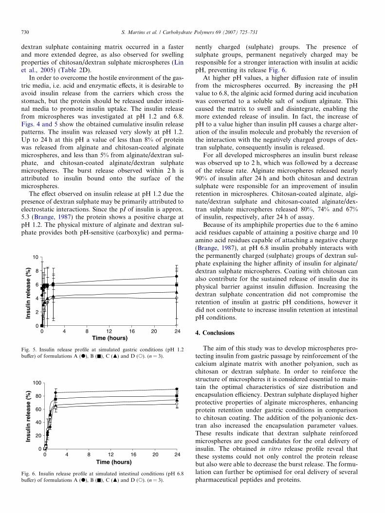
In order to overcome the hostile environment of the gas-tric media, i.e. acid and enzymatic effects, it is desirable toavoid insulin release from the carriers which cross thestomach, but the protein should be released under intesti-nal media to promote insulin uptake. The insulin releasefrom microspheres was investigated at pH 1.2 and 6.8.Figs. 4 and 5 show the obtained cumulative insulin releasepatterns. The insulin was released very slowly at pH 1.2.Up to 24 h at this pH a value of less than 8% of proteinwas released from alginate and chitosan-coated alginatemicrospheres, and less than 5% from alginate/dextran sul-phate, and chitosan-coated alginate/dextran sulphatemicrospheres. The burst release observed within 2 h isattributed to insulin bound onto the surface of themicrospheres.
The effect observed on insulin release at pH 1.2 due thepresence of dextran sulphate may be primarily attributed toelectrostatic interactions. Since the pI of insulin is approx.5.3 (Brange, 1987) the protein shows a positive charge atpH 1.2. The physical mixture of alginate and dextran sul-phate provides both pH-sensitive (carboxylic) and perma-
0
2
4
6
8
10
0 4 8 12 16 20 24Time (hours)
Insu
lin r
elea
se (
%)
Fig. 5. Insulin release profile at simulated gastric conditions (pH 1.2buffer) of formulations A (d), B (n), C (m) and D (s). (n = 3).
0
20
40
60
80
100
0 4 8 12 16 20 24
Time (hours)
Insu
lin r
elea
se (
%)
Fig. 6. Insulin release profile at simulated intestinal conditions (pH 6.8buffer) of formulations A (d), B (n), C (m) and D (s). (n = 3).
nently charged (sulphate) groups. The presence ofsulphate groups, permanent negatively charged may beresponsible for a stronger interaction with insulin at acidicpH, preventing its release Fig. 6.
At higher pH values, a higher diffusion rate of insulinfrom the microspheres occurred. By increasing the pHvalue to 6.8, the alginic acid formed during acid incubationwas converted to a soluble salt of sodium alginate. Thiscaused the matrix to swell and disintegrate, enabling themore extended release of insulin. In fact, the increase ofpH to a value higher than insulin pH causes a charge alter-ation of the insulin molecule and probably the reversion ofthe interaction with the negatively charged groups of dex-tran sulphate, consequently insulin is released.
For all developed microspheres an insulin burst releasewas observed up to 2 h, which was followed by a decreaseof the release rate. Alginate microspheres released nearly90% of insulin after 24 h and both chitosan and dextransulphate were responsible for an improvement of insulinretention in microspheres. Chitosan-coated alginate, algi-nate/dextran sulphate and chitosan-coated alginate/dex-tran sulphate microspheres released 80%, 74% and 67%of insulin, respectively, after 24 h of assay.
Because of its amphiphile properties due to the 6 aminoacid residues capable of attaining a positive charge and 10amino acid residues capable of attaching a negative charge(Brange, 1987), at pH 6.8 insulin probably interacts withthe permanently charged (sulphate) groups of dextran sul-phate explaining the higher affinity of insulin for alginate/dextran sulphate microspheres. Coating with chitosan canalso contribute for the sustained release of insulin due itsphysical barrier against insulin diffusion. Increasing thedextran sulphate concentration did not compromise theretention of insulin at gastric pH conditions, however itdid not contribute to increase insulin retention at intestinalpH conditions.
4. Conclusions
The aim of this study was to develop microspheres pro-tecting insulin from gastric passage by reinforcement of thecalcium alginate matrix with another polyanion, such aschitosan or dextran sulphate. In order to reinforce thestructure of microspheres it is considered essential to main-tain the optimal characteristics of size distribution andencapsulation efficiency. Dextran sulphate displayed higherprotective properties of alginate microspheres, enhancingprotein retention under gastric conditions in comparisonto chitosan coating. The addition of the polyanionic dex-tran also increased the encapsulation parameter values.These results indicate that dextran sulphate reinforcedmicrospheres are good candidates for the oral delivery ofinsulin. The obtained in vitro release profile reveal thatthese systems could not only control the protein releasebut also were able to decrease the burst release. The formu-lation can further be optimised for oral delivery of severalpharmaceutical peptides and proteins.

S. Martins et al. / Carbohydrate Polymers 69 (2007) 725–731 731
Acknowledgements
The authors wish to thank Lilly Portugal for insulin sup-ply and Rui Fernandes from the Institute for Molecularand Cell Biology (IBMC, Porto, Portugal) for TEManalyses.
References
Arbit, E. (2004). The physiological rationale for oral insulin administra-tion. Diabetes Technology and Therapeutics, 6(4), 510–517.
Bajpai, J., Bajpai, A., & Mishra, S. (2006). Dynamics of controlled releaseof potassium nitrate from a highly swelling binary biopolymeric blendof alginate and pectin. Journal of Macromolecular Science, Part A:
Pure and Applied Chemistry, 43(1), 165–186.Bradford, M. M. (1976). A rapid and sensitive method for the quantitation
of microgram quantities of protein utilizing the principle of protein–dye binding. Analytical Biochemistry, 72, 248–254.
Brange, J. (1987). Galenics of insulin: The physico-chemical and pharma-
ceutical aspects of insulin and insulin preparations. Springer-Verlag.Brange, J., Anderson, L., Laursen, E. D., Meyn, G., & Rasmussen, E.
(1997). Toward understanding insulin fibrilation. Journal of Pharma-
ceutical Sciences, 86, 517–525.Dumitriu, S., & Chornet, E. (1998). Inclusion and release of proteins from
polysaccharide-based polyion complexes. Advanced Drug Delivery
Reviews, 31, 223–246.El-Kamel, A. H., Al-Gohary, O. M. N., & Hosny, E. A. (2003). Alginate-
diltiazem hydrochloride beads: optimization of formulation factors,in vitro and in vivo availability. Journal of Microencapsulation, 20(2),211–225.
Fuentes, M., Pessela, B. C. C., Maquiese, J. V., Ortiz, C., Segura, R. L.,Palomo, J. M., et al. (2004). Reversible and strong immobilization ofproteins by ionic exchange on supports coated with sulfate–dextran.Biotechnology Progress, 20(4), 1134–1139.
Gombotz, W. R., & Wee, S. F. (1998). Protein release from alginatematrices. Advanced Drug Delivery Reviews, 31, 267–285.
Halder, A., Maiti, S., & Sa, B. (2005). Entrapment efficiency and releasecharacteristics of polyethyleneimine-treated or -untreated calciumalginate beads loaded with propranolol-resin complex. International
Journal of Pharmaceutics, 302(1–2), 84–94.
Haug, A. (1964). Composition and properties of alginates. Thesis Trond-heim Norwegian Institute of Technology.
Janes, K. A., & Alonso, M. J. (2003). Depolymerized chitosan nanopar-ticles for protein delivery: preparation and characterization. Journal of
Applied Polymer Science, 88, 2769–2776.Lin, W. C., Yu, D. G., & Chien, M. (2005). pH-sensitive polyelectrolyte
complex gel microspheres composed of chitosan/sodium tripolyphos-phate/dextran sulfate: swelling kinetics and drug delivery properties.Colloids and Surfaces B: Biointerfaces, 44(2–3), 143–151.
Onal, S., & Zihnioglu, F. (2002). Encapsulation of insulin in chitosan-coated alginate beads: oral therapeutic peptide delivery. Artificial
Cells, Blood Substitutes, and Immobilization. Biotechnology, 30(3),229–237.
Quong, D., Yeo, J. N., & Neufeld, R. J. (1999). Stability of chitosanand poly-L-lysine membranes coating DNA-alginate beads whenexposed to hydrolytic enzymes. Journal of Microencapsulation, 16(1),73–82.
Ramadas, M., Paul, W., Dileep, K. J., Anitha, Y., & Sharma, C. P. (2000).Lipoinsulin encapsulated alginate–chitosan capsules: intestinal deliveryin diabetic rats. Journal of Microencapsulation, 17(4), 405–411.
Setty, C. M., Sahoo, S. S., & Sa, B. (2005). alginate-coated alginate–polyethyleneimine beads for prolonged release of furosemide insimulated intestinal fluid. Drug Development and Industrial Pharmacy,
31, 435–446.Shu, X. Z., & Zhu, K. J. (2002). The release behavior of brilliant blue from
calcium–alginate gel beads coated by chitosan: the preparation methodeffect. European Journal of Pharmaceutical Sciences, 53(2), 193–201.
Silva, C. M., Ribeiro, A. J., Figueiredo, I. V., Goncalves, A. R., & Veiga,F. (2006). Alginate microspheres prepared by internal gelation:development and effect on insulin stability. International Journal of
Pharmaceutics, 311(1-2), 1–10.Silva, C., Ribeiro, A., Veiga, F., & Sousa, A. (2006). Insulin release from
alginate microspheres reinforced with dextran sulfate. Chemical
Industry and Chemical Engineering Quarterly, 12, 40–46.Sluzky, V., Tamada, J. A., Klibanov, A. M., & Langer, R. (1991).
Kinetics of insulin aggregation in aqueous solutions upon agitationin the presence of hydrophobic surfaces. Proceedings of the National
Academy of Sciences of the United States of America, 88(21),9377–9381.
Yu, S., Zhao, Y., Wu, F., Zhang, X., Lu, W., Zhang, H., et al. (2004).Nasal insulin delivery in the chitosan solution: in vitro and in vivostudies. International Journal of Pharmaceutics, 281, 11–23.