Instruction Manual - Bio-Rad Laboratories Powder-free latex, vinyl, or nitrile gloves 1.5. Reagents...
27
Oriole ™ Fluorescent Gel Stain Instruction Manual Catalog # 161-0495, 1x solution, 200 ml 161-0496, 1x solution, 1 L 161-0497, kit for 5 L For technical support call your local Bio-Rad office. In the U.S. call 1-800-424-6723.
Transcript of Instruction Manual - Bio-Rad Laboratories Powder-free latex, vinyl, or nitrile gloves 1.5. Reagents...
Oriole™ Fluorescent Gel Stain
Instruction Manual
Catalog # 161-0495, 1x solution, 200 ml161-0496, 1x solution, 1 L161-0497, kit for 5 L
For technical support call your local Bio-Rad office. In the U.S. call 1-800-424-6723.
Table of Contents
Section 1: Introduction and General Information 1
1.1 Introduction 11.2 Product Description 21.3 Storage 21.4 Materials and Equipment Required but Not Supplied 31.5 Reagents Required but Not Supplied 31.6 Safety Considerations 41.7 Disposal Considerations 41.8 Fluorescence Characteristics 4
Section 2: Instructions 6
2.1 General Considerations 62.2 Stain Solution Preparation 82.3 Gel Staining 92.4 Gel Imaging 11
Section 3: Technical Information 14
3.1 Sensitivity of Staining — 1-D Gels 14 3.2 Compatibility with Mass Spectrometry 153.3 Protein-to-Protein Variability 153.4 Dynamic Range 163.5 2-D Gel Staining 17
Section 4: Troubleshooting 18
Section 5: Product Information 21
5.1 Oriole™ Fluorescent Gel Stain 215.2 Related Products 21

1
Section 1
Introduction and General Information1.1. Introduction
Oriole™ fluorescent gel stain is an easy to use, rapid, and sensitive stain for visualization and quantitation of proteins separated by SDS-PAGE. The product is available in three configurations. The 200 ml and 1 L sizes are provided ready to use. The product is also available as a kit containing components to make 5 L of ready to use staining solution.
The staining procedure is a simple one-step protocol that can be completed in as little as 90 minutes. Gels stained with Oriole fluorescent gel stain may be visualized with a variety of different UV-based fluorescence imaging systems.
Oriole fluorescent gel stain gives exceptional sensitivity and dynamic range (see pages 14–16) and is compatible with subsequent analysis by enzymatic digestion and mass spectrometry. It is thus particularly well suited to proteomics applications.
2
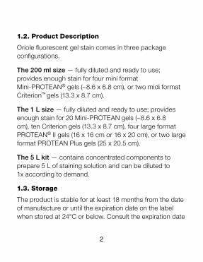
1.2. Product Description
Oriole fluorescent gel stain comes in three package configurations.
The 200 ml size — fully diluted and ready to use; provides enough stain for four mini format Mini-PROTEAN® gels (~8.6 x 6.8 cm), or two midi format Criterion™ gels (13.3 x 8.7 cm).
The 1 L size — fully diluted and ready to use; provides enough stain for 20 Mini-PROTEAN gels (~8.6 x 6.8 cm), ten Criterion gels (13.3 x 8.7 cm), four large format PROTEAN® II gels (16 x 16 cm or 16 x 20 cm), or two large format PROTEAN Plus gels (25 x 20.5 cm).
The 5 L kit — contains concentrated components to prepare 5 L of staining solution and can be diluted to 1x according to demand.
1.3. Storage
The product is stable for at least 18 months from the date of manufacture or until the expiration date on the label when stored at 24°C or below. Consult the expiration date
3
before using. Avoid prolonged exposure to temperatures greater than 37°C and protect from light.
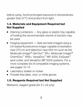
1.4. Materials and Equipment Required but Not Supplied
n Staining containers — Any glass or plastic tray capable of holding the recommended volume of solution may be used
n Imaging equipment — Gels are best imaged using a UV-based fluorescence imager capable of excitation near 270 nm and detection near 604 nm such as the Molecular Imager® Gel Doc™ XR+, Molecular Imager® ChemiDoc™ XRS+, VersaDoc™ MP 4000, ExQuest™ spot cutter, and VersaDoc MP 5000 systems. For a more complete list of compatible imaging systems, see pages 12–13
n Laboratory shaker or rockern Powder-free latex, vinyl, or nitrile gloves
1.5. Reagents Required but Not Supplied
Methanol, reagent grade (for 5 L kit only)
4
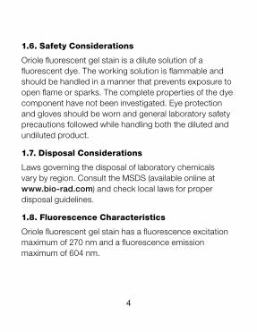
1.6. Safety Considerations
Oriole fluorescent gel stain is a dilute solution of a fluorescent dye. The working solution is flammable and should be handled in a manner that prevents exposure to open flame or sparks. The complete properties of the dye component have not been investigated. Eye protection and gloves should be worn and general laboratory safety precautions followed while handling both the diluted and undiluted product.
1.7. Disposal Considerations
Laws governing the disposal of laboratory chemicals vary by region. Consult the MSDS (available online at www.bio-rad.com) and check local laws for proper disposal guidelines.
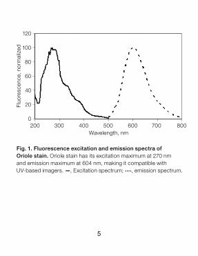
1.8. Fluorescence Characteristics
Oriole fluorescent gel stain has a fluorescence excitation maximum of 270 nm and a fluorescence emission maximum of 604 nm.
5
Fluo
resc
ence
, nor
mal
ized
120
100
80
60
40
20
0200 300 400 500 600 700 800
Wavelength, nm
Fig. 1. Fluorescence excitation and emission spectra of Oriole stain. Oriole stain has its excitation maximum at 270 nm and emission maximum at 604 nm, making it compatible with UV-based imagers. —, Excitation spectrum; ...., emission spectrum.

6
Section 2
Instructions2.1. General Considerations
Best results are obtained by using clean technique. Any dust or dirt transferred to the surface of the gel may appear in the fluorescence image as smudges or speckles. Oriole™ fluorescent gel stain is exceptionally sensitive. Contaminant proteins such as keratin will appear in the gel image if care is not taken to minimize such contamination.
All glassware used should be cleaned with laboratory glassware cleaner and rinsed with distilled or deionized water. Use dust-free gloves and limit dust exposure by keeping reagent vessels and gel trays covered as much as possible. If gels are cast in the laboratory, the glass plates used should be thoroughly cleaned with lint-free laboratory wipes.
Oriole fluorescent gel stain is very sensitive, and less protein can be visualized than what is possible using a visible stain like Coomassie Blue. Sensitivity is of the
7
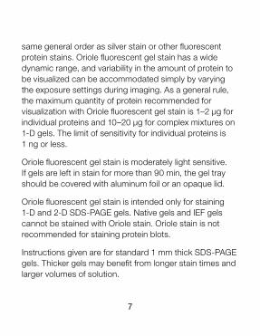
same general order as silver stain or other fluorescent protein stains. Oriole fluorescent gel stain has a wide dynamic range, and variability in the amount of protein to be visualized can be accommodated simply by varying the exposure settings during imaging. As a general rule, the maximum quantity of protein recommended for visualization with Oriole fluorescent gel stain is 1–2 µg for individual proteins and 10–20 µg for complex mixtures on 1-D gels. The limit of sensitivity for individual proteins is 1 ng or less.
Oriole fluorescent gel stain is moderately light sensitive. If gels are left in stain for more than 90 min, the gel tray should be covered with aluminum foil or an opaque lid.
Oriole fluorescent gel stain is intended only for staining 1-D and 2-D SDS-PAGE gels. Native gels and IEF gels cannot be stained with Oriole stain. Oriole stain is not recommended for staining protein blots.
Instructions given are for standard 1 mm thick SDS-PAGE gels. Thicker gels may benefit from longer stain times and larger volumes of solution.
8
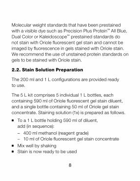
Molecular weight standards that have been prestained with a visible dye such as Precision Plus Protein™ All Blue, Dual Color or Kaleidoscope™ prestained standards do not stain with Oriole fluorescent gel stain and cannot be imaged by fluorescence in gels stained with Oriole stain. We recommend the use of unstained protein standards on gels to be stained with Oriole stain.
2.2. Stain Solution Preparation
The 200 ml and 1 L configurations are provided ready to use.
The 5 L kit comprises 5 individual 1 L bottles, each containing 590 ml of Oriole fluorescent gel stain diluent, and a single bottle containing 50 ml of Oriole gel stain concentrate. Staining solution (1x) is prepared as follows.
n To a 1 L bottle holding 590 ml of diluent, add (in sequence):
– 400 ml methanol (reagent grade) – 10 ml of Oriole fluorescent gel stain concentraten Mix well by shakingn Stain is now ready to be used
9
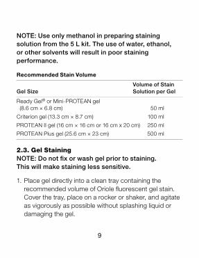
NOTE: Use only methanol in preparing staining solution from the 5 L kit. The use of water, ethanol, or other solvents will result in poor staining performance.
Recommended Stain Volume
Volume of Stain Gel Size Solution per Gel
Ready Gel® or Mini-PROTEAN gel (8.6 cm × 6.8 cm) 50 ml
Criterion gel (13.3 cm × 8.7 cm) 100 ml
PROTEAN II gel (16 cm × 16 cm or 16 cm x 20 cm) 250 ml
PROTEAN Plus gel (25.6 cm × 23 cm) 500 ml
2.3. Gel StainingNOTE: Do not fix or wash gel prior to staining. This will make staining less sensitive.
1. Place gel directly into a clean tray containing the recommended volume of Oriole fluorescent gel stain. Cover the tray, place on a rocker or shaker, and agitate as vigorously as possible without splashing liquid or damaging the gel.
10
2. Stain for 90 min for maximum sensitivity.
3. Cover the tray to exclude light if the gel is stained longer than 90 min.
NOTE: Best results are obtained if the gels are left in staining solution no longer than 2 hr.
4. Transfer gel to water prior to imaging. This step prevents exposure of the imaging equipment to moderately corrosive staining solution.
NOTE: Destaining is not necessary. Staining intensity persists when the gel is stored in water.
Stained gels can be stored in water for up to 6 months and imaged without significant loss of sensitivity if protected from light and stored at 2–8°C.
11
2.4. Gel Imaging
Gels stained with Oriole stain are visualized using UV light excitation. Bio-Rad Molecular Imager Gel Doc XR+, Molecular Imager ChemiDoc XRS+, EXQuest spot cutter, VersaDoc MP 4000, or VersaDoc MP 5000 systems are recommended for imaging gels stained with Oriole stain. If the imaging equipment has no preprogrammed imaging function for Oriole fluorescent gel stain, the imaging setting for SYPRO Ruby stain or ethidium bromide that uses UV transillumination is recommended.
Any imaging system using UV light excitation may be used to image Oriole fluorescent gel stain. Such imaging systems almost always have midrange (300 nm, 306 nm, or 312 nm) UV excitation and red or orange emission filters as standard options for imaging ethidium bromide–stained gels. The excitation and emission properties of Oriole stain are very compatible with ethidium bromide, therefore imager settings for ethidium bromide can be used when imaging gels stained with Oriole stain.
Imaging systems capable of imaging gels stained with Oriole fluorescent gel stain include the following:
12
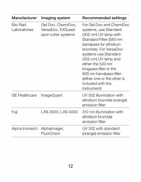
Manufacturer Imaging system Recommended settings
Bio-Rad Gel Doc, ChemiDoc, For Gel Doc and ChemiDoc Laboratories VersaDoc, EXQuest systems, use Standard spot cutter systems (302 nm) UV lamp with Standard Filter (580 nm bandpass for ethidium bromide). For VersaDoc systems use Standard (302 nm) UV lamp and either the 520 nm longpass filter or the 605 nm bandpass filter (either one or the other is included with the instrument)
GE Healthcare ImageQuant UV 302 illumination with ethidium bromide (orange) emission filter
Fuji LAS 3000, LAS 4000 312 nm illumination with ethidium bromide emission filter
Alpha Innotech AlphaImager, UV 302 with standard FluorChem (orange) emission filter
13
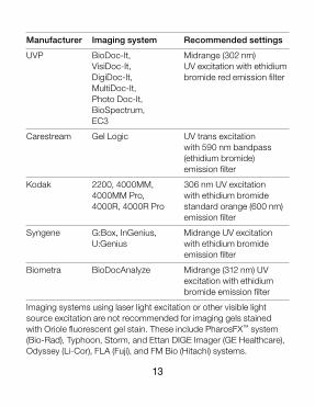
Manufacturer Imaging system Recommended settings
UVP BioDoc-It, Midrange (302 nm) VisiDoc-It, UV excitation with ethidium DigiDoc-It, bromide red emission filter MultiDoc-It, Photo Doc-It,
BioSpectrum,
EC3
Carestream Gel Logic UV trans excitation with 590 nm bandpass (ethidium bromide) emission filter
Kodak 2200, 4000MM, 306 nm UV excitation 4000MM Pro, with ethidium bromide 4000R, 4000R Pro standard orange (600 nm) emission filter
Syngene G:Box, InGenius, Midrange UV excitation U:Genius with ethidium bromide emission filter
Biometra BioDocAnalyze Midrange (312 nm) UV excitation with ethidium bromide emission filter
Imaging systems using laser light excitation or other visible light source excitation are not recommended for imaging gels stained with Oriole fluorescent gel stain. These include PharosFX™ system (Bio-Rad), Typhoon, Storm, and Ettan DIGE Imager (GE Healthcare), Odyssey (Li-Cor), FLA (Fuji), and FM Bio (Hitachi) systems.
14
Section 3
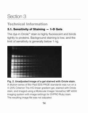
Technical Information3.1. Sensitivity of Staining — 1-D Gels
The dye in Oriole™ stain is highly fluorescent and binds tightly to proteins. Background staining is low, and the limit of sensitivity is generally below 1 ng.
Fig. 2. Unadjusted image of a gel stained with Oriole stain. A dilution series of Bio-Rad SDS-PAGE standards was run on a 4–20% Criterion Tris-HCl linear gradient gel, stained with Oriole stain, and imaged using a Molecular Imager VersaDoc MP 4000 imaging system with image settings for SYPRO Ruby stain. The resulting image file was not adjusted.
15
3.2. Compatibility With Mass Spectrometry
Oriole fluorescent protein gel stain is fully compatible with downstream proteolysis and mass spectrometric analysis.
3.3. Protein-to-Protein Variability
Oriole fluorescent gel stain will stain most proteins and exhibits little protein-to-protein variability in staining intensity.
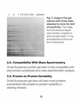
Fig. 3. Image of the gel stained with Oriole stain, adjusted to show the limit of sensitivity. The image from the previous figure was inverted, cropped to show protein loads ≤ 4 ng, and adjusted to show the limit of sensitivity.
4 2 1 0.5 0.25 0.125 ng/band
16
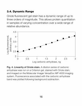
Fig. 4. Linearity of Oriole stain. A dilution series of carbonic anhydrase was run on a Criterion gel, stained with Oriole stain, and imaged on the Molecular Imager VersaDoc MP 4000 imaging system. Fluorescence associated with the carbonic anhydrase band was plotted following background subtraction.
Log
(fluo
resc
ence
), ar
bitr
ary
units
3.4. Dynamic Range
Oriole fluorescent gel stain has a dynamic range of up to three orders of magnitude. This allows protein quantitation in samples of varying concentration over a wide range of relative abundance.
4.5
4
3.5
3
2.5
20 0.5 1 1.5 2 2.5 3
Log carbonic anhydrase, ng
17
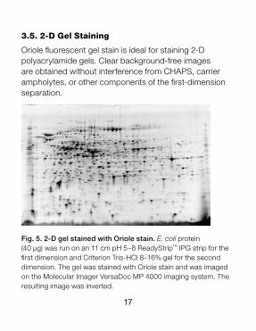
3.5. 2-D Gel Staining
Oriole fluorescent gel stain is ideal for staining 2-D polyacrylamide gels. Clear background-free images are obtained without interference from CHAPS, carrier ampholytes, or other components of the first-dimension separation.
Fig. 5. 2-D gel stained with Oriole stain. E. coli protein (40 μg) was run on an 11 cm pH 5–8 ReadyStrip™ IPG strip for the first dimension and Criterion Tris-HCl 8–16% gel for the second dimension. The gel was stained with Oriole stain and was imaged on the Molecular Imager VersaDoc MP 4000 imaging system. The resulting image was inverted.
18
Section 4
Troubleshooting
continued
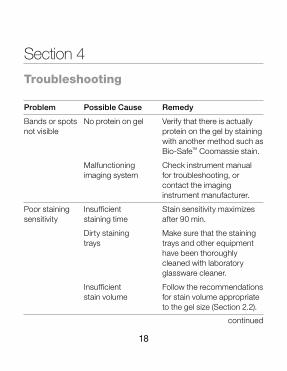
Problem Possible Cause Remedy
Bands or spots No protein on gel Verify that there is actually not visible protein on the gel by staining with another method such as Bio-Safe™ Coomassie stain.
Malfunctioning Check instrument manual imaging system for troubleshooting, or contact the imaging instrument manufacturer.
Poor staining Insufficient Stain sensitivity maximizes sensitivity staining time after 90 min.
Dirty staining Make sure that the staining trays trays and other equipment have been thoroughly cleaned with laboratory glassware cleaner.
Insufficient Follow the recommendations stain volume for stain volume appropriate to the gel size (Section 2.2).
19
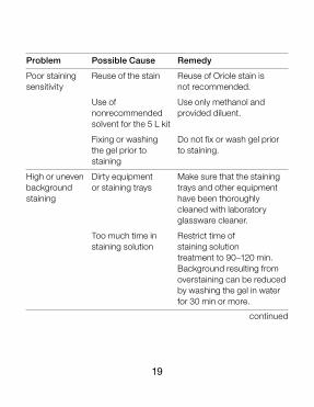
Problem Possible Cause Remedy
Poor staining Reuse of the stain Reuse of Oriole stain is sensitivity not recommended.
Use of Use only methanol and nonrecommended provided diluent. solvent for the 5 L kit
Fixing or washing Do not fix or wash gel prior the gel prior to to staining. staining
High or uneven Dirty equipment Make sure that the staining background or staining trays trays and other equipment staining have been thoroughly cleaned with laboratory glassware cleaner.
Too much time in Restrict time of staining solution staining solution treatment to 90–120 min. Background resulting from overstaining can be reduced by washing the gel in water for 30 min or more.
continued
20
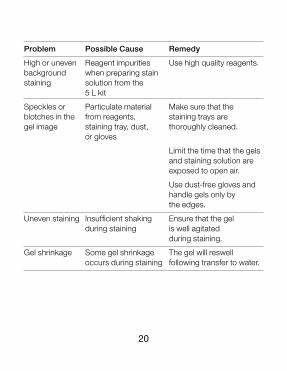
Problem Possible Cause Remedy
High or uneven Reagent impurities Use high quality reagents. background when preparing stain staining solution from the 5 L kit
Speckles or Particulate material Make sure that the blotches in the from reagents, staining trays are gel image staining tray, dust, thoroughly cleaned. or gloves
Limit the time that the gels and staining solution are exposed to open air.
Use dust-free gloves and handle gels only by the edges.
Uneven staining Insufficient shaking Ensure that the gel during staining is well agitated during staining.
Gel shrinkage Some gel shrinkage The gel will reswell occurs during staining following transfer to water.
21
Section 5
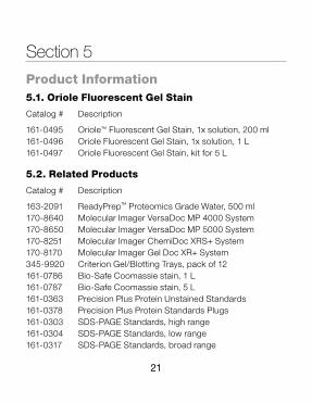
Product Information5.1. Oriole Fluorescent Gel Stain
Catalog # Description
161-0495 Oriole™ Fluorescent Gel Stain, 1x solution, 200 ml 161-0496 Oriole Fluorescent Gel Stain, 1x solution, 1 L 161-0497 Oriole Fluorescent Gel Stain, kit for 5 L
5.2. Related Products
Catalog # Description
163-2091 ReadyPrep™ Proteomics Grade Water, 500 ml170-8640 Molecular Imager VersaDoc MP 4000 System170-8650 Molecular Imager VersaDoc MP 5000 System170-8251 Molecular Imager ChemiDoc XRS+ System170-8170 Molecular Imager Gel Doc XR+ System345-9920 Criterion Gel/Blotting Trays, pack of 12161-0786 Bio-Safe Coomassie stain, 1 L161-0787 Bio-Safe Coomassie stain, 5 L161-0363 Precision Plus Protein Unstained Standards161-0378 Precision Plus Protein Standards Plugs161-0303 SDS-PAGE Standards, high range161-0304 SDS-PAGE Standards, low range161-0317 SDS-PAGE Standards, broad range
22
Bio-Rad Laboratories, Inc. is licensed by Invitrogen Corporation to sell SYPRO products for research use only, under U.S. patent 5,616,502.
AlphaImager and FluorChem are trademarks of Alpha Innotech Corporation.BioDoc-It, BioSpectrum, DigiDoc-It, Doc-It, and MultiDoc-It are trademarks of UVP, LLC. Coomassie is a trademark of BASF Aktiengesellschaft. Ettan, ImageQuant, Typhoon, and Storm are trademarks of GE Healthcare Group Companies.Odyssey is a trademark of LI-COR, Inc.
Life Science Group
10-0361 0510 Sig 110910017295 Rev B US/EG
Bio-Rad Laboratories, Inc.
Web site www.bio-rad.com USA 800 424 6723 Australia 61 2 9914 2800 Austria 01 877 89 01 Belgium 09 385 55 11 Brazil 55 31 3689 6600 Canada 905 364 3435 China 86 20 8732 2339 Czech Republic 420 241 430 532 Denmark 44 52 10 00 Finland 09 804 22 00 France 01 47 95 69 65 Germany 089 31 884 0 Greece 30 210 777 4396 Hong Kong 852 2789 3300 Hungary 36 1 459 6100 India 91 124 4029300 Israel 03 963 6050 Italy 39 02 216091 Japan 03 6361 7000 Korea 82 2 3473 4460 Mexico 52 555 488 7670 The Netherlands 0318 540666 New Zealand 0508 805 500 Norway 23 38 41 30 Poland 48 22 331 99 99 Portugal 351 21 472 7700 Russia 7 495 721 14 04 Singapore 65 6415 3188 South Africa 27 861 246 723 Spain 34 91 590 5200 Sweden 08 555 12700 Switzerland 061 717 95 55 Taiwan 886 2 2578 7189 United Kingdom 020 8328 2000