Inspection of the patient - 193.105.7.56
157
Transcript of Inspection of the patient - 193.105.7.56


Inspection of the patient: Is simple method, 80% of information comes through your eyes; Allows to evaluate objectively the state of the patient (status present objectivus); Helps to organize the algorithm of patient’s examination; Sometimes its possible to diagnose at a moment (diagnosis ad oculos) (acromegaly, hyperthyreoidosis); To evaluate the data of subjective examination; Important part of the physician-patient relationship development;

Rules of inspection
The patient is fully or partially exposed;
Positioned at a distance of 2-3 steps from the doctor
Gradually turning the patient's doctor examines him in direct and lateral illumination;
Inspection of thorax is best done in a vertical position and the abdomen in the vertical and horizontal

Direct light
Lateral illumination
Rules of inspection
Use daylight; Use direct and lateral illumination; In artificial light there are possible problems in the evaluation of color skin and mucus, skin elements;
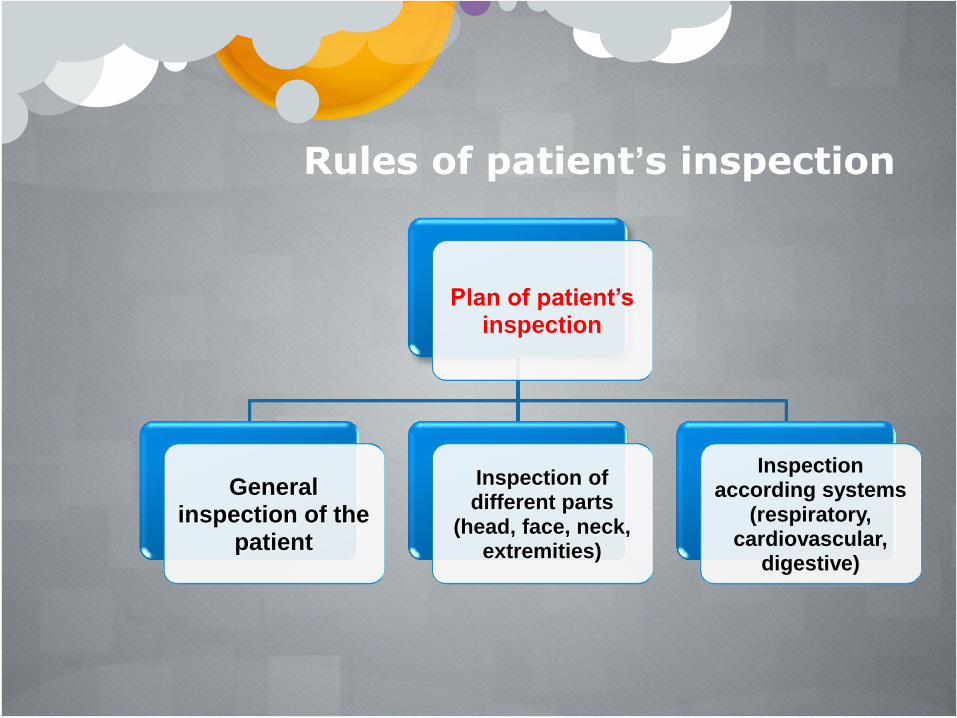
Plan of patient’s inspection
General inspection of the
patient
Inspection of different parts
(head, face, neck, extremities)
Inspection according systems
(respiratory, cardiovascular,
digestive)
Rules of patient’s inspection

Topographic regions and lines of the chest
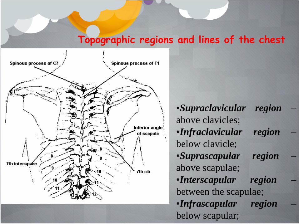
Topographic regions and lines of the chest
•Supraclavicular region –
above clavicles;
•Infraclavicular region –
below clavicle;
•Suprascapular region –
above scapulae;
•Interscapular region –
between the scapulae;
•Infrascapular region –
below scapular;
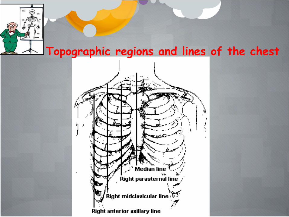
Topographic regions and lines of the chest

Topographic regions and lines of the chest

Topographic regions and lines of the chest

Topographic regions of the abdomen
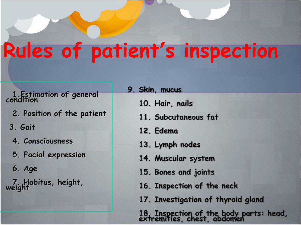
Rules of patient’s inspection
1.Estimation of general condition
2. Position of the patient
3. Gait
4. Consciousness
5. Facial expression
6. Age
7. Habitus, height, weight
9. Skin, mucus
10. Hair, nails
11. Subcutaneous fat
12. Edema
13. Lymph nodes
14. Muscular system
15. Bones and joints
16. Inspection of the neck
17. Investigation of thyroid gland
18. Inspection of the body parts: head, extremities, chest, abdomen

General condition of the patient The criteria ’ s of patient ’ s condition are the next
clinical features:
consciousness;
posture;
gait;
the facial expression;
weight;
mood (mental condition);

good; satisfactory; moderate severe; severe; extremely severe;
General condition of the patient

Good patient’s condition: •clear consciousness •active posture •free gait •sensible facial expression •sufficient weight and good mood (patients with remission of chronic disease favorable course of a disease, or during recovery).

Satisfactory patients condition (status morboacili): •clear consciousness; •active or active with restriction posture; •free or partial deranged (specific) gait; •sensible facial expression; •adequate mental reaction; (patients with remission of prolong chronic disease, or during recovery from acute disease).
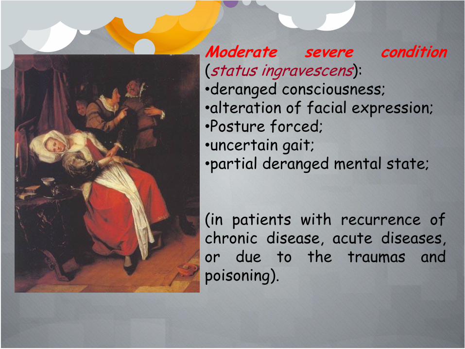
Moderate severe condition (status ingravescens): •deranged consciousness; •alteration of facial expression; •Posture forced; •uncertain gait; •partial deranged mental state; (in patients with recurrence of chronic disease, acute diseases, or due to the traumas and poisoning).

Severe condition (status morbogravi): •deranged consciousness; •changed facial expression (fear, suffer, hopelessness, indifference); •forced posture; •loss of weight; •edema; •inadequate mental state; (in patients with infections and oncological diseases, heart failure, disorders of renal, liver functions, abnormalities of nervous and endocrine systems, after operations, traumas).

Extremely severe condition (status gravissimus): •unconsciousness; •passive posture; •indifferent facial expression; observed in patient with coma, shock, and agony.

Consciousness (sensorium) may be clear or deranged.
The criteria’s of consciousness condition are the following
features:
• orientation to the surroundings,
• adequate answers,
• concentrated attention,
• reflex,
• pupil reaction on light.

CONSCIOUSNESS
EXCITED
Irritative disorder Delirium
DEPRESSED
Cloudiness Stupor Sopor Coma

Clear consciousness (sensorium lucidum):
adequate behavior,
• correct orientation to
the surroundings,
• timely answer to the
question,
• preservation of all
reflexes.

The deranged consciousness develop due to the different causes:
– disorders of cerebral or cardiac circulation;
– endogenic and exogenic intoxication; – infectious affections; – hormonal, mineral, metabolic
abnormalities; – traumas of the brain.

Disorientation in space, indifference, the answers adequate, but delayed, reflexes are present
Cloudiness

Stupor
Disorientation in space, surroundings,
the answers inadequate and delayed,
reflexes are present

Sopor
Disorientation in time, space, surroundings, own personality.
Pathological deep sleep from which patient wake up only for short periods of time when called loudly or roused by an external stimulus,
reflexes are present, but delayed

Coma
Unconsciousness with absence of response to external stimuli,
absence of reflexes,
deranged vital function

The following forms of coma are most common.
Coma due to the disorders of cerebral or cardiac circulation: – apoplexy coma resulted from stroke, thromboemboly of cerebral vessels asymmetric face, noisy, deep breathing, narrowed pupil. Duration from several hours to several days;

Endotoxic coma:
diabetic coma occurs in patients with diabetes mellitus due to metabolic carbohydrate and lipid disorders.
The causes of coma with ketoacidosis: too little or no insulin; an infection; digestive disturbance.

Endotoxic coma:
Hypoglycemic coma occurs in patients with
diabetes mellitus commonly treated with insulin due to sudden decreasing a blood glucose concentration of less than 2,5 mmol/l.

Endotoxic coma:
Hepatic coma develops in patients with acute and subacute dystrophy and necrosis of the liver parenchyma (acute viral hepatitis, acute drug-induced liver disease); in patient patients with final stage of liver cirrhosis related to disorders of bilirubin, protein, and carbohydrate metabolism.

Endotoxic coma:
Uraemic coma develops slowly in patients with congenital and inherited renal diseases, glomerular and interstitial diseases, obstructive uropathy, as complication of vascular systemic diseases, in condition which destroyed the normal structure and function of the kidney, acute and chronic renal failure develops.

Exotoxic
Exotoxical coma is relevant to acute poisoning.
Poisoning substances may give rise to primary toxic effects, which may result in organ damage of a nonspecific type.
The organ damage may then lead to respiratory or metabolic disturbance or a combination of these, hence to a variety of clinical features.
The forms of exited consciousness
Twilight state is characterized by disorientation in
surroundings, loss of memory (amnesia), patient is exited,
has pathologically high spirits, is anxious, sometimes even
aggressive. This state may observe in patients with
epilepsy.

The forms of exited consciousness
Delirium is characterized with visual and acoustic
hallucinations, inadequacy of emotions, anxiety,
intermittent thinking. There are some kinds of delirium:
alcoholic (delirium tremens), infection senile, traumatic,
pharmacogenic, epileptic.

Posture of the patients
Forms Definition Pathological state
Active Patient has ability to walk,
stand, to change his posture
Mild disease
Onset of a severe
disease
Passive Patient is motionless, he lies,
his head and the limbs hand
down
Unconscious state
Forced Forced posture assumed by the patient to relieve or remove pain, cough, dyspnoea, or other signs of disease
Severe disease

Forced posture differs relevant to the process which cause specific
patients position.
Standing upright position is observed in patient with attacks of angina pectoris and atheromatous peripheral vascular disease.
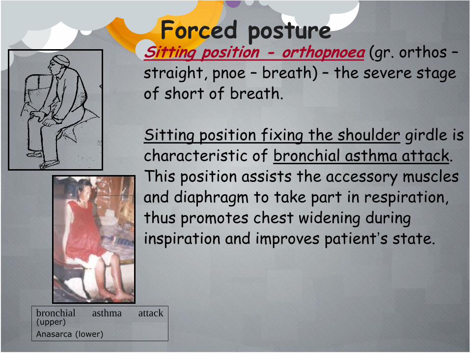
Sitting position - orthopnoea (gr. orthos – straight, pnoe – breath) – the severe stage of short of breath. Sitting position fixing the shoulder girdle is characteristic of bronchial asthma attack. This position assists the accessory muscles and diaphragm to take part in respiration, thus promotes chest widening during inspiration and improves patient’s state.
bronchial asthma attack (upper)
Anasarca (lower)
Forced posture
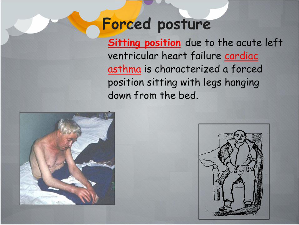
Sitting position due to the acute left ventricular heart failure cardiac asthma is characterized a forced position sitting with legs hanging down from the bed. .
Forced posture

Sitting position and inclines forward may observe in patient with pericarditis, which produce a pericardiac effusion resulted restriction a diastolic heart function.
Forced posture

The supine posture is characteristic of strong pain in the abdomen acute appendicitis acute cholecistitis perforated ulcer of stomach or duodenum. Sometimes patient bends the leg in knee joint for decreasing marked strain of muscle of the abdominal wall. The supine posture with complete immobility is observed in patients with acute rheumatic polyartritis due to the severe pain; patients with sclerodermia and patients with severe fatigue.
Forced posture

The forced posture on the side:
– on the affected side lie the patients with the lung, pleura diseases. The patients with dry pleurisy prefer to lie on the affected side because the limitation of the pleural layers movement relieves the pain. The patients with pneumonia, massive lung tumor, effusive pleurisy prefer to lie on the affected side for decreasing dyspnoea resulted decline pressure and hyperventilation of healthy lung;
Forced posture

The forced posture on the side:
– on the healthy side often lie the patients with fractured ribs, intercostal neuralgia, herpes zoster, because pain intensifies if the affected side is pressed against bed.
Forced posture

The position lying on the side with head thrown back and the thighs and legs flexed on the abdomen is characteristic of cerebrospinal meningitis due to the rigidity and contraction of the muscles at the neck and trunk of varying degree.
Forced posture
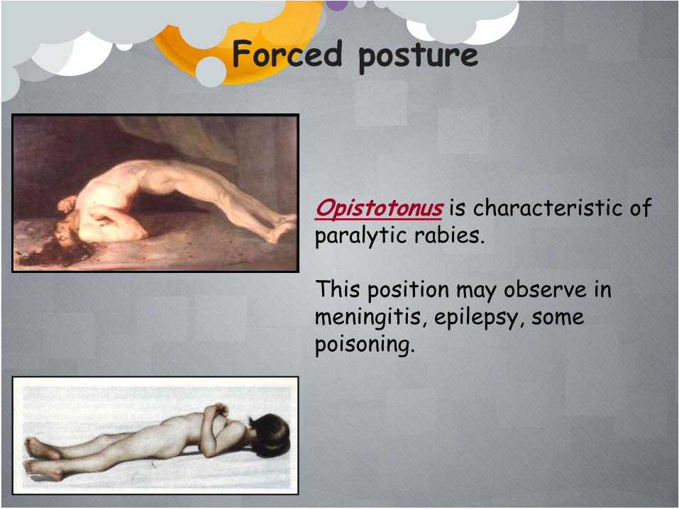
Opistotonus is characteristic of paralytic rabies. This position may observe in meningitis, epilepsy, some poisoning.
Forced posture

The prone position (lying with face down) : •tumor of pancreas; •gastric ulcer (in the posterior wall of the stomach is affected); •acute thrombosis of lien vein; •trauma and tuberculosis of spine; •trophic ulcer, placed on the skin of back and buttocks.
Forced posture

The forced “knee-elbow position” with bend trunk forward may observe in patients with effusive pericarditis. The state of restless, anxiety, occur in patient due to the urinary tract calculi and nephrocalcinosis.
Forced posture

GAIT Gait - combination of the pose and movement during
walking.
Gait depends from the state of nervous system
connective tissue, muscles, joint and bones.
Gait of the healthy person is firm, free, and straight.

•hemiglegic/circumductive
gait is characterized by
abundance (superfluous) leg
draw aside and the arm from
the same side bond to the
trunk;
There are some specific gaits according to the pathological processes:

•paretic gait is characterized by slow
movement with difficulties walking due to
the development of flexor spasm and
contractures in the limbs;
•peroneal gait, stoppage is characterized
by high climb of leg, sharp drawing;
•ataxic gait – (origin from Greek ataktos –
confused) is characterized by high rising of
climb, reach the floor, limb continue to
search fulcrum.;

spastic gait is characterized by small
step with difficulties during bend of
limb in knee and hell clinging due to the
a pyramidal tract lesion;
•doll ’ s/puppet gait is observed in
patients is with Parkinsonism, which
includes three main components: tremor,
muscular rigidity and hypokinesis;
GAIT
cerebellar gait, wobbly/tottering/reeling gait is characterized by
incoordination of ipsilateral limbs: decomposition of movements,
impaired alternating movements; loss of balance: broad based gait,
leaning towards of lesion; hypotonia of limbs; head tremor;
gait with forced movements femur nerve fibula neuritis is
observed in patients with child central paralysis;
retarded gait is characterized by small snuffle step with uncertain
and uncoordinative movement of arms;
“proud” gait is characterized by putting trunk backward for
support balance relevant to pregnancy, ascitis, or great tumor of
abdominal cavity

gait as “a duck” – is characterized by
small, slow step with compensatory
inclination trunk to the opposite side due
to the hypotonia of pelvis muscle;
HABITUS
The concept of habitus includes: •bodybuild; •height; •body weight;
In addition to general inspection it is necessary, to perform some anthropometry measurement.

Body-build is determined by morphological bodily features and divided into two groups: •a) correct habitus with a well proportioned parts of the body: trunk, head, limbs without deformity; •b) incorrect habitus with different deformity and disproportion of the trunk, limbs, chest, abdomen;

HEIGHT
The normal height of
males varies from 165 to 180 cm
females 155-170 cm.

Dwarfism may be due to hypofunction of
the anterior lobe of the pituitary (nanism)
or of the thyroid gland (cretinism).
Gigantism can be due to dysfunction and of
the anterior lobe of the pituitary or
hypofunction of the sex gland.
Patients height and the length of his trunk
are important for the assessment of both
his physical grow and proportions of his
separate parts, which can be upset in some
congenital diseases and disease acquired in
childhood.

WEIGHT
Body Mass Index (BMI) was proposed in order to assess the weight in adults. BMI may calculate using formula: BMI = weight (kg) height2 (m2)

According to the BMI it is possible to reveal the overweight and obesity. (WHO classification of overweight and obesity in adults) Category Body Mass Index (BMI), kg/m Underweight < 18.5 Normal weight 18.5–24.9 Overweight 25.0 Pre-obesity 25.0–29.9 Obesity I degree 30.0–34.9 Obesity II degree 35.0–39 Obesity III degree 40.0

Gain of weight may be in persons without weight
control, having eating habits with increased intake of carbohydrates, saturated fat and alcohol. Endocrine disorders are potential contributors to obesity (Gushing ’s syndrome, hypofunction of thyroid gland, diabetes mellitus 2 type).

Loss of weight is observed in persons during
starvation, in patients with severe diseases, oncology pathology, endocrine dysfunction - hyperfunction of thyroid gland

FACE OF THE PATIENT
Face in patients with diseases of respiratory system: •– facies pneumonica – one-sided blush on the same cheek as affected lung, cyanosis, often herpes on the lips and nose; •– facies asthmatica – pale, cyanotic face, sweating, cool extremities an unproductive cough, accelerated breathing rate;
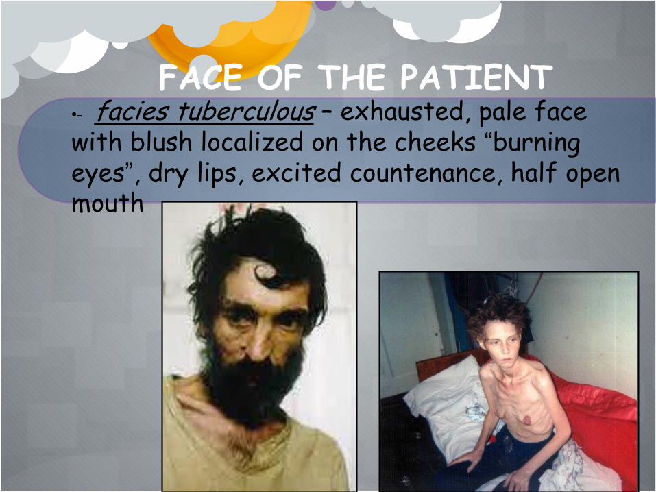
FACE OF THE PATIENT •- facies tuberculous – exhausted, pale face with blush localized on the cheeks “burning eyes”, dry lips, excited countenance, half open mouth

FACE OF THE PATIENT Face in patients with diseases of respiratory system: •–– facies adenoidea – half open or full open mouth, loose-hanging lower lip, noisy breathing.
Diffuse cyanosis in patient
with lung-heart failure

Face in patients with disease of cardiovascular system: •– facies aortale - pale skin, rhythmical movements of the head, simustaneosly with aortic regurgitation; •– facies mitrale – the patient looks younger his age, face with blush, localized on the cheeks, cyanotic color of the tip nose, ears, dyspnoea. The face is observed in patients with mitral stenosis ;
FACE OF THE PATIENT

Face in patients with disease of cardiovascular system:
•– facies Corvisara, facies cardiaca – is characteristic of heart failure. The face is edematous, pale, and yellowish with a cyanotic hue. The mouth is always half open, the lips are cyanotic, the yeas are dull;
FACE OF THE PATIENT

facies plethorica – hyperemic and cyanotic skin, puffy face due to the excessive circulated blood in patients, plethora with hypertensive crises.
FACE OF THE PATIENT

FACE OF THE PATIENT Face in patients with endocrine disorders:
•– facies acromegalica – due to the hyperproduction of growth hormone by anterior lobe of hypophysis. There are enlarged superciliary arches zygomatic bones, ears, auricles nose, lips, tongue, growth and putting forward of low jaw (prognotism) ;

FACE OF THE PATIENT Face in patients with endocrine disorders: •– facies in patients with Cushing’s syndrome due to the increased excessive cortisone production in patients with adrenal tumor or prolonged glucocorticoid administration is characterized by round or “moon-like” face, plethora, red cheeks, excessive hair growth (hirsutism in women);

FACE OF THE PATIENT Face in patients with endocrine disorders: – facies myxoedemica in patient with severe hypothyroidism (myxedema) due to the thyroid hypofunction has a dull, puffy face, with purplish lips and malar flush;

FACE OF THE PATIENT Face in patients with endocrine disorders: facies basedovica are observed in patients with hyperthyroidism which results from exposure of the body tissues to excess circulating levels of free thyroid hormones. The face is lively with widened eye slits and abnormally sparkling eyes (exophtalmus); •– facies in patients with hypogonadism is characterized by dry skin, wrinkled, absence of hair in men, thin eyebrows, looks as “baked apple”.

FACE OF THE PATIENT Face in patients with diseases of nervous system: – facies amimica, Parkinson’s mask in patients with blunts expression. A mask like, amimic face may result, with decreased blanking and a characteristic stare. Since the neck and upper trunk tend to flex forward, the patient seems to peer upward toward the observer. Facial skin becomes oily and drooling may occur.

FACE OF THE PATIENT
Face in patients with diseases of nervous system: – facies myophtica – with half-open month, without wrangle on the forehead, amimic, halt open eyes is characteristic of progressive myophathy;
FACE OF THE PATIENT
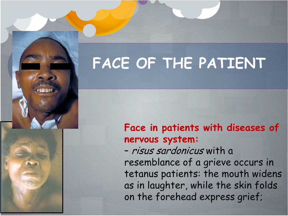
Face in patients with diseases of nervous system: – risus sardonicus with a resemblance of a grieve occurs in tetanus patients: the mouth widens as in laughter, while the skin folds on the forehead express grief;
FACE OF THE PATIENT
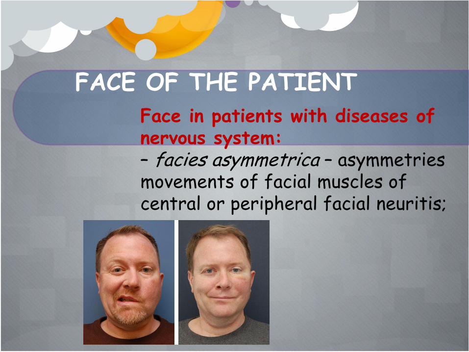
Face in patients with diseases of nervous system: – facies asymmetrica – asymmetries movements of facial muscles of central or peripheral facial neuritis;
Faces in patients with diseases of digestive system:
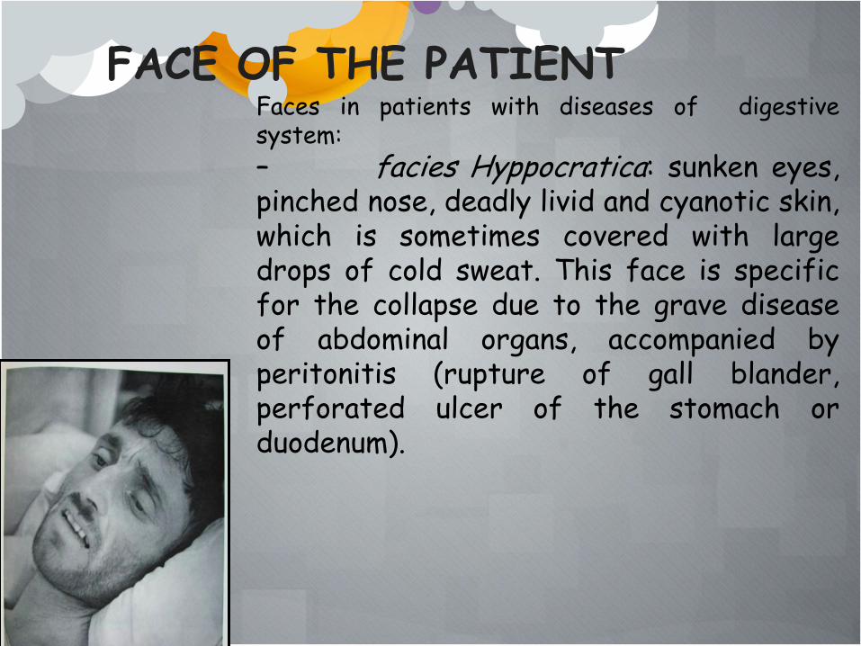
– facies Hyppocratica: sunken eyes, pinched nose, deadly livid and cyanotic skin, which is sometimes covered with large drops of cold sweat. This face is specific for the collapse due to the grave disease of abdominal organs, accompanied by peritonitis (rupture of gall blander, perforated ulcer of the stomach or duodenum).
FACE OF THE PATIENT
.

FACE OF THE PATIENT
Face in patients with diseases of kidney: •– facies nefritica – the face is edematous and often pale. Swelling usually appears first around the eyes and in the morning. The eyes may become slit like when edema is pronounced.

FACE OF THE PATIENT Face in patients with infectious disease: •– facies fibrilis is characterized hyperemic skin sparkling eyes and excited expression; •– facies in patients with louse-borne typhus: general hyperemia, the sclera is injected (“rabbit eye”); •– facies in patients with typhoid fever: slightly icteric yellow color; •– facies in patients with meningitis: the countenance of the read, anisocoria (different size of pupils), ptosis; •– facies in patients with cholera – frequent blinking, changing grimace, disorderly (irregular)mobility of face; •– facies leontina with nodular thickening of the skin under the eyes and over the eyebrows, with flattered nose is observed in leprosy; •– facies in patients with parotitis (mumps) swelling of parotid glands, which are visible above the angles of the jaw. At first it may be unilateral swelling, gradually become bilateral swelling due to the parotid gland enlargement; •– facies in patients with whooping cough: puffy face with edematous eyelids, conjunctiva hemorrhage, constant tears.

Faces in patients with diseases of blood system: •– facies anemic – very pale, with greenish tint in patients with iron deficiency anaemia •– facies as a “wax-doll”: very pale with yellowish tint and seemingly translucent skin.
FACE OF THE PATIENT

FACE OF THE PATIENT
Faces in patients with another
pathology:
•– facies cachectica – very
pale, with like earth like tint,
pinched nose. Patient loss of
weight significantly. These
features are observed in patients
with malignant tumor of digestive
system;

FACE OF THE PATIENT Faces in patients with another pathology:
– facies potatorice – hyperemic skin, especially
nose, cyanosis of the nose, lips, cheek, observed on
alcohol abuse;
•– Stokes collar – means edematous neck with
associated with edematous face due to the
compression of lymph ducts and veins with enlarged
mediastinal lymph nodes, tumor of mediastinum,
adhesive mediastinopericarditis, excessive effusion
in the pleural and pericardial cavity.

FACE OF THE PATIENT Faces in patients with another pathology:
•– Stokes collar – means edematous neck with
associated with edematous face due to the
compression of lymph ducts and veins with enlarged
mediastinal lymph nodes, tumor of mediastinum,
adhesive mediastinopericarditis, excessive effusion
in the pleural and pericardial cavity.

Examination of the skin:
•visual;
•palpative;
SKIN

SKIN
Order of inspection of the skin:
color;
eruption of the skin;
turgor and elasticity (visual and palpative methods);
moist of the skin (visual and palpative methods);
edema;
temperature of the skin;
subcutaneous veins;

COLOR OF THE SKIN
In healthy person skin has corporeal color (cutis coloris
somatici), without eruption, moderate moisture and elasticity,
preserved turgor.
There are next pathological changes of the skin
color:
pale, red, cyanotic,
yellow and bronze.

NB !
The pale and red color of the skin related to the
thickness, blood circulation, innervation and may be
transient character in physiological condition
(fear, high and low temperature of the air).
The yellow, cyanotic and bronze color of the skin
are due to the changing of the chemical blood content
and are observed only in pathological condition except
physiological jaundice at newborn.

Pale color of the skin (cutis
pallide)
Can be physiological and pathological.
Transient physiological pallid skin pear is due
to the vasomotor reaction of central (fear) and
peripheral origin (effect of low temperature).
Constant physiological pallid skin is observed
in patients with thick skin, insufficient
development of subcutaneous vessels.

Pathological pallid skin is connected with amount and
quality of blood.
observed in patients with decreased number of
erythrocytes and/or hemoglobin content in a blood unit
volume, classified as anemia, which accompanied such
diseases :
•hemoblastosis,
•different forms of anemia;
•acute and chronic infections with hemolysis (malaria, sepsis)
•chronic toxicity.
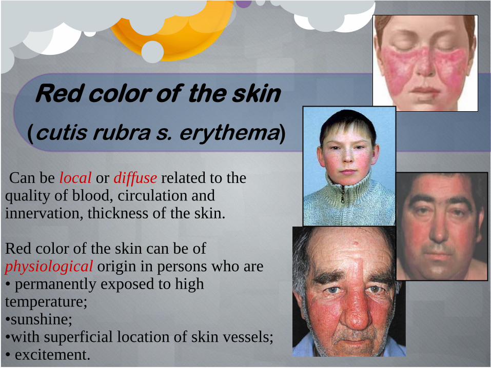
Red color of the skin
(cutis rubra s. erythema) Can be local or diffuse related to the quality of blood, circulation and innervation, thickness of the skin. Red color of the skin can be of physiological origin in persons who are • permanently exposed to high temperature; •sunshine; •with superficial location of skin vessels; • excitement.
Pathological red color may be transient in fever. In patient with pneumonia the redness is located on checks, more pronounced on the side of the affected lung. Local erythema as two-sided blush is characteristic of mitral stenosis (“mitral batter fly” with cyanotic tint), lupus hemoglobin concentration erythematous (“lupus buffer fly”) and tuberculosis. Constant diffuse erythema is observed in polycythemia (erythraemia) - excessive production by bone marrow erythroid precursor and cousuquasly the erythrocyte count hemoglobin concentration increases in peripheral blood.

Cyanosis
(cutis cyanotica)
Can be due to the changing the quality of
blood – (accumulation of the carbon dioxide
and reduced restored hemoglobin) and
venous congestion.
There are three forms of cyanosis: •central or diffuse, •peripheral or acrocyanosis •local .

Central or diffuse cyanosis (cyanosis diffuse) may be observed in such pathological states as: – chronic lung diseases (chronic bronchitis, acute pneumonia, emphysema, pneumosclerosis, bronchial asthma, atelectasis, thromboembolism of the pulmonary artery); – poisoning of the hemolytic substances; – congenital heart disease.

Peripheral or acrocyanosis is observed in patients with congestive heart failure. The blue color appears in the lips, cheeks, ear auricles, tip of the nose, and fingers. Local cyanosis (cyanosis localis) is observed in patients with thrombosis of artery or vein.
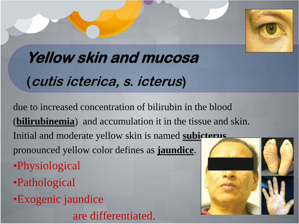
Yellow skin and mucosa
(cutis icterica, s. icterus)
due to increased concentration of bilirubin in the blood
(bilirubinemia) and accumulation it in the tissue and skin.
Initial and moderate yellow skin is named subicterus,
pronounced yellow color defines as jaundice.
•Physiological
•Pathological
•Exogenic jaundice
are differentiated.

Physiological jaundice
is observed in newborn
at first 5-7 days and resulted from hemolysis of
excessive erythrocyte amount
during transition to external respiration.

Pathological jaundice are divided into three types
according to their etiology:
1. hemolytic or suprahepatic jaundice (icterus colore
citricoluteo s. icterus suprahepatica) is characterized by lemon-
yellow tint due to the excessive hemolysis of erythrocytes in the
cells of the reticulohistocytic system (spleen, liver, bone marrow);
2. parenchymatous or hepatic jaundice (icterus colore
rubiginoso s. icterus hepatica) is characterized by orange-yellow
tint due to the damage of hepatocytes and disorders of their
function (inversion of unbound bilirubin to bound), observe in
acute and chronic hepatitis, poisoning;
3. obstructive or subhepatic jaundice (icterus colore luteoviridi s.
icterus infrahepatica) is characterized by greenish-yellow tint due
to the accumulation of bilirubin (the product of gradual oxidation
of bilirubin) resulted from partial or complete obstruction of the
common bile duct in patients with stones in the gall bladder, cancer
of the head of the pancreas, cancer of the major duodenal papilla.

Brown or bronze skin can observe in physiological and
pathological condition.
Physiological brown color
is of transient character and observes
during prolonged exposure of sunshine
(gelioxanthosis) and in pregnancy (as a
separate brown points).
Pathological brown or bronze color
•In Addison’s disease or bronze disease
resulted from the adrenal insufficiency in
patients with hypofunction of adrenal
gland .
•Hemochromatosis (bronzed diabetes or
pigmentary cirrhosis of the liver). The
disease is associated with inherited
disorder of iron metabolism, excessive
absorption of iron in the intensive and
accumulation of hemosiderin in various
tissues and organs, in the first instance in
the liver and pancreas

DEPIGMENTATION
vitiligo,
leukoderma
albinismus

Decreased pigmentation
Albinism – at birth the whole skin is white and pigment is also deficient in the hair, iris, and retina. Vitiligo – complete loss of melanocytes from affected parts. Segmental vitiligo is restricted to one part of the body. Generalized vitiligo is often symmetrical and frequently involves the hands, wrists, knees and neck as well as around the body orifices. Hypopigmentation is due to the decreased production of pituitary melanotrophic hormone in the patients with hypopituitarism. The complexion has a pale, yellow tinge; there is skin atrophy.
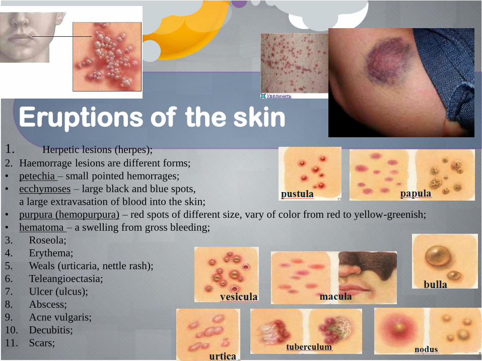
Eruptions of the skin 1. Herpetic lesions (herpes);
2. Haemorrage lesions are different forms;
• petechia – small pointed hemorrages;
• ecchymoses – large black and blue spots,
a large extravasation of blood into the skin;
• purpura (hemopurpura) – red spots of different size, vary of color from red to yellow-greenish;
• hematoma – a swelling from gross bleeding;
3. Roseola;
4. Erythema;
5. Weals (urticaria, nettle rash);
6. Teleangioectasia;
7. Ulcer (ulcus);
8. Abscess;
9. Acne vulgaris;
10. Decubitis;
11. Scars;
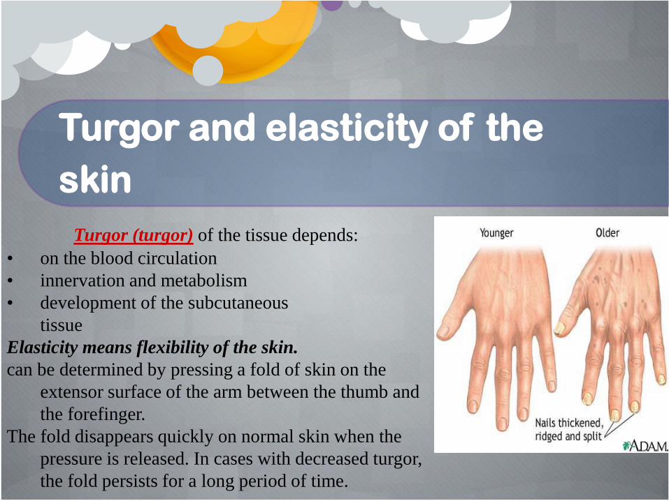
Turgor and elasticity of the
skin
Turgor (turgor) of the tissue depends:
• on the blood circulation
• innervation and metabolism
• development of the subcutaneous
tissue
Elasticity means flexibility of the skin.
can be determined by pressing a fold of skin on the
extensor surface of the arm between the thumb and
the forefinger.
The fold disappears quickly on normal skin when the
pressure is released. In cases with decreased turgor,
the fold persists for a long period of time.
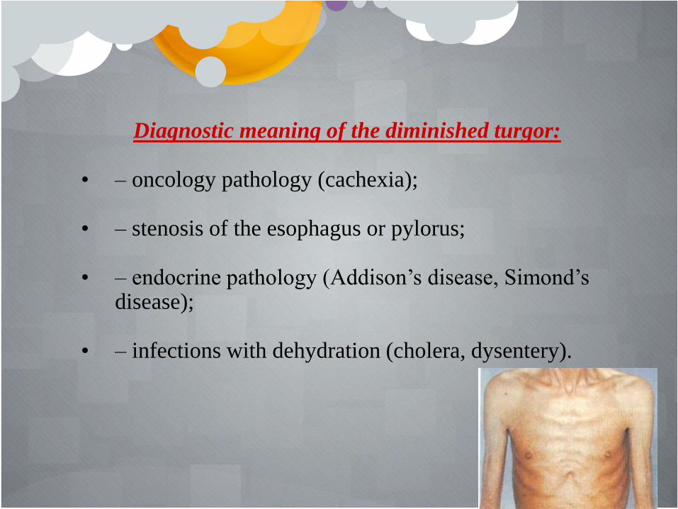
Diagnostic meaning of the diminished turgor:
• – oncology pathology (cachexia);
• – stenosis of the esophagus or pylorus;
• – endocrine pathology (Addison’s disease, Simond’s disease);
• – infections with dehydration (cholera, dysentery).

Edema may be caused by penetration of
fluid through the capillary walls and its
accumulation in tissues.
According to the pathogenic and location
factors, edema may be general and local.
General edema associated with disease of the heart,
kidneys, and endocrine disorders is characterized by
symmetrical localization in some regions of the body
or general overspreading of edema throughout the
entire body.
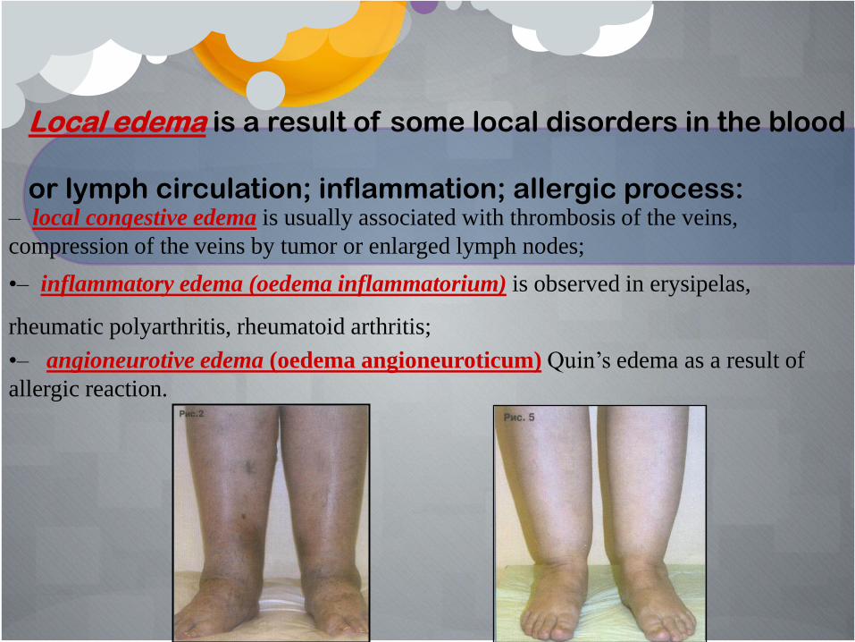
Local edema is a result of some local disorders in the blood
or lymph circulation; inflammation; allergic process: – local congestive edema is usually associated with thrombosis of the veins,
compression of the veins by tumor or enlarged lymph nodes;
•– inflammatory edema (oedema inflammatorium) is observed in erysipelas,
rheumatic polyarthritis, rheumatoid arthritis;
•– angioneurotive edema (oedema angioneuroticum) Quin’s edema as a result of
allergic reaction.

General edema overspread throughout the
entire body is named anasarca.
If edema is generalized fluid may accumulate
in the body’s cavities:
•in the abdomen (ascitis),
•in pleural cavity (hydrothorax),
•in pericardium (hydropericardium).

Subcutaneous fat.
In order to assess the degrees of subcutaneous fat
you should take a fold of the skin wrinkle and fat
over Traube’s cavity and measure the thickness.
In normosthenic person this size is 1,5-2 cm.
more than 2 cm it reflects the excessive accumulation of subcutaneous fat;
less than 1,5 cm – deficiency;
less than 0,5 cm – the sign of cachecxia;
Excessive accumulation of fat in the cells and tissue is defined as obesity (adipositas).

Types of obesity: –ginoid types of obesity is characterized by uniformly fat distribution with more pronounced accumulation at the buttock and hip. Another terms of this type of obesity: peripheral, buttock-thigh; –android types of obesity is characterized by accumulation of fat mainly at the upper part of the body, stomach (abdomen), and completely absence of fat at the buttock and legs. Another terms obesity: abdominal, central, male obesity.

Emaciation (macies) is divided into three groups: •loss of weight (demetritio);
•disturbances of weight (dysthrophia);
•acute and excessive loss of weight (cachexia).
Lymph nodes
are reveal during general inspection, using palpation.
Regional lymph nodes include: submandibular, parotid, occipital, posterior and anterior cervical, supraclavicular, subclavicular, axillary, cubital, inguinal, popliteal.
The examination of lymph nodes is performed by simple inspection and superficial palpation of the symmetrical region following the certain consequence: location, size, consistency, pain, mobility, color of the skin over the lymph nodes.
Normal lymph nodes cannot be detected visually or by palpation.

The main causes of the enlargement of the lymph nodes: –infections (tuberculosis, AIDS, brucellosis, infectious mononucleosis, tularemia, plague; –inflammation (local or generalized); –diseases of the blood (leukemia, lymphogranulomatosis); –lymphatic metastatic spread.

Muscular system
The main methods of examination are inspection and palpation.
During examination of the muscular system doctor should assess:
the level of development;
gender and age correspondence;
pain muscular tonus;
evidence of cramps;
In normal condition the muscular system develops
•corresponding to sex and age,
•the muscular tonus is present,
•painlessness,
•cramps and atrophy are absent.

Disorders of voluntary muscles include: – muscular dystrophy; – metabolic and endocrine myopathy; – congenital myopathy; – toxic myopathy; – disorders of the neuromuscular junction.

Bones system The main methods of examination of bones system
are inspection and palpation.
The attention should be paid to the
• development of the skeleton,
• correspondence to the age and sex,
• the presence of visible deformities (facture, curvature, enlargement).

Deformity of extremities Hyppocratic fingers or clubbing of the terminal phalanges of the fingers and toes, which resembles clock glass. Pseudohypoparathyidism as a congenital tissue resistance to the effects of parathyroid hormone. The patients have skeletal abnormalities such a small stature and short 4th and 5th metacarpals.

Scleroderma – the disorders of connective tissue characterized by fibrosis and degenerative changes in the skin and extremities. “Claw foot” is characterized by the presence of a high medial arch and secondary metatarsal collositis with clawing of the toes. Raynaud’s disease is characterized by “dead fingers” – transient pallor of the fingers and toes, sometimes nail fold infarction, leg ulcers or purpura. Large areas of skin necrosis or digital gangrene may observe.

Congenital deformity of extremities
Syndactyly – union of fingers or toes;
polydactyly – fingers more than five;
hemimelia – absence of the distal segment of an extremity;
amelia – absence of an extremity;

talipomanus – congenital club hand;congenital radio-ulnar synostosis; deformity of the femoral neck, congenital dislocation of the femur; club foot; bilateral club foot; valgus foot; congenital flat/splay foot, brachydactily – inherited abnormality shortening of finger detected during child birth.

The common causes of the bones system
abnormalities Pathology
Examples
Endocrine
pathology
Acromegaly, achondroplasia, hypopituitarism,
hypothyroidism, Cushing’s syndrome
Infections
Tuberculosis, syphilis
Hypo- and
avitaminosis
Rickets (Rachitis)
Diseases of the
blood system
Leukemia, congenital hemolytic anemia,
lymphoglanulomatosis, polycythaemia, multiple myeloma
Oncology
Sarcoma, metastatic spread
Congenital
pathology
Deformity of the thorax, cleft lip, Cherechewski-Turner syndrome, Marfan’s syndrome, Klinefelter’s syndrome, Edwards syndrome, mucopolysaccharidoses, Ehlers-Danlos syndrom
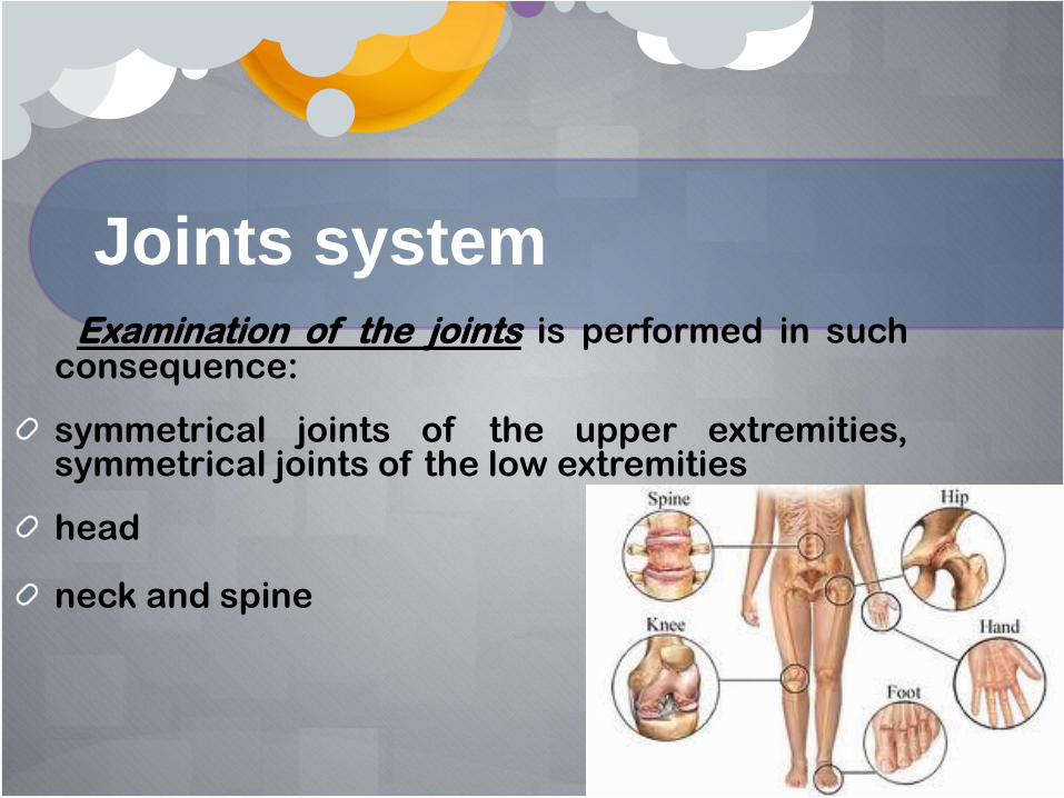
Joints system Examination of the joints is performed in such
consequence:
symmetrical joints of the upper extremities, symmetrical joints of the low extremities
head
neck and spine

Attention should be paid to : •Shape
•Configuration
•Swelling
•Hyperemia
Using palpation you should reveal possible pain, fluid in the join cavity, and crackle.

In clinical practice it is important to assess
joints movements.
Two kinds of movement are distinguished:
active movement, which is fulfilled by patient according to the doctor’s instruction
passive movement, which perform doctor.
Simultaneous restriction active and passive
movement suggests the affection of the joints
(rheumatoid arthritis, rheumatic polyarthritis).

Restriction of active, but preservation of passive movements is observed in patients with coma and during disorders of local joint circulation and innervation.

Examination of the spine
In normal condition there are four physiological curvature of the spine:
– cervical curvature of the spine column with forward convexity – lordosis;
– thoracic backward curvature of the spinal column – kyphosis;
– lumbar forward curvature – lordosis;
– pelvic kyphosis.

During inspection of the head you should pay
attention on the
size,
• shape,
• position,
• movement and state of eyes, nose, mouth, tongue, and teeth.
Head
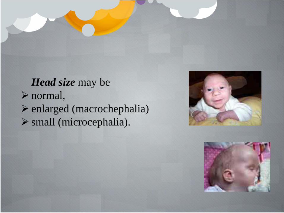
Head size may be
normal,
enlarged (macrochephalia)
small (microcephalia).

A “square” head with “bossing” of the frontal and parietal
bones and delayed closure of the anterior fontanelle in
childhood is characteristic of rickets.
May be craniotabes – small round unossified areas in the
membranous bones of the skull.
“Bossing” of the skull, prominent malar bones and protuberant
teeth are development in sickle-cell anemia.
A head bossing due to the bone marrow hyperplasia appears in
child with beta-thalassemia. The skull radiograph shows a
“hair on end” appearance and general widening of the
medullary spaces, which may interfere with the development
of the paranasal sinuses;
Head Shape

“tringle” head develops due to the intrauterus
craniosynostosis, clossing the skull fissura, elevation of
intracerebral pressure. The characheristical features: high
forehead, exophtalmus, ptosis, and nose as beak;
Local osteomalacia of skull bone is characteristic of multiple
myeloma (disease of blood);
A “square” head, flattened on top, with prominent frontal
tubers, can indicate congenital syphilis.

depends on state of spine and nervous system:
•immovable head is observed at ankylosing spondyloarthritis,
Bechterew-Strumpal-Mari’s disease, verterbal osteochon-drosis,
myositis, myopathy, fusion of cervical vertebral (Klippel-Fell’s
disease cervical ribs);
•torcicollis (stiff-neck) – turning head in one side may be acquired
and occur due to the myositis. More commonly the reason are
congenital muscular torticollis (Grisel’s disease), spastic torticollis;
•neck stiffness with head throw back is specific for meningitis or
tetanus.
stiff-neck
Position of the Head

Involuntary shaking head associated with tremor of the
hands occurs at patients with Parkinsonis disease;
Non-rhythmic tremor of the head is the symptom of
chorea, st. Vitus’s dance;
Shaking head synchronous with heart function (with
pulse wave a head throw back) named as Musset’s sign is
observed at aortic regurgitation
Movement of the head

Inspection of the eyes can reveal some essential
diagnostic signs.
It is necessary to exam :
eyelids
eye slit
eyeball
sclera
cornea of the eye
pupils
Eyes

•swallowing and pigmentation is the sign of
dermatomyositis;
•edema of the eyelids is the first indication of the nephritis;
•narrowing of the eye slit occurs in myxedema and general
edema (anasarca);
•dark eyelids are the characteristic of Addison’s disease
and diffuse toxic goiter;
•xanthomas at eyelids indicate deranged cholesterol
metabolism – atherosclerosis, liver cirrosis, cholestatic
jaundice;
•persistent drooping of the upper eyelids (ptosis) may be
congenital or acquired origin.
xanthomas
Eyelids:

• anophtalmia – bilateral or unilateral absence of eye
or presence of rudimentary eye due to the inherited
disorders;
• microphtalmia – small size of eyeball is observed in
some inherited syndromes (gingival fibromatosis,
depigmentation and microphtalnia, Cohem syndrome:
hemifacial microsomia);
Eyeball:

EXOPHTALMIA (protrusion of the eyeball) is observed:
bilateral - in thyrotoxicosis, strong myopia, some inherited
abnormalities (Klleeblattsehadel anomaly; cranio-facial
dysostosis – Cronzon syndrom);
unilateral – in patient with retrobulbar tumor, brain abscess,
parsis of nervus faciales;
Eyeball:

ENOPHTALMIA – (recession of the eyeball) is
observed :
bilateral – in hypothyroidism, peritonitis
(Hyppocrates face), agonia, cholera;
unilateral – Klod-Bernard-Horner syndrom,
enophtalmia is a sign of inherited pathology (hemifacial
atrophy progressive, Cockayne syndrome).
Eyeball:

Movement of the eyeball is synchronous, is fulfilled in
some direction (horizontal, vertical, circular) resulted from
coordinate function of 6 pairs muscle.
Disorders of eyeball movement may be in a form of
squint heterotropia (strabismus) and nystagmus.
Eyeball:

-depends on the eyeball and eyelid position.
Narrowing of the eye slit may be observed in acute
glomerulonephritis, Quenke oedema, myxoedema,
peritonitis, and congenital pathology.
Widening of the eye slit may be observed in
thyrotoxicosis, retrabulbar abscesses.
Asymmetry of the eye slit may be at unilateral ptosis,
tumor of the brain.
Eye slit

JELLOW SCLERA is early sign of jaundice;
bleeding into the conjunctive and sclera at bacterial
endocarditis, epilepsy, deficiency of vitamin C;
RED “AS A RABBIT” conjunctivae ocular injection at
typhus;
single brown sport at conjunctiva may observe at
Addison’s disease.
SCLERA, CONJUNCTIVA ,
CORNEA OF THE EYE

CORNEA in normal condition is bright, clear and
transparent.
Dull cornea ulceration, corneal erosion may by at
congenital syphilis, bacterial, mycotic, herpetic keratitis.
Aging arcus (arcus senilis) – white-grey ring 1-2 mm at
cornea is the typical symptom of old age.
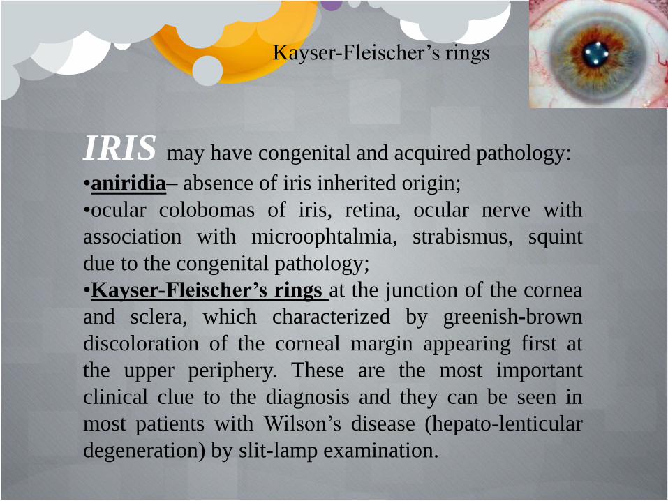
IRIS may have congenital and acquired pathology:
•aniridia– absence of iris inherited origin;
•ocular colobomas of iris, retina, ocular nerve with
association with microophtalmia, strabismus, squint
due to the congenital pathology;
•Kayser-Fleischer’s rings at the junction of the cornea
and sclera, which characterized by greenish-brown
discoloration of the corneal margin appearing first at
the upper periphery. These are the most important
clinical clue to the diagnosis and they can be seen in
most patients with Wilson’s disease (hepato-lenticular
degeneration) by slit-lamp examination.
Kayser-Fleischer’s rings

PUPILS:
examination of the size
Shape
reaction to convergence, light
accommodation

MYOSIS – papillary constriction is observed in uremia,
intracranial hemorrhages, brain tumor, neurosyphilis,
typhus, chronic poisoning.
In persons with morphine abuse – point-like pupil are
typical.
MYDRIASIS– papillary dilation is observed in patients
with coma (except uremic and apoplectic), syphilis,
sometimes at aortic aneurysm.
ANISOCORIA – asymmetrical pupils is observed in
syphilis, Argyll Robertson’s syndrome.
PUPILS
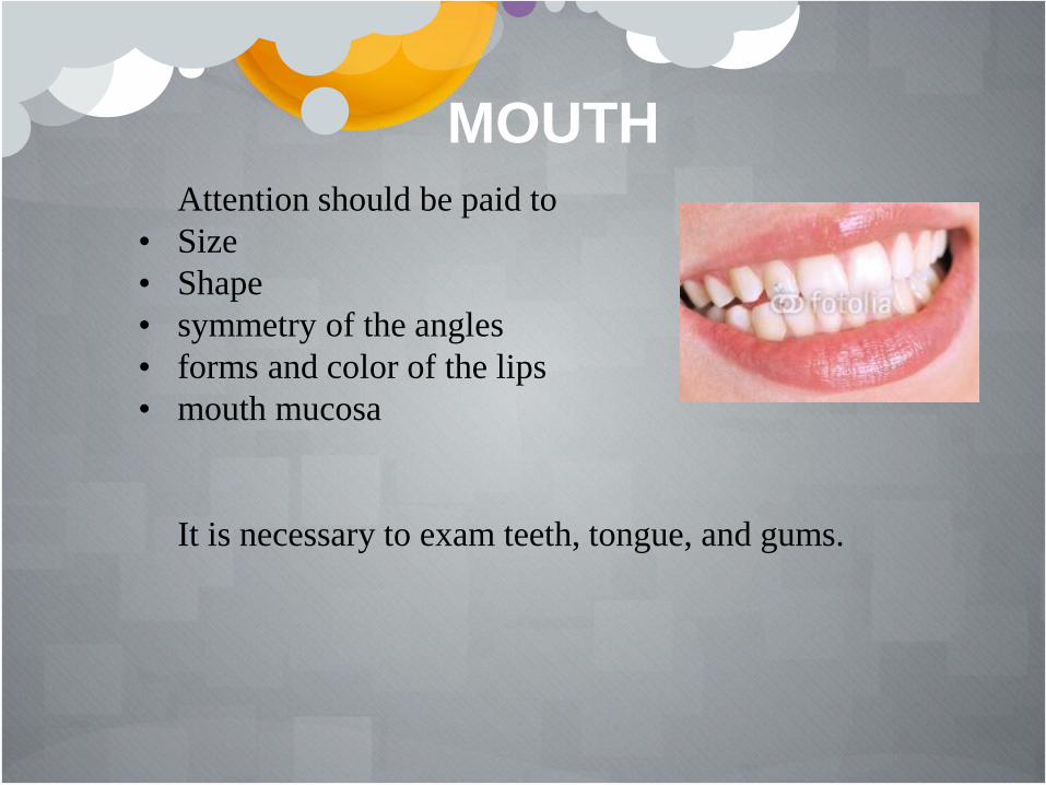
Attention should be paid to
• Size
• Shape
• symmetry of the angles
• forms and color of the lips
• mouth mucosa
It is necessary to exam teeth, tongue, and gums.
MOUTH

Mouth shape in pathological condition may
be in a form of macrostomia and microstomia.
Macrostomia is a result of congenital
pathology.
Microstomia has inhereted origin and may
be acquired (mouth in patients with
sclerodermia and hypoparathyroidism).

Asymmetry position of angles mouth
observed in
• local inflammatory process and in patients
with lesions, affecting the trygeminus system;
• paralyses of the facial nerve;
• stroke with such clinical feature: unilateral
loss of nasolabial fold, mouth deviates to
normal size and salvia may drool from it.

Size and deformity
Macrocheilia – pathological enlargement of lips
observe in acromegaly, hypothyroidism, allergic edema,
and congenital pathology.
Cheilietis – inflammatory process commonly at
mouth angles is a symptom of iron deficiency anemia,
and hypovitaminosis B2.
Procheilia – protruding lips in acromegaly.
Opistocheilia – retracted lips in patients without teeth
and peritonitis.
Cheilietis
LIPS

Acheilia (absence of lips), syncheilia (adhesion of
lateral portion of the lips), brachycheilia (shortening of
the middle portion of the upper lip) are the signs of
congenital pathology.
Cheiloshisis – labial cleft (“hare”-lip) is a symptom of
congenital pathology (Bixler syndrome – hypertelorism,
microtonia, facial clefting and conductive deafness; cleft
lip with or without cleft palate, median cleft-face
syndrome).
Bumpy lips and tongue neuromas are in patients with
multiple endocrine neoplasia (bilateral
pheochromocytomas and medullary carcinoma of thyroid
gland).

COLOR OF THE LIPS Acrocyanosis – blue color of the lips is
characteristic of mitral stenosis, heart failure.
Hyperemic lips – high temperature, inflammatory
processes.
Pale lips – acute and chronic bleeding, oncological
process, leukemia, hypo- and aplastic anemia.

In health the tongue is moist with only slight
white fur on the dorsum.
The papillae are readily seen.
With inspecting the tongue attention should be
paid to its shape and size, surface, movement,
color, and the state of papillae.
Tongue

Shape and size
Macroglossia – enlargement of the tongue is a
sign of congenital pathology (Daun’s disease;
Beckwith-Wiedeman syndrome – macroglossia,
visceromegalia, ompalocele; glycogenosi type II –
macroglossia, cardiomegalia, myotonia; cerebral
gigantism).
Microglossia – decreased tongue observe in the
patients with cholera, typhus, starvation, and vitamin
B12-deficiency.
Unilateral atrophia of the tongue occurs at
pyramidal tract lesion.
Flat tongue due to the atrophy of it base resulted
from ulcerative stomatitis and scarification of the soft
palate and pharynx in secondary syphilis.

Fur of the tongue in pathological conditions has
diagnostic significance:
Coated (furred) tongue is observed at gastritis, peritonitis,
colitis, fever, infections (hepatitis), and pneumonia;
Coated in the center and at the base bur clear the tip and
margins of the tongue is typical to typhoid fever.
Additional diagnostic meaning is a fur character and color:
White fur observe at typhus, pneumonia and peritonitis;
White-gray fur observe at gastritis, virus hepatitis, and some
infections;
White-dirty or yellow fur – at peritonitis;
White-blue – in rheumatic polyarthritis;
White-yellow fur in those who smoke excessively;

Crimson-red (strawberry/raspberry) tongue observe in
scarlet fever;
Dry tongue is an indication of dehydration with
followed formation of erosion hemorrhage and
observed in peritonitis, and severe infections. Dryness
of the mouth (xerostomia) may be caused by
anticholinergic or antidepressant drugs; bur commonly
it is due to anxiety.
Glossitis may be a prominent feature of stomatitis
resulting from nutrition deficiency and overdose of
antibiotics.

Surface of the tongue
Atrophy of tingual papilla cause smooth (as if polished) crimson
tongue, Hunter’s glossitis, which may observe in the patients with B12-
deficiency anemia.
The glassy tongue is characteristic of gastric cancer, pellagra, and
sprue.
The local thickening of the tongue with chronic migrating superficial
glossitis named as geographical tongue is found in the patients with
hyperacidity of gastric juice.
Grooved (fluted) tongue, with multiple wrinkles is characteristic of
acromegaly.
There are some patches, ulceration at the mouth.
Leucoplakia is white, firm, smooth patches beginning at the side of
the tongue and later spreading over the dorsum. In the early stages the
tongue is not painful but later fissures split the patches with tenderness.
Hairy leucoplakia occurs in AIDS.
Syphilis may present as a painful solitary ulcer usually on the
tongue or palate.

Neck Attention should be paid to the shape and size,
symmetry skin colour presence of scars and visible
pulsation.
Changing neck shape and sized depend on constitutional type, the state of
lymph nodes, thyroid gland, cervical column and musels development.
Short thick neck is observed in hyperstenic persons. In pathological condition
such neck may be in patients with lung emphysema, obesity, hypothyreoidism,
Gushing’s disease, and pronounced enlargement of thyreoid gland.
Long slim neck with prominent cartilage is observed in asthenic persons. In
pathology, slim neck may be in patients with disorders of pituitary (sex) gland,
starvation, cachexia.

Skin colour at the neck region indicates to some pathology:
• pigmentation with outlined border is observed in Addison’s disease;
• multiple pound white sport (syphilitic leucoderma) as a neclace (collar Veneris)in
syphilis;
• legible limited red-brown line due to the skin atrophia is observed in pellagra
(hypovitaminos) column casae;
• scars at neck indicate to the previous tuberculous lymphadenitis.
Prerugium-syndrom – a skin fold placed on the side neck surface from mastoid
process is a sign of Shereshevsky-Turner syndrome.
Pulsation of the carotid artery (carotid shudder, saltus carotidum) appears due to the
changing of blood pressure and filling of arteries during systole and diastole in patients
with aortic regurgitation, hyperthyroidism, and fever.
Swollen and pulsation of jugular veins is explained by difficulty of blood flow to
right atrium in tricuspid regurgitation, pericarditis, and chronic lung diseases.
Thyroid gland placed on anterior surface of cricoid cartilage. In normal condition in
health persons thyroid gland is impalpable. The thyroid isthmus is often but not always
palpable. The lobes are more lateral than isthmus and harder to feel.
Coiter is a general term for an enlarged thyroid gland and observed in Basedovica
disease (hyperthyroidism), autoimmune thyroiditis (Shasimoto goiter) and endemic
goiter.
The diffuse enlargement of thyroid gland is observed in hyperthyroidism, cancer of
the thyroid gland is characterized by asymmetric separate nodes with unequal surface.

NAILS
it is a combination of both .....
leuco and koilonychia
Typical nail changes of
pachyonychia congenita: yellow-
gray discoloration and thickening
of nail plates, subungual
hyperkeratosis, upward angulation
of the nail and pincer nail
deformity

NAILS
Pale nails