
Insights into Living with Kidney Disease -...
53
BioMed Research International Insights into Living with Kidney Disease Guest Editors: Veronica Swallow, Houry Puzantian, Leah Krischock, and Ulf Gunnar Bronas
Transcript of Insights into Living with Kidney Disease -...

BioMed Research International
Insights into Living with Kidney Disease
Guest Editors: Veronica Swallow, Houry Puzantian, Leah Krischock, and Ulf Gunnar Bronas

Insights into Living with Kidney Disease

BioMed Research International
Insights into Living with Kidney Disease
Guest Editors: Veronica Swallow, Houry Puzantian,Leah Krischock, and Ulf Gunnar Bronas

Copyright © 2017 Hindawi. All rights reserved.
This is a special issue published in “BioMed Research International.” All articles are open access articles distributed under the CreativeCommons Attribution License, which permits unrestricted use, distribution, and reproduction in any medium, provided the originalwork is properly cited.

Contents
Insights into Living with Kidney DiseaseVeronica Swallow, Houry Puzantian, Leah Krischock, and Ulf Gunnar BronasVolume 2017, Article ID 2805684, 2 pages
Cognitive Impairment in Chronic Kidney Disease: Vascular Milieu and the PotentialTherapeutic Roleof ExerciseUlf G. Bronas, Houry Puzantian, and Mary HannanVolume 2017, Article ID 2726369, 10 pages
Posttransplant Anemia as a Prognostic Factor of Mortality in Kidney-Transplant RecipientsMaria Majernikova, Jaroslav Rosenberger, Lucia Prihodova, Miriam Jarcuskova, Robert Roland,Johan W. Groothoff, and Jitse P. van DijkVolume 2017, Article ID 6987240, 8 pages
Etiology of End-Stage Renal Disease and Arterial Stiffness among Hemodialysis PatientsBalsam El Ghoul, Yazan Daaboul, Serge Korjian, Andrew El Alam, Anthony Mansour, Essa Hariri,Salam Samad, Pascale Salameh, Georges Dahdah, Jacques Blacher, Michel E. Safar, and Sola Aoun BahousVolume 2017, Article ID 2543262, 6 pages
How Should Disaster Base Hospitals Prepare for DialysisTherapy after Earthquakes? Introduction ofDouble Water Piping Circuits Provided byWell Water SystemNaoki Ikegaya, George Seki, and Nobutaka OhtaVolume 2016, Article ID 9647156, 5 pages
The Experience of Older People in the Shared Decision-Making Process in Advanced Kidney CareNicola Thomas, Karen Jenkins, Breeda McManus, and Brian GraceyVolume 2016, Article ID 7859725, 8 pages
Piloting Psychology Annual Reviews as a Method of Measuring Psychological Distress and Quality ofLife in Paediatric Renal Transplant PatientsJade Bamford and Lucy WirzVolume 2016, Article ID 1685362, 9 pages

EditorialInsights into Living with Kidney Disease
Veronica Swallow,1,2 Houry Puzantian,3 Leah Krischock,4 and Ulf Gunnar Bronas3
1School of Healthcare, University of Leeds, Baines Wing, Leeds LS2 9JT, UK2School of Nursing, Midwifery, and Indigenous Health, Faculty of Science, Charles Sturt University, Boorooma St, Wagga Wagga,NSW 2650, Australia3Department of Biobehavioral Health Science, College of Nursing, University of Illinois at Chicago, Chicago, IL, USA4Sydney Children’s Hospital, Randwick, Sydney, NSW, Australia
Correspondence should be addressed to Veronica Swallow; [email protected]
Received 23 April 2017; Accepted 23 April 2017; Published 16 May 2017
Copyright © 2017 Veronica Swallow et al. This is an open access article distributed under the Creative Commons AttributionLicense, which permits unrestricted use, distribution, and reproduction in any medium, provided the original work is properlycited.
There are many implications for those across the life-coursewho are living with kidney disease and their relatives/carers[1, 2]. Chronic kidney disease (CKD) is an important causeof reduced quality of life, morbidity, and death, so early iden-tification is needed to help reduce these dire consequences.Public health approaches to enabling early identification are,therefore, receiving increasing attention. As part of theseapproaches, self-care is an integral part of daily life forpersons across the life-course who are living with kidneydisease and oftenmeans that patients and/or their carers takeresponsibility for day-to-daymanagement of complex clinicalinterventions and treatment regimens, with support (thatis often provided remotely) from the health professionalsinvolved in their care [3]. People living with kidney diseasecan benefit enormously if they receive appropriate support forself-care. However, to understand the best ways to supportthem we need detailed insights into the challenges of livingwith kidney disease.The papers presented in this special issuereport and discuss current evidence and new innovations;these insights will help professionals who are managingpatients living with kidney disease.
In the management of patients receiving hemodialysistherapy, it is notable (as reported by B. El Ghoul et al.)that the etiology of end-stage renal disease is indepen-dently associated with arterial stiffness. This is higher amongpatients who developed renal sequelae of either diabetesmellitus or hypertension as compared with those with ahistory of either diabetes mellitus or hypertension alone.The clinical implication of this finding can translate intoearlier interventions to reduce end-organ complications.
Moreover, M. Majernikova et al. note that clinical eval-uation and treatment of even mild anemia might reducethe higher risk of mortality in patients with posttransplantanemia in early stages of CKD after kidney transplantation.Furthermore, understanding the cerebro-vascular-renal axispathophysiological link and its interconnection with thepossible protective role of exercise is important for clinicianssupporting patients with CKD in order to minimize therisk of loss of independence and improve quality of life (asreported by U. G. Bronas et al.)
Two papers in this issue focus on the psychosocial impactof CKD, in relation to dialysis for adult patients (N. Thomaset al.), and posttransplantation for children and young people(J. Bamford and L. Wirz). Posttransplant psychology annualreviews introduced into one Pediatric Renal Service enabledmeasurement of psychological distress and quality of life andhelped identify those families most likely to benefit frompsychological intervention. In N. Thomas et al.’s study, mostpatients were satisfied with the amount of information theyreceived, although it was recommended that the quality ofthe information they received could have been improved, inparticular concerning the effect of dialysis on individuals’day-to-day life.
Finally, the importance of lobbying policy makers andlocal water departments to ensure the availability of robustinfrastructures support to sustain dialysis, the life-savingtherapy for many people living with end-stage renal disease,including at times of natural disasters such as earthquakes, ishighlighted by N. Ikegaya et al.
HindawiBioMed Research InternationalVolume 2017, Article ID 2805684, 2 pageshttps://doi.org/10.1155/2017/2805684

2 BioMed Research International
Overall, the newknowledge contained in this special issuemakes an important contribution to the literature and canhelp shape the services and support offered to patients andfamilies living with kidney disease.
Veronica SwallowHoury PuzantianLeah Krischock
Ulf Gunnar Bronas
References
[1] V. Jha, G. Garcia-Garcia, K. Iseki et al., “Chronic kidney disease:global dimension and perspectives,” The Lancet, vol. 382, no.9888, pp. 260–272, 2013.
[2] L. S. Chawla, R. Bellomo, A. Bihorac et al., “Acute kidney diseaseand renal recovery: consensus report of the acute disease qualityinitiative (ADQI) 16 workgroup,” Nature Reviews Nephrology,vol. 13, no. 4, pp. 241–257, 2017.
[3] S. W. Ong, S. V. Jassal, E. Porter, A. G. Logan, and J. A. Miller,“Using an electronic self-management tool to support patientswith chronic kidney disease (CKD): a CKD clinic self-caremodel,” Seminars in Dialysis, vol. 26, no. 2, pp. 195–202, 2013.
Review ArticleCognitive Impairment in Chronic Kidney Disease: VascularMilieu and the Potential Therapeutic Role of Exercise
Ulf G. Bronas, Houry Puzantian, andMary Hannan
College of Nursing, Department of Biobehavioral Health Science, University of Illinois at Chicago, Chicago, IL, USA
Correspondence should be addressed to Ulf G. Bronas; [email protected]
Received 27 November 2016; Accepted 28 February 2017; Published 19 April 2017
Academic Editor: Hiroyuki Shimada
Copyright © 2017 Ulf G. Bronas et al. This is an open access article distributed under the Creative Commons Attribution License,which permits unrestricted use, distribution, and reproduction in any medium, provided the original work is properly cited.
Chronic kidney disease (CKD) is considered a model of accelerated aging. More specifically, CKD leads to reduced physicalfunctioning and increased frailty, increased vascular dysfunction, vascular calcification and arterial stiffness, high levels of systemicinflammation, and oxidative stress, as well as increased cognitive impairment. Increasing evidence suggests that the cognitiveimpairment associated with CKD may be related to cerebral small vessel disease and overall impairment in white matter integrity.The triad of poor physical function, vascular dysfunction, and cognitive impairment places patients living with CKD at an increasedrisk for loss of independence, poor health-related quality of life, morbidity, and mortality. The purpose of this review is to discussthe available evidence of cerebrovascular-renal axis and its interconnection with early and accelerated cognitive impairment inpatients with CKD and the plausible role of exercise as a therapeutic modality. Understanding the cerebrovascular-renal axispathophysiological link and its interconnection with physical function is important for clinicians in order to minimize the riskof loss of independence and improve quality of life in patients with CKD.
1. Introduction
Chronic kidney disease (CKD) affects 45% of adults olderthan 70 years of age in the US [1]. The incidence of CKDwill increase significantly over the next decade due to theincreasing incidence of diabetes and hypertension in therapidly aging US population. The economic cost of CKD isstaggering with Medicare spending for patients with CKDaged 65 and older exceeding $50 billion in 2013, representing20% of all Medicare spending in this age group [2]. Con-tributing to the high cost of CKD is the remarkably highprevalence of cognitive impairment or overt dementia thatranges 20–50% in older patients with moderate CKD [3–8] and may reach as high as 70% in severe CKD/dialysis[9]. Cognitive impairment impacts patients negatively bycontributing to functional dependence and behavioral symp-toms that result in poor outcomes and decreased medicationand medical care compliance. These negative consequencesresult in a downward spiral of functional decline and anaccelerated loss of independence, which leads to prematureinstitutionalization [10–16]. The negative impact of cognitiveimpairment on quality of life and emotional wellbeing is
significant, and it even affects employment rates negatively[17–20]. Moreover, cognitive function for incident dialysispatients has been found to be correlated with frailty andmeasures of depression [21]. Additionally, it more thandoublesmortality risk and increases days spent in the hospital[15, 22], contributing to the tremendous individual, societal,and economical burden of CKD. We will review the vascularmilieu as it is associated with cognitive decline in patientswith kidney disease and the potential therapeutic role ofexercise.
2. Measurement of Cognitive Impairment
Cognitive impairment is commonly referred to as a reductionin global cognition that is new and affects at least 2 areasof cognitive function that can be measured using a stan-dard cognitive function test (e.g., Mini Mental State Exam(MMSE) or the Montreal Cognitive Assessment (MOCA))[23, 24]. Impairment can be evident in various cognitivedomains: executive function (judgement and planning), lan-guage, attention, memory, and visual-spatial learning. Mildcognitive impairment (MCI) is commonly defined as a deficit
HindawiBioMed Research InternationalVolume 2017, Article ID 2726369, 10 pageshttps://doi.org/10.1155/2017/2726369

2 BioMed Research International
in global cognition that is not consistent with aging andhas not progressed to overt dementia. Although there is alack of a consensus for a standard definition of MCI, it isknown to be present with a performance of 1.5–1.99 standarddeviations below the standard norm on a given cognitive test.MCI is mostly manifested in short-term memory loss butcan also be manifested as impaired language and executivefunctions [25, 26]. Importantly, progression from MCI toovert dementia is approximately 15%per year in older patients[26]. Dementia on the other hand is used as the umbrella termfor moderate/severe progressive cognitive impairment oftendefined as scoring 2 standard deviations below populationnorms in at least 2 cognitive domains [27]. Importantly,overt dementia leads to a loss of independent daily functionwhereas MCI does not appear to significantly affect indepen-dent daily function.
3. Cognitive Impairment andDementia in CKD
It is well established that patients with kidney disease com-monly have some degree of cognitive impairment and thatkidney dysfunction is associated with a more rapid decline inmental function than in age matched comparisons [28, 29].As many as 20–50% of patients with moderate CKD haveestablished cognitive impairment or overt dementia [3–8, 30].It should be noted that the actual population prevalence andincidence of cognitive impairment are likely underreportedbecause published studies are primarily clinic-based and nottrue population studies.TheUnited States Renal Data SystemAnnual Data Report found a lower prevalence of cognitiveimpairment in CKD patients (7.6–16.8%) [22]. However,the true population prevalence is likely substantially higher.This is evidenced by Kurella et al. [6] who reported a23–28% prevalence of cognitive impairment in stages 3-4CKD patients (𝑛 = 80) seen in clinical practice and Murrayet al. [9] who reported a prevalence of MCI or dementia in87% of older dialysis patients. Most published studies havereported a prevalence of cognitive impairment of 20–50% inCKD patients and up to 70% in older patients on dialysis [3–9]. Unfortunately, less than 5% of all renal disease patientswith cognitive impairment have been screened or received amedical diagnosis [9, 31].This suggests that cognitive impair-ment in this group of patients is severely underdetected andnot adequately addressed.
The degree of renal dysfunction appears to be correlatedwith the degree of cognitive impairment. Cognitive impart-ment has been shown inmultiple studies to be associatedwithdeteriorating renal function well before requiring dialysis,although this association is particularly strong in patientsrequiring dialysis [31]. Increased serum cystatin C and albu-minuria are also associated with accelerated cognitive decline[32–34]. Studies have shown a 15-25% increased risk of cog-nitive impairment for every 10 ml/min per 1.73m2 reductionin the estimated glomerular filtration rate (eGFR). Further,there is an increased odds ratio of 2.43 (95% CI 1.38 to 4.29)for cognitive impairment in patients with an eGFR of <45ml/min/per 1.73m2 even after adjustment for confounders [7,35].Thus, patients with CKD appear to have at least a twofold
increased risk of cognitive impairment than those withoutCKD [7, 8, 30]. This risk increases to fourfold with furtherreductions in eGFR to <30ml/min per 1.73m2 independentof potential confounders [8, 35]. These findings translate topatients with CKD having an increased and accelerated riskof cognitive aging equivalent to 3.6–7 years compared to thegeneral population [32, 36]. However, current physical exam-ination and medical history for patients with CKD or end-stage renal disease (ESRD) do not include cognitive functionmeasures.
In terms of the clinical implications of cognitive impair-ment in CKD, improving support and access to psychol-ogy and social professionals, support groups, and patienteducation are likely to improve outcomes, although this hasyet to be determined. Support of patients with CKD shouldalso include counseling with pharmacists and providersregarding the risk of polypharmacy and potential inter-actions with patient-initiated supplements. The prevalenceof cognitive impairment and dementia in the growingCKD population is likely to cause strain on the healthcaresystem, individuals, and family members. It is imperativethat clinicians recognize the risk of cognitive impairmentin the CKD population and include screening for cogni-tive impairment and initiate prompt treatment and copingstrategies.
4. Brain Structure in Renal Disease
Magnetic resonance imaging (MRI) techniques have beenused to assess brain structure and function in patients withCKD. Older MRI techniques have shown general cerebralatrophy of the hippocampus, cortical atrophy, and prominentlesions of the frontal lobes [37–39]. More recent MRI studieshave been able to show deterioration of functional struc-tures including reduced deep white matter volume, whitematter hyperintensities representing small vessel disease,white matter lesions, and overt white matter disease [40–44]. Moreover, white matter lesions (degeneration of cellsin the white matter) are frequent (up to 70% in dialysispatients) in CKD patients, even before requiring dialysis,suggesting that structural alterations begin early in the CKDdisease process [40–44]. White matter lesions likely reflectvascular damage and cerebral ischemic areas. AdvancedMRItechniques including diffusion tensor imaging (DTI) haveshown subtle alterations in brain structural connectivity ofthe white matter via mean diffusivity (MD) and fractionalanisotropy (FA). The white matter is important for coordi-nating interactions between different regions of the brainand is essential for normal functioning of the brain [45–49]. Impaired white matter integrity appears to be a primarycontributor to cognitive decline in CKD and is stronglyaffected by the internal vascular milieu [45, 46]. Severalstudies have reported a correlation between MD and FAvalues and neuropsychiatric testing for patients with CKD,on hemodialysis, and after transplant [50–52]. The use ofadvanced MRI measures such as DTI may provide a methodto diagnose early risk of cognitive decline before symptompresentation [53]. Moreover, several newer MRI techniques

BioMed Research International 3
are emerging such as multicomponent relaxometry tech-niques that may provide a tool to understand the etiologyand the impact of risk factor contribution to cognitive declinein patients with renal disease [47, 50–55]. Notably, structuraland functional brain changes appear to occur in conjunctionwith reduced cerebral blood flow, likely related to systemicand cerebral endothelial dysfunction and arterial calcification[45, 56, 57]. Interestingly, Zhang et al. (2016) attempted toevaluate potential changes in white matter integrity in a smallnonrandomized single arm study by assessing patients’ brainfunctional connectivity before and after kidney transplanta-tion [55]. They reported that structural connectivity valueswere abnormal before transplantation but returned close tonormal values one-month after transplantation. Radic et al.(2011) observed improvement in cognitive function followingtransplantation, which was maintained at 2-year follow-up [58]. The reasons for these findings are not clear andneed to be confirmed in appropriately powered randomized,controlled trials. However, these studies are encouragingand suggest that the adverse brain structural changes maybe susceptible to reversal although it is clear that muchadditional research is needed before any conclusions can bemade.
5. Etiology of Cognitive Decline inKidney Disease
The most common type of dementia in the general popula-tion is neurodegenerative dementia (as seen in Alzheimer’sdisease) often manifested as atrophy of the hippocampus.Patients with renal disease are more likely to have largeand small blood vessel disease, which causes white matterdisease and reduced white matter integrity related cognitiveimpairment that often is superimposed on neurodegenerativedisease. This vascular disease results in a high rate andsusceptibility of cerebrovascular disease including subclinicalmicrovascular cerebral disease and overt stroke [59–62].Cerebral microbleeds occur in up to 60% of all patientswith CKD and appear to be more frequent in patients withblack ethnicity [63, 64]. Moreover, CKD patients have afivefold increased risk of developing clinical and subclin-ical cerebrovascular disease, and the annual incidence ofstroke is approximately 10%, compared to 2.5% in an ageand sex matched population without CKD [22, 65]. Thisrate is even higher in the dialysis population and mayreach as high as a tenfold increased incidence of strokecompared to the general population [66, 67]. There istherefore a strong likelihood that patients with CKD are atan increased risk for cognitive impairment due to vasculardisease-related causes, manifested as cerebral microinfarctsand white matter disease, and not overt Alzheimer’s diseaseper se [68]. The cerebral vascular disease appears to actin conjunction with a neurodegenerative disease processmediated in part by uremic toxins, creatinine levels, andeven cystatin C levels [62]. It should be noted that thepathophysiology and etiology of cognitive decline in CKDare complex and multifaceted, and far from completelyunderstood.
6. Risk Factors Associated with CognitiveDecline in CKD
Risk factors for cognitive decline in patients with CKD arelisted as follows:
Demographic Factors
African AmericanHispanicFemale sexOlder ageLow education
Clinical Factors
HypertensionDiabetesDyslipidemiasPolypharmacySleep qualityDepression
Vascular Milieu
Oxidative stressInflammationHyperhomocysteinemiaUremiaAlbuminuria
Dialysis Procedure Specific Risk Factors for CognitiveImpairment in End-Stage Renal Disease
Volume and electrolyte fluctuationCerebral edemaCerebral hypoperfusionHypotension during dialysisExcessive cytokine releaseMicroembolismDelirium
The traditional risk factors for cerebrovascular diseaseinclude African American and Hispanic ethnicity, dyslipi-demia, hypertension, diabetes mellitus, female sex, educationstatus, and older age [7, 31, 35, 69–75]. Vascular risk factorswill be discussed in detail below. Various clinical factorsthat are unique to the CKD population contribute to cogni-tive impairment. These include a high rate of undiagnoseddepression and polypharmacy-related side effects or inter-actions [19, 20]. Patients with renal disease often have sig-nificant fatigue and daytime sleepiness related to poor sleepquality, which could contribute to further cognitive decline[76]. It should be noted that patients undergoing hemodial-ysis have many additional risk factors predisposing them to

4 BioMed Research International
Endothelial dysfunction,
arterial calci�cation
Aortic sti�ness
Angiotensin II
Systemic vasculature
Decreased total brain volumewhite matter lacunae, lesions
Cortical, hippocampal atrophyCerebral vascular disease
Brain
Carotidarteries
Comorbidities (DM, HTN, dyslipidemia)
Other factors:In�ammation,
oxidative stress, uremic toxicity, homocysteine, cystatin-c
CKD milieu
Renin
Kidney
Renal vasculature
Cognition
Pulsatile pressure
Pulsatile pressure
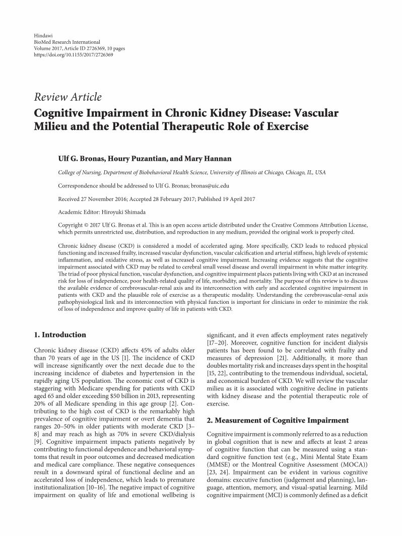
Figure 1: Systemic and cerebral vasculature and cognition in CKD.
cognitive impairment, including the dialysis procedure itself.The dialysis procedure predisposes patients to potential riskfactors for cognitive impairment and cerebrovascular dis-ease including volume and electrolyte fluctuations, cerebraledema and hypoperfusion, and excessive cytokine release[60]. Interestingly, the frequency of hypotensive episodesduring dialysis has been associated with cerebral atrophy andlacunae frequency, while microembolisms may contribute tothe burden of both large and small vessel cerebrovasculardisease althoughmuch research is needed in this area [77, 78].Moreover, secondary and recurrent delirium (often relatedto hypoperfusion) and encephalopathy (i.e., untreated renalfailure related neurotoxicity) appear to be associated withthe development of cognitive impairment [79]. Finally, werecognize that anemia and derangements in serum vitaminD levels also contribute to the CKD milieu and potentiallycognitive decline. In cross-sectional studies, anemia has beenfound to be associated with cognition in ESRD; however, in alongitudinal study, anemia was not an independent predictorof cognitive decline in elderly patients with CKD [80]. Interms of vitamin D, a review by Cheng et al. (2016) notes thatreduced levels of 25(OH)-vitamin D may be contributing tocognitive impairment in CKD [81]. Clinical trials are neededto investigate the effect of vitamin D supplementation oncognitive outcomes.
7. The Cerebrovascular-Renal Axis (Figure 1)
The accelerated cognitive decline in older CKD patientsappears to be due, in part, to the CKD disease process itself,which creates a toxic vascular and metabolic milieu that con-sists of chronic inflammation, oxidative stress, uremia, andsystemic vascular endothelial dysfunction [82–88].This toxicinternal vascular and metabolic milieu is postulated to causevascular dysfunction related impairment of the white matterthat is superimposed on neurodegenerative damage caused
by homocysteine, uremic toxins, creatinine, and cystatin C[62, 89]. Homocysteine appears to be an especially strongrisk factor for stroke in CKD patients via a direct neurotoxiceffect, initiation of systemic inflammation, and endothe-lial dysfunction [90–94]. The increase in homocysteine isprobably due to reduced renal clearance. Unfortunately,interventions with folate to reduce homocysteine levels havethus far been conflicting and disappointing [95–97]. Patientswith CKD have increased levels of oxidative stress, caused byuremia, production of reactive oxygen species via physiolog-ical pathways (e.g., impaired/damaged/malfunctioningmito-chondria), and an inability to produce adequate antioxidativeenzymes [98, 99]. These changes all contribute to a vascularmilieu that consists of systemic inflammation, high levels ofoxidative stress, and endothelial dysfunction that is unique tothe CKD patient and creates a vascular pathway to cognitivedecline.
8. Vascular Mechanisms Related to CognitiveDecline in CKD
Patients with CKD are at an increased risk for vasculardisease-related cognitive impairment rather thanAlzheimer’sdisease per se as described above. Vascular calcification inadvanced stages of CKD, possibly including intracranial cal-cification, could be influencing cognition. Interestingly, thereappears to be a significant influence of chronic hypertensionon the progression of cognitive decline. This may be relatedto the high volume of blood flow and pressure that the brainand the kidney are exposed to.
The association between systemic arterial stiffness andcognitive performance has been established in cross-sectionalstudies [100]. More recently, Pase et al. (2016) studied theFraminghamOffspring cohort and found that aortic stiffnesspredicts incident mild cognitive impairment and incidentdementia in nondiabetic patients over 10 years [101]. Apart

BioMed Research International 5
from aortic (central arterial) stiffness, stiffness in arteries inclose proximity to the brain may need to be considered. Onestudy reports that, in swines, carotid artery stiffness seemsto be associated with impaired memory [102]. Additionally,although intracranial artery stiffness is evenmore challengingto measure, it may also be associated with cognitive decline[103].
Hypertension is associated with changes in brain tissueand cerebral vasculature. For example, mean arterial pressurewas associated with white matter hyperintensity volume inthe Framingham Offspring cohort, even in the absence ofassociations between changes in brain tissue and tonometrymeasures (such as arterial stiffness or central pulse pressure)[104]. Importantly, increased duration of hypertension isan important contributor to cognitive outcomes. Midlifehypertension has a significant impact on long-term cognitiveimpairment, as reviewed by Iadecola et al. (2016) [105].Although some studies have shown a relationship betweenelevated blood pressure and cognitive impairment in theabsence of a stroke, whether intensive hypertension controlresults in prevention or reversal of cognitive outcomes isunclear [106]. Upcoming results from the SPRINT-MINDtrial (Systolic Blood Pressure Intervention Trial,Memory andCognition in Decreased Hypertension) may address some ofthese unanswered questions.
The Strain Vessel Hypothesis states that “strain vessels”found in vital organs play a protective role [107]. Strain vesselshelp maintain a pressure gradient between the larger arteriesand capillaries. High-pressure flow from large arteries, inaddition to low resistance to flow in small vessels in vitalorgans, causes subsequent damage to vessels exposed to highpulsatility [108]. In the brain, small perforating arteries areexposed to high pressure; cerebral hemorrhage and infarc-tion occur frequently in these small arteries [107, 109]. Asdecreasing kidney function is associatedwith arterial stiffness[110] and high blood pressure, patients with CKD are likelyto have their blood vessels exposed to high pulsatility flow.Therefore, CKD patients are at high risk of developing injuryto cerebral vasculature. The latter, in turn, would impactcognitive function.
It is challenging for drugs to influence the aorta andlarge arteries; and thus interventionsmay target other conduitarteries to reduce wave reflection. Although drugs suchas angiotensin-converting enzyme inhibitors and calciumchannel blockers seem to be beneficial in hypertensive elderlyindividuals, blood pressure levels that are optimal for cog-nitive function are yet to be identified [106]. Apart frommedications, regular exercise may be employed to target thisphenomenon.
9. Exercise as a PotentialTherapeutic Approach
Higher levels of physical activity and cardiorespiratory fit-ness levels are associated with increased levels of cognitivefunction in healthy individuals. Exercise appears to preventcerebral atrophy or even increase hippocampal volume inthe general population [111, 112]. It is conjectured that theseobservations are related to an increase in brain-derived
neurotrophic factor and an exercise-induced increase inangiogenesis, neurogenesis, and synaptogenesis. It is con-ceivable that physical activity and fitness levels are relatedto cognitive function in patients with kidney disease, butthere have been a minimal number of studies in this areaand the results are inconsistent. Patients on hemodialy-sis with the highest self-reported activity levels had thehighest cognitive scores in one study [113]. Conversely,one smaller study found no association between maximaloxygen consumption and scores on the MMSE in patients onhemodialysis [114]. Several exercise intervention studies haveshown promise in improving cognition in healthy elderlyparticipants with and without MCI [111, 115–126], whereasothers have reported no improvement [127–130]. Regularexercise and higher fitness levels in non-CKD patients withand without cognitive impairment have been associated withimproved cognitive function, white matter integrity, andhippocampal volume suggesting a possible neuroprotectiveeffect of exercise [111, 113, 115–125]. This is conjectured to bedue to an improvement in vascular function-related increasesin cerebral blood flow [116]. It is therefore plausible that exer-cise training may improve the vascular milieu and therebycontribute to improved cognitive function in patients withCKD. However, the impact of exercise on cognitive functionin the CKD population is currently unknown. Only onestudy has reported on cognitive function following exercisetraining in the dialysis population. Martins et al., 2011,reported an improvement in cognitive functionmeasured viathe MMSE following exercise training [131]. Unfortunately,this study was not randomized and the MMSE is not asensitive measure for change in global cognition, whichlimits any conclusions. Studies investigating the impact ofexercise on cognition in CKD patients are needed. Exercisetraining appears to improve the vascular milieu in patientswith CKD by reducing systemic inflammation and oxidativestress, arterial stiffness, and improving vascular function[132]. However, not all studies have shown improvementsin these vascular risk factors, likely due to differences insample characteristics, exercise program, and outcome mea-sures. Moreover, exercise trainingmay reduce traditional riskfactors for cerebrovascular disease such as blood pressure andlipid profile although it should be noted that randomized con-trolled trials are scarce in patients with CKD and most dataare based on secondary analyses from smaller trials. Exercisetraining is also known to improve glucose control in diabeticpatients and may reduce homocysteine levels. Importantly,the pleiotropic effect of exercise provides additional benefitsthat are important to patients with CKD including improvedquality of life, improved physical function, and reducedrisk of frailty. Moreover, higher levels of physical activityhave been associated with reduced risk of initiation of renalreplacement therapy and higher survival rate in patientswith CKD stages 3–5 [133]. Finally, emerging studies suggestthat there is an independent association between prolongedsedentary time and kidney function decline, whereas higherlevels of physical activity are associated with reduced levelsof creatinine and lower risk of kidney impairment [134, 135].Thus, it is plausible that exercise may affect renal functionitself and thereby provide a protective effect. Despite lack of

6 BioMed Research International
data on the impact of exercise on cognitive function, it isprudent for clinicians to recommend that patients with CKDconsider initiating an exercise program and increase theirdaily physical activity levels to gain the mental and physicalhealth benefits of exercise.
10. Summary and Conclusions
Cognitive impairment is common in patients with CKDand negatively affects health-related quality of life and otherhealth-related outcomes. It is imperative that clinicians rec-ognize the value of early screening for cognitive impairmentand initiate preventive and treatment measures. Importantly,the decline in cognitive function appears to be multifacetedwith a major involvement of vascular dysfunction in aunique CKD metabolic milieu that predisposes patients toan accelerated cognitive decline. Multidisciplinary health-care teams are needed to provide psychosocial support andpatient education on essential topics such as control of bloodpressure, risks of polypharmacy, and other individualizedself-care practices. Current research investigating exercise-induced improvement in cognition in non-CKD populationis promising, but there are conflicting reports in the literature.As exercise trainingmay be a plausible adjunctive therapeuticapproach to improve cognitive outcomes and quality of life inpatients with CKD, further research should focus on exerciseas a promising approach that may retard the progression ofcognitive impairment in CKD.
Conflicts of Interest
The authors declare that they have no conflicts of interest.
References
[1] J. Coresh, E. Selvin, L. A. Stevens et al., “Prevalence of chronickidney disease in the United States,” Journal of the AmericanMedical Association, vol. 298, no. 17, pp. 2038–2047, 2007.
[2] U.S. Renal Data System, “USRDS 2013 annual data report,” Atlasof Chronic Kidney Disease & End Stage Renal Disease in theUnited States, 2013.
[3] C. E. Rodriguez-Angarita, R. M. Sanabria-Arenas, J. D. Vargas-Jaramillo, and I. Ronderos-Botero, “Cognitive impairment anddepression in a population of patients with chronic kidneydisease in colombia: a prevalence study,” Canadian Journal ofKidney Health and Disease, vol. 3, article 26, 2016.
[4] M. K. Tamura, K. Yaffe, C.-Y. Hsu et al., “Cognitive Impairmentand Progression of CKD,” American Journal of Kidney Diseases,vol. 68, no. 1, pp. 77–83, 2016.
[5] M. Kurella Tamura, V. Wadley, K. Yaffe et al., “Kidney func-tion and cognitive impairment in US adults: the reasons forgeographic and racial differences in stroke (REGARDS) study,”American Journal of Kidney Diseases, vol. 52, no. 2, pp. 227–234,2008.
[6] M. Kurella, G. M. Chertow, J. Luan, and K. Yaffe, “Cognitiveimpairment in chronic kidney disease,” Journal of the AmericanGeriatrics Society, vol. 52, no. 11, pp. 1863–1869, 2004.
[7] M. Kurella, G. M. Chertow, L. F. Fried et al., “Chronic kidneydisease and cognitive impairment in the elderly: the health,
aging, and body composition study,” Journal of the AmericanSociety of Nephrology, vol. 16, no. 7, pp. 2127–2133, 2005.
[8] K. Yaffe, L. Ackerson, M. K. Tamura et al., “Chronic kidneydisease and cognitive function in older adults: findings fromthe chronic renal insufficiency cohort cognitive study,” Journalof the American Geriatrics Society, vol. 58, no. 2, pp. 338–345,2010.
[9] A. M. Murray, D. E. Tupper, D. S. Knopman et al., “Cognitiveimpairment in hemodialysis patients is common,” Neurology,vol. 67, no. 2, pp. 216–223, 2006.
[10] A. Phinney, “Living with dementia from the patient’s perspec-tive,” Journal of Gerontological Nursing, vol. 24, no. 6, pp. 8–15,1998.
[11] E. Steeman, I. L. Abraham, and J. Godderis, “Risk profiles forinstitutionalization in a cohort of elderly people with dementiaor depression,” Archives of Psychiatric Nursing, vol. 11, no. 6, pp.295–303, 1997.
[12] S. Andrieu, E. Reynish, F. Nourhashemi et al., “Predictivefactors of acute hospitalization in 134 patients with Alzheimer’sdisease: a one year prospective study,” International Journal ofGeriatric Psychiatry, vol. 17, no. 5, p. 422, 2002.
[13] R. Newcomer, K. E. Covinsky, T. Clay, and K. Yaffe, “Predicting12-month mortality for persons with dementia,” Journals ofGerontology—Series B Psychological Sciences and Social Sciences,vol. 58, no. 3, pp. S187–S198, 2003.
[14] J. H. Wlodarczyk, H. Brodaty, and G. Hawthorne, “The rela-tionship between quality of life, mini-mental state examination,and the instrumental activities of daily living in patients withAlzheimer’s disease,”Archives of Gerontology andGeriatrics, vol.39, no. 1, pp. 25–33, 2004.
[15] A. R. Sehgal, S. F. Grey, P. B. DeOreo, and P. J. Whitehouse,“Prevalence, recognition, and implications of mental impair-ment amonghemodialysis patients,”American Journal of KidneyDiseases, vol. 30, no. 1, pp. 41–49, 1997.
[16] D. E. Weiner and S. L. Seliger, “Cognitive and physical functionin chronic kidney disease,” Current Opinion in Nephrology andHypertension, vol. 23, no. 3, pp. 291–297, 2014.
[17] E. P. Sorensen, M. J. Sarnak, H. Tighiouart et al., “The kidneydisease quality of life cognitive function subscale and cognitiveperformance in maintenance hemodialysis patients,” AmericanJournal of Kidney Diseases, vol. 60, no. 3, pp. 417–426, 2012.
[18] B. A. Bremer, K. M. Wert, A. L. Durica, and A. Weaver,“Neuropsychological, physical, and psychosocial functioning ofindividuals with end-stage renal disease,” Annals of BehavioralMedicine, vol. 19, no. 4, pp. 348–352, 1997.
[19] S. Jung, Y.-K. Lee, S. R. Choi, S.-H. Hwang, and J.-W. Noh,“Relationship between cognitive impairment and depression indialysis patients,”YonseiMedical Journal, vol. 54, no. 6, pp. 1447–1453, 2013.
[20] L. Feng, K. B. Yap, and T. P. Ng, “Depressive symptoms inolder adults with chronic kidney disease: mortality, quality oflife outcomes, and correlates,” American Journal of GeriatricPsychiatry, vol. 21, no. 6, pp. 570–579, 2013.
[21] M. A.McAdams-Demarco, J. Tan,M. L. Salter et al., “Frailty andcognitive function in incident hemodialysis patients,” ClinicalJournal of the American Society of Nephrology, vol. 10, no. 12, pp.2181–2189, 2015.
[22] U.S. Renal Data System,USRDS 2006Annual Data Report: Atlasof Chronic Kidney Disease & End Stage Renal Disease in theUnited States, 2006.

BioMed Research International 7
[23] M. F. Folstein, S. E. Folstein, and P. R. McHugh, “‘Mini-mentalstate’. A practical method for grading the cognitive state ofpatients for the clinician,” Journal of Psychiatric Research, vol.12, no. 3, pp. 189–198, 1975.
[24] D. S. Knopman, B. F. Boeve, and R. C. Petersen, “Essentials ofthe proper diagnoses of mild cognitive impairment, dementia,and major subtypes of dementia,”Mayo Clinic Proceedings, vol.78, no. 10, pp. 1290–1308, 2003.
[25] R. C. Petersen, R. Doody, A. Kurz et al., “Current concepts inmild cognitive impairment,” Archives of Neurology, vol. 58, no.12, pp. 1985–1992, 2001.
[26] R. C. Petersen, “Mild cognitive impairment as a diagnosticentity,” Journal of Internal Medicine, vol. 256, no. 3, pp. 183–194,2004.
[27] American Psychiatric Associtation, Diagnostic and StatisticalManual of Mental Disorders (DSM-IV), American PsychiatricAssociation, Washington, DC, USA, 4th edition, 1994.
[28] M. Bossola, M. Antocicco, E. Di Stasio et al., “Mini MentalState Examination over time in chronic hemodialysis patients,”Journal of Psychosomatic Research, vol. 71, no. 1, pp. 50–54, 2011.
[29] A. S. Buchman, D. Tanne, P. A. Boyle, R. C. Shah, S. E. Leurgans,and D. A. Bennett, “Kidney function is associated with the rateof cognitive decline in the elderly,”Neurology, vol. 73, no. 12, pp.920–927, 2009.
[30] T. Etgen, D. Sander,M. Chonchol et al., “Chronic kidney diseaseis associated with incident cognitive impairment in the elderly:The INVADE study,” Nephrology Dialysis Transplantation, vol.24, no. 10, pp. 3144–3150, 2009.
[31] M. Kurella, D. L. Mapes, F. K. Port, and G. M. Chertow,“Correlates and outcomes of dementia among dialysis patients:the dialysis outcomes and practice patterns study,” NephrologyDialysis Transplantation, vol. 21, no. 9, pp. 2543–2548, 2006.
[32] A. M. Murray, J. I. Barzilay, J. F. Lovato et al., “Biomarkersof renal function and cognitive impairment in patients withdiabetes,” Diabetes Care, vol. 34, no. 8, pp. 1827–1832, 2011.
[33] K. Yaffe, K. Lindquist, M. G. Shlipak et al., “Cystatin C as amarker of cognitive function in elders: findings from theHealthABC Study,” Annals of Neurology, vol. 63, no. 6, pp. 798–802,2008.
[34] M. K. Georgakis, N. G. Dimitriou, M. A. Karalexi et al.,“Albuminuria in Association with Cognitive Function andDementia: A Systematic Review and Meta-Analysis,” Journal ofthe American Geriatrics Society, 2017.
[35] M. Kurella, K. Yaffe, M. G. Shlipak, N. K. Wenger, and G. M.Chertow, “Chronic kidney disease and cognitive impairment inmenopausal women,” American Journal of Kidney Diseases, vol.45, no. 1, pp. 66–76, 2005.
[36] I. Sajjad, F. Grodstein, J. H. Kang, G. C. Curhan, and J. Lin,“Kidney dysfunction and cognitive decline in women,” ClinicalJournal of the American Society of Nephrology, vol. 7, no. 3, pp.437–443, 2012.
[37] J. A. Passer, “Cerebral atrophy in end-stage uremia,” Proceedingsof the Clinical Dialysis and Transplant Forum, vol. 7, pp. 91–94,1977.
[38] T. Kamata, A. Hishida, T. Takita et al., “Morphologic abnormal-ities in the brain of chronically hemodialyzed patients withoutcerebrovascular disease,” American Journal of Nephrology, vol.20, no. 1, pp. 27–31, 2000.
[39] G. M. Savazzi, F. Cusmano, and S. Musini, “Cerebral imagingchanges in patients with chronic renal failure treated conserva-tively or in hemodialysis,”Nephron, vol. 89, no. 1, pp. 31–36, 2001.
[40] A.-H. Cho, S. B. Lee, S. J. Han, Y.-M. Shon, D.-W. Yang, and B.S. Kim, “Impaired kidney function and cerebral microbleeds inpatients with acute ischemic stroke,” Neurology, vol. 73, no. 20,pp. 1645–1648, 2009.
[41] G. Fazekas, F. Fazekas, R. Schmidt, P. Kapeller, H. Offenbacher,and G. J. Krejs, “Brain MRI findings and cognitive impairmentin patients undergoing chronic hemodialysis treatment,” Jour-nal of the Neurological Sciences, vol. 134, no. 1-2, pp. 83–88, 1995.
[42] H. Shima, E. Ishimura, T. Naganuma et al., “Cerebral microb-leeds in predialysis patients with chronic kidney disease,”Nephrology Dialysis Transplantation, vol. 25, no. 5, pp. 1554–1559, 2010.
[43] M. A. Ikram, M.W. Vernooij, A. Hofman, W. J. Niessen, A. VanDer Lugt, and M. M. B. Breteler, “Kidney function is related tocerebral small vessel disease,” Stroke, vol. 39, no. 1, pp. 55–61,2008.
[44] M. Wada, H. Nagasawa, C. Iseki et al., “Cerebral small vesseldisease and chronic kidney disease (CKD): results of a cross-sectional study in community-based Japanese elderly,” Journalof the Neurological Sciences, vol. 272, no. 1-2, pp. 36–42, 2008.
[45] S. Sedaghat, L. G. M. Cremers, M. De Groot et al., “Kidneyfunction and microstructural integrity of brain white matter,”Neurology, vol. 85, no. 2, pp. 154–161, 2015.
[46] A. M. Tuladhar, A. G. van Norden, K. F. de Laat et al., “Whitematter integrity in small vessel disease is related to cognition,”NeuroImage: Clinical, vol. 7, pp. 518–524, 2015.
[47] H. J. Chen, L. J. Zhang, and G. M. Lu, “Multimodality MRIfindings in patients with end-stage renal disease,” BioMedResearch International, vol. 2015, Article ID 697402, 12 pages,2015.
[48] L. E. Oberlin, T. D. Verstynen, A. Z. Burzynska et al., “Whitematter microstructure mediates the relationship between car-diorespiratory fitness and spatial working memory in olderadults,” NeuroImage, vol. 131, pp. 91–101, 2016.
[49] M. W. Vernooij, M. A. Ikram, H. A. Vrooman et al., “WhiteMatter microstructural integrity and cognitive function in ageneral elderly population,” Archives of General Psychiatry, vol.66, no. 5, pp. 545–553, 2009.
[50] M.-C. Chou, T.-J. Hsieh, Y.-L. Lin et al., “Widespread whitematter alterations in patients with end-stage renal disease: avoxelwise diffusion tensor imaging study,” American Journal ofNeuroradiology, vol. 34, no. 10, pp. 1945–1951, 2013.
[51] X. Kong, J.-Q. Wen, R.-F. Qi et al., “Diffuse interstitial brainedema in patients with end-stage renal disease undergoinghemodialysis: A Tract-Based Spatial Statistics Study,”Medicine,vol. 93, no. 28, article e313, 2014.
[52] R. Zhang, K. Liu, L. Yang et al., “Reduced white matterintegrity and cognitive deficits in maintenance hemodialysisESRD patients: a diffusion-tensor study,” European Radiology,vol. 25, no. 3, pp. 661–668, 2014.
[53] H. S. Kim, J. W. Park, D. S. Bai et al., “Diffusion tensor imagingfindings in neurologically asymptomatic patients with end stagerenal disease,” NeuroRehabilitation, vol. 29, no. 1, pp. 111–116,2011.
[54] M. Lamar, X. J. Zhou, R. A. Charlton, D. Dean, D. Little, and S.C.Deoni, “In vivo quantification ofwhitemattermicrostructurefor use in aging: a focus on two emerging techniques,”AmericanJournal of Geriatric Psychiatry, vol. 22, no. 2, pp. 111–121, 2014.
[55] L. J. Zhang, J. Wen, X. Liang et al., “Brain default mode networkchanges after renal transplantation: a diffusion-tensor imagingand resting-state functional MR imaging study,” Radiology, vol.278, no. 2, pp. 485–495, 2016.

8 BioMed Research International
[56] S. Lavi, D. Gaitini, V. Milloul, and G. Jacob, “Impaired cerebralCO
2vasoreactivity: association with endothelial dysfunction,”
American Journal of Physiology—Heart and Circulatory Physiol-ogy, vol. 291, no. 4, pp. H1856–H1861, 2006.
[57] S. Sedaghat, M.W. Vernooij, E. Loehrer et al., “Kidney functionand cerebral blood flow: The Rotterdam Study,” Journal of theAmerican Society of Nephrology, vol. 27, no. 3, pp. 715–721, 2016.
[58] J. Radic, D. Ljutic, M. Radic, V. Kovacic, K. Dodig-Curkovic,and M. Sain, “Kidney transplantation improves cognitive andpsychomotor functions in adult hemodialysis patients,” Ameri-can Journal of Nephrology, vol. 34, no. 5, pp. 399–406, 2011.
[59] W. L. Lau, B. N. Huisa, and M. Fisher, “the cerebrovascular-chronic kidney disease connection: perspectives and mecha-nisms,” Translational Stroke Research, vol. 8, no. 1, pp. 67–76,2017.
[60] R. Lu, M. C. Kiernan, A. Murray, M. H. Rosner, and C. Ronco,“Kidney-brain crosstalk in the acute and chronic setting,”Nature Reviews Nephrology, vol. 11, no. 12, pp. 707–719, 2015.
[61] M. Madero, A. Gul, and M. J. Sarnak, “Cognitive function inchronic kidney disease,” Seminars in Dialysis, vol. 21, no. 1, pp.29–37, 2008.
[62] J.-M. Bugnicourt, O. Godefroy, J.-M. Chillon, G. Choukroun,and Z. A. Massy, “Cognitive disorders and dementia in CKD:the neglected kidney-brain axis,” Journal of the American Societyof Nephrology, vol. 24, no. 3, pp. 353–363, 2013.
[63] M. Kobayashi, N. Hirawa, K. Yatsu et al., “Relationship betweensilent brain infarction and chronic kidney disease,” NephrologyDialysis Transplantation, vol. 24, no. 1, pp. 201–207, 2009.
[64] B. Ovbiagele, J. J. Wing, R. S. Menon et al., “Association ofchronic kidney disease with cerebral microbleeds in patientswith primary intracerebral hemorrhage,” Stroke, vol. 44, no. 9,pp. 2409–2413, 2013.
[65] A. M. Murray, “Cognitive impairment in the aging dialysisand chronic kidney disease populations: an occult burden,”Advances in Chronic Kidney Disease, vol. 15, no. 2, pp. 123–132,2008.
[66] H.-H. Wang, S.-Y. Hung, J.-M. Sung, K.-Y. Hung, and J.-D.Wang, “Risk of stroke in long-term dialysis patients comparedwith the general population,” American Journal of KidneyDiseases, vol. 63, no. 4, pp. 604–611, 2014.
[67] R. N. Foley, D. T. Gilbertson, T. Murray, and A. J. Collins, “Longinterdialytic interval and mortality among patients receivinghemodialysis,” The New England Journal of Medicine, vol. 365,no. 12, pp. 1099–1107, 2011.
[68] C. Helmer, B. Stengel, M. Metzger et al., “Chronic kidneydisease, cognitive decline, and incident dementia: the 3C Study,”Neurology, vol. 77, no. 23, pp. 2043–2051, 2011.
[69] C. Gaxatte, M. Daroux, J. Bloch, F. Puisieux, V. Deramecourt,and E. Boulanger, “Cognitive impairment and chronic kidneydisease: which links?”Nephrologie etTherapeutique, vol. 7, no. 1,pp. 10–17, 2011.
[70] T. Umemura, T. Kawamura, H. Umegaki et al., “Association ofchronic kidney disease and cerebral small vessel disease withcognitive impairment in elderly patients with type 2 diabetes,”Dementia and Geriatric Cognitive Disorders Extra, vol. 3, no. 1,pp. 212–222, 2013.
[71] A. F. Perna, D. Ingrosso, E. Violetti et al., “Hyperhomocysteine-mia in uremia—a red flag in a disrupted circuit,” Seminars inDialysis, vol. 22, no. 4, pp. 351–356, 2009.
[72] S. L. Seliger, D. L. Gillen, D. Tirschwell, H. Wasse, B. R. Kesten-baum, and C. O. Stehman-Breen, “Risk factors for incident
stroke among patients with end-stage renal disease,” Journal ofthe American Society of Nephrology, vol. 14, no. 10, pp. 2623–2631, 2003.
[73] J. B. Wetmore, E. F. Ellerbeck, J. D. Mahnken et al., “Atrialfibrillation and risk of stroke in dialysis patients,” Annals ofEpidemiology, vol. 23, no. 3, pp. 112–118, 2013.
[74] P. B. Gorelick, J. Brody, D. Cohen et al., “Risk factors fordementia associated with multiple cerebral infarcts. A case-control analysis in predominantly African-American hospital-based patients,”Archives of Neurology, vol. 50, no. 7, pp. 714–720,1993.
[75] M. K. Tamura, D. Xie, K. Yaffe et al., “Vascular risk factors andcognitive impairment in chronic kidney disease: the ChronicRenal InsufficiencyCohort (CRIC) study,”Clinical Journal of theAmerican Society of Nephrology, vol. 6, no. 2, pp. 248–256, 2011.
[76] E. A. Iliescu, H. Coo, M. H. McMurray et al., “Quality of sleepand health-related quality of life in haemodialysis patients,”Nephrology Dialysis Transplantation, vol. 18, no. 1, pp. 126–132,2003.
[77] D. W. Droste, K. Kuhne, R. M. Schaefer, and E. B. Ringelstein,“Detection of microemboli in the subclavian vein of patientsundergoing haemodialysis and haemodiafiltration using pulsedDoppler ultrasound,” Nephrology Dialysis Transplantation, vol.17, no. 3, pp. 462–466, 2002.
[78] T.Mizumasa, H. Hirakata, T. Yoshimitsu et al., “Dialysis-relatedhypotension as a cause of progressive frontal lobe atrophyin chronic hemodialysis patients: a 3-year prospective study,”Nephron Clinical Practice, vol. 97, no. 1, pp. c23–c30, 2004.
[79] R. Brouns and P. P. De Deyn, “Neurological complications inrenal failure: a review,”ClinicalNeurology andNeurosurgery, vol.107, no. 1, pp. 1–16, 2004.
[80] M. Kurella Tamura, E. Vittinghoff, J. Yang et al., “Anemia andrisk for cognitive decline in chronic kidney disease,” BMCNephrology, vol. 17, no. 1, article 226, 2016.
[81] Z. Cheng, J. Lin, and Q. Qian, “Role of vitamin D in cognitivefunction in chronic kidney disease,” Nutrients, vol. 8, no. 5,article 291, 2016.
[82] J. Himmelfarb, “Uremic toxicity, oxidative stress, and hemodial-ysis as renal replacement therapy,” Seminars in Dialysis, vol. 22,no. 6, pp. 636–643, 2009.
[83] S. L. Seliger and W. T. Longstreth Jr., “Lessons about brainvascular disease from another pulsating organ, the kidney,”Stroke, vol. 39, no. 1, pp. 5–6, 2008.
[84] J.M.Wardlaw, P. A. G. Sandercock,M. S. Dennis, and J. Starr, “Isbreakdown of the blood-brain barrier responsible for lacunarstroke, leukoaraiosis, and dementia?” Stroke, vol. 34, no. 3, pp.806–811, 2003.
[85] H. Kalimo, “Does chronic brain edema explain the conse-quences of cerebral small-vessel disease?” Stroke, vol. 34, no. 3,pp. 806–812, 2003.
[86] D. E. Weiner, K. Bartolomei, T. Scott et al., “Albuminuria,cognitive functioning, and white matter hyperintensities inhomebound elders,” American Journal of Kidney Diseases, vol.53, no. 3, pp. 438–447, 2009.
[87] C. Berr, B. Balansard, J. Arnaud, A. M. Roussel, and A.Alperovitch, “Cognitive decline is associated with systemicoxidative stress: the EVA study. etude du vieillissement arteriel,”Journal of the American Geriatrics Society, vol. 48, no. 10, pp.1285–1291, 2000.
[88] T. A. Ikizler, J. D. Morrow, L. J. Roberts et al., “PlasmaF2-isoprostane levels are elevated in chronic hemodialysispatients,” Clinical Nephrology, vol. 58, no. 3, pp. 190–197, 2002.

BioMed Research International 9
[89] P. P. De Deyn, R. Vanholder, S. Eloot, and G. Glorieux,“Guanidino compounds as uremic (neuro)toxins,” Seminars inDialysis, vol. 22, no. 4, pp. 340–345, 2009.
[90] F. Anan, N. Takahashi, T. Shimomura et al., “Hyperhomocys-teinemia is a significant risk factor for silent cerebral infarctionin patients with chronic renal failure undergoing hemodialysis,”Metabolism, vol. 55, no. 5, pp. 656–661, 2006.
[91] K. Faßbender, O. Mielke, T. Bertsch, B. Nafe, S. Froschen, andM. Hennerici, “Homocysteine in cerebral macroangiographyand microangiopathy,”The Lancet, vol. 353, no. 9164, pp. 1586–1587, 1999.
[92] S. A. Lipton, W.-K. Kim, Y.-B. Choi et al., “Neurotoxicityassociated with dual actions of homocysteine at the N-methyl-D-aspartate receptor,” Proceedings of the National Academy ofSciences of the United States of America, vol. 94, no. 11, pp. 5923–5928, 1997.
[93] S. Seshadri, P. A. Wolf, A. S. Beiser et al., “Association of plasmatotal homocysteine levels with subclinical brain injury: cerebralvolumes, white matter hyperintensity, and silent brain infarctsat volumetric magnetic resonance imaging in the FraminghamOffspring Study,” Archives of Neurology, vol. 65, no. 5, pp. 642–649, 2008.
[94] C. B.Wright,M. C. Paik, T. R. Brown et al., “Total homocysteineis associated with white matter hyperintensity volume: theNorthernManhattan study,” Stroke, vol. 36, no. 6, pp. 1207–1211,2005.
[95] M. Lee, K.-S. Hong, S.-C. Chang, and J. L. Saver, “Efficacyof homocysteine-lowering therapy with folic acid in strokeprevention: ameta-analysis,” Stroke, vol. 41, no. 6, pp. 1205–1212,2010.
[96] C.A.De Jager, A.Oulhaj, R. Jacoby,H. Refsum, andA.D. Smith,“Cognitive and clinical outcomes of homocysteine-lowering B-vitamin treatment in mild cognitive impairment: a randomizedcontrolled trial,” International Journal of Geriatric Psychiatry,vol. 27, no. 6, pp. 592–600, 2012.
[97] T. Kwok, J. Lee, C. B. Law et al., “A randomized placebocontrolled trial of homocysteine lowering to reduce cognitivedecline in older demented people,” Clinical Nutrition, vol. 30,no. 3, pp. 297–302, 2011.
[98] K. Fujisaki, K. Tsuruya, M. Yamato et al., “Cerebral oxidativestress induces spatial working memory dysfunction in uremicmice: neuroprotective effect of tempol,” Nephrology DialysisTransplantation, vol. 29, no. 3, pp. 529–538, 2014.
[99] D. Salisbury and U. Bronas, “Reactive oxygen and nitrogenspecies: impact on endothelial dysfunction,” Nursing Research,vol. 64, no. 1, pp. 53–66, 2015.
[100] A. Zeki Al Hazzouri and K. Yaffe, “Arterial stiffness andcognitive function in the elderly,” Journal of Alzheimer’s Disease,vol. 42, supplement 4, pp. S503–S514, 2014.
[101] M. P. Pase, J. J. Himali, G. F.Mitchell et al., “Association of aorticstiffness with cognition and brain aging in young and middle-aged adults: The FraminghamThird Generation Cohort Study,”Hypertension, vol. 67, no. 3, pp. 513–519, 2016.
[102] T. D. Olver, D. Klakotskaia, B. S. Ferguson et al., “Carotidartery vascular mechanics serve as biomarkers of cognitivedysfunction in aortic-banded miniature swine that can betreated with an exercise intervention,” Journal of the AmericanHeart Association, vol. 5, no. 5, Article ID e003248, 2016.
[103] X. Sun and T. Rundek, “Does increased arterial stiffness heraldcognitive impairment?” Stroke, vol. 47, no. 9, pp. 2171–2172, 2016.
[104] C.W. Tsao, J. J. Himali, A. S. Beiser et al., “Association of arterialstiffness with progression of subclinical brain and cognitivedisease,” Neurology, vol. 86, no. 7, pp. 619–626, 2016.
[105] C. Iadecola, K. Yaffe, J. Biller et al., “Impact of hypertension oncognitive function: a scientific statement from the AmericanHeart Association,” Hypertension, vol. 68, no. 6, pp. e67–e94,2016.
[106] M. Tadic, C. Cuspidi, and D. Hering, “Hypertension andcognitive dysfunction in elderly: blood pressure managementfor this global burden,” BMC Cardiovascular Disorders, vol. 16,article 208, 2016.
[107] S. Ito, T. Nagasawa, M. Abe, and T. Mori, “Strain vesselhypothesis: a viewpoint for linkage of albuminuria and cerebro-cardiovascular risk,” Hypertension Research, vol. 32, no. 2, pp.115–121, 2009.
[108] M. F. O’Rourke and M. E. Safar, “Relationship between aorticstiffening and microvascular disease in brain and kidney: causeand logic of therapy,” Hypertension, vol. 46, no. 1, pp. 200–204,2005.
[109] G. F. Mitchell, M. A. Van Buchem, S. Sigurdsson et al., “Arterialstiffness, pressure and flow pulsatility and brain structure andfunction:TheAge, Gene/Environment Susceptibility-ReykjavikStudy,” Brain, vol. 134, no. 11, pp. 3398–3407, 2011.
[110] R. R. Townsend, N. J. Wimmer, J. A. Chirinos et al., “AorticPWV in chronic kidney disease: a CRIC ancillary study,”American Journal of Hypertension, vol. 23, no. 3, pp. 282–289,2010.
[111] K. I. Erickson, M. W. Voss, R. S. Prakash et al., “Exercisetraining increases size of hippocampus and improves memory,”Proceedings of the National Academy of Sciences of the UnitedStates of America, vol. 108, no. 7, pp. 3017–3022, 2011.
[112] J. Weuve, J. H. Kang, J. E. Manson, M. M. B. Breteler, J. H.Ware, and F. Grodstein, “Physical activity, including walking,and cognitive function in olderwomen,” Journal of theAmericanMedical Association, vol. 292, no. 12, pp. 1454–1461, 2004.
[113] F. Stringuetta-Belik, F. G. Shiraishi, V. R. Oliveira e Silva etal., “Greater level of physical activity associated with bettercognitive function in hemodialysis in end stage renal disease,”Jornal Brasileiro de Nefrologia, vol. 34, no. 4, pp. 378–386, 2012.
[114] R.-L. Hsieh, W.-C. Lee, and C.-H. Chang, “Maximal cardio-vascular fitness and its correlates in ambulatory hemodialysispatients,”American Journal of Kidney Diseases, vol. 48, no. 1, pp.21–27, 2006.
[115] J. E. Ahlskog, Y. E. Geda, N. R. Graff-Radford, and R.C. Petersen, “Physical exercise as a preventive or disease-modifying treatment of dementia and brain aging,”Mayo ClinicProceedings, vol. 86, no. 9, pp. 876–884, 2011.
[116] S. M. Hayes, M. L. Alosco, and D. E. Forman, “The effects ofaerobic exercise on cognitive and neural decline in aging andcardiovascular disease,” Current Geriatrics Reports, vol. 3, no. 4,pp. 282–290, 2014.
[117] J. C. Smith, K. A. Nielson, J. L. Woodard et al., “Does physicalactivity influence semanticmemory activation in amnesticmildcognitive impairment?” Psychiatry Research, vol. 193, no. 1, pp.60–62, 2011.
[118] R. Ruscheweyh, C. Willemer, K. Kruger et al., “Physical activityand memory functions: an interventional study,” Neurobiologyof Aging, vol. 32, no. 7, pp. 1304–1319, 2011.
[119] L. D. Baker, L. L. Frank, K. Foster-Schubert et al., “Effects ofaerobic exercise on mild cognitive impairment: a controlledtrial,” Archives of Neurology, vol. 67, no. 1, pp. 71–79, 2010.

10 BioMed Research International
[120] L. S. Nagamatsu, A. Chan, J. C. Davis et al., “Physical activityimproves verbal and spatial memory in older adults withprobable mild cognitive impairment: a 6-month randomizedcontrolled trial,” Journal of Aging Research, vol. 2013, Article ID861893, 10 pages, 2013.
[121] N. J. Kirk-Sanchez and E. L. McGough, “Physical exercise andcognitive performance in the elderly: current perspectives,”Clinical Interventions in Aging, vol. 9, pp. 51–62, 2013.
[122] M.W. Voss, K. I. Erickson, R. S. Prakash et al., “Neurobiologicalmarkers of exercise-related brain plasticity in older adults,”Brain, Behavior, and Immunity, vol. 28, pp. 90–99, 2013.
[123] S. J. Colcombe, A. F. Kramer, K. I. Erickson et al., “Cardiovas-cular fitness, cortical plasticity, and aging,” Proceedings of theNational Academy of Sciences of the United States of America,vol. 101, no. 9, pp. 3316–3321, 2004.
[124] D. E. Barnes, W. Santos-Modesitt, G. Poelke et al., “The mentalactivity and exercise (MAX) trial: a randomized controlled trialto enhance cognitive function in older adults,” JAMA InternalMedicine, vol. 173, no. 9, pp. 797–804, 2013.
[125] N. T. Lautenschlager, K. L. Cox, L. Flicker et al., “Effect ofphysical activity on cognitive function in older adults at risk forAlzheimer disease: a randomized trial,” JAMA, vol. 300, no. 9,pp. 1027–1037, 2008.
[126] P. J. Smith, J. A. Blumenthal, B.M.Hoffman et al., “Aerobic exer-cise and neurocognitive performance: ameta-analytic review ofrandomized controlled trials,” Psychosomatic Medicine, vol. 72,no. 3, pp. 239–252, 2010.
[127] J. A. Blumenthal and D. J. Madden, “Effects of aerobic exercisetraining, age, and physical fitness on memory-search perfor-mance,” Psychology and Aging, vol. 3, no. 3, pp. 280–285, 1988.
[128] D. Laurin, R. Verreault, J. Lindsay, K. MacPherson, and K.Rockwood, “Physical activity and risk of cognitive impairmentand dementia in elderly persons,” Archives of Neurology, vol. 58,no. 3, pp. 498–504, 2001.
[129] J. D.Williamson,M. Espeland, S. B. Kritchevsky et al., “Changesin cognitive function in a randomized trial of physical activity:results of the lifestyle interventions and independence for elderspilot study,” Journals of Gerontology, Series A: Biological Sciencesand Medical Sciences, vol. 64, no. 6, pp. 688–694, 2009.
[130] K. Okumiya, K. Matsubayashi, T. Wada, S. Kimura, Y. Doi, andT. Ozawa, “Effects of exercise on neurobehavioral function incommunity-dwelling older people more than 75 years of age,”Journal of theAmericanGeriatrics Society, vol. 44, no. 5, pp. 569–572, 1996.
[131] C. T. Martins, G. S. Ramos, S. A. Guaraldo, C. B. Uezima, J.P. Martins, and E. Ribeiro Junior, “Comparison of cognitivefunction between patients on chronic hemodialysis who carryout assisted physical activity and inactive ones,” Jornal Brasileirode Nefrologia, vol. 33, no. 1, pp. 27–30, 2011.
[132] U.G. Bronas, “Exercise training and reduction of cardiovasculardisease risk factors in patients with chronic kidney disease,”Advances in Chronic Kidney Disease, vol. 16, no. 6, pp. 449–458,2009.
[133] I.-R. Chen, S.-M. Wang, C.-C. Liang et al., “Association ofwalkingwith survival andRRT among patientswithCKD stages3–5,” Clinical Journal of the American Society of Nephrology, vol.9, no. 7, pp. 1183–1189, 2014.
[134] V. Y. Guo, S. Brage, U. Ekelund, S. J. Griffin, and R. K. Simmons,“Objectively measured sedentary time, physical activity andkidney function in people with recently diagnosed Type 2diabetes: a prospective cohort analysis,” Diabetic Medicine, vol.33, no. 9, pp. 1222–1229, 2016.
[135] S. Lee, H. Shimada, S. Lee et al., “Association between seden-tary time and kidney function in community-dwelling elderlyJapanese people,” Geriatrics & Gerontology International, 2016.

Research ArticlePosttransplant Anemia as a Prognostic Factor ofMortality in Kidney-Transplant Recipients
Maria Majernikova,1,2 Jaroslav Rosenberger,1,2,3,4 Lucia Prihodova,2 Miriam Jarcuskova,5
Robert Roland,1,4 JohanW. Groothoff,6 and Jitse P. van Dijk2,6
1Fresenius Medical Care-Dialysis Services Slovakia, Kosice, Slovakia2Graduate School Kosice Institute for Society and Health, Faculty of Medicine, Safarik University, Kosice, Slovakia3Department of Health Psychology, Faculty of Medicine, Safarik University, Kosice, Slovakia4Transplantation Department, Faculty of Medicine and University Hospital, Safarik University, Kosice, Slovakia5St. Lukas Geriatric Centre, Kosice, Slovakia6Department of Community & Occupational Health, University Medical Centre Groningen,University of Groningen, Groningen, Netherlands
Correspondence should be addressed to Maria Majernikova; [email protected]
Received 19 May 2016; Revised 29 August 2016; Accepted 26 February 2017; Published 19 March 2017
Academic Editor: Houry Puzantian
Copyright © 2017 Maria Majernikova et al. This is an open access article distributed under the Creative Commons AttributionLicense, which permits unrestricted use, distribution, and reproduction in any medium, provided the original work is properlycited.
Background. Findings on the association between posttransplant anemia (PTA) and mortality in posttransplant patients are scarce.This study explored whether PTA shortly after kidney transplantation (KT) predicts mortality at up to 10 years’ follow-up, stratifiedfor chronic kidney disease (CKD) stages. Methods. PTA was divided into 3 categories according to the hemoglobin (Hb) value:severe (Hb < 10 g/dl), mild (10.0 g/dl ≤ Hb < 11.9 g/dl), or no PTA (Hb ≥ 12 g/dl). CKD stages were estimated using the CKD-EPIformula and divided into 2 groups: CKD1-2 and CKD3–5. Cox regression, stratified according to CKD, was performed to identifywhether different categories of PTA predicted mortality in KT recipients. Results. Age, being female, and both mild and severePTA contributed significantly to the Cox regression model on mortality in CKD1-2. In the Cox regression model for mortalityin CKD3–5, age and severe PTA contributed significantly to this model. Conclusion. PTA shortly after KT increased the risk ofmortality at up to 10 years’ follow-up. Even mild PTA is associated with a 6-fold higher risk of mortality and severe PTA with a10-fold higher risk of mortality in CKD1-2. Clinical evaluation and treatment of anemia might reduce the higher risk of mortalityin patients with PTA in early stages of CKD after KT.
1. Introduction
Thedefinition and grades of anemia were established decadesago by the World Health Organization (WHO) as beingamong the important factors influencing health outcomes:decreased hemoglobin concentration predicts morbidity andmortality in the general population [1], and this definitionwas consequently adopted by nephrologists. According to“The National Kidney Foundation Disease Outcomes Qual-ity Initiative” (NKF/KDOQI), “Kidney Disease ImprovingGlobal Outcomes” (KDIGO), and “European Best PracticeGuidelines” (EBPG), anemia is defined as a target hemoglobin(Hb) <13.5 g/dl in adult males/postmenopausal females,
<12.0 g/dl in premenopausal females, and <5th percentile forchildren [2–4]; alternatively, the target Hb should generallybe <11.0 g/dl [5, 6].
However, in most individuals there is a considerableamount of variation in theHb-value over time, and the conse-quences of this variability in Hb-levels have been thoroughlystudied in dialysis patients, though not in transplant recipi-ents [7]. Renal anemia after transplantation, or posttransplantanemia (PTA), has a multifactorial etiology including theprogress of transplant kidney failure, comorbidity, infections,inflammation, angiotensin-converting enzyme inhibitor/angiotensin receptor blocker (ACEi/ARB), and immunosup-pressive treatment [2–6, 8–10]. Thus far, some evidence has
HindawiBioMed Research InternationalVolume 2017, Article ID 6987240, 8 pageshttps://doi.org/10.1155/2017/6987240

2 BioMed Research International
Base
line
Patients died in the �rst 3 months a�er KT (n = 3)
Patients refused to participate (n = 35)
Patients with no PTA = 219, 68.9%
Patients died before follow-up= 26, 11.9
Patients alive at follow-up = 193, 88.1
Patients alive at follow-up = 62, 86.1
Patients died before follow-up= 10, 13.9
Patients alive at follow-up = 12, 44.4
Patients died before follow-up= 15, 55.6
Patients with severe PTA = 27, 8.5
Patients with mild PTA = 72, 22.6
Follo
w-up
Patients lost their transplanted kidney in the �rst 3 months a�er KT (n = 6)
Transplanted patient s (N = 362)
Patients invited at baseline a�er KT (N = 353)
Patients responded at baselin e (N = 318, 22 = 90.1%)
(N %) (N %) (N %)
(n %) (n %) (n %)
(n %)(n %)(n %)
Figure 1: Flow-chart diagram of the participants.𝑁/𝑛: number; RR: response rate; KT: kidney transplantation; PTA: posttransplant anemia.
been found suggesting that kidney-transplant recipients mayhave Hb-level lower than what can be expected based on thelevel of their kidney function [2].
Guidelines from NKF/KDOQI, KDIGO, and EBPG rec-ommend treating anemia of renal origin in order to reduceboth morbidity and mortality [2–5]. KDIGO guidelines forkidney-transplant recipients state that treatment should bedirected at the underlying cause. In contrast, regular testingfor anemia is not recommended by the above-mentionedguidelines, and treatment of posttransplant anemia shouldbe managed according to the guidelines for chronic kidneydisease (CKD) in the predialysis period, with no specificrecommendation for treatment of this specific population [2].
Additionally, the impact of the variability of hemoglobinover time in transplant recipients as compared with dialyzedpatients has been considered in only a few studies. Somerelationships between rejection episodes, immunosuppres-sant use, and increased anemia prevalence [9, 10], as well asbetween anemia of renal origin and mortality [11–13], havebeen shown.
Renal anemia after transplantation and its associationwith transplant outcomes have not been sufficiently explored;moreover, longitudinal studies on the association betweenanemia and mortality are rather rare, and PTA is still anunderestimated problem [7, 13–15]. Therefore, the aim of thisstudy was to explore whether anemia shortly after kidneytransplantation predictsmortality at up to 10 years’ follow-up.
2. Materials and Methods
2.1. Sample and Procedure. A total of 362 consecutive patientswho underwent KT between January 2001 and January 2011at the Transplant Centre of Kosice in the eastern region of
Slovakia were enrolled in the study. The presented findingsare part of a bigger study focused on quality of life measuredusing several questionnaires; the collection of medical datafor this study took place during the collection of the ques-tionnaires. The baseline examination of the participants tookplace between the 3rd and 12th month after successful KTduring regular outpatient clinical visits in our centre. Theinclusion criterion was graft survival at 3 months after KT,because the first 3 months after KT are usually consideredas the most problematic period associated with dramaticchanges and increased morbidity and even mortality [16]. Allrecipients were previously included on the waiting list for akidney transplant. Therefore, they were tested for all seriouscomorbidities, such as cancer, which is always an exclusioncriterion for transplantation; thus, no transplanted recipientshad a cancer diagnosis at baseline. Additionally, the degreeof renal anemia during a period shorter than 3 months aftersuccessful transplantation depends on the pre- and peritrans-plantation period [2]. In the case of any severe medical prob-lem (infection, rejection, surgery, etc.) data collection waspostponed by one month after overall clinical stabilization.
Nine patients dropped out prior to reaching 3 monthsafter transplantation: 3 (0.8%) died and 6 (1.7%) lost theirtransplanted kidney. In total 353 kidney-transplant recipientsafter successful transplant surgery were invited to participate.Out of these, 35 (9.9%) refused to participate, resulting in atotal of 318 patients (an effective response rate of 90.1%) at thestart of the study. Figure 1 presentsmore detailed informationabout the sample (Figure 1). Only patients who signed aninformed consent form prior to the study were included.TheInstitutional Ethics Committee of the University Hospital inKosice approved the study. All data and information usedfrom the documentation, including demographic and clinical
BioMed Research International 3
ones, were used in accordance with the ethical standards aslaid down in the 1964 Declaration of Helsinki and its lateramendments or comparable ethical standards.
2.2. Measures
2.2.1. Sociodemographic Data. Sociodemographic dataincluded age and gender. Age was treated as a continuousvariable. Male gender was the reference category.
2.2.2. Clinical Data. Clinical data were retrieved from med-ical files. These included serum hemoglobin, creatinine(laboratory methods by Scheffe), primary kidney diagnosis,previous duration of dialysis (in years), source of transplantedkidney, comorbidities, current and antirejection immuno-suppressive treatment, acute rejection episodes, chronicrenal allograft dysfunction, uroinfection (which includedpyelonephritis and diagnosis of graft loss), andmortality.Theestimated glomerular filtration rate (eGFR) was calculatedusing the CKD-EPI formula in milliliters per minute and1.73m2 [17]. CKD stages were determined as recommendedby the “Kidney Disease Initiative for Global Outcomes”(KDIGO) guideline. This proposes a classification of chronickidney disease [2, 6]; the classification reflects the impact ofCKD (stages) for risk evaluation, diagnosis, patient manage-ment, and treatment options. In order to explore the effect ofthe CKD stages on anemia, we stratified the sample into twogroups, as groups consisting of the separate CKD stages weretoo small for stratification: CKD stages 1-2 versus CKD stages3–5, according to the known impact of deceased kidney func-tion on increasing anemia of renal origin from CKD stage 3[2, 6]. Acute rejection episodes and chronic renal allograftdysfunction were diagnosed from a biopsy according to theBanff 2009 update of diagnostic categories for renal allograftbiopsies [18]. Depending on the hemoglobin value, PTA wasdivided into 3 categories: (1) severe PTA (Hb< 10 g/l), (2)mildPTA (10 ≤Hb < 12 g/l), and (3) no PTA (Hb ≥ 12 g/dl) accord-ing to the “European Renal Best Practice” (ERBP) Guidelines[3, 4]. Severe cardiac failure was classified by the NewYork Heart Association (NYHA) Functional Classification asClasses III and IV [19].
2.2.3. Mortality Data. Mortality data were obtained from ourdatabase ofmedical reports and completedwith data from the“Health Care Surveillance Authority of the Slovak Republic”up to 10 years after KT.
2.3. Statistical Analyses. Frequencies, means, and standarddeviations were calculated for the sample description. TheMann–Whitney 𝑈 test and 𝜒2 test were used to identifythe association between mortality and the following baselinevariables: age, gender, duration on dialysis before KT (inyears), eGFR and CKD stages, PTA (severe, mild, andno anemia, which was the reference category), uroinfec-tion (pyelonephritis included), number of acute rejectionepisodes, chronic renal allograft dysfunction, source oftransplanted kidney, cardiovascular disease (coronary arterydisease, cardiac failure, myocardial infarction), hypertension,and categories of diabetes mellitus (no diabetes mellitus,
already existing diabetes mellitus, and new-onset diabetesmellitus after transplantation). Stratification by CKD wasperformed with regard to the known impact of decreasedkidney function on anemia of renal origin [6] in orderto study the potentially different associations between PTAand other incorporated variables independently of kidneyfunction.These variables were included in the analysis due tothe past research evidence outlined in the Introduction [2, 5].Kaplan–Meier plots and log-rank test were used to displaythe differences between mortality risks by PTA categoriesseparately for CKD stages 1-2 compared to CKD stages 3–5.Cox regressionwas performed in order to identify the predic-tors of mortality (censored for graft loss). The independentvariables in both stratified Cox regression models were allvariables with a level of significance set at 𝑝 < 0.1 in theMann–Whitney 𝑈 test and the 𝜒2 test, as appropriate. TheStatistical Package for the Social Science (IBM SPSS Inc.,Chicago, IL, USA) version 24 was used for statistical analyses.
3. Results
No significant differences were found at baseline betweenparticipants and nonparticipants regarding age, gender, graftloss, and mortality. The observation period of follow-up wasfrom 1 to 10 years (mean 5.6 ± 2.7); the mean period forsevere PTA was 4.3 ± 2.6 years, for mild PTA 5.3 ± 2.6 years,and for the category without PTA 5.9 ± 2.7 years. The preva-lence of renal anemia therapy was 14% with Erythropoiesis-Stimulating Agents (ESA), 48% iron supplementation, 33%folic acid, 14% ascorbic acid, 10% pyridoxine, and 6% cobal-amin. Table 1 displays detailed information about the charac-teristics of the sample (𝑁 = 318) (Table 1).
The Mann–Whitney 𝑈 test found that those who diedwere older (𝑝 < 0.001) and had a lower eGFR (𝑝 < 0.001)in the first year after KT. 𝜒2 test indicated that they were alsomore likely to be of female gender (𝑝 < 0.1), to have a higherdegree of PTA (𝑝 < 0.001), to have a more advanced stage ofCKD (𝑝 < 0.001), and to show the presence of severe cardiacfailure (𝑝 < 0.1) in the first year after KT (Table 1).The risk ofdeath in patients with mild and severe PTA in CKD stages 1-2compared with no PTA starts to rise at 3 years after KT; thehazard ratio for mild and severe PTA compared with no PTAincreased after this period independently of kidney function.However, the risk of death in patients with severe PTA inCKD stages 3–5 starts to rise already at 2 years after KT.Figures 2 and 3 display the differences in mortality betweenthose with severe PTA, those with mild PTA, and those withno PTA. (Figures 2 and 3).
3.1. Model 1: Cox RegressionModel forMortality in CKD Stages1-2. Age (HR 1.1, 𝑝 ≤ 0.001), female gender (HR 0.1, 𝑝 ≤0.05), mild PTA (HR 6.2, 𝑝 ≤ 0.05), and severe PTA (HR 9.8,𝑝 ≤ 0.001) contributed significantly to Cox regression model1 for mortality in CKD stages 1-2. The risk of death increasedby 10% for each year of age, while, on the other hand, the riskof death decreased by 90% among females. In addition, the

4 BioMed Research International
Table 1: Characteristics of the sample (𝑁 = 318).
Characteristics of the sample at baseline Died after transplant (𝑛 = 51) Survived after transplant (𝑛 = 267)𝑝 value
𝑛 (%) or mean ± SD 𝑛 (%) or mean ± SDAge 48.4 ± 6.8 46.9 ± 7.4 0.001Gender
Male 24 (47.1%) 159 (59.6%) 0.05Female 27 (52.9%) 108 (40.4%)
Duration on dialysis before KT (in years) 3.7 ± 2.1 3.5 ± 2.9 n.s.Primary diagnosis of kidney failure
Glomerulonephritis 19 (37.3%) 96 (36.0%)
n.s.
Tubulointerstitial nephritis 12 (23.5%) 66 (24.7%)Vascular disease 3 (5.9%) 28 (10.5%)Polycystic kidneys adult type 2 (3.9%) 19 (7.1%)Diabetic nephropathy 8 (15.7%) 13 (4.9%)Others or unknown 7 (13.7%) 45 (16.8%)
Source of transplanted kidneyDeceased donor 47 (92.2%) 256 (95.9%) n.s.Living donor 4 (7.8%) 11 (4.1%)
Function immediately after KTImmediate function 27 (52.9%) 150 (56.2%) n.s.Delayed function 24 (47.1%) 117 (43.8%)
Estimated glomerular filtration rate (ml/min/1.73m2) 61.2 ± 19.8 63.8 ± 20.1 0.001CKD stage
1 5 (9.8%) 18 (6.7%)
0.072 20 (39.2%) 115 (43.2%)3a + 3b 21(41.2%) 101 (37.8%)4 3 (5.9%) 11 (4.1%)5 2 (3.9%) 22 (8.2%)
Hemoglobin value (g/dl) 11.9 ± 1.9 12.7 ± 2.1 0.001Posttransplant anemia
Severe (Hb < 10.0 g/dl) 8 (15.8%) 19 (7.1%)0.001Mild (10.0 ≤Hb < 12.0 g/dl) 17 (33.3%) 55 (20.6%)
No anemia (Hb 12.0 g/dl) 26 (50.9%) 193 (72.3%)Therapy for anemia
ESA 3 (5.9%) 11 (4.1%)
n.s.
Iron 8 (15.7%) 39 (14.6%)Folic acid 14 (27.4%) 19 (7.1%)Cobalamin 2 (3.9%) 4 (1.5%)Pyridoxine 1(2.0%) 9 (3.4%)Ascorbic acid 3 (5.9%) 11 (4.1%)
Acute rejection episodes 17 (33.3%) 78 (29.2%) n.s.Type of rejection treatment
Steroids 9 (17.6%) 63 (23.6%)
n.s.Antithymocyte globulin 2 (3.9%) 8 (3.0%)Plasmapheresis 1(2.0%) 5 (1.9%)Plasmapheresis + i.v. immunoglobuline 1(2.0%) 6 (2.2%)
Chronic renal allograft dysfunction 8 (15.7%) 34 (12.7%) n.s.Uroinfection (including pyelonephritis of graft) 14 (27.4%) 71 (26.6%) n.s.Immunosuppression treatment
CsA + P 7 (13.7%) 29 (10.9%)
n.s.CsA + AZA/CsA + AZA + P 8 (15.7%) 16 (6.0%)CsA + MMF/CsA + MMF + P 23 (45.1%) 131 (49.0%)Tac + MMF/Tac + MMF + P 11 (21.6%) 86 (32.2%)SIR + MMF + P/EVER + CsA + MMF 2 (3.9%) 5 (1.9%)

BioMed Research International 5
Table 1: Continued.
Characteristics of the sample at baseline Died after transplant (𝑛 = 51) Survived after transplant (𝑛 = 267)𝑝 value
𝑛 (%) or mean ± SD 𝑛 (%) or mean ± SDComorbidities
Coronary artery disease 11 (21.6%) 67 (25.1%) n.s.Severe cardiac failure 15 (29.4%) 57 (21.3%) 0.09Myocardial infarction 3 (5.9%) 14 (5.2%) n.s.Hypertension 37 (72.5%) 189 (70.8%) n.s.Diabetes mellitus identified before KT 8 (15.7%) 22 (8.2%) n.s.NODAT 3 (5.9%) 14 (5.2%) n.s.CKD-MBD 23 (45.1%) 140 (52.4%) n.s.Other comorbidities: ≥2 1(2.0%) 8 (3.0%) n.s.
Level of significance 𝑝 < 0.1;𝑁/𝑛: number, SD: standard deviation, AZA: azathioprine, CKD: chronic kidney disease, MBD: mineral bone disorder, NODAT:new-onset diabetesmellitus after transplantation,CsA: cyclosporineA, ESA: erythropoiesis-stimulating agents, EVER: everolimus,Hb: hemoglobin,KT: kidneytransplantation, n.s.: not significant, MMF: mycophenolate mofetil/mycophenolate sodium, P: prednisone, SIR: sirolimus, and Tac: tacrolimus.
0,0
0,1
0,2
0,3
0,4
Mor
talit
y (in
per
cent
age)
2 4 6 8 100Time since transplantation (in years)
Categories of PTA
No PTAMild PTASevere PTA
Figure 2: Differences in mortality between severe PTA, mild PTA,and no PTA over 10 years in CKD stages 1-2. Kaplan–Meier plotsshowing higher mortality during 10 years after transplantation inpatients with CKD stages 1-2 with mild or severe anemia comparedwith patients without PTA. Log-rank test: 𝜒2 = 39.62 and 𝑝 value ofthe model < 0.001.
presence of mild PTA increased the risk 6-fold and severePTA 10-fold (Table 2).
3.2. Model 2: Cox Regression Model for Mortality in CKDStages 3–5. Age (HR 1.1, 𝑝 ≤ 0.01) and severe PTA (HR 10.8,𝑝 ≤ 0.001) contributed significantly to Cox regression model2 for mortality in CKD stages 3–5.The risk of death increasedby 10% for each year of age, and the presence of severe PTAincreased the risk 10-fold (Table 2).
0,0
0,2
0,4
0,6
0,8
1,0
Mor
talit
y (in
per
cent
age)
2 4 6 8 100Time since transplantation (in years)
Categories of PTANo PTAMild PTASevere PTA
Figure 3: Differences in mortality between severe PTA, mild PTA,and no PTA over 10 years in CKD stages 3–5. Kaplan–Meier plotsshowing higher mortality during 10 years after transplantation inpatients with CKD stages 3–5 with severe PTA compared withpatients without PTA. Log-rank test: 𝜒2 = 32.09 and 𝑝 value of themodel < 0.001.
4. Discussion
We explored the independent effect of anemia onmortality inthe early period after kidney transplantation.Mild and severePTA in the first year after transplantation increased the higherrisk of mortality independently of kidney function at up to 10years’ follow-up. Mild PTA predicted a 6-fold higher risk ofmortality and severe PTA a 10-fold higher risk of mortalitycompared with no PTA in CKD stages 1-2. However, patientswith more advanced stages of CKD showed no association ofmild PTA with mortality, probably as this only reflects their

6 BioMed Research International
Table 2: Final models of Cox regression [stratified due to 2 CKDgroups (CKD stages 1-2 and 3–5)] containing predictors ofmortality.
Models for mortality(𝑁 = 318) HR 95% CI for
HR 𝑝 value
Model 1 in CKD stages1-2 (𝑛 = 158)Age 1.12 1.05; 1.20 0.000Gender
Male ReferenceFemale 0.09 0.09; 0.82 0.033
Severe cardiac failureNo ReferenceYes 1.82 0.60; 4.23 0.316
PTANo ReferenceMild 6.16 1.12; 34.33 0.038Severe 9.79 2.57; 37.26 0.001
Model 2 in CKD stages3–5 (𝑛 = 160)Age 1.06 1.02; 1.09 0.003Gender
Male ReferenceFemale 0.59 0.29; 1.20 0.593
Severe cardiac failureNo ReferenceYes 1.92 0.82; 5.56 0.835
PTANo ReferenceMild 1.29 0.56; 2.98 0.554Severe 10.78 4.15; 28.08 0.000
PTA: posttransplant anemia, CI: Confidence Interval, HR: hazard ratio.
worse kidney function; however, severe PTA predicted a 10-fold higher risk of mortality. The other factor associated withincreased risk of mortality was advanced age and that withdecreased mortality was female gender, which is in line withthe other studies [20, 21].
NKF/KDOQI and KDIGO guidelines for diagnosis andtreatment of renal anemia recommend a global assessment ofthe patient, which should consist of an inventory of complica-tions of the dialyzed, perioperative, and posttransplantationperiod, including inflammatory diseases, rejections, comor-bidities, ACEi/ARB, and immunosuppressant treatment [2,3, 6]. In our study uroinfection (including pyelonephritis),rejection episodes, chronic renal allograft dysfunction, car-diovascular disease, already existing diabetes mellitus, new-onset diabetes mellitus after transplantation, and the totalnumber of other comorbidities were not associated withmortality in patients after KT.
In line with our results, Amaral et al. showed that patientswith mild and severe anemia of renal origin independently ofCKD had an increased risk for mortality [22]. A few other
studies have also shown that a low level of Hb is stronglyassociated with mortality [7, 12, 13]. In our sample, 31.1% ofrespondents had various grades of anemia, fitting in the rangefrom 20 to 57%, as was found earlier for Central Europe [23].
The study of Lawler et al. (2010), with CKD in the predia-lyzed period, showed that anemia in this group was underes-timated, with an absence of relevant blood tests and a lack oftreatment [24]. Iseki and Kohagura showed that renal anemiais a marker of kidney failure and is associated with a higherincidence of stroke and heart failure and relevant lower qual-ity of life and survival [25]. Regarding the above-mentionedoutcomes, Amaral et al. suggested that the hazard ratio ofmortality is increased proportionally according to the sever-ity of the anemia. They discovered that a serum hemoglobinconcentration of 11.0 g/dl andhigher showed a 60–70% reduc-tion in the risk of mortality [22].
The prevalence of renal anemia treatment after KT is,according to theNKF/KDOQI guidelines, relevant to CKD inthe predialyzed period [5]. Surprisingly, Molnar et al. foundin ten renal transplant units across Europe that the prevalenceand management practices related to renal anemia aftertransplantationwere quite variable and overall have remainedlargely unchanged over the last 5 years [11]. In our sample,more than two-thirds of the patients were treated by a combi-nation of two drugs. Similar to our results, Spiegel and Cher-tow (2009) showed the benefit of renal anemia treatment byESA and iron therapy [8].
The most recent studies regarding renal anemia therapyhave shown that there is a narrow boundary between safetreatment and therapy causing increased morbidity andmortality risk [26]. Reports on the relative adequate serumconcentration of hemoglobin and iron and other essentialcomponents have shown a higher risk of stroke, thrombosis,and progression of cancer [26, 27]. These conclusions bringup newquestions about dosing algorithms aimed at achievingand maintaining optimal target hemoglobin levels withoutendangering the patient.
4.1. Strengths and Limitations. The main strength of thisstudy is the prospective follow-up for 10 years, which enabledus to explore anemia and other factors as predictors of mor-tality in kidney-transplant recipients. Moreover, all consec-utive patients originating from one major transplant centrein Slovakia over a number of years were asked to participatein the study to prevent selection bias. Additional strengthsof this study are the exclusion of problems in the first 3months, the long follow-up, and the exclusion of preexistingcardiovascular disease and rejection episodes. Moreover, thisstudy compares the PTA impact on mortality separately forwell-functioning graft versus advanced stages of CKD andaddresses the underreporting and undertreating of renalanemia in patients with a well-functioning graft (CKD1-2).Thefindings demonstrated the ability of usingPTA inpatientswith a well-functioning graft for predicting mortality.
In contrast, stratification of the sample into only twogroups according to CKD stages is the main limitation of thisstudy. We were unable to compare each CKD stage regardingthe mortality prediction by anemia separately due to the

BioMed Research International 7
low number of participants in CKD stages four and five.Additionally, the level of PTA was based on the Hb-valueat baseline. The next limitation of the study might be thelack of certain other biomarkers (serum concentration ofESA, iron, ferritin, transferrin, vitamins, etc.) and additionalinformation (inflammatory markers, catabolism). These fac-tors, therapy, and inflammatory markers are associated witha decrease in the hemoglobin value; in addition, they play animportant role in exploring the causes of PTA and in its inci-dence.Therefore, these factors have to be considered in futureresearch.The variable observation period betweenminimumand maximum (1 and 10 years) is also a limitation. Testingfor anemia in this study was not conducted immediately aftertransplantation to prevent false findings due to perioperativecomplications. Therefore, patients who died or lost theirtransplanted kidney within the first 3 months after KT werenot included in the study. It could be of interest to control fora potential effect of pretransplantation anemia and potentialprogression of PTA, as they may predict mortality and graftloss, as well.
4.2. Recommendations. Our findings imply that mild andsevere anemia in CKD stages 1-2 may be an independentelement of the pathway to survival in kidney-transplant recip-ients. In line with our results, we suggest treating mild andsevere anemia in patients after the thirdmonth following suc-cessful transplantation to increase their probability for sur-vival. Further studies should also be carried out to shed morelight on this important pathway. It would be worthwhile toplan a similar study based on the individual CKD stages in thefuture. According to these results, a randomized controlledtrial in ESA treatment of posttransplant anemia with a targetHb-value above 10.0 g/dl would be appropriate. We couldthen verify whether treatment of anemia after KT decreasesmortality in kidney-transplant recipients and thus fills a gapin the guidelines for ESA in posttransplant anemia regardingthe Hb-value. Furthermore, the pathways between othermedical determinants associated with anemia and mortalityshould be studied as well.
5. Conclusion
Posttransplant anemia in an early period after transplantationincreased the risk of mortality independently of kidney func-tion at up to 10 years’ follow-up in CKD stages 1-2. Mild PTAis associated with a 6-fold higher risk of mortality and severePTAwith a 10-fold higher risk of mortality compared with noPTA inCKD stages 1-2.Thus, patients with awell-functioningtransplanted kidney but with posttransplant anemia mightbenefit from clinical evaluation as well as treatment (e.g.,Erythropoiesis-Stimulating Agents, iron therapy) to reducetheir higher risk of mortality. However, patients with moreadvanced stages of CKD showed no association of mild PTAwith mortality, probably as this may only reflect their worsekidney function; however, severe PTA predicted a 10-foldhigher risk of mortality.
Disclosure
(1) Maria Majernikova, Jaroslav Rosenberger, and RobertRoland are employees of Dialysis Services, Fresenius MedicalCare, Slovakia; (2) none of the authors have had any relation-ship with any company or funding source that might have aninterest in the submitted work during the previous 3 years;and (3) their spouses, partners, or children do not have anyfinancial relationships that may be relevant to the submittedwork.
Conflicts of Interest
The authors declare that there are no conflicts of interestregarding the publication of this article.
Acknowledgments
The authors would like to thank Professor Dr. Daniele Mar-celli for his comments regarding the methodology.This workwas supported by the Slovak Research and DevelopmentAgency under Contract no. APVV-0220-10 (70%) and by theAgency of the Slovak Ministry of the Education, Science,Research and Sport of the Slovak Republic for the StructuralFunds of the EUunder Project no. ITMS: 26220120058 (20%).Furthermore, this work was supported by the Medical Boardof Fresenius Medical Care Germany (10%).
References
[1] World Health Organization, Haemoglobin Concentrations forthe Diagnosis of Anaemia and Assessment of Severity, vol. 6,World Health Organization, Geneva, Switzerland, 2011.
[2] Kidney Disease: Improving Global Outcomes (KDIGO) Trans-plant Work Group, “KDIGO Clinical Practice Guideline forthe care of kidney transplant recipients,” American Journal ofTransplantation, vol. 9, supplement 3, pp. S1–S157, 2009.
[3] A. Mikhail, R. Shrivastava, and D. Richardson, Clinical PracticeGuidelines Anaemia of CKD UK Renal Association, FinalVersion, 5th edition, 2009–2012, http://www.renal.org/docs/default-source/guidelines-resources/old-guidelines/anaemia-in-ckd-5th-editionef0fa231181561659443ff000014d4d8.pdf?sfvrsn=2.
[4] F. Locatelli, A. Covic, K.-U. Eckardt, A. Wiecek, and R. Van-holder, “Anaemia management in patients with chronic kidneydisease: a position statement by the Anaemia Working Groupof European Renal Best Practice (ERBP),” Nephrology DialysisTransplantation, vol. 24, no. 2, pp. 348–354, 2009.
[5] M. Abecassis, S. T. Bartlett, A. J. Collins et al., “Kidneytransplantation as primary therapy for end-stage renal disease: aNational Kidney Foundation/Kidney Disease Outcomes Qual-ity Initiative (NKF/KDOQI�) conference,” Clinical Journal ofthe American Society of Nephrology, vol. 3, no. 2, pp. 471–480,2008.
[6] “KDIGO clinical practice guideline for anemia in chronickidney disease,” Kidney International Supplements, vol. 2, no. 4,pp. 331–335, 2012.
[7] A. Kainz, J. Wilflingseder, R. Fugger, R. Kramar, and R.Oberbauer, “Hemoglobin variability after renal transplantationis associated with mortality,” Transplant International, vol. 25,no. 3, pp. 323–327, 2012.

8 BioMed Research International
[8] D. M. Spiegel and G. M. Chertow, “Lost without directions:lessons from the anemia debate and the drive study,” ClinicalJournal of the American Society of Nephrology, vol. 4, no. 5, pp.1009–1010, 2009.
[9] J. J. Augustine, T. C. Knauss, J. A. Schulak, K. A. Bodziak, C.Siegel, and D. E. Hricik, “Comparative effects of sirolimus andmycophenolate mofetil on erythropoiesis in kidney transplantpatients,”American Journal of Transplantation, vol. 4, no. 12, pp.2001–2006, 2004.
[10] H. Kreis, J.-M. Cisterne,W. Land et al., “Sirolimus in associationwith mycophenolate mofetil induction for the prevention ofacute graft rejection in renal allograft recipients,” Transplanta-tion, vol. 69, no. 7, pp. 1252–1260, 2000.
[11] M. Z. Molnar, I. Mucsi, I. C. Macdougall et al., “Prevalenceandmanagement of anaemia in renal transplant recipients: datafrom ten European centres,”Nephron—Clinical Practice, vol. 117,no. 2, pp. c127–c134, 2011.
[12] C. Ponticelli, J. Morales, D. Abramowicz, K. Baboolal, andY. Vanrenterghem, “Prevalence and management of anemiain renal transplant recipients: a European survey,” AmericanJournal of Transplantation, vol. 3, no. 7, pp. 835–845, 2003.
[13] H. Jones, M. Talwar, J. M. Nogueira et al., “Anemia after kidneytransplantation; its prevalence, risk factors, and independentassociation with graft and patient survival: a time-varyinganalysis,” Transplantation, vol. 93, no. 9, pp. 923–928, 2012.
[14] U. Ott, M. Busch, T. Steiner, and G. Wolf, “Anemia after renaltransplantation: an underestimated problem,” TransplantationProceedings, vol. 40, no. 10, pp. 3481–3484, 2008.
[15] A. Mehrotra, “ESAs in transplant anemia: one size does not “fitall”,” Journal of the American Society of Nephrology, vol. 23, no.2, pp. 192–193, 2012.
[16] R. A. Wolfe, V. B. Ashby, E. L. Milford et al., “Comparison ofmortality in all patients on dialysis, patients on dialysis awaitingtransplantation, and recipients of a first cadaveric transplant,”New England Journal ofMedicine, vol. 341, no. 23, pp. 1725–1730,1999.
[17] A. S. Levey and L. A. Stevens, “Estimating GFR using the CKDepidemiology collaboration (CKD-EPI) creatinine equation:More accurate GFR estimates, lower CKDprevalence estimates,and better risk predictions,” American Journal of Kidney Dis-eases, vol. 55, no. 4, pp. 622–627, 2010.
[18] B. Sis, M. Mengel, M. Haas et al., “Banff ’09 meeting report:antibody mediated graft deterioration and implementation ofBanff working groups,” American Journal of Transplantation,vol. 10, no. 3, pp. 464–471, 2010.
[19] The Criteria Committee of the New York Heart Association,Nomenclature and Criteria for Diagnosis of Diseases of the Heartand Great Vessels, Little, Brown & Co, Boston, Mass, USA, 9thedition, 1994.
[20] A. Djamali, M. Samaniego, J. Torrealba, J. Pirsch, and B. L.Muth, “Increase in proteinuria >200 mg/g after late rejectionis associated with poor graft survival,” Nephrology DialysisTransplantation, vol. 25, no. 4, pp. 1300–1306, 2010.
[21] E. Nemati, V. Pourfarziani, A. M. Jafari et al., “Predictionof inpatient survival and graft loss in rehospitalized kidneyrecipients,” Transplantation Proceedings, vol. 39, no. 4, pp. 974–977, 2007.
[22] S. Amaral, W. Hwang, B. Fivush, A. Neu, D. Frankenfield,and S. Furth, “Association of mortality and hospitalizationwith achievement of adult hemoglobin targets in adolescentsmaintained on hemodialysis,” Journal of the American Societyof Nephrology, vol. 17, no. 10, pp. 2878–2885, 2006.
[23] O. Viklicky, L. Janouek, and P. Balaz, Kidney Transplantation inClinical Practice, Grada, Prague, Czech Republic, 2008.
[24] E. V. Lawler, D. R. Gagnon, J. Fink et al., “Initiation of anaemiamanagement in patients with chronic kidney disease not ondialysis in the Veterans Health Administration,” NephrologyDialysis Transplantation, vol. 25, no. 7, pp. 2237–2244, 2010.
[25] K. Iseki and K. Kohagura, “Anemia as a risk factor for chronickidney disease,”Kidney International, vol. 72, no. 107, pp. S4–S9,2007.
[26] D. Goldsmith, “2009: A requiem for rHuEPOs—but should wenail down the coffin in 2010?” Clinical Journal of the AmericanSociety of Nephrology, vol. 5, no. 5, pp. 929–935, 2010.
[27] B. Kestenbaum, “Reevaluating erythropoiesis-stimulatingagents,” New England Journal of Medicine, vol. 362, no. 18, pp.1742–1744, 2010.

Research ArticleEtiology of End-Stage Renal Disease and Arterial Stiffnessamong Hemodialysis Patients
Balsam El Ghoul,1 Yazan Daaboul,2 Serge Korjian,3 Andrew El Alam,4
Anthony Mansour,4 Essa Hariri,4 Salam Samad,1 Pascale Salameh,5 Georges Dahdah,1
Jacques Blacher,6 Michel E. Safar,6 and Sola Aoun Bahous4,7
1North Hospital Center, Zgharta, Northern Lebanon, Lebanon2Department of Medicine, Tufts Medical Center, Tufts University School of Medicine, Boston, MA, USA3Department of Medicine, Beth Israel Deaconess Medical Center, Harvard Medical School, Boston, MA, USA4Lebanese American University School of Medicine, Byblos, Lebanon5Lebanese University, School of Public Health, Beirut, Lebanon6Diagnosis Center, Hotel-Dieu Hospital, Paris, France7University Medical Center-Rizk Hospital, Beirut, Lebanon
Correspondence should be addressed to Sola Aoun Bahous; [email protected]
Received 21 November 2016; Revised 15 January 2017; Accepted 31 January 2017; Published 19 February 2017
Academic Editor: Veronica Swallow
Copyright © 2017 Balsam El Ghoul et al. This is an open access article distributed under the Creative Commons AttributionLicense, which permits unrestricted use, distribution, and reproduction in any medium, provided the original work is properlycited.
Background. Prior studies have demonstrated that conventional and emerging CV risk factors are associated with worseningarterial stiffness among end-stage renal disease (ESRD) patients on hemodialysis. The present cross-sectional study evaluates theassociation between the etiology of ESRD and arterial stiffness among a cohort of hemodialysis patients.Methods. Etiology of ESRDwas identified from patients’ medical records and classified as either vascular renal disease, diabetic nephropathy, nondiabeticglomerulopathy, tubular interstitial nephropathy, hereditary nephropathy, or ESRD of unconfirmed etiology. Results. A total of 82subjects were enrolled. cfPWV was independently associated with the composite of either diabetic nephropathy or vascular renaldisease (𝑝 = 0.022), pulse pressure (𝑝 = 0.001), and a history of CV events (𝑝 = 0.025), but not history of hypertension or diabetesmellitus alone. The median cfPWVs in diabetic nephropathy and vascular renal disease were comparable and significantly higherthan median cfPWVs in other etiologies of ESRD. Conclusion. The study suggests that the etiology of ESRD is independentlyassociated with arterial stiffness among hemodialysis patients. Furthermore, arterial stiffness was higher among patients whodeveloped renal sequelae of either diabetes mellitus or hypertension as compared with those who have a history of either diabetesmellitus or hypertension alone.
1. Introduction
Cardiovascular (CV) disease is the most common cause ofmorbidity andmortality among patients with end-stage renaldisease (ESRD) on hemodialysis (HD) [1]. Compared withthe general population, the incidence of CV events amongthese patients is significantly higher, but it does not seem tobe fully explained by the increased incidence of conventionalrisk factors alone. It has been hypothesized that HD patientsare exposed to unique renal- and HD-related risk factors thatpredispose them to an increased rate of CV events [2, 3].
Arterial stiffness, a functional marker of arterial disease,may be measured by carotid-femoral pulse wave velocity(cfPWV), a noninvasive and reproducible technique to eval-uate large artery stiffness [4]. The independent associationbetween cfPWV and CV risk has been previously demon-strated in several patient populations, including those on HD[2, 5]. Compared with age-matched controls, HD patientsare at an increased risk of developing arterial stiffness andvascular calcifications, both of which may contribute tothe development of CV disease and subsequent CV events[3, 4, 6]. While the physiological interaction between the
HindawiBioMed Research InternationalVolume 2017, Article ID 2543262, 6 pageshttps://doi.org/10.1155/2017/2543262

2 BioMed Research International
kidney and the vascular system has been well established,the etiology of ESRD among HD patients has been poorlyinvestigated in the context of arterial stiffness and CV risk.The present cross-sectional study evaluates the associationbetween the etiology of ESRD and cfPWV in a cohort of HDpatients.
2. Materials and Methods
2.1. Study Participants. A total of 93 subjects with ESRDundergoing HD at the North Hospital Center in Zgharta,Lebanon, and who had been stable for at least 3 months wereinvited to participate in this cross-sectional study. Of those,82 subjects consented and were enrolled. All proceduresperformed were in accordance with the ethical standards ofthe institutional and/or national research committee andwiththe 1964 Declaration of Helsinki and its later amendments.All subjects providedwritten informed consent, and the studywas approved by the North Hospital Center institutionalreview board.
2.2. Measurements. Relevant medical and biological patientinformation was obtained from patients’ electronic medi-cal records and confirmed by means of a personal inter-view, which included demographic information as well asconfirmation of personal and family medical and socialhistory and adherence with home medications. Etiologyof ESRD was classified as either vascular renal disease,diabetic nephropathy, nondiabetic glomerulopathy, tubuloin-terstitial nephropathy, hereditary nephropathy, or ESRD ofunconfirmed etiology. Vascular disease was defined as eithernephroangiosclerosis (defined as either biopsy-proven orpresumed based on either the presence of long-standinghypertension of nonrenal origin for >10 years, developmentof hypertensive target organ damage, or normal baselineglomerular filtration rate with progressive worsening of renalfunction) or ESRD due to renal artery stenosis. Diabeticnephropathy was defined as either severely increased albu-minuria that is macroalbuminuria (urinary albumin excre-tion rate > 300mg/24 hr) among subjects with prior historyof diabetes mellitus or moderately increased albuminuriathat is microalbuminuria (urinary albumin excretion ratebetween 30 and 300mg/24 hr) among subjects with diabeticretinopathy or type I diabetes mellitus for more than 10 years[7]. ESRD secondary to diabetic nephropathy was defined asESRDwith a history of diabetic nephropathy and no evidenceof other renal or systemic diseases. Nondiabetic glomeru-lopathy was biopsy-proven and was defined as glomerulopa-thy with no evidence of diabetes mellitus or features of dia-betic nephropathy on biopsy. Tubulointerstitial nephropathywas defined as ESRD secondary to either pyelonephritis, anal-gesic nephropathy, congenital obstructive or nonobstructivemalformations, such as reflux nephropathy, renal dysplasia,or acquired obstructive uropathy. Hereditary nephropathywas defined as either ESRD secondary to polycystic kidneydisease, ESRD secondary to confirmed genetic etiology, orearly-onset (<40 years) ESRD of undetermined etiologyamong patients with first-degree family history of early-onsetESRD.
Hypertension was defined as either intake of antihy-pertensive therapy or an average systolic blood pressure ≥140mmHg and/or diastolic blood pressure ≥90mmHg over 6consecutive measurements. Diabetes mellitus was defined aseither HbA1c > 6.4% or intake of antidiabetic drugs. Historyof CV events was defined as either prior acute coronarysyndrome or stroke.
Aortic stiffness was estimated by the measurement ofcfPWV using an automated device (Complior, France) aspreviously described and validated in the hemodialysis pop-ulation [8]. All measurements were obtained one hour fol-lowing a hemodialysis session by the same trained operators.Simultaneously recorded pulse waveforms were obtainedtranscutaneously over the common carotid and femoralarteries. cfPWV was then calculated automatically by thedevice using the distance between the carotid and femoralartery recording sites divided by the time interval of thepressure waves (averaged over 10 cardiac cycles).
2.3. Statistical Analysis. All statistical analyses were per-formed using Stata� 13 (StataCorp LP, Texas, USA). Baselinecharacteristics of the study population were evaluated usingdescriptive statistics. Data were expressed as frequenciesand percentages for categorical variables, means ± SD forparametric continuous variables, and median (IQR) for non-parametric continuous variables. The associations betweencfPWV and patients’ clinical and biological variables weretested using Spearman test for means comparison between2 groups and Kruskal-Wallis test for associations with cate-gorical variables. To evaluate differences in cfPWV betweenvarious etiologies of ESRD, one-way ANOVA with post hocBonferroni method of pairwise comparison was performed.Variables with a 𝑝 value ≤ 0.10 in the univariate analysis wereheld for inclusion in the multivariate regression model, inaddition to parameters of interest, and those with historicalassociation with cfPWV. Testing for multicollinearity wasperformed, and it was predetermined that when variableswere strongly correlated with each other, only the variablewith the stronger correlation with cfPWV would be retainedin the multivariate regression model. All tests were double-sided. A two-sided 𝑝 value ≤ 0.05 was considered statisticallysignificant.
3. Results
3.1. Patient Characteristics. Patient characteristics are sum-marized in Table 1. Mean patient age was 52.8 ± 18.5 years,56% of patients were males, 70% had hypertension, and 23%had diabetes mellitus. The etiology of ESRD was identifiedamong 78% (𝑛 = 64) of patients and was distributed asfollows: vascular renal disease (18.3%, 𝑛 = 15), diabeticnephropathy (18.3%, 𝑛 = 15), nondiabetic glomerulopathy(11%; 𝑛 = 9), tubulointerstitial disease (11%; 𝑛 = 9), andhereditary nephropathy (19.4%; 𝑛 = 16). Among those withvascular renal disease, nephroangiosclerosis was the mainetiology of ESRD among 13/15 patients (87%), whereas only2/15 (13%) were diagnosed with renal artery stenosis. Theetiology could not be confirmed among 18 patients due to thelack of identifying clinical features and/or renal biopsy.

BioMed Research International 3
Table 1: Baseline characteristics of the study population.
Characteristic Value (𝑁 = 82)Age (years), mean ± SD 52.8 ± 18.5Male gender, % (𝑛) 55% (45)Age at dialysis initiation (years), mean ±SD 47.8 ± 19.2
Length of time on dialysis (months),median (IQR) 49.4 (23.7, 87.5)
Consanguinity, % (𝑛) 34% (28)Etiology of ESRD
Vascular renal disease, % (𝑛) 18.3% (15)Diabetic nephropathy, % (𝑛) 18.3% (15)Nondiabetic glomerulopathy, % (𝑛) 11.0% (9)Tubulointerstitial disease, % (𝑛) 11.0% (9)Hereditary nephropathy, % (𝑛) 19.4 (16)Unconfirmed etiology of ESRD, % (𝑛) 22.0% (18)
History of HTN, % (𝑛) 71.0 (58)Pulse pressure (mmHg), mean ± SD 52.4 ± 14.5Mean blood pressure (mmHg), mean ±SD 83.7 ± 12.4
History of diabetes mellitus, % (𝑛) 23.2% (19)History of CV events, % (𝑛) 34.2% (28)History of coronary artery disease, % (𝑛) 34.2% (28)History of stroke, % (𝑛) 2.4% (2)History of significant PAD, % (𝑛) 6.1% (5)Hypercholesterolemia, % (𝑛) 52.4% (43)Active smoking, % (𝑛) 34.2% (28)Calcium (mg/dL), mean ± SD 8.52 ± 0.92Phosphorus (mg/dL), mean ± SD 5.20 ± 1.86iPTH (ng/L), mean ± SD 494.7 ± 378.3CV: cardiovascular; ESRD: end-stage renal disease; HTN: hypertension;iPTH: intact parathyroid hormone level; IQR: interquartile range; PAD:peripheral artery disease; SD: standard deviation.
3.2. Association between cfPWV and Etiology of ESRD.Univariate analysis demonstrated a significant associationbetween cfPWV and patient age, etiology of ESRD, ageat dialysis initiation, length of time on dialysis, weeklydialysis hours, history of hypertension, history of diabetesmellitus, history of hypercholesterolemia, history of CVevents, systolic blood pressure, pulse pressure, and iPTHconcentration. Multivariate regression adjusted for patientage, length of time on dialysis, weekly dialysis hours, historyof hypercholesterolemia, and serum iPTH concentrationdemonstrated a significant association between cfPWV andvascular renal disease (𝑝 = 0.036), pulse pressure (𝑝 =0.001), and history of CV events (𝑝 = 0.025), but notdiabetic nephropathy (𝑝 = 0.084), history of hypertension(𝑝 = 0.58), or history of diabetes mellitus (𝑝 = 0.74).One-way ANOVA with post hoc Bonferroni method ofpairwise comparison demonstrated that median cfPWVswere comparable between patients with diabetic nephropathy(14.3m/s) and vascular renal disease (13.8m/s) (Figures 1
Dia
betic
neph
ropa
thy
Non
diab
etic
glom
erul
opat
hy
Tubu
loin
ters
titia
lne
phro
path
y
Her
edita
ryne
phro
path
y
Unc
onfir
med
9.5 10.310.3
14.3
8.8
0
2
4
6
8
10
12
14
16
18
20
cfPW
V (m
/s),
med
ian
p = 0.003
p = 0.001
p < 0.001
p = 0.003
Figure 1: Median cfPWV among patients with diabetic nephropa-thy as compared with median cfPWV among patients with nondia-betic, nonvascular renal disease.
Vasc
ular
rena
l dise
ase
Non
diab
etic
glom
erul
opat
hy
Tubu
loin
ters
titia
lne
phro
path
y
Her
edita
ryne
phro
path
y
Unc
onfir
med
10.3
13.8
10.3 9.58.8
0
2
4
6
8
10
12
14
16
18
20
cfPW
V (m
/s),
med
ian
p = 0.05
p = 0.012
p = 0.003
p = 0.032
Figure 2: Median cfPWV among patients with vascular renaldisease as compared with median cfPWV among patients withnondiabetic, nonvascular renal disease.
and 2), both of which were significantly higher than themedian cfPWVs observed among other etiologies of ESRD(9.9m/s) (𝑝 < 0.001) (Figure 3). Accordingly, the etiologies ofESRDwere then dichotomized into diabetic nephropathy andvascular renal disease versus all other etiologies. Multivariateregression demonstrated a significant, positive associationbetween cfPWV and the composite of either diabetic orvascular nephropathy (𝑝 = 0.02), when adjusted for all otherparameters previously described. Similarly, neither history ofhypertension, history or diabetes mellitus, nor the composite

4 BioMed Research International
Diabetic nephropathy/vascularrenal disease
Other or unconfirmedetiology of ESRD
14.1
9.9
0
2
4
6
8
10
12
14
16
18
cfPW
V (m
/s),
med
ian
p < 0.001
Figure 3: Median cfPWV among patients with either diabeticnephropathy or vascular renal disease as compared with mediancfPWVamong patients with nondiabetic, nonvascular renal disease.
Table 2: Association between cfPWV and the composite of eitherdiabetic nephropathy or vascular renal disease.
VariablecfPWV (m/s)𝑅2 = 0.55, 𝑝 ≤ 0.001
Coefficient 95% CI 𝑝 valueComposite of eitherdiabetic nephropathy orvascular renal disease
2.4 0.4, 4.4 0.022
Pulse pressure 0.1 0.03, 0.1 0.001History of CV events 1.7 0.2, 3.2 0.025History of hypertension 0.3 −0.9, 1.6 0.59History of diabetes mellitus −0.3 −3.1, 2.6 0.84Multivariate model adjusted for age, length of time on dialysis, weekly dial-ysis hours, history of hypercholesterolemia, and serum iPTH concentration.CI: confidence interval; CV: cardiovascular; cfPWV: carotid femoral pulsewave velocity.
of bothwas significantly associatedwith cfPWV(Tables 2 and3).
4. Discussion
The impact of increased arterial stiffness on all-cause andCV-relatedmortality amongHDpatients has been previouslydescribed, and it suggests that the strongest correlates of CVmortality among these patients relate to large artery structureand function [2, 9–12]. The present study provides primarydata on the association between the etiology of ESRD andcfPWVamongHDpatients, where vascular renal disease anddiabetic nephropathy were independently associated withhigher cfPWV as compared with other etiologies of ESRD.Prior studies have demonstrated that a history of eitherhypertension or diabetesmellitus is associated with increasedarterial stiffness among HD patients [13, 14], but investiga-tions that address the association between the renal sequelaeof both diseases and arterial stiffness in this population arescarce [15, 16]. The current results suggest that developmentof renal disease due to either chronic hypertension (vascular
Table 3: Association between cfPWVand either diabetic nephropa-thy alone or vascular renal disease alone.
VariablecfPWV (m/s)𝑅2 = 0.55, 𝑝 ≤ 0.001
Coefficient 95% CI 𝑝 valueVascular renal disease 2.3 0.2, 4.5 0.036Diabetic nephropathy 2.5 −0.4, 5.4 0.084Pulse pressure 0.08 0.03, 0.1 0.001History of CV events 1.7 0.2, 3.2 0.025History of hypertension 0.4 −0.9, 1.6 0.58History of diabetes mellitus −0.4 −2.9, 2.1 0.74Multivariatemodel adjusted for age, duration of dialysis, hours of dialysis perweek, history of hypercholesterolemia, and serum iPTH concentration.CI: confidence interval; CV: cardiovascular; cfPWV: carotid femoral pulsewave velocity.
renal disease) or diabetes (diabetic nephropathy) is morestrongly associated with arterial stiffness than either diseasealone, demonstrating the direct role of the kidneys onarterial stiffness that extends beyond the risks attributed tohypertension and diabetes alone.
Accordingly, these findings reflect the impact of thekidneys on the structure and function of large arteries and thebidirectional relationship between the renal and the vascularsystems. More importantly, results from this analysis mayhelp confirm the association between arterial stiffness andCV risk factors and additionally be hypothesis-generating tosuggest that more in the clinical context, a more aggressivemanagement may be indicated among ESRD patients due todiabetic or vascular nephropathy as compared with eitherESRD patients due to nondiabetic, nonhypertensive etiologyor patients with diabetes and hypertension alone.
The role of managing modifiable CV risk factors afterHD is not as well understood as it is among patients withCKD without ESRD. Unfortunately, it has been speculatedthat once patients are on HD, further control of diabetes andblood pressure becomes less pressing, and patient complianceto further control diabetes and blood pressure decreasesfollowing the onset of HD. However, this study provides earlyhypothesis-generating insight to both healthcare profession-als and ESRD patients that continuing aggressive control ofmodifiable CV risk factors is independently associated withimproved arterial stiffness and possibly reduced risk of CVevents after the onset of HD. Interestingly, the study demon-strated that arterial stiffness among patients who developESRD due to modifiable etiologies, such as diabetes andhypertension, is significantly higher than among those withhereditary, nonmodifiable causes. The study suggests thatfurther research is needed to evaluate the role of healthcareprofessionals in closely monitoring and supporting patientson HD following the onset of HD, and stratification ofpatients with regard to risk of CV outcomes remains possibleafter the development of ESRD.
Consistent with prior studies, both pulse pressure anda history of CV events were also associated with cfPWV[8, 17, 18]. Pulse pressure is a result of the intermittent

BioMed Research International 5
ventricular ejection of blood, which is minimized by theinherent elasticity of large conduit arteries, such as theaorta. This physiological mechanism supports the positivecorrelation between pulse pressure and arterial stiffness,where stiffer large arteries have a diminished capacity toreduce vascular pulsatility resulting in an increase in pulsepressure [18]. With stiffer arteries, the risk of future CVevents increases significantly, and population-based studieshave previously demonstrated that an elevated cfPWV isindependently associated with an increased CV risk. While ahistory of CV events is a well-established predictor of futureCV events, the current findings confirm the associationbetween a prior history of CV events and arterial stiffness,another surrogate of future CV risk.
In this study, 18HDpatients had ESRDwith unconfirmedetiology. Among these patients, there were no clinical indicesof either a diagnosis of diabetes mellitus or a history oflong-standing hypertension to suggest a diabetic or vascularetiology of ESRD. In addition, the mean age of patientswith unconfirmed ESRD etiology was also significantly lowerthan those with either vascular renal disease or diabeticnephropathy (67.5 years versus 47 years, 𝑝 < 0.001) butwas similar to those with ESRD due to either nondiabeticnephropathy, tubulointerstitial nephropathy, or hereditarynephropathy. Accordingly, given their clinical profiles, theetiologies of ESRD in this group of patients were mostlikely nondiabetic and nonvascular in origin. Compared withcfPWV of patients with vascular renal disease and diabeticnephropathy, cfPWV among patients with unconfirmedrenal disease was significantly lower when these patients wereanalyzed alone and when they were combined with othernondiabetic, nonvascular ESRD etiologies.
The cross-sectional nature of the data should not beconsidered sufficient to support any recommendation onCKD management based on ESRD etiology or to shed lighton the effect of aggressive CV risk factors management onarterial stiffness in HD patients. Results from the currentinvestigation may be considered hypothesis-generating forfuture research. Although the study is limited by its cross-sectional design, small sample size, monocentric model,and its missing data, the limitations are not thought tosignificantly affect the validity of the study results. Given thefindings from the additional analysis among the 18 patientswithout a confirmed etiology of ESRD, the interpretationof the missing data does not seem to necessarily alter theinterpretation of the study results.
In conclusion, the study suggests that the etiology ofESRD is independently associated with arterial stiffnessamong hemodialysis patients, and patients with either dia-betic nephropathy or vascular renal disease have significantlyhigher cfPWV than those with other etiologies of ESRD.Furthermore, arterial stiffness was higher among patientswho developed renal sequelae of either diabetes mellitus orhypertension as compared with those who have a history ofdiabetes mellitus or hypertension alone.
Competing Interests
The authors declare they have no conflict of interests.
Authors’ Contributions
Balsam El Ghoul and Yazan Daaboul contributed equally tothis paper.
References
[1] R. N. Foley, P. S. Parfrey, and M. J. Sarnak, “Clinical epi-demiology of cardiovascular disease in chronic renal disease,”American Journal of KidneyDiseases, vol. 32, no. 5, pp. S112–S119,1998.
[2] J. Blacher, A. P. Guerin, B. Pannier, S. J. Marchais, M. E. Safar,and G. M. London, “Impact of aortic stiffness on survival inend-stage renal disease,” Circulation, vol. 99, no. 18, pp. 2434–2439, 1999.
[3] J. Blacher, M. E. Safar, A. P. Guerin, B. Pannier, S. J. Marchais,and G. M. London, “Aortic pulse wave velocity index andmortality in end-stage renal disease,” Kidney International, vol.63, no. 5, pp. 1852–1860, 2003.
[4] J. Blacher, K. Demuth, A. P. Guerin, M. E. Safar, N. Moatti,and G. M. London, “Influence of biochemical alterations onarterial stiffness in patients with end-stage renal disease,”Arteriosclerosis, Thrombosis, and Vascular Biology, vol. 18, no.4, pp. 535–541, 1998.
[5] A. P. Guerin, J. Blacher, B. Pannier, S. J. Marchais, M. E. Safar,and G. M. London, “Impact of aortic stiffness attenuation onsurvival of patients in end-stage renal failure,” Circulation, vol.103, no. 7, pp. 987–992, 2001.
[6] W.G.Goodman, J. Goldin, B. D. Kuizon et al., “Coronary-arterycalcification in young adults with end-stage renal disease whoare undergoing dialysis,” New England Journal of Medicine, vol.342, no. 20, pp. 1478–1483, 2000.
[7] “K/DOQI clinical practice guidelines for chronic kidney dis-ease: evaluation, classification, and stratification,” AmericanJournal of Kidney Diseases, vol. 39, pp. S1–266, 2002.
[8] S. A. Bahous, A. Stephan, W. Barakat, J. Blacher, R. Asmar,and M. E. Safar, “Aortic pulse wave velocity in renal transplantpatients,” Kidney International, vol. 66, no. 4, pp. 1486–1492,2004.
[9] J. Blacher, A. P. Guerin, B. Pannier, S. J. Marchais, and G. M.London, “Arterial calcifications, arterial stiffness, and cardio-vascular risk in end-stage renal disease,” Hypertension, vol. 38,no. 4, pp. 938–942, 2001.
[10] M. E. Safar, J. Blacher, B. Pannier et al., “Central pulse pressureand mortality in end-stage renal disease,”Hypertension, vol. 39,no. 3, pp. 735–738, 2002.
[11] J. Blacher, M. E. Safar, B. Pannier, A. P. Guerin, S. J. Marchais,and G. M. London, “Prognostic significance of arterial stiffnessmeasurements in end-stage renal disease patients,” CurrentOpinion in Nephrology and Hypertension, vol. 11, no. 6, pp. 629–634, 2002.
[12] J. Blacher, B. Pannier, A. P. Guerin, S. J. Marchais, M. E. Safar,and G. M. London, “Carotid arterial stiffness as a predictorof cardiovascular and all-cause mortality in end-stage renaldisease,” Hypertension, vol. 32, no. 3, pp. 570–574, 1998.
[13] M. F. O’Rourke and G. Mancia, “Arterial stiffness,” Journal ofHypertension, vol. 17, no. 1, pp. 1–4, 1999.
[14] R. R. Townsend, N. J. Wimmer, J. A. Chirinos et al., “AorticPWV in chronic kidney disease: a CRIC ancillary study,”American Journal of Hypertension, vol. 23, no. 3, pp. 282–289,2010.

6 BioMed Research International
[15] E. Imai, S. Matsuo, H. Makino et al., “Chronic Kidney DiseaseJapan Cohort study: baseline characteristics and factors asso-ciated with causative diseases and renal function,” Clinical andExperimental Nephrology, vol. 14, pp. 558–570, 2010.
[16] I. Kesoi, B. Sagi, O. I. Toth et al., “Different effect of IgAnephropathy and polycystic kidney disease on arterial stiffness,”Kidney and Blood Pressure Research, vol. 34, no. 3, pp. 158–166,2011.
[17] W. W. Nichols, S. J. Denardo, I. B. Wilkinson, C. M. McEniery,J. Cockcroft, and M. F. O’Rourke, “Effects of arterial stiffness,pulse wave velocity, and wave reflections on the central aorticpressure waveform,” Journal of Clinical Hypertension, vol. 10, no.4, pp. 295–303, 2008.
[18] M. E. Safar, B. I. Levy, and H. Struijker-Boudier, “Current per-spectives on arterial stiffness and pulse pressure in hypertensionand cardiovascular diseases,” Circulation, vol. 107, no. 22, pp.2864–2869, 2003.

Review ArticleHow Should Disaster Base Hospitals Prepare forDialysis Therapy after Earthquakes? Introduction of DoubleWater Piping Circuits Provided by Well Water System
Naoki Ikegaya,1 George Seki,2 and Nobutaka Ohta3
1Department of Medicine, Yaizu City Hospital, 1000 Dobara, Yaizu, Shizuoka 425-8505, Japan2Department of Nephrology, Yaizu City Hospital, 1000 Dobara, Yaizu, Shizuoka 425-8505, Japan3Department of Urology, Yaizu City Hospital, 1000 Dobara, Yaizu, Shizuoka 425-8505, Japan
Correspondence should be addressed to Naoki Ikegaya; [email protected]
Received 27 May 2016; Revised 10 October 2016; Accepted 24 October 2016
Academic Editor: Veronica Swallow
Copyright © 2016 Naoki Ikegaya et al. This is an open access article distributed under the Creative Commons Attribution License,which permits unrestricted use, distribution, and reproduction in any medium, provided the original work is properly cited.
After earthquakes, continuing dialysis for patients with ESRD and patients suffering from crush syndrome is the serious problem. Inthis paper, we analyzed the failure of the provision of dialysis services observed in recent disasters and discussed how to prepare fordisasters to continue dialysis therapy. Japan has frequently experienced devastating earthquakes. A lot of dialysis centers could notcontinue dialysis treatment owing to damage caused by these earthquakes. The survey by Japanese Society for Dialysis Treatment(JSDT) after the Great East Japan Earthquake in 2011 showed that failure of lifelines such as electric power and water supply wasthe leading cause of the malfunction of dialysis treatment. Our hospital is located in Shizuoka Prefecture, where one of the biggestearthquakes is predicted to occur in the near future. In addition to reconstructing earthquake-resistant buildings and facilities, wetherefore have adopted double electric and water lifelines by introducing emergency generators and well water supply systems. It isvery important to inform politicians, bureaucrats, and local water departments that dialysis treatment, a life sustaining therapy forpatients with end stage renal diseases, requires a large amount of water. We cannot prevent an earthquake but can curb the extentof a disaster by preparing for earthquakes.
1. Introduction
Mass natural disasters cause widespread and severe damageon the delivery of dialysis services. Disasters such as earth-quakes may result in two nephrologic patient populationsto manage those with crush syndrome and AKI and thosealready undergoing chronic dialysis. On August 29, 2005,Hurricane Katrina resulted in the worst urban disaster inmodern American history. Katrina forced the closure of 94dialysis facilities in New Orleans and the Gulf region, largelydue to loss of electrical power or flooding [1]. Nearly 6,000dialysis patients were affected, and most missed dialysis careand had to find another dialysis clinics [2, 3]. The eventhighlighted the vulnerability of the poor, the elderly, andpatients with chronic diseases, and especially of patients withend stage renal disease (ESRD).
In addition, major earthquakes are followed by a con-siderable numbers of patients with crush syndrome. Theincidence of crush syndrome has been assessed at 2 to 5% atleast. Approximately 50%of the patientswith crush syndromedevelop AKI, and approximately 50% of those with AKI willneed dialysis. In the 1999 Marmara earthquake, 477 patientswith crush syndrome needed dialysis therapy [4]. In the 1995Hanshin-Awaji earthquake in Japan, 123 patients with crushsyndrome were dialyzed [5, 6].
On the other hand, disasters result in the damage ofinfrastructure such as medical facilities, lifelines, and trans-portation routes. The ability to provide hemodialysis tochronic dialysis patients and patients with AKI is impacted[7–10].
In April 2016, this year, a series of earthquakes struckKumamoto Prefecture in Japan’s Kyusyu Region and dozens
Hindawi Publishing CorporationBioMed Research InternationalVolume 2016, Article ID 9647156, 5 pageshttp://dx.doi.org/10.1155/2016/9647156

2 BioMed Research International
of people died. Twenty-two out of 94 dialysis facilities inKumamoto Prefecture could not continue dialysis therapybecause of damage in medical infrastructures [11]. Fromnow on, the precise data about the damage caused bythese earthquakes will be announced. Japan, located in anarea with frequent earthquakes, has experienced devastatingearthquakes in the past. The aim of this article is to reviewthe damage in dialysis facilities caused by the recent earth-quakes in Japan, where hemodialysis (HD) is much morepopular than peritoneal dialysis [12]. In addition, we wish toemphasize the importance of preparedness for earthquakesby introducing well water supply system.
2. Previous Earthquakes inJapan and Dialysis: The Great HanshinEarthquake and Dialysis
In January 1995, the Hanshin-Awaji Earthquake, with a mag-nitude of 7.2 on the Richter scale, struck the Hanshin areaaround the city of Kobe, where the social and economicfunctions are concentrated, and killed more than 6,000people. About 50 out of 104 dialysis facilities were affected[17]. Of all the dialysis centers in the affected areas, twowere completely destroyed, and 28 lightly damaged. Only twocenters escaped destruction. Public facilities, such as waterpipes, communication lines, and gas pipes, were damaged.Electric power failures occurred all over the city, but 80%of the affected areas recovered within 24 h and within 5days the problem was solved. Every HD center or hospitalwas provided with a water tank, which has a capacity tomeet about half the normal daily consumptions. During theearthquake, more than 20% of centers lost the major part ofstored water. Restoring water supply and drainage required12.8 days on average, themaximumbeing 37 days. Forty-threehospitals received water wagon services. It was not sufficientso that 15 hospitals had to keep water by their own efforts. Atthat time, the local government and the local water authoritydid not fully recognize the necessity of such a large quantityof water for dialysis treatment, a life sustaining therapy forpatients with end stage renal diseases. Since then, manynephrologists who have experienced this disaster at dialysiscenters thought it very important that the local governmentand water department recognize and share the idea thatdialysis treatment is a life sustaining therapy requiring about120 L of pure water for a single standard dialysis. After TheGreat Hanshin-Awaji earthquake, the Ministry of Health,Labour and Welfare (MHLW) instituted the disaster basehospital in 1996.The disaster base hospital is expected to playa key role in the most acute phase of a large-scale disaster.The requirement criteria as a disaster hospital by MHLW aredescribed as follows [18].
Disaster Base Hospital Designation Requirements [18]
(1) Accepting all seriously injured or ill patients from thestricken area around the clock
(2) Conducting the aeromedical shuttling by helicopterfor patients andmedical supplies between the disaster
base hospital in the stricken area and disaster basehospital outside the stricken area
(3) Holding disaster dispatch medical care team (DMAT(disaster medical assistance team))
(4) Having surge capacity (two times for inpatients andfive times for outpatients)
(5) Earthquake-resistant structure(6) In-hospital generator, capable of operation 60% of the
hospital’s electrical needs, and with fuel for three days(7) Tray water tank of appropriate capacity and posses-
sion of the well(8) Helicopter landing pad at the hospital site(9) Having the following practice equipment:
(i) Satellite phone(ii) Satellite line Internet(iii) Multiple means of communication(iv) EmergencyMedical InformationSystem(EMIS)(v) Lifesaving medical care kits for the seriously ill
emergency patients(vi) Carrying-type lifesaving medical care equip-
ment, medical supplies, tent, generator, drink-ing water, food, life supply, and triage tag
(vii) Emergency vehicle or ambulance
By April 2016, 712 hospitals including ours had been desig-nated as disaster base hospitals [19].
3. Previous Earthquakes inJapan and Dialysis: The Great East JapanEarthquake and Dialysis
OnMarch 11, 2011, the Great East Japan Earthquake triggereda massive tsunami along the Pacific Coast of northeasternJapan and killed nearly 20,000 people. The survey performedby Japanese Society for Dialysis Therapy (JSDT) showedthat 315 out of 3886 dialysis facilities in the Eastern Japanwere nonfunctional by damage caused by the earthquake[20]. They identified eight causes for HD center failure atthat time: (1) structural damage to buildings by earthquake,(2) damage to dialysis equipment by earthquake, (3) failureof the electrical power supply, (4) damage by tsunami, (5)damage by nuclear power plant accident, (6) interruption ofthe water supply, (7) shortage of dialysis materials, and (8)lack of dialysis workforce. They showed that the leading twocauses for long-term (more than 3 days) malfunction wereinterruption of water supply and damage to buildings. Thesurvey concluded that serious effects on HD centers wereclassified into three types of failures: (1) failure of lifelinessuch as electric power and water supply (80%), (2) damageto buildings (15%), and (3) damage by special causes suchas tsunami, nuclear power plant accident, and shortage ofsupply such as dialysis material and fuel forth (5%). HenceHD centers must prepare for disasters in terms of these threefactors.

BioMed Research International 3
treatmentsystem
Well waterWell water
tank
Watersubtank
Mixed tank
Main pipeline
Backup pipeline
Well waterPurified water
Tap waterMixed water
HD center21 m3/day
Tapwater
day95m3/
Hospital507 m3/day
Wellwater
day433m3/
Figure 1: Water treatment system in Yaizu City Hospital. Double water pipelines for the HD center have been installed in our hospital.
4. Damage in Water Supply System inthe Great East Japan Earthquake
Matsumura et al. reported the damage to water supply facil-ities and state of water resource operation at disaster basehospitals in Miyagi Prefecture (Japan) in the wake of theGreat East Japan Earthquake [21]. Nine out the 14 hospitalsexperienced cuts to their water supplies, with a median valueof three days (ranging from one to 20 days). Three out of 4hospitals, which had well water supply facilities, were ableto obtain water from their wells even after the disaster. Thevolume of water from these wells was quite comparable tothat supplied under normal operating conditions.The surveyproved that well facilities that can produce purewater are veryeffective in the event of an emergency.They concluded that itis possible tominimize the disruption or reduction of hospitalfunctions in the event of a disaster through the propermaintenance ofwater facilities by securing alternative sourcesof water, such as well water. At the same time, it is desir-able to conclude water supply agreements and to formulatewater allocation plans. However, frequent personnel shifts inJapanese local governments sometimes make it difficult tocontinue such agreements or plans. For this reason, severaldialysis centers in Fukushima Prefecture, after experiencingthe difficulty in getting water for dialysis therapy at the GreatEast Japan Earthquake, proposed a bill to the municipalcouncil to secure sufficient water for dialysis treatment fromthe local government with priority at a disaster [22].
5. Future Earthquakes
Shizuoka Prefecture, where our hospital is situated, is con-sidered to have one of the biggest earthquakes in the nearfuture. In 1976, Mr. Katsuhiko Ishibashi, an assistant at theFaculty of Science, University of Tokyo, theorized: “It would
not be surprising if a large-scale earthquake occurred in theTokai area centering around Shizuoka Prefecture tomorrow[23].” After a serious social problem brought about by thepublication of this earthquake theory, it has become themost urgent task for the prefecture, cities, and families toprepare for a Tokai Earthquake. Over forty years have passedwithout a major earthquake in this area since this theory waspublished, but seismologists are in agreement with each otherthat “the occurrence of a Tokai Earthquake is more likely dayby day.”The recent data suggest that amegathrust earthquakeis predicted to occur in not only Tokai but also Nankaiarea almost at the same time [15]. The local governmentreconstructed earthquake-resistant hospitals, schools, andsocial welfare facilities. Japanese Ministry of Health, Labourand Welfare reported in 2015 that the rate of earthquake-resistant buildings in disaster base hospitals is 95.5% inShizuoka Prefecture, highest in Japan, while it is 85.2% inJapan, on average.
6. How Should We Prepare for Earthquakes?
Above results have suggested a means by which HD centerof disaster base hospitals can prepare for earthquakes. Inaddition to earthquake-resistant buildings and facilities, it isessential to secure water supply systems. Since our hospitalis a disaster base hospital and maintained by the city, thedirector of our hospital regularly meets the mayor andgovernmental officials and informs them of the necessity ofa large amount of water for hospital functions. Now our citybureaucrats well recognize the requirement of water supplyfor maintaining hospital roles. In addition to securing waterallocation plans by the city, a well water facility and doublewater piping circuits have been installed in our hospital(Figure 1) [13]. Our hospital, having 471 inpatient beds and35 HD beds, usually consumes about 500m3 of water daily.

4 BioMed Research International
Table 1: Online resources for dialysis units and patients.
Organization Contact Target audience ResourcesCenters for Disease Controland Prevention http://www.cdc.gov/disasters/dialysis.html Dialysis providers Dialysis Care After a Disaster [13]
National Kidney Foundation https://www.kidney.org Patients Planning for Emergencies [14]Kidney CommunityEmergency Response (KCER)Program
http://www.kcercoalition.com Dialysis providersDisaster Preparedness:
A Guide for Chronic DialysisFacilities [15]
The ISN Renal Disaster ReliefTask Force (RDRTF) http://www.theisn.org Dialysis providers Renal Disaster Relief [16]
This well water system equipped with a reverse-osmosispurification devise can supply ultrapure water in a volumecorresponding to 85% of daily water consumption in ourhospital. Moreover, emergency electric power supply systemshave been employed, making our lifelines even more stable.
7. Discussion
In this paper, we presented damage of dialysis facilitiesobserved in recent earthquakes. Earthquakes resulted in thedamage of infrastructure such as buildings, lifelines, andtransportation routes. The leading two causes for the long-term malfunction of dialysis facilities were interruption ofwater supply and damage to buildings. Hence we recon-structed earthquake-resistant buildings and facilities, and wehave adopted double water lifelines by introducing well watersupply systems in our hospital. In addition, the director of ourhospital regularly meets the officials of the water departmentin the city and stresses that a large amount of pure water isvital in the dialysis treatment.
In 2005, Hurricane Katrina disclosed serious problemsin preparedness for dialysis patients, facilities, and ESRDnetworks. In 2006, The Kidney Community EmergencyResponse (KCER) Coalition was organized in 2006 [14]. Themission of KCER is to collaboratively develop, disseminate,implement, and maintain a coordinated preparedness andresponse framework for the kidney community. KCER pro-vides resources specifically for dialysis facilities to help themprepare, respond, and recover from disasters. One of theresources for dialysis facilities is “Disaster Preparedness: AGuide for Chronic Dialysis Facilities” in their HP. The guidedescribes that dialysis units with access to on-site water viaa well were able to function during the Hurricane Katrinaand it is necessary to establish a good working relationshipwith the municipal water supplier, educate them about theimportance of water to your patients, and teach them aboutyour water treatment system and its limitations. There areseveral resources to guide planning preparation for a disaster[14, 16, 24–26] and a list of online resources are shown inTable 1.
Irrespective of the event of Hurricane Katrina, mostdialysis patients in North Carolina were unprepared for adisaster. Dialysis providers should educate dialysis patientsand rehearse a disaster plan with patients regularly [26].
8. Implications for Practice
Dialysis providers should be aware that a disaster resultsin the failure of the provision of hemodialysis services. Inaddition, patients with crush syndrome and AKI may appearafter major earthquakes.The leading two causes for the long-term malfunction of dialysis facilities were interruption ofwater supply and damage to buildings. It is necessary fordialysis providers to prepare in advance for a disaster usingonline resources such as KCER.
9. Conclusions
An earthquake is a geological event that is beyond humancontrol. But we can at least curb the extent of a disaster. Nowis the time to draw lessons frommalfunction inHD treatmentwe have experienced during the previous large earthquakes.
Competing Interests
The authors declare that there is no conflict of interestsregarding the publication of this paper.
Acknowledgments
The authors are grateful to Toshiaki Suzuki (Department ofClinical Engineering, Yaizu cityHospital) for his kind advice.
References
[1] J. B. Kopp, L. K. Ball, A. Cohen et al., “Kidney patient care indisasters: lessons from the hurricanes and earthquake of 2005,”Clinical Journal of the American Society of Nephrology, vol. 2,no. 4, pp. 814–824, 2007.
[2] A. H. Anderson, A. J. Cohen, N. G. Kutner, J. B. Kopp, P. L.Kimmel, and P. Muntner, “Missed dialysis sessions and hos-pitalization in hemodialysis patients after Hurricane Katrina,”Kidney International, vol. 75, no. 11, pp. 1202–1208, 2009.
[3] J. B. Kopp, L. K. Ball, A. Cohen et al., “Kidney patient carein disasters: emergency planning for patients and dialysisfacilities,”Clinical Journal of the American Society of Nephrology,vol. 2, no. 4, pp. 825–838, 2007.
[4] R. Vanholder, M. S. Sever, M. De Smet, E. Erek, and N. Lameire,“Intervention of the renal disaster relief task force in the 1999

BioMed Research International 5
Marmara, Turkey earthquake,”Kidney International, vol. 59, no.2, pp. 783–791, 2001.
[5] J. Oda, H. Tanaka, T. Yoshioka et al., “Analysis of 372 patientswith Crush syndrome caused by the Hanshin-Awaji earth-quake,” Journal of Trauma, vol. 42, no. 3, pp. 470–476, 1997.
[6] Y. Kuwagata, J. Oda, H. Tanaka et al., “Analysis of 2,702traumatized patients in the 1995 Hanshin-Awaji Earthquake,”Journal of Trauma - Injury, Infection and Critical Care, vol. 43,no. 3, pp. 427–432, 1997.
[7] G. Raman, R. M. Perkins, and B. G. Jaar, “Acute kidney injuryand ESRD management in austere environments,” Advances inChronic Kidney Disease, vol. 19, no. 3, pp. 149–157, 2012.
[8] N. A. Gray, M. Wolley, A. Liew, and M. Nakayama, “Naturaldisasters and dialysis care in the Asia-Pacific,” Nephrology, vol.20, no. 12, pp. 873–880, 2015.
[9] R. Zoraster, R. Vanholder, and M. S. Sever, “Disaster manage-ment of chronic dialysis patients,” American Journal of DisasterMedicine, vol. 2, no. 2, pp. 96–106, 2007.
[10] M. S. Sever, N. Lameire, W. V. Biesen, and R. Vanholder,“Disaster nephrology: a new concept for an old problem,”Clinical Kidney Journal, vol. 8, no. 3, pp. 300–309, 2015.
[11] Japanese Association of Dialysis Physicians, “Information Net-work at disasters,” https://www.saigai-touseki.net//.
[12] I. Masakane, S. Nakai, S. Ogata et al., “An overview of regulardialysis treatment in japan (as of 31 December 2013),”Therapeu-tic Apheresis and Dialysis, vol. 19, no. 6, pp. 540–574, 2015.
[13] N. Ohta, R. Kobayashi, S. Koike et al., “Double water pipingcircuits for a disaster; Introduction of well water system,” in Pro-ceedings of the 17th Japan Society of Health Care Management,vol. 16, supplement, p. 257, 2015.
[14] Kidney Community Emergency Response (KCER), “DisasterPreparedness: A Guide for Chronic Dialysis Facilities,” http://www.therenalnetwork.org/home/resources/Disaster Prepared-ness - A Guide for Chronic Dialysis Facilities - Second Edi-tion.pdf.
[15] Y. Yokota, T. Ishikawa, S.-I. Watanabe, T. Tashiro, and A. Asada,“Seafloor geodetic constraints on interplate coupling of theNankai Trough megathrust zone,”Nature, vol. 534, pp. 374–377,2016.
[16] Centers for Disease Control and Prevention, “Natural Disastersand Severe Weather,” http://www.cdc.gov.
[17] H. Naito, “Renal replacement therapy in a disaster area: theHanshin earthquake experience (invited report),” NephrologyDialysis Transplantation, vol. 11, no. 11, pp. 2135–2138, 1996.
[18] M. Homma, “Development of the Japanese national disastermedical system and experiences during the great east Japanearthquake,” Yonago Acta Medica, vol. 58, no. 2, pp. 53–61, 2015.
[19] http://www.mhlw.go.jp/stf/seisakunitsuite/bunya/0000089060.html.
[20] The Japanese Society for Dialysis Therapy, “The academicreport about the Great East Japan Earthquake,” http://www.jsdt.or.jp/jsdt/1641.html.
[21] T. Matsumura, S. Osaki, D. Kudo et al., “Water supply facilitydamage and water resource operation at disaster base hospitalsin miyagi prefecture in the wake of the great East JapanEarthquake,” Prehospital and Disaster Medicine, vol. 30, no. 2,pp. 193–198, 2015.
[22] K. Suzuki, K. Ujiie, A. Nagamine et al., “Securing municipalwater supply in dialysis centers with priority at disasters,”Journal of Japanese Society for Dialysis Therapy, vol. 48, no. 10,pp. 617–619, 2015.
[23] Shizuoka Prefecture, “Earthquake Preparedness in ShizuokaPrefecture, Japan,” http://www.pref.shizuoka.jp/bousai/seisak-u/documents/shizuoka-zishintaisaku-english.pdf.
[24] The National Kidney Foundation, “Planning for Emergencies”.[25] The ISN Renal Disaster Relief Task Force (RDRTF), “Renal
relief,” 2016.[26] M. Foster, J. H. Brice, F. Shofer et al., “Personal disaster prepar-
edness of dialysis patients in North Carolina,” Clinical Journalof the American Society of Nephrology, vol. 6, no. 10, pp. 2478–2484, 2011.
Research ArticleThe Experience of Older People in the SharedDecision-Making Process in Advanced Kidney Care
Nicola Thomas,1 Karen Jenkins,2 Breeda McManus,3 and Brian Gracey4
1London South Bank University, 103 Borough Road, London SE1 0AA, UK2Kent Kidney Care Centre, East Kent Hospitals University NHS Foundation Trust, Canterbury, UK3Barts Health NHS Trust, London, UK4Patient and Carer Group, Barts Health NHS Trust, London, UK
Correspondence should be addressed to Nicola Thomas; [email protected]
Received 1 August 2016; Accepted 20 October 2016
Academic Editor: Houry Puzantian
Copyright © 2016 Nicola Thomas et al.This is an open access article distributed under the Creative Commons Attribution License,which permits unrestricted use, distribution, and reproduction in any medium, provided the original work is properly cited.
Introduction.This qualitative descriptive studywas designed to understand the experiences of older people (>70 years)whenmakinga decision about renal replacement therapy. This was a coproduced study, whereby patients and carers were involved in all aspectsof the research process.Methods. A Patient and Carer Group undertook volunteer and research training. The group developed theinterview questions and interviewed 29 people who had commenced dialysis or made a decision not to have dialysis. Interview datawere transcribed and analysed, and common themes were identified. Results. 22 men and 7 women (mean age 77.4 yrs) from twohospitals were interviewed. 18 had chosen haemodialysis, 6 peritoneal dialysis, and 5 supportive care.Themajority of patients wereinvolved in the dialysis decision. Most were satisfied with the amount of information that they received, although some identifiedthat the quality of the information could be improved, especially how daily living can be affected by dialysis. Conclusion. Ourfindings show that overall older patients were involved in the dialysis decision along with their families. Our approach is innovativebecause it is the first time that patients and carers have been involved in a coproduced study about shared decision-making.
1. Introduction
Shared decision-making (SDM) is the conversation thathappens between a patient and their health professional toreach a healthcare choice together. This conversation needspatients and professionals to understand what is importantto the other person when choosing a treatment [1]. Thepurpose of this study is to find out about the experiences ofolder people who have recently made a choice about havingdialysis or not, in order to contribute to the evidence base foradvanced kidney care practice.
It is possible that some people who have advanced kidneydisease (AKD) may not wish to participate in decision-making; some may wish to share their decisions solely withtheir family; others may prefer to use a structured decisionaid. In a systematic review of the factors that influencedecision-making in adults living with kidney disease, Murrayet al. [2] suggested in their conclusion that although patientdecision aids and implementation of shared decision-making
have been evaluated in patients with other medical condi-tions, little is known about interventions to support patientswith AKD when making quality decisions.
It is hoped that the findings will inform the practice ofmembers of local AKD teams, to provide a range of differentdecision-making models which are individualised accordingto patient preference.
2. Literature Review
Shared decision-making is a process by which “cliniciansand patients work together to select tests, treatments, man-agement, or support packages, based on clinical evidenceand the patient’s informed preferences” [3]. Shared decision-making (SDM) is important for patients because of potentialbenefits such as improved physiological parameters throughself-management programmes [4] and increased patientsatisfaction [5].
Hindawi Publishing CorporationBioMed Research InternationalVolume 2016, Article ID 7859725, 8 pageshttp://dx.doi.org/10.1155/2016/7859725

2 BioMed Research International
People with AKD have to make difficult and complexdecisions about their future care including which type ofdialysis may suit them best or whether to have dialysis atall. A recent systematic review into the needs of older peoplewith advanced kidney disease [6]made recommendations forfurther study into this area, as their review revealed a lack ofresearch regarding the education requirements of the olderperson with AKD who has been asked to make a decisionregarding dialysis or supportive care.
Two systematic reviews in kidney care found that patients’participation in the decision-making process was influencedby relationships with others; possible disruption to currentlifestyle; desired degree of control (such as whether to self-care or not); and personal views on the benefits and risks ofthe treatment [2, 7].
A National US Study of Public Preferences [8] found thatolder people tended to prefer a physician-directed style ofcare, independent of health status. A survey of patients inSweden [9] also found that older patients were more likelyto defer to physicians for decisions about treatment indepen-dent of the presence of chronic illness. Whilst a number ofother studies have suggested this pattern, limitations in studydesign have confounded age-related decline in health withpreferences for a physician-directed style [8].
There is anecdotal evidence that people of different agesand also those from different cultures and ethnic groupshave differing approaches to SDM. However, there is littleevidence to back up this assertion. In the systematic reviewof the factors that influence decision-making in adults livingwith kidney disease, Murray et al. [10] suggested in theirconclusion that more research is required to enhance ourunderstanding of how these factors vary across the course ofthe disease by culture. Some studies undertaken in other con-ditions and contexts which suggest that patient preferences inSDMmay be based on cultural-ethnic beliefs. Various studieshave identified a family-centred model of decision-making,in contrast with an informative or shared model of decision-making [11]. Tariman et al. [12] found a diverse group offactors that informed clinical decision in older people withcancer, including personal beliefs and values, ethnicity, andprevious health-related experience.
This research is important as it is the first study to ourknowledge that aims to understanding of the experience ofolder people who have recently had to make difficult choicesabout their long term renal therapy.
3. Aims/Objectives
The overall aim of this study was to explore, during face-to-face interviews, the experiences of older people (>70 years) inthe shared decision-making process in advanced kidney care.The objectives were
(i) to convene a specific Patient and Carer Group (PCG)to colead the project;
(ii) to train the PCG members on the research processand in addition to facilitate volunteer training for thegroup members as per each hospital’s requirements;
(iii) for members of the PCG to develop interview ques-tions and to then undertake semistructured inter-views with older people who have had to make adecision about dialysis care in the past six months;
(iv) to transcribe and analyse the data using thematic dataanalysis;
(v) to evaluate the role of the PCG in the project.
Findings will indicate if adaptations of existing decision-making models or decision aids and/or available materialare required for the older person. Learning from this project(e.g., key messages about patient and carer training needs)will inform future projects that will involve service users ascoresearchers.
4. Methods
This was a qualitative descriptive study, carried out in twohospitals in the UK: a large inner-city teaching hospital witha diverse population and a smaller hospital with a mostlywhite population. Ethical approval was obtained from thenational Integrated Research Application System (IRAS) inthe UK and, in addition, the local ethics committee in eachhospital. Data on age, gender, ethnicity, and type of therapy(haemodialysis, peritoneal dialysis, or supportive care) wererecorded but anonymised. Transcripts of interviews wereidentified by code (not name).
4.1. Patient and Carer Group Involvement. Coproduction isa term that refers to a way of working whereby decision-makers and service providers and users work together tocreate a decision or a service which works for them all [13].The consultant nurses in each hospital approached membersof their existing patient participation groups (such as thosealready involved in patient education) and invited them totake part in the PCG for this study. The PCG memberswere an integral part of the research team and participatedin the research design, ethical approval process, design ofthe interview questions, undertaking of the interviews, dataanalysis, and dissemination of findings.
Members of the PCG had a role description and werereimbursed for their involvement, according to Universityand INVOLVE [14] guidance. Members of the PCG alsounderwent training for volunteers in each hospital andheld a volunteer’s contract which included a confidentialityagreement. They were supported by a renal counsellor andmembers of the research team throughout.
The training programme of three half-day sessions wasdelivered separately at each site over a four-week period.An outline of the training programme is shown in Table 1and was delivered by both the university and hospital-basedresearchers.
4.2. Recruitment of Participants. In early 2015, participantswere recruited by invitation letter. The inclusion criteriawere those who were 70 years of age or older and who hadcommenced dialysis or made a decision not to dialyse withinthe previous six months. Those who were acutely ill and

BioMed Research International 3
Table 1: Patient and Carer Group training sessions.
Learning Outcomes Content(i) To understand the role of theAdvisory Group members inundertaking interviews
Aims of project and rationalefor method (group work)
(ii) To be aware of theknowledge, skills, and attitudesrequired to be a competentinterviewer
What makes a good interviewer(brainstorm)
(iii) To be confident inundertaking an interview alone
Peer review of interviewingskills (role play)
(iv) To identify the practicalissues involved in interviewing
Practicalities of interviewing(group discussion)
(v) To be competent and feelconfident to carry outone-to-one interviews withsupport
Preparation for interviews:individualised learning needs(one-to-one support)
those who did not have the capacity to make a decision wereexcluded.
4.3. Interviews. One-to-one semistructured interviews withparticipants were undertaken. Semistructured interviewswere chosen as the most appropriate method, as the alter-native, which is structured interviews, can force respondentsto choose from answers already provided and there is littleopportunity for free expression [15]. In addition, semistruc-tured interviews can allow the interviewer to focus on issuesthat are of particular importance to the research question, toprobe and clarify comments made by the informant and touse prior knowledge to help him or her in this process [16].
Semistructured interview questions were originallydevised from the literature review and based on a shareddecision-making model as suggested by Stacey et al. [17].Later, during the training sessions, the questions andprompts were more fully developed by the PCG (see Table 2),to enable the interviewers to encourage the participants totell their story, as if in informal conversation.
Interviews were undertaken in the local hospital, usuallyin a private clinic room, and were scheduled accordingto patient preference (e.g., on dialysis days). There wasprovisionmade for interviewees who preferred to converse intheir native language (e.g., Bengali), to have their interviewsconducted by a bilingual member of the PCG and thentranslated into English. A renal counsellor was available forpastoral support of interviewees if required.
Participant identifiable data were only accessed by thosein the direct health care team. Once participants wererecruited, they were allocated a participant number (P1, P2,etc.).
Data on age, gender, ethnicity, and type of therapy(haemodialysis, peritoneal dialysis, or supportive care) wererecorded but anonymised.
4.4. Analysis. Interviews were recorded using a digital re-corder and transcribed by an external transcriber. Interviewdata were analysed by clinical members of the research
Table 2: Interview questions.
Theory of SDM(adapted from [17]) Interview questions
Recognition of thedecision
Can you remember when you were toldabout having dialysis or not?Can you remember how you felt?
Knowledgetransfer andexchange
Were you given any information?How far did you understand all theinformation you were given?
Expression ofvalues andpreferences
How much input did the doctors and/ornurses have in making the decision?How far do you think the decision wasshared between you and your family?
Deliberation
Were you given an opportunity to thinkabout the information?Was anyone with you to help explain theinformation?Did you try and find out anything moreyourself?
Implementation ofthe decision
Did you feel supported in making yourchoice?Did you feel your own views were taken intoaccount?Do you feel you made the right decision?Do you have any suggestions for improvingthe process?
team and the PCG together, using thematic data analysis.This is a conventional practice in qualitative research whichinvolves searching through data to identify any recurrentpatterns [18, p. 205].Themes are a cluster of linked categoriesthat convey similar meanings and usually emerge throughan inductive analytic process, undertaken by the person(s)who has conducted the interview. Themes were identifiedusing the technique of thematic analysis [19]. See “ThematicAnalysis (adapted from [19])” below. The researchers andPCG members were provided with anonymised transcriptsof the interviews. These transcripts were read by eachresearcher/PCG member separately (Stage 1) and codes weregenerated (Stage 2). A PCG meeting then facilitated Stages 3and 4. Stage 5 was carried out by electronic communication.
Thematic Analysis (adapted from [19])
Stage 1. Becoming familiar with the data.
Stage 2. Generating initial codes.
Stage 3. Searching for themes.
Stage 4. Reviewing themes.
Stage 5. Defining and naming themes.
4.5. Evaluation of Service User Involvement in the Project.Additionally, there needed to be a greater understandingon exactly what differences a PCG can make to the overallresearch process, especially implementation. A recent reviewhas provided evidence of a range of benefits to researchers

4 BioMed Research International
and participants of public and patient involvement (PPI) inhealth research [20]. We undertook an informal evaluationof the impact of the PCG on the research process (adaptedfrom [20]) but explored how far PCGmembers felt valued asequal members of the research team and how far there wasa measurable impact on the research design and delivery. Arenal counsellor facilitated the evaluation of the focus groupusing some key questions to provoke discussion.
5. Results
5.1. Sample. In total 22 men and 7 women were interviewed,with a mean age of 77.4 years. One participant had anunplanned start to dialysis whilst the remainder had beenreferred in a timely way to nephrology services. 18 had chosenhaemodialysis (HD), 6 had chosen peritoneal dialysis (PD),and 5 had chosen supportive care.
There were challenges in recruiting people who were rep-resentative of the diverse patient population in one hospital.Although more than 50% of adult incident population forRRT in one hospital is non-white [21], only 4/14 (28.5%)participants were non-white.
5.2. Themes. Overall the majority of participants felt thatthe medical and nursing staff were professional and caringand provided a range of comprehensive information sources(such as education sessions, written material, and DVDs) toaid in the decision-making process. There was much positivefeedback from attendance at the regular seminars/patientdays to enable questions to be asked face-to-face. The major-ity had the view that the dialysis decision was shared and thatthere was no undue influence from the medical or nursingstaff.
Three main themes were identified following data anal-ysis. These were Delaying the Decision, The Decision-MakingContinuum, andThe Reality of Dialysis.
5.3. Delaying theDecision to Start Dialysis. Many participantsdid not want too much information about dialysis too earlyin the decision-making process. In addition, if a decision tostart dialysis did not need to be made within the forthcomingcouple of months, then a number of participants said theywould prefer to delay the decision until nearer the time.
One participant saidHe wanted to put me on dialysis for about ninemonths before I eventually went on because I keptputting him off, because we’re great holiday peopleand we like to go on holiday and everything and Iwas trying to put him off all the time. (P1)
Other participants realised that they were putting the deci-sion off because they were anxious about dialysis and indenial of their symptoms.
Yes, again I had the opportunity to go into thedialysis room and I thought, no, I’m putting thatoff tonight. It’s like the execution day isn’t it? (P5)
Many participants spoke of feeling very scared about dialysis,especially following education input, and this may be one
reason for putting off the decision to start replacementtherapy.
When I saw what happens on this big machine,wow, I had the fright ofmy life.” and “I saw this bigthing on his hand (the fistula), and got the frightof my life. (P26)
5.4. The Decision-Making Continuum. The continuum ofshared decision-making was demonstrated by the majoritysaying they had been involved in the decision, yet one ortwo saying that they trusted the doctor to make the decisionfor them. Many people explained how the dialysis decisionwas truly shared, often between the individual and the healthcare professional (HCP). Participants felt that they had theopportunity to ask questions which were answered clearlyand were also able to include members of their family/carersin the discussions.
When interviewees were asked whether their questionswere answered, one participant said the following:
They tell you to ask questions, they say is thereanything you want to know, but you don’t knowthe right questions to ask because you have no ideawhat the hell you're getting into. (P16).
Anumber of participants explained the importance of havinga familymemberwith them,when dialysis optionswere beingexplained.
They (family members) come with me. When Icome here, they bring me. . .and they are educatedpeople - they understand better than I do. (P29)
We asked the participants if they felt their family influencedtheir decision; the response varied with one person saying
Well I’ve only got one daughter and she’s a nurseanyway, you know, she gave me advice on it withregards to was it worth it or such, and her verdicton it was that, yes, go for it. (P1)
Another participant explained that family members did notinfluence their decision.
I’ve got two daughters and a son and I spoke tothem. His wife said to me, (because she’s a littlebit on the bossy side), you’ve got to have dialysisand I said why and she said you’ve got to go downthat road and I said no. I said if I was younger Iwouldn’t think twice about it. (P2)
However, some felt that although there were discussionsthey did not always understand what the doctor said andjust trusted them to guide the decision. A smaller numberdescribed a process whereby the HCP had influenced thedecision, with age being a particular theme in the context ofthe decision about whether to have dialysis or not. One said
I did ask questions, and I said to him ‘The thing is,you tell me my age is against me’, he said ‘Yes, it isagainst you’, but he said it’s entirely up to me, so Idecided not to have it (dialysis). (P2)

BioMed Research International 5
Some participants found the information provided aboutdialysis was too complex and was not easily understood.Several felt that although the information was good it wasdifficult to take it all in.
I suspect that I had all the information at the time.What I didn’t have was understanding. (P23)
Others said there was too much information, especially theinformation gleaned from casual discussions in the waitingareas. One participant commented
. . .so it’s a waste of time listening to them or talkingto them about it, because a lot of things I thoughtit’s not as bad as it was. They all come here. . .andI thought I wish you hadn’t said that because I’vegot to sit here listening to this, am I going to bealright, so the more information you’ve got is notreally helping you at all. (P7)
However, another found this type of casual informationuseful.
When I was waiting to see the specialist, the onesthat had come for dialysis, I listened to theirconversations, and you can learn a lot that way.(P11)
Others commented that peer support would not be useful.
The only thing mentioned in that line is that therewas a Patient’s Association or some such name likethat, that you could join and talk to other people inthe same situation as you basically.Well I’m afraidI’m not one to join these sorts of clubs because Idon’t want to be bored with their hard luck storiesand evenmore than that, I don’t want to bore themwith my hard luck stories. They are my problems,nobody else’s problems and vice versa. (P6)
5.5. The Reality of Dialysis. Although the availability ofinformation was generally thought to be comprehensive,sometimes it appeared difficult to get answers about activitiesof daily living. One comment was that the one-to-oneconsultations and the group education sessions did not coverthe bad points of dialysis. Particular reference was madeto the need to hold large amounts of stock at home whenundertaking peritoneal dialysis.
Unfortunately I didn’t realise there were otherboxes as well with the tubes,. . .and of course itmean there were more boxes than I anticipatedand as I say, it more or less took over the house.(P16)
This participant also mentioned travel arrangements.
Anyway I think the hardest part, the most aggra-vating part for me is the getting here, the parking,because I live in the middle of nowhere and I haveto drive. (P16)
The impact of dialysis was mentioned by a few participants tobe understated.
Both of them gave the impression that it wasvery easy, but nobody actually used the words lifechanging, and it is life changing. (P22)
Although they did glamorise it (the dialysis) a bit,they give the impression that you could sit andcrochet for the four hours. (P22)
A few participants asked for a single point of contact whilston their kidney journey.
I think if there was a bit more continuity, andI don’t mean a personal relationship, but a bestpersonal treatment where you could get the samenurse or staff nurse or sister on a more regularbasis, because each time you go in you neverknow who you’re going to get and they all seem toperform the function slightly differently from eachother. (P17)
And
I think the only thing is you end up seeing so manydifferent people, so there isn’t anyone that actuallycontrols your own situation, apart from yourself Isuppose. So it might be helpful to have some sort ofa managing agent effect, if you like, who’s dealingwith your treatment. . .. (P18)
In summary, the participants in this study generally felt thatthe decisionwas shared and valued their family’s involvementin the decision. However, at times, the information that wasrequired to make the decision was sometimes provided tooearly and on occasion did not reflect the reality of dialysis.
5.6. Focus Group with PCG Members. The aim of the focusgroup was to evaluate, in a qualitative way, the impact thatthe PCG had on the research process [22]. Questions weredeveloped by the research team following a review by Staley[22], and questions focused specifically on the impact ofPCG involvement on the research delivery and the impact onthe participants. The focus group was facilitated by a renalcounsellor, who was part of the research team but not directlyinvolved in the study.The focus group lasted for one hour andallmembers of the PCG (𝑛 = 6) were present.The focus groupwas recorded and thematic analysis was undertaken. Threemain themes that arose from the focus groupwere Experienceof Interviewing; Personal Gain; and Feeling Valued.
5.7. Experience of Interviewing. For the majority of the PCGmembers, this was the first type of research project they hadbeen involved in, although two had acquired interviewingskills from previous jobs.
I’d interviewed all my career, but this is a com-pletely different type of interview. . . .As the inter-views progressed I felt I was able to get moreinformation out of people because I became morecomfortable with the whole process.

6 BioMed Research International
And
What was different in this whole experience wasasking people about their emotional experience. . .for me, finding the right questions and actuallylistening to myself making those questions wasquite important because you don’t want to comeacross at interrogatory, you want to come acrossas caring.
Another member of the PCG spoke of how it was challengingto make the interviewees feel comfortable and trying not topry into their personal lives. Another found it difficult to dealwith emotional responses and said
There was one person who started crying and it’sa tough deal and you have to realise that peoplehave had a life changing experience and listeningto people saying I’ve just had enough of all this it’sreally quite tough. I came away feeling quite sadfrom a couple of the interviews.
Overall the PCG feedback on the interview process was verypositive and all members felt they had been as well preparedas they could have been. They learned a lot about themselvesin the process and it helped them understand about listeningand how to ask questions to this particular client group.
5.8. Personal Gain. Another theme that arose from thefocus group was the variety of positive outcomes that aroseserendipitously during the study. One member of the PCGsaid
I was quite inspired by some people becauseyou think when you’re going through somethingyourself you suddenly realise that personally allI’ve got is kidney failure, I haven’t got diabetes,I haven’t got cancer or other organ failure. Ifound them just so positive and quite inspiringjust to think my God, these people are makingthis decision not to have treatment at all and theyseemed to be incredible people.
Another said
I thoroughly enjoyed it and glad I’ve been involvedand hopefully the patients will have got somethingout of it, I certainly have and if nothing else metother people.
5.9. Feeling Valued. The feedback from the focus group high-lighted how much the members had benefited from beingable to participate in a study which they could contributeto, rather than just being a participant. They felt they werelistened to by the healthcare professionals, and their opinionmattered. They had an equal voice and one member of thePCG said
We felt part of the whole project from the verybeginning, although at times we found it hard tounderstand some of the processes such as ethicalapproval and the volunteer training.
The PCG members talked about the advantages and dis-advantages of involving patients in research, including animmediate camaraderie between interviewer and intervie-wee.
It helped because they felt that you understoodwhat they were telling you, there’s almost thatimmediate bond.
There were some issues with illness amongst PCG members.This did not delay the study, although it meant that oneinterviewer only undertook one interview.
6. Discussion
In general terms the findings suggested that participants werehappy with the dialysis decision they had made and had beeninvolved in the decision with their family and HCPs. Thiscontrastswith previous studies that suggest that older patientsfeel insufficiently involved in the treatment choice [23].
6.1. Comparison with Other Study Findings. There are fewpreviously published papers with a similar research questionwith which to compare findings. Harwood & Clark [24]suggested that dialysis modality decision-making processesfor the older adult are very similar to how younger adultswith CKD make modality decisions. A survey study in theUSA [23] found that a significantly higher percentage ofolder patients felt the dialysis decision was made by thedoctor rather than on their own or with their family orcollaboratively with the doctor. In Harwood and Clark’s[25] study, gender differences were noted between men andwomen. Compared with the women, the men in that studywere less likely to seek out information regarding dialysis andmore likely to delay making modality decisions.
Our study findings suggest that decision-making pro-cesses in our older population appear no different froma generic population, with few participants wanting aphysician- or nurse-directed decision. Gender differenceswere not apparent, although only seven women were inter-viewed.
6.2. Impact on Research Design and Delivery. This is the firsttime a study on shared decision-making has been completelydesigned with patients and patients/carers being directlyinvolved in interviewing patients who are from similarbackgrounds. The PCG were able to empathise with theparticipants. There were occasions when information wasprovided by the interviewees about their dialysis experienceswhich (it was felt) may not otherwise have been forthcomingif a HCP had been interviewing them, in particular, withregard to the interviewees’ personal and sometimes difficultdecisions theymade.The PGC felt that their relationshipwiththe interviewees significantly reduced barriers that mightotherwise have not been overcome.We also felt that it helpedto put the interviewees at their ease when recalling what musthave been a very difficult time in their lives.

BioMed Research International 7
Some PCG members did not undertake as many inter-views as originally planned, with one interviewer undertak-ing just one interview because of illness. This did not impactour study as we had other interviewers who could step in;however this could potentially impact the trustworthiness ofthe data [26] if there is only one other interviewer available.It is important for coproduced projects to have processes inplace for when coinvestigators are unwell. However, solutionsto this potential issue do not appear to be well-reported, as noidentified studies on user involvement in research appearedto discuss this in detail.
There were some issues with recruitment of participantsfrom different ethnic groups, and, on reflection, these issuesmight have been ameliorated if the PCG members had beenable to recruit participants directly. However, as Brett etal. [27] identified from a systematic review of 66 studiesinvolving patients and the public, much of the evidence baseconcerning impact of patients and carers on research remainsweak and needs significant work over the next ten years.
6.3. Limitations of Study. Rigour was maintained in thedesign and conduct of this study as far as possible usingverification methods to ensure reliability and validity [28].As with any novice interviewer, members of the PCG weresupported by the research team and feedback given on theearly interviews concerning interview technique and depthof probing.
An additional limitation was the recruitment difficultyin the inner-city trust with ethnic representation. This issuehas been reported before [29] with individuals fromminorityethnic backgrounds remaining frequently underrepresentedin clinical studies. As these authors suggest, the forming oftrusting relationships is pivotal to the successful recruitmentof minority ethnic groups into research, and if a similar studyis funded in future, members of the PCG would be involvedin recruitment of participants.
6.4. Recommendations for Practice. The themes from theinterviews suggest that healthcare professionals should con-sider tailoring information and patient education sessionsto meet the needs of older people to support them inthe decision-making process regarding renal replacementtherapy. As Williams [30] suggests, there are many specificquestions for older people who aremaking a dialysis decision:predictions about survival, quality of life, burden of thetherapy, and the amount of recovery expected that mustbe answered by HCPs. At the same time, it must not beassumed that simply sharing this information equates toshared decision-making.
One specific way in which the findings of this studycould inform clinical practice is involving peer supporters indisseminating the findings. Peer supporters are more widelybeing used in UK renal units to support those who arestarting on their dialysis journey. One way in which peersupporters could help patients in their dialysis-decision isto provide real-life stories of others who have been throughthe decision, especially those who have delayed startingdialysis. For example, this quote “when I walk out of here (the
dialysis unit), I feel like a youngster again” could be used toillustrate how much better people feel once they start renalreplacement.
6.5. Recommendations for Future Research. It is recom-mended to repeat the study with younger patients andcompare the findings with those from different age groups. Inaddition, it is important to measure the impact that patientsand carers have on the research process. As Brett et al. [27]suggested that few studies have attempted any quantitativemeasurement of impact of patient involvement, reflecting thelack of robust tools available.
7. Conclusion
This coproduced study has highlighted the benefits of involv-ing patients and carers in the design and process of qualitativeresearch. It has enriched the findings of the study and enabledus to have a greater insight into the information needs of olderpeople approaching dialysis. The involvement of patients ascoresearchers has been a significant factor in its success.Therehas been a strong sense of ownership and responsibility tomake it work and this is reflected in the comments from thePCG.
Competing Interests
The authors declare that there is no conflict of interestsregarding the publication of this paper.
Acknowledgments
The authors would like to thank the participants who tookpart in the study and members of the Patient and CarerGroup.The study was funded by East Kent Hospitals Univer-sity NHS Foundation Trust.
References
[1] Right Care, Decision Aids, 2012, http://sdm.rightcare.nhs.uk/pda/established-kidney-failure/.
[2] M. A. Murray, G. Brunier, J. O. Chung et al., “A systematicreview of factors influencing decision-making in adults livingwith chronic kidney disease,” Patient Education and Counseling,vol. 76, no. 2, pp. 149–158, 2009.
[3] Kings Fund, Making shared decision making a reality: no deci-sion about me, without me, http://www.kingsfund.org.uk/sites/files/kf/Making-shared-decision-making-a-reality-paper-Angela-Coulter-Alf-Collins-July-2011 0.pdf.
[4] A. Coulter and J. Ellins, “Effectiveness of strategies for inform-ing, educating, and involving patients,” British Medical Journal,vol. 335, no. 7609, pp. 24–27, 2007.
[5] A. Edwards and G. Elwyn, “Inside the black box of shareddecisionmaking: distinguishing between the process of involve-ment and who makes the decision,” Health Expectations, vol. 9,no. 4, pp. 307–320, 2006.
[6] J. Moustakas, P. N. Bennett, J. Nicholson, and S. Tranter, “Theneeds of older people with advanced chronic kidney disease

8 BioMed Research International
choosing supportive care: a review,” Renal Society of AustralasiaJournal, vol. 8, no. 2, pp. 70–75, 2012.
[7] R. L. Morton, A. Tong, K. Howard, P. Snelling, and A. C.Webster, “The views of patients and carers in treatment decisionmaking for chronic kidney disease: systematic review and the-matic synthesis of qualitative studies,” British Medical Journal,vol. 340, no. 7742, article c112, p. 350, 2010.
[8] W. Levinson, A. Kao, A. Kuby, and R. A. Thisted, “Not allpatients want to participate in decisionmaking: a national studyof public preferences,” Journal of General Internal Medicine, vol.20, no. 6, pp. 531–535, 2005.
[9] P. Rosen, A. Anell, and C. Hjortsberg, “Patient views on choiceand participation in primary health care,”Health Policy, vol. 55,no. 2, pp. 121–128, 2001.
[10] E. Murray, L. Pollack, M. White, and B. Lo, “Clinical decision-making: patients’ preferences and experiences,” Patient Educa-tion and Counseling, vol. 65, no. 2, pp. 189–196, 2007.
[11] L. J. Blackhall, S. T. Murphy, G. Frank, V. Michel, and S. Azen,“Ethnicity and attitudes toward patient autonomy,” Journal ofthe American Medical Association, vol. 274, no. 10, pp. 820–825,1995.
[12] J. D. Tariman, D. L. Berry, B. Cochrane, A. Doorenbos, and K.G. Schepp, “Physician, patient, and contextual factors affectingtreatment decisions in older adults with cancer and models ofdecision making: a literature review,” Oncology Nursing Forum,vol. 39, no. 1, pp. E70–E83, 2012.
[13] C. Needham and S. Carr, SCIE Research Briefing 31: Co-Production: An Emerging Evidence Base for Adult Social CareTransformation, Social Care Institute for Excellence, London,UK, 2009.
[14] Mental Health Research Network and INVOLVE, Budgeting forInvolvement: Practical Advice on Budgeting for Actively Involvingthe Public in Research Studies,MentalHealth ResearchNetwork,London and INVOLVE, Eastleigh, UK, 2013.
[15] R. Newell, “The structured interview,” Nurse Researcher, vol. 1,no. 3, pp. 14–20, 1994.
[16] C. Earnley, “A reflection on the use of semi-structured inter-views,” Nurse Researcher, vol. 13, no. 1, pp. 19–28, 2005.
[17] D. Stacey, F. Legare, S. Pouliot, J. Kryworuchko, and S. Dunn,“Shared decisionmakingmodels to inform an interprofessionalperspective on decision making: a theory analysis,” PatientEducation and Counseling, vol. 80, no. 2, pp. 164–172, 2010.
[18] I. Holloway and S.Wheeler,Qualitative Research in Nursing andHealth Care, Wiley-Blackwell, Oxford, UK, 2009.
[19] V. Braun andV. Clarke, “Using thematic analysis in psychology,”Qualitative Research in Psychology, vol. 3, no. 2, pp. 77–101, 2006.
[20] K. Staley, Exploring Impact: Public Involvement in NHS, PublicHealth and Social Care Research, National Institute for HealthResearch, 2009.
[21] J. Gilg, F. Caskey, andD. Fogarty, “UK renal registry 18th annualreport: chapter 1 UK renal replacement therapy incidence in2014: national and centre-specific analyses,” Nephron, vol. 132,supplement 1, pp. 9–40, 2016.
[22] K. Staley, “‘Is it worth doing? Measuring the impact of patientand public involvement in research,” Research Involvement andEngagement, vol. 1, no. 1, article 6, 2015.
[23] M.-K. Song and S. E. Ward, “The extent of informed decision-making about starting dialysis: does patients’ age matter?”Journal of Nephrology, vol. 27, no. 5, pp. 571–576, 2014.
[24] L. Harwood and A. M. Clark, “Understanding pre-dialysismodality decision-making: ameta-synthesis of qualitative stud-ies,” International Journal of Nursing Studies, vol. 50, no. 1, pp.109–120, 2013.
[25] L. Harwood and A. M. Clark, “Dialysis modality decision-making for older adults with chronic kidney disease,” Journalof Clinical Nursing, vol. 23, no. 23-24, pp. 3378–3390, 2014.
[26] H. Noble and J. Smith, “Issues of validity and reliability inqualitative research,” Evidence-Based Nursing, vol. 18, no. 2, pp.34–35, 2015.
[27] J. Brett, S. Staniszewska, C. Mockford et al., “Mapping theimpact of patient and public involvement on health and socialcare research: a systematic review,” Health Expectations, vol. 17,no. 5, pp. 637–650, 2014.
[28] W. Darawsheh, “Reflexivity in research: promoting rigour,reliability and validity in qualitative research,” InternationalJournal of Therapy & Rehabilitation, vol. 21, no. 12, pp. 560–568,2014.
[29] L. K. Rooney, R. Bhopal, L. Halani et al., “Promoting recruit-ment of minority ethnic groups into research: qualitative studyexploring the views of SouthAsian people with asthma,” Journalof Public Health, vol. 33, no. 4, pp. 604–615, 2011.
[30] A. W. Williams, “Older adults with CKD and acute kidney fail-ure: do we know enough for critical shared decision making?”Journal of the American Society of Nephrology, vol. 25, no. 1, pp.5–8, 2014.

Research ArticlePiloting Psychology Annual Reviews as a Method ofMeasuring Psychological Distress and Quality of Life inPaediatric Renal Transplant Patients
Jade Bamford and Lucy Wirz
Great North Children’s Hospital Kidney Team, Royal Victoria Infirmary, Newcastle upon Tyne Hospitals NHS Foundation Trust,Queen Victoria Road, Newcastle upon Tyne NE1 4LP, UK
Correspondence should be addressed to Lucy Wirz; [email protected]
Received 25 May 2016; Revised 13 August 2016; Accepted 19 October 2016
Academic Editor: Ulf Bronas
Copyright © 2016 J. Bamford and L. Wirz. This is an open access article distributed under the Creative Commons AttributionLicense, which permits unrestricted use, distribution, and reproduction in any medium, provided the original work is properlycited.
Psychosocial distress and poorer quality of life after renal transplantation are common in children and young people. This hasimplications for medication adherence and survival. Posttransplant psychology annual reviews were introduced in one PaediatricRenal Service in the UK as a means of measuring psychological distress and quality of life, as well as facilitating identificationof patients and parents/carers who would benefit from psychological intervention. The process of completing posttransplantpsychology annual reviews is discussed within this paper. The posttransplant psychology annual review appointments identifiedpatients experiencing depression and/or anxiety and problems in quality of life.These assessments have led to appropriate referralsto, and engagement with, the renal psychology service as well as with community tier 3 child and adolescent mental health services.The posttransplant psychology annual review will continue to be completed at this UK site and discussions will be undertaken withother paediatric renal transplant services to consider whether these could be introduced at a national level to facilitate collectionof longitudinal data regarding long-term psychosocial impact of paediatric renal transplantation and its effect on quality of life.
1. Introduction
There has been a growing body of research into the psychoso-cial impact of renal transplantation and its impact on qualityof life. Children and young people after renal transplantationreport higher mental health difficulties and lower quality oflife compared with their healthy peers [1]. Examples of thedifficulties experienced by children and young people includedepression [2, 3], generalised, social and health anxiety [3,4], cognitive difficulties [3, 5], body image concerns [4,6, 7], difficulties adjusting to a “healthy” status [4], sleepdisturbances [4], and pain [3]. All of these difficulties canmake it harder for young people to attend school and spendtime with peers [8] thus further impacting on quality of life.
Greater levels of psychological distress have been foundto be correlated with poorer medication adherence aftertransplantation [9]. This suggests that psychological well-being can have a direct impact on the longer-term outcome of
a transplant. Therefore maintaining and promoting positivepsychological well-being also has positive implications formedical outcomes.
Risk factors associated with psychological distress afterrenal transplantation include shorter time since transplant;younger age at time of transplantation; congenital disease;existing psychological diagnosis; neurological disease; lowsociodemographic status; family conflict; parental factors, forexample, parental anxiety, psychosocial distress, difficulties inparents’ physical functioning, or lower parental quality of life;and individual/transplant factors, for example, adherence,frequency of rescheduled appointments, and presence of atransplant rejection episode [8, 10].
In order to provide holistic care to paediatric renaltransplant patients, it is important to identify those who areexperiencing psychological distress. Within the PaediatricRenal Service at this UK site, pretransplant assessments ofpsychosocial well-being are carried out routinely for all renal
Hindawi Publishing CorporationBioMed Research InternationalVolume 2016, Article ID 1685362, 9 pageshttp://dx.doi.org/10.1155/2016/1685362

2 BioMed Research International
transplant patients. Pretransplant assessment is an opportu-nity to identify the areas of strengths and needs for a patientand their family and offer timely intervention for thoseexperiencing psychological distress. Having an increasedawareness of the risk factors for psychological distress assistsbut does not ensure reliable prediction of patients who willexperience psychological difficulties after transplant [9]. Anassessment of psychosocial well-being after transplant is,therefore, recommended to identify signs of psychologicaldistress [4, 9].
The Paediatric Renal Service at this UK site introducedpsychology annual review appointments for all posttrans-plant patients as a method of screening for psychosocialissues.The aim of the psychology annual review appointmentis to facilitate a more holistic model of care by identifyingareas of difficulties for patients and families to enable psychol-ogists to provide timely and targeted intervention to improvequality of life and reduce symptoms of psychological distress.
This paper outlines and evaluates the process of introduc-ing psychology annual reviews for paediatric renal transplantpatients at oneUK site. It aims to investigatewhether psychol-ogy annual reviews have facilitated identification of patientsand families experiencing psychosocial difficulties. Reportedpsycho-social difficulties were compared with accessing ofpsychological services (e.g. renal psychology service, Childand Adolescent Mental Health Services) to assess whetherthe psychology annual reviews were predictive of need forpsychological input.
2. Materials and Methods
2.1. Procedure. Apsychology annual review appointmentwasoffered to all patients with a kidney transplant engaged in thePaediatric Renal Service at one UK site and this formed partof the medical transplant annual review. Medical transplantannual reviews are completed one year after kidney transplantand then each consecutive year until the patient transitions toadult renal services, usually aged 17-18 years. 31 patients wereeligible to complete the psychology annual review betweenJanuary and December 2014.
The psychology annual review was conducted by a super-vised assistant psychologist and consisted of a combinationof clinical interview and psychometric measures. Firstly,the assistant psychologist discussed the psychology annualreview process with the parent and/or patient and requestedcompletion of a consent form. The assistant psychologistgave instructions on how to complete all psychometricmeasures. Most psychometric measures were completed ina clinic room supervised by the assistant psychologist. Theassistant psychologist reviewed the psychometric measureswith the parents and/or patient and conducted a clinicalinterview. Several patients and parents/carers did not havetime to complete the consent form and measures during theappointment and took them home. All measures taken homewere accompanied by a pre-paid stamped addressed envelopeto return them in the post once completed. Following com-pletion of the clinical interview and psychometric measures,a letter was written to patients/families to provide an outlineof the discussion and any key issues identified. With parental
consent, this letter was copied to the medical and nurs-ing team for the purpose of information sharing. Previousinvolvement with psychological services and/or involvementwithChild andAdolescentMentalHealth Services (CAMHS)was recorded for all consented patients.
2.2. Psychometric Measures. Psychology annual reviewappointments utilised a battery of six psychometric measuresto screen for psychosocial concerns. These measures wereselected because they have been developed and validatedfor measuring patient and carer psychological well-beingand quality of life in the context of physical health problems[11–15].
The measures completed by patients and/or parents/carers are detailed below (see Table 1 to identify question-naires administered dependent on age of patient).
2.2.1. Paediatric Quality of Life Generic Core Scales: ParentVersion (Peds-QL Version 4.0) [12, 16]. The Peds-QL mea-sures parents’/carers’ feelings about their child’s quality of life.There are specific questionnaires for 0–12 months and 13–24months. It provides information about physical functioning,physical symptoms, emotional functioning, social function-ing and cognitive functioning.
A 5-point response scale is utilised (0 = never a problem; 1= almost never a problem; 2 = sometimes a problem; 3 = oftena problem; 4 = almost always a problem).
2.2.2. Paediatric Quality of Life Transplant Module: ParentVersion (Peds-QL Version 4.0) [12, 16]. The Peds-QL mea-sures parents’/carers’ feelings about their child’s health relatedquality of life following a transplant. There are specific ques-tionnaires for parents/carers of children aged 5–7 years, 8–12years, and 13–18 years. It provides information about barriersto medical regimen adherence, medication side effects, socialrelationships and transplant, physical discomfort, worriesrelated to health status, treatment anxiety, impact of trans-plant on appearance and communication with medical staffand others regarding transplant issues.
A 5-point response scale is utilised (0 = never a problem; 1= almost never a problem; 2 = sometimes a problem; 3 = oftena problem; 4 = almost always a problem).
2.2.3. Paediatric Quality of Life Transplant Module: PatientVersion (Peds-QL Version 4.0) [12, 16]. The Peds-QL mea-sures patients’ feelings about their health related quality of lifefollowing a transplant. There are specific questionnaires forpatients aged 5–7 years, 8–12 years, and 13–18 years. It pro-vides information about barriers to medical regimen adher-ence, medication side effects, social relationships and trans-plant, physical discomfort, worries related to health status,treatment anxiety, impact of transplant on appearance, andcommunication with medical staff and others regardingtransplant issues.
For the questionnaires for 5–7 years a 3-point responsescale is utilised (0 = not at all a problem; 2 = sometimes aproblem; 4 = almost always a problem). For the question-naires for 8–12 years and 13–18 years, a 5-point response scaleis utilised (0 = never a problem; 1 = almost never a problem;

BioMed Research International 3
2 = sometimes a problem; 3 = often a problem; 4 = almostalways a problem).
2.2.4. Paediatric Inventory for Parents (PIP) [13]. The PIP isa parent report questionnaire, measuring the frequency anddifficulty of different events which parents/carers of childrenwho have a serious illness sometimes face. The questionnairelooks at four domains: communication, medical care, emo-tional distress, and role function.
Parents are asked to rate how frequent and how difficulteach event is using a 5-point response scale (frequency: 1 =never, 2 = rarely, 3 = sometimes, 4 = often, and 5 = very often;difficulty: 1 = not at all, 2 = a little, 3 = somewhat, 4 = verymuch, and 5 = extremely).
2.2.5. Paediatric Index of Emotional Distress (PI-ED) [17]. ThePI-ED is a questionnaire completed by patients (8–15 years)to assess if they are experiencing symptoms of emotionaldistress. The questionnaire is designed to be used withchildren with physical health problems that is, in paediatricclinics and hospitals, but can also be used with the generalpopulation. Patients are asked to rate the frequency of positiveand negative feelings and emotions.
A 4-point response scale is utilised (always, a lot of thetime, sometimes, and not at all).
2.2.6. Hospital Anxiety and Depression Scale (HADS) [18].The HADS is completed by patients (16–18 years) to assess ifthey are experiencing symptoms of anxiety and depression.The questionnaire is designed to be used with adults withphysical health problems that is, in clinics and hospitals butcan also be used with the general population. The measurealso provides an indication of symptom severity.
A 4-point response scale is utilised (e.g., most of the time;a lot of times; from time to time; not at all).
2.3. Clinical Interview. Following completion of psycho-metric measures, the assistant psychologist reviewed theresponses with the parents and/or patients; this formed partof the clinical interview. The assistant psychologist raisedwith the parent and/or patient any responses suggestingclinical concern. Clinical concern and further explorationwas deemed to be required in the following circumstances.
2.3.1. Peds-QL Transplant Parent and Patient Version. Anyindividual questions in the Peds-QL which received a scoreof 2 (sometimes a problem), 3 (often a problem) or 4 (almostalways a problem) were further explored with the responder(parent or patient) to assess for psychological distress ornegative impact on quality of life.
2.3.2. PIP. Any individual questions in the PIP whichreceived a score of 3 (sometimes), 4 (often) or 5 (very often)for frequency of events and/or which received a score of 3(somewhat), 4 (very much) or 5 (extremely) for difficulty ofevents were further explored with the parent. This provideda forum for parents to discuss the challenges presented tothemselves as carers for a child or young person with a physi-cal illness.
2.3.3. PI-ED. Total scores above the level of clinical signif-icance (10 for boys, 11 for girls) were explored further withthe patient and discussed with the parents providing theopportunity to assess the mood of the child/young person.
2.3.4. HADS. Total scores above the level of clinical signifi-cance (8) for anxiety and/or depression were explored furtherwith the patient and discussed with the parents providing theopportunity to assess the mood of the young person.
Supplementary information was gathered from the par-ent and/or patient during the clinical interview. Topicsexplored included challenging behaviour and conduct diffi-culties; sleep difficulties; toileting; obsessive and compulsivebehaviours; eating and feeding; peer relationships and socialskills; confidence and self-esteem; and impact on parents,siblings, and other family members.
The combination of psychometric measures and sup-plementary information allowed the assistant psychologist,under the supervision of a consultant clinical psychologist,to determine whether or not a patient and/or family requiredongoing psychological support. This has been termed the“psychology outcome.” There were 5 possible psychologyoutcomes following the completion of the psychology annualreviews:
(1) Patient already seeing renal psychologist: patientswith this outcome are actively seeing an assistantpsychologist or clinical psychologist within the Pae-diatric Renal Service.
(2) Patient referred to renal psychology: patients withthis outcome are newly referred to the psychologyservice within the Paediatric Renal Service as a resultof their psychology annual review appointment andhave opted in to an appointment with an assistantpsychologist or clinical psychologist.
(3) Patient referred to renal psychology but did notengage: patients with this outcome are newly referredto the psychology service within the Paediatric RenalService as a result of their psychology annual reviewappointment but have opted out of an appointmentwith an assistant psychologist or clinical psychologist.
(4) Patient referred to CAMHS: patients with this out-come are newly referred to CAMHS following anidentified complex mental health need that is notrelated to their kidney disease and/or kidney trans-plant, for example, assessments for Autism SpectrumDisorder or Obsessive Compulsive Disorder. Alter-natively, due to the nature of the service providingregional care, some parents/patients may request areferral to CAMHS, rather than the renal psychologyservice, if the distance of travel between home and theregional hospital base is felt to be too burdensome.
(5) No further action/referral needed: patients with thisoutcome were identified to not require any ongoingpsychological support or assessment at the currenttime. They may have received support from the psy-chology service within the Paediatric Renal Service

4 BioMed Research International
Table 1: Overview of psychometric measures administered to patients and/or parents/carers depending upon patient age.
Age of patient Parent/carer to complete Patient to completePeds-QL Generic Core Scales Peds-QL Transplant Module PIP Peds-QL Transplant Module PI-ED HADS
0–2 years √ √
2–4 years √ √
5–7 years √ √ √
8–12 years √ √ √ √
13–15 years √ √ √ √
16–18 years √ √ √ √
Table 2: Number of completed questionnaires and percentagecompleted from those eligible within the sample that completed apsychology annual review.
Questionnaires
Number of questionnairescompleted by parents/patients(percentage of parents/patientscompleted from eligible age
range)Peds-QL Generic Core Scales,parent-rated 0 (100%)
Peds-QL Transplant Module,parent-rated 16 (80%)
Paediatric Inventory forParents 12 (60%)
Peds-QL Transplant Module,patient-rated 17 (100%)
Paediatric Index of EmotionalDistress
11 (100% + 2 patients aged 16years)
Hospital Anxiety andDepression Scale
6 (75% remaining, 25%completed PI-ED)
or from CAMHS previously but are not activelyreceiving support at present.
3. Results
3.1. Demographics. In 2014, there were 31 transplant patientsin the Paediatric Renal Service at this UK site. Psychologyannual reviews were conducted with 20 (64.5%; 9 male, 11female) patients and/or parents/carers. 11 patients and par-ents/carers did not complete the psychology annual reviewbecause questionnaires were taken home by families and notreturned (4); patient did not attend their scheduled annualreview appointment in 2014 (2); no psychologist was ableto be present in clinic during the patient’s annual reviewappointment (2); and patients transitioned to adult renalservices before their annual review appointment in 2014(3). The age of patients who completed, or whose familycompleted, questionnaires ranged from 3 to 17 years (mean =13.4 years) and number of years post-transplant ranged from1 to 13 years (mean = 6.1 years).
As summarised in Table 1, the psychometric measuresadministered depended upon patient age. Table 2 outlineshow many questionnaires of each type were completed by
Table 3: Psychological involvement before patients’ 2014 psychol-ogy annual review appointment.
Psychology involvement before psychologyannual review
Number of patients(out of 20∗)
Patient already sees renal psychologist 4Patient already sees CAMHS 0Patient has previously seen renalpsychologist but is now discharged 12
Patient has previously seen CAMHS but isnow discharged 3
No previous psychological interventionneeded 4∗Note: 2 patients have previously seen renal psychologist and CAMHSbut are now discharged from both services. 1 patient was seeing renalpsychologist at time of review and has previously seen CAMHS but is nowdischarged from CAMHS.
Table 4: Psychological involvement following completion ofpatients’ 2014 psychology annual review appointment.
Psychology outcome as a result ofpsychology annual review
Number of patients(out of 20)
Patient already sees renal psychologist 4Patient referred to renal psychology 3Patient referred to renal psychology but didnot engage 2
Patient referred to CAMHS 2No further action/referral needed 9
patients and by parents/carers. Table 3 outlines patients’psychological involvement prior to the psychology annualreview appointment in 2014.
3.2. Psychology Outcome. Table 4 demonstrates that as aresult of the psychology annual review appointment, threepatients have been newly referred to renal psychology andtwo patients have been newly referred to CAMHS. A furthertwo patients were identified to have ongoing psychologicalneed but the patient and their family chose to not engage withrenal psychology services.
Presenting psychological needs which led to referral torenal psychology services included: patient adjustment totheir health condition, child behaviour issues, procedural

BioMed Research International 5
68.881.4
62.7 66.878.4
Patie
nt al
read
yse
es re
nal
psyc
holo
gist
Patie
nt re
ferr
ed to
rena
l psy
chol
ogy
Patie
nt re
ferr
ed to
CAM
HS
No
furt
her
actio
n/re
ferr
alne
eded
Patie
nt re
ferr
ed to
rena
l psy
chol
ogy
but d
id n
ot en
gage
Psychology outcome
0102030405060708090
100
Tota
l Ped
s-Q
L sc
ore
Figure 1:Mean total scaled score, as rated by parents/carers, for eachpsychological outcome.
anxiety and self-harm. Patients were referred to CAMHS ifthey presented with complex mental health needs not specif-ically related to their physical health condition and/or atparental request when the families geographical location didnot enable them to routinely access the regional hospital base.
3.3. Measures
3.3.1. Parent Questionnaire: Paediatric Quality of Life GenericCore Scales (Peds-QL). No patient included in this study wasaged 2 years or below and therefore the Peds-QL GenericCore Scales was not administered and shall not be discussedfurther in the results or discussion. However, it is helpfulmoving forward to highlight the methodology for assessingyounger children using the psychology annual review pro-cess.
3.3.2. Parent Questionnaire: Paediatric Quality of Life Trans-plant Module (Peds-QL). Scores on individual items on thePeds-QL are converted into a total scaled score ranging from0 to 100. Higher scores indicate fewer problems with qualityof life. Total scaled scores as rated by parents, ranged from40.2 to 96.2 (mean = 73.9). Figure 1 displays the mean totalscaled score for each psychological outcome.
Figure 1 suggests that lower scores on the parent reportPeds-QL is not predictive of need for psychological interven-tion.
3.3.3. Patient Questionnaire: Paediatric Quality of Life Trans-plant Module (Peds-QL). Scores on individual items areconverted into a total scaled score ranging from 0 to 100.Higher scores indicate fewer problems. Total scaled scoresas rated by patients, ranged from 25.3 to 96.6 (mean =79.6). Figure 2 displays the mean total scaled score for eachpsychological outcome.
Figure 2 suggests that patients that did not require furtherpsychological intervention have the highest quality of life,in the sample of 17 patients that completed the question-naire. Patients who were already accessing renal psychologyservices had higher quality of life relative to those whowere newly referred to renal psychology services. Patientsreferred to CAMHS reported higher quality of life on the
79.9
25.3
59.776.6
90.5
Patie
nt al
read
yse
es re
nal
psyc
holo
gist
No
furt
her
actio
n/re
ferr
alne
eded
Psychology outcome
0102030405060708090
100
Tota
l Ped
s-Q
L sc
ore
Patie
nt re
ferr
ed to
CAM
HS
Patie
nt re
ferr
ed to
rena
l psy
chol
ogy
but d
id n
ot en
gage
Patie
nt re
ferr
ed to
rena
l psy
chol
ogy
Figure 2: Mean total scaled score, as rated by patients, for eachpsychological outcome.
Peds-QL relative to patients referred to renal psychologyservice, suggesting that patients referred to CAMHS wereexperiencing mental health difficulties not directly relatedto their physical health condition. The patient rated Peds-QL therefore appears to be more predictive of need forpsychological intervention and can distinguish betweenpatients with psycho-social difficulties specific to their healthcondition and patients with genericmental health difficulties.
Figure 3 demonstrates that the greatest areas of concernfor children and young people in relation to their transplantare their perceived physical appearance as a result of trans-plantation; physical discomfort, pain and hurt; communica-tion withmedical staff and others regarding transplant issues;and how their transplant affects their social relationships withothers. Figure 3 suggests that the areas of least concern forchildren and young people were adherence to medication;side effects of medication; worry about future ill-health; andtreatment anxiety.
3.3.4. Paediatric Index of Emotional Distress (PI-ED). Eachresponse on the PI-ED has a corresponding score between 0and 3. All scores are summed together to get a total distressscore. Higher scores indicate greater levels of emotional dis-tress. Elevated emotional distress (clinical caseness) is 11 andover for girls and 10 and over for boys. 11 patients completedthe PI-ED.
Nine patients (82%) who completed the PI-ED scoredwithin the clinically significant range for symptoms of anxietyand/or depression. Figure 4 summarises the psychologyoutcome for these nine patients who scored within the clini-cally significant range. Figure 4 shows that 66.6% of patientswho reported that they were experiencing clinically sig-nificant levels of emotional distress were already receivingsupport from the renal psychologists or were newly referredto psychology services in the renal team or in CAMHS. Twopatients (22.2%) were referred to psychology services but didnot engage. Only 1 patient (11.1%) scoring clinically signifi-cant for symptoms of emotional distress did not require anypsychological follow-up or intervention as determined bytheir clinical interview. In this case, patient’s responses on the

6 BioMed Research International
91.3 89.077.5 76.0
85.5 84.270.1 76.8
Adhe
renc
eto
med
icin
es
Side
effec
tsof
med
icin
es
Tran
spla
ntan
d ot
hers
Pain
and
hurt
Wor
ry
Trea
tmen
t anx
iety
Perc
eive
dph
ysic
al ap
pear
ance
Com
mun
icat
ion
Domains within Peds-QL
0102030405060708090
100
Mea
n sc
ore o
n Pe
ds-Q
Las
rate
d by
pat
ient
s
Figure 3: Mean score on Peds-QL as rated by patients for each ofthe 8 domain scores.
PI-EDmay have been elevated as a result of other factors suchas sleep difficulties or physical symptoms.
3.3.5. Hospital Anxiety and Depression Scale (HADS). Res-ponses to each question in the HADS has a correspondinga score between 0 and 3. Scores are summed together to getan overall anxiety symptom score and an overall depressionsymptom score. Total symptom scores indicate the severity ofanxiety and depression (0–7 = normal; 8–11 = mild; 12–14 =moderate; 15–21 = severe).
Six patients completed the HADS. All six patients (100%)scored within the normal range for symptoms of depressionand four patients (67%) within the normal range for anxietysymptoms. Two patients (33%) reported experiencing mildsymptoms of anxiety, but neither required any psychologyfollow-up or intervention following further exploration dur-ing the clinical interview. Scores between 8 and 10 are in theborderline range and thus not decisively indicative of mooddisorder [19]. Therefore, our findings that mild anxiety didnot require psychological intervention are consistent withother studies.
3.3.6. Paediatric Inventory for Parents (PIP). Scores for eachevent are totaled together to give a total frequency and totaldifficulty score. Higher scores indicate greater frequency andgreater perceived difficulty of events. Scores can range from42 to 210. Total frequency scores ranged from 45 to 138 (mean= 78.5). Total difficulty scores ranged from 44 to 112 (mean =69.4).This highlights that all parents were experiencing someevents (i.e. reported events occur at least “rarely”) related totheir child’s medical condition and that all of them found thisto be at least a little difficult (i.e. rated events to be above “notat all difficult”).The difficulty of events relating to their child’smedical condition was further explored during the clinicalinterview. Parents were signposted to relevant agencies asrequired to support them with their personal well-being, forexample, signposting to GP, Paediatric Renal Service socialworker or adult mental health practitioner.
Patie
nt al
read
yse
es re
nal
psyc
holo
gist
Patie
nt re
ferr
ed to
rena
l psy
chol
ogy
but d
id n
ot en
gage
Patie
nt re
ferr
ed to
rena
l psy
chol
ogy
Patie
nt re
ferr
ed to
CAM
HS
No
furt
her
actio
n/re
ferr
alne
eded
4
3
2
1
0
Num
ber o
f pat
ient
s
Psychology outcome
Figure 4: Psychology outcomes for patients in the clinically signif-icant range for symptoms of emotional distress as reported on thePI-ED.
4. Discussion
Posttransplant psychology annual review appointments havebeen successfully introduced within the Paediatric RenalTransplant Service as a routine element of the posttransplantannual review process. Studies have previously demonstratedthat there is stigma attached with acknowledging psycholog-ical difficulties [20–22]. Familiarity with the psychologistsand the role of psychologists through routine psychologyappointments at annual review may help to reduce thisstigma within the patient/parent group, enabling psychoso-cial concerns to be raised more openly and targeted psy-chological intervention to be provided. However, the timethat is required to complete the psychology element of theannual review could be perceived as an additional demandby some families. It would be beneficial, in the future,to gather feedback from patients and parents/carers aboutthe acceptability of the psychology annual review processincluding the appropriateness of questionnaires.
The questionnaires, in combination with clinical inter-views, used in the psychology annual review appointmenthave helped to identify patients experiencing psychologicaldistress and problems in quality of life. This has increasedopportunities to offer advice and psychological support,including engaging new patients with the renal psychologyservice, and new referrals to CAMHS. These patients havepreviously not presented overtly with concerns when seenby the medical and nursing team and therefore identifyingpsycho-social issues that have been overlooked on otheroccasions is a positive outcome. For those patients whowere already engaged with the renal psychology service, thepsychology annual review has acted as an opportunity toevaluate patient progress. Some patients were identified ashaving psychological need during their annual review butdid not wish to engage in psychology support at this time.Identifying this group of patients allowed all members ofthe multidisciplinary renal team to be mindful of potentialpsychological risk and to rediscuss issues at subsequent clinicappointments, where appropriate. Of those patients who didnot require further intervention following the psychologyannual review appointment, two thirds have previouslyseen a psychologist for psycho-social issues related to their

BioMed Research International 7
transplant and thus having amechanism for yearly follow-upswith this patient group is beneficial.
Incorporating the psychology annual review into themedical posttransplant annual review has increased recog-nition within the team of the psychosocial impact of kidneytransplantation. Copying letters to the medical and nursingstaff to outline key issues presented by patients has beenvaluable information sharing to support patients. On thePeds-QL, patients frequently reported difficulties communi-cating with doctors and nurses.Therefore, young people maynot always feel able to raise issues relating to their trans-plant or psychological well-being with a medical or nursingmember of the team. For this reason, it could be helpfulto have the psychology annual review appointment as analternative forum for these discussions. Delivering more psy-choeducation and training to themedical/nursing teamaboutthe psychosocial impact of renal transplantation may alsobe helpful in further promoting appropriate discussion andengagement with young people.
There was some consistency between the main issuesreported by patients on the Peds-QL and the literature [4, 6,7], for example, physical discomfort and physical appearance.Having the mechanism to identify these issues has facilitatedappropriate and timely psychological intervention. The areason the Peds-QL in which patients reported fewest concerns(adherence to medication, side effects of medication, worryabout future ill-health, and treatment anxiety) are most fre-quently monitored by themedical team due to the immediateimpact on a young person’s physical health. It is thereforenot surprising that there were fewest concerns reported inthese areas. A young person with treatment anxiety, forexample, would have an immediate impact on their receiptof medical intervention and therefore it is likely that a referralwould have already have beenmade to the renal psychologistsfor support regarding these issues. Previous studies haveidentified that patients over-estimate treatment adherence[23] and therefore, on the Peds-QL, patients may have chosento not acknowledge that there is a problem with adherence.In the adult transplant population, disclosures regardingmedication adherence were found to be more accurate whenthis was disclosed to an independent researcher, rather thanto clinical staff [24]. This suggests that there may be a benefitof having a non-medicalmember of the team to also ask aboutmedication adherence.
The literature highlighted that paediatric kidney trans-plant patients are at an increased risk of experiencing symp-toms of depression and anxiety [2–4]. During the psychologyannual review appointment, 11 (64%) patients who completedeither the PI-ED or the HADS scored within the clinicalrange for symptoms of depression and/or anxiety. Of these 11patients, only three (28%) did not require ongoing psycholog-ical support when this was further explored during the clini-cal interview. This suggests that the PI-ED and the HADS,accompanied by the clinical interview, were effective inidentifying low mood and anxiety in this patient population.It also underlines the significance of psychological serviceswithin Paediatric Renal Services nationally, and the perti-nence of enquiring actively about more internalising psy-chological difficulties such as anxiety and low mood. Often
these difficulties are less readily highlighted by children andyoung people, and are usually less obvious to others includingparents/carers, schools andmedical teams, compared tomoreexternalising difficulties such as aggressive or oppositionalbehaviour.
The questionnaires used within the psychology annualreviews did not measure all of the problem areas reportedwhen clinical interviews were conducted with young peopleand their families. For example, a common difficulty reportedby parents during the clinical interview was behaviouralissues. None of the questionnaires administered account forbehaviour and thus it could be helpful to consider incor-porating an additional questionnaire such as the Strengthsand Difficulties Questionnaire (SDQ) [25], which assessesemotional-behavioural difficulties more generally. Incorpo-rating this questionnaire would increase the demands placedon parents and may affect the acceptability and engagementwith the annual review process and therefore should be con-sidered carefully. Patterns of referrals to the paediatric renalpsychology service suggest that parents/carers and the med-ical team are more able to recognise where input is neededaround externalising issues such as challenging behaviour.
Parental well-being in the context of having a child withhealth issues was assessed using the PIP. All parents reportedthat they experienced events which they found difficult inrelation to their child’s physical health condition. Parentshave been signposted, as appropriate, to GP, adult mentalhealth services, benefits advisers and to the Paediatric RenalService social worker as a result of responses on psychometricmeasures and information collected during clinical interview.Details of signposting and engagement with services have notbeen recorded as part of this study and so cannot be reportedat the present time. Moving forward, it would be useful torecord advice and signposting given to parents during thepsychology annual review appointment with the researchdata to monitor informal psychological support provided. Itis also important to continue to collect information regardingparental well-being as this has been identified as a risk factorfor psychological distress post-kidney transplantation [8, 10].
The administration of questionnaires and conductingclinical interviews requires the use of psychology staffresource which is limited within paediatric renal psychologyservices nationally. The psychometric measures provide anindication of patients who are experiencing emotional dis-tress and lower quality of life. However, administration ofthe measures alone does not reliably identify patients whodo and do not require ongoing psychological support orwhich service would be best placed to provide that support.The PI-ED and the HADS are designed to be suggestive ofmood and anxiety disorders and therefore require furtherexploration by an experienced clinician. Currently, thereare no established cut-off points for clinical significance onthe Peds-QL Transplant and the PIP. Further collection ofPeds-QL and PIP data in the paediatric renal transplantpopulation may facilitate the development of normative datafor this population which would provide an indication forwhen scores are elevated and therefore when patients/parentsare experiencing greater levels of distress. This may help inidentifying patients requiring psychological input; however,

8 BioMed Research International
the incorporation of the clinical interview to gain additionalqualitative information from families is invaluable.
5. Conclusion
The introduction of psychology annual reviews has enhancedthe quality of care provided to patients and families accessingthe Paediatric Renal Service at this UK site. The posttrans-plant psychology annual review appointments have enabledpatients and families who are experiencing psychosocialdifficulties to be identified and supported. Referrals to therenal psychology service and to CAMHS have been madeas a direct result of the questionnaires and clinical interviewadministered at psychology annual review appointments.
The conclusions which can be drawn from this studyat the present time are somewhat limited due to the smallsample size (sample size = 20) and collection of data from2014 only. Continuing to collect this data over an extendedtime-frame would provide longitudinal information aboutthe difficulties young people face post-kidney transplant andprovide an indication of whether psychological interventionshave improved psychological well-being and quality of life.The authors have begun discussions with psychologists work-ing within paediatric renal transplant services across the UKto consider whether post-transplant annual reviews could beintroduced at a national level. This would facilitate collectionof longitudinal data from a larger sample regarding long-termpsychosocial impact of paediatric renal transplantation andits effect on quality of life.
Competing Interests
The authors declare that there is no conflict of interestsregarding the publication of this paper.
Acknowledgments
This project could not have been completed without thechildren, young people, and families within the PaediatricRenal Service sharing their thoughts and experiences. Theauthors would like to thank them for their participation.
References
[1] T. H. Diseth, T. Tangeraas, T. Reinfjell, and A. Bjerre, “Kidneytransplantation in childhood: mental health and quality of lifeof children and caregivers,” Pediatric Nephrology, vol. 26, no. 10,pp. 1881–1892, 2011.
[2] R. Karaminia, S. A. Tavallaii, M. Lorgard-Dezfuli-Nejad et al.,“Anxiety and depression: a comparison between renal trans-plant recipients and hemodialysis patients,” TransplantationProceedings, vol. 39, no. 4, pp. 1082–1084, 2007.
[3] A. Tong, L. Tjaden, K. Howard, G. Wong, R. Morton, and J. C.Craig, “Quality of life of adolescent kidney transplant recipi-ents,”The Journal of Pediatrics, vol. 159, no. 4, p. 670, 2011.
[4] P. R. D. Ranawaka, C. K. Abeysekera, M. P. Gamage et al., “Psy-chosocial outcomes of children and parents after renal trans-plantation,” Sri Lanka Journalof Child Health, vol. 44, no. 1, pp.38–44, 2015.
[5] A. Haavisto, M. Korkman, C. Holmberg, H. Jalanko, and E.Qvist, “Neuropsychological profile of children with kidney
transplants,” Nephrology Dialysis Transplantation, vol. 27, no. 6,pp. 2594–2601, 2012.
[6] P.Morel, P. S. Almond,A. J.Matas et al. et al., “Long-termqualityof life after kidney transplantation in childhood,” Transplanta-tion, vol. 52, pp. 47–53, 1999.
[7] R. T. Krmar, A. Eymann, J. A. Ramirez, and J. R. Ferraris, “Qual-ity of life after kidney transplantation in children,” Transplanta-tion, vol. 64, no. 3, pp. 540–541, 1997.
[8] A. Haavisto, M. Korkman, H. Sintonen et al., “Risk factors forimpaired quality of life and psychosocial adjustment after pedi-atric heart, kidney, and liver transplantation,” Pediatric Trans-plantation, vol. 17, no. 3, pp. 256–265, 2013.
[9] M. A. Dew, A. D. Dabbs, L. Myaskovsky et al., “Meta-analysis ofmedical regimen adherence outcomes in pediatric solid organtransplantation,” Transplantation, vol. 88, no. 5, pp. 736–746,2009.
[10] K. A. Devine, B. Reed-Knight, K. A. Loiselle, L. E. Simons, L. L.Mee, and R. L. Blount, “Predictors of long-term health-relatedquality of life in adolescent solid organ transplant recipients,”Journal of Pediatric Psychology, vol. 36, no. 8, pp. 891–901, 2011.
[11] J. Varni, M. Seid, and P. Kurtin, “PedsQL 4.0: reliability andvalidity of the paediatric quality of life inventory version 4.0generic scales in health and patients populations,”Medical Care,vol. 39, pp. 800–812, 2001.
[12] J. Weissberg-Benchell, T. E. Zielinski, S. Rodgers et al., “Pedi-atric health-related quality of life: feasibility, reliability andvalidity of the PedsQL� transplant module,” American Journalof Transplantation, vol. 10, no. 7, pp. 1677–1685, 2010.
[13] R. Streisand, S. Braniecki, K. P. Tercyak, andA. E.Kazak, “Child-hood illness-related parenting stress: the pediatric inventory forparents,” Journal of Pediatric Psychology, vol. 26, no. 3, pp. 155–162, 2001.
[14] S. O’Connor, E. Ferguson, T. Carney, E. House, and R. C.O’Connor, “The development and evaluation of the PaediatricIndex of Emotional Distress (PI-ED),” Social Psychiatry andPsychiatric Epidemiology, vol. 51, no. 1, pp. 15–26, 2016.
[15] I. Bjelland, A. A. Dahl, T. T. Haug, and D. Neckelmann, “Thevalidity of the Hospital Anxiety and Depression Scale. Anupdated literature review,” Journal of Psychosomatic Research,vol. 52, no. 2, pp. 69–77, 2002.
[16] J. W. Varni, M. Seid, and C. A. Rode, “The PedsQL�: measure-ment model for the pediatric quality of life inventory,” MedicalCare, vol. 37, no. 2, pp. 126–139, 1999.
[17] S. O’Connor, T. Carney, E. House, E. Ferguson, F. Caldwell, andR. O’Connor, “Revision of the Hospital Anxiety and DepressionScale (HADS) to produce the Paediatric Index of EmotionalDistress (PI-ED),” Patient Reported Outcomes Newsletter, vol.43, pp. 2–4, 2010.
[18] A. S. Zigmond and R. P. Snaith, “The hospital anxiety anddepression scale,” Acta Psychiatrica Scandinavica, vol. 67, no. 6,pp. 361–370, 1983.
[19] R. P. Snaith, “The hospital anxiety and depression scale,”Healthand Quality of Life Outcomes, vol. 1, article 29, 2003.
[20] A. H. Crisp, M. G. Gelder, S. Rix, H. I. Meltzer, and O. J. Row-lands, “Stigmatisation of people with mental illnesses,” BritishJournal of Psychiatry, vol. 177, pp. 4–7, 2000.
[21] N. J. Reavley and A. F. Jorm, “Recognition of mental disordersand beliefs about treatment and outcome: findings from anAustralian national survey ofmental health literacy and stigma,”Australian and New Zealand Journal of Psychiatry, vol. 45, pp.947–956, 2011.

BioMed Research International 9
[22] J. Wang and D. Lai, “The relationship between mental healthliteracy, personal contacts and personal stigma against depres-sion,” Journal of Affective Disorders, vol. 110, no. 1-2, pp. 191–196,2008.
[23] D. M. Waterhouse, K. A. Calzone, C. Mele, and D. E. Brenner,“Adherence to oral tamoxifen: a comparison of patient self-report, pill counts, and microelectronic monitoring,” Journal ofClinical Oncology, vol. 11, no. 6, pp. 1189–1197, 1993.
[24] S. De Geest, L. Borgermans, H. Gemoets et al., “Incidence,determinants, and consequences of subclinical noncompliancewith immunosuppressive therapy in renal transplant recipi-ents,” Transplantation, vol. 59, no. 3, pp. 340–347, 1995.
[25] R. Goodman, “The strengths and difficulties questionnaire: aresearch note,” Journal of Child Psychology and Psychiatry andAllied Disciplines, vol. 38, no. 5, pp. 581–586, 1997.