Insider information on successful covalent protein ...
19
CHAPTER SEVENTEEN Insider information on successful covalent protein coupling with help from SpyBank Anthony H. Keeble, Mark Howarth* Department of Biochemistry, University of Oxford, Oxford, United Kingdom *Corresponding author: e-mail address: mark.howarth@bioch.ox.ac.uk Contents 1. Introduction 444 2. SpyDesign—Construction of successful SpyTag/SpyCatcher reagents 449 2.1 SpyBank database 449 2.2 Construct design 451 2.3 Cellular expression and purification 454 3. Analysis of SpyTag/SpyCatcher covalent coupling reactions 456 3.1 Reaction of components 456 3.2 Analysis of reaction 456 3.3 Conjugate purification 457 4. Concluding remarks 458 Acknowledgment 458 Conflicts of interest 458 References 458 Abstract New biological properties can stem from the freedom to link, multimerize, or multiplex protein building blocks. The peptide SpyTag on one protein irreversibly reacts with SpyCatcher on another protein, through spontaneous isopeptide bond formation. Reac- tion is specific in a wide range of cellular environments and all components are genet- ically encoded, making this chemistry accessible to molecular biologists. SpyTag/ SpyCatcher has been widely used for enzyme immobilization, colocalization of different enzymatic activities, and increasing enzyme resilience. Here we present routes and advice for efficient design, expression, and purification of SpyTag/SpyCatcher constructs in bacterial and eukaryotic environments, including the latest 002 variants, and how to analyze reaction efficiency. The SpyInfo webpage collates the different publications and patents using SpyTag/SpyCatcher, while the SpyBank database lists their sequences and expression routes. The ability of SpyTag/SpyCatcher to react in a broad range of situa- tions creates diverse opportunities for augmenting the function of enzymes and other biomolecules. Methods in Enzymology, Volume 617 # 2019 Elsevier Inc. ISSN 0076-6879 All rights reserved. https://doi.org/10.1016/bs.mie.2018.12.010 443
Transcript of Insider information on successful covalent protein ...

CHAPTER SEVENTEEN
Insider information on successfulcovalent protein coupling withhelp from SpyBankAnthony H. Keeble, Mark Howarth*Department of Biochemistry, University of Oxford, Oxford, United Kingdom*Corresponding author: e-mail address: [email protected]
Contents
1. Introduction 4442. SpyDesign—Construction of successful SpyTag/SpyCatcher reagents 449
2.1 SpyBank database 4492.2 Construct design 4512.3 Cellular expression and purification 454
3. Analysis of SpyTag/SpyCatcher covalent coupling reactions 4563.1 Reaction of components 4563.2 Analysis of reaction 4563.3 Conjugate purification 457
4. Concluding remarks 458Acknowledgment 458Conflicts of interest 458References 458
Abstract
New biological properties can stem from the freedom to link, multimerize, or multiplexprotein building blocks. The peptide SpyTag on one protein irreversibly reacts withSpyCatcher on another protein, through spontaneous isopeptide bond formation. Reac-tion is specific in a wide range of cellular environments and all components are genet-ically encoded, making this chemistry accessible to molecular biologists. SpyTag/SpyCatcher has been widely used for enzyme immobilization, colocalization of differentenzymatic activities, and increasing enzyme resilience. Here we present routes andadvice for efficient design, expression, and purification of SpyTag/SpyCatcher constructsin bacterial and eukaryotic environments, including the latest 002 variants, and how toanalyze reaction efficiency. The SpyInfo webpage collates the different publications andpatents using SpyTag/SpyCatcher, while the SpyBank database lists their sequences andexpression routes. The ability of SpyTag/SpyCatcher to react in a broad range of situa-tions creates diverse opportunities for augmenting the function of enzymes and otherbiomolecules.
Methods in Enzymology, Volume 617 # 2019 Elsevier Inc.ISSN 0076-6879 All rights reserved.https://doi.org/10.1016/bs.mie.2018.12.010
443
1. Introduction
Covalent coupling enables the construction of protein assemblies
beyond nature or conventional genetic fusion. The SpyTag/SpyCatcher
system has been termed a genetically encoded click chemistry, without the
complication of using any unnatural amino acids (Wang & Zhang, 2018).
SpyCatcher fused to one protein irreversibly reacts with the SpyTag on
another protein to form a spontaneous isopeptide bond (Fig. 1A). The reac-
tion is irreversible and specific in a range of cellular environments (Bedbrook
et al., 2015;Keeble et al., 2017;Zakeri et al., 2012). In this chapterwe describe
methods and principles to design and generate successful SpyTag/SpyCatcher
constructs. There are various other approaches for covalent coupling of pro-
teins with other proteins, which we have recently summarized (Banerjee &
Howarth, 2018).
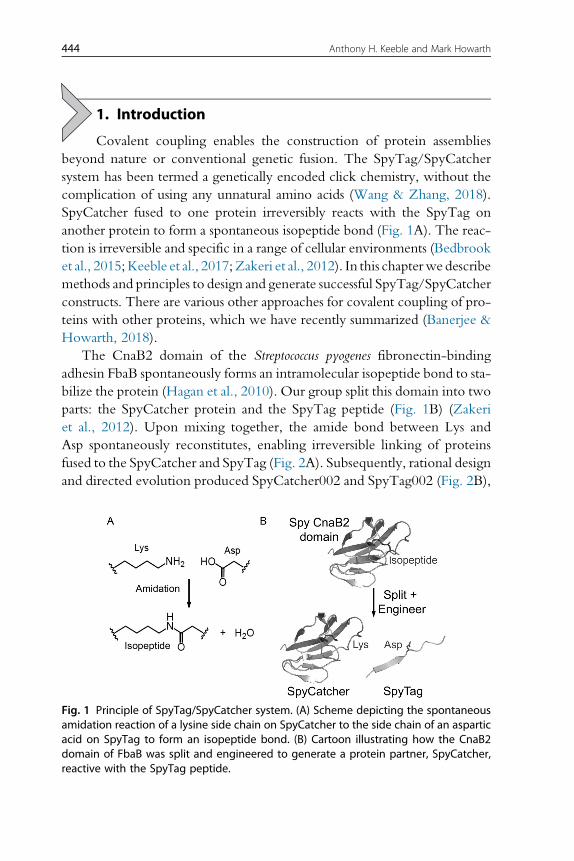
The CnaB2 domain of the Streptococcus pyogenes fibronectin-binding
adhesin FbaB spontaneously forms an intramolecular isopeptide bond to sta-
bilize the protein (Hagan et al., 2010). Our group split this domain into two
parts: the SpyCatcher protein and the SpyTag peptide (Fig. 1B) (Zakeri
et al., 2012). Upon mixing together, the amide bond between Lys and
Asp spontaneously reconstitutes, enabling irreversible linking of proteins
fused to the SpyCatcher and SpyTag (Fig. 2A). Subsequently, rational design
and directed evolution produced SpyCatcher002 and SpyTag002 (Fig. 2B),
Fig. 1 Principle of SpyTag/SpyCatcher system. (A) Scheme depicting the spontaneousamidation reaction of a lysine side chain on SpyCatcher to the side chain of an asparticacid on SpyTag to form an isopeptide bond. (B) Cartoon illustrating how the CnaB2domain of FbaB was split and engineered to generate a protein partner, SpyCatcher,reactive with the SpyTag peptide.
444 Anthony H. Keeble and Mark Howarth

with an order of magnitude faster reactivity, so reacting to completion even
at low (100nM) protein concentrations (Fig. 2B) (Keeble et al., 2017). The
sequence relationships between the most common versions of SpyTag/
SpyTag002 and SpyCatcher/SpyCatcher002 in the published literature
Fig. 2 SpyTag/SpyCatcher purification and reactivity. (A) SpyTag/SpyCatcher purifica-tion. 16% SDS-PAGE with Coomassie staining showing post-induction cell lysate forSpyCatcher002 (lane 2) and SpyTag002-MBP (lane 3) expressed in E. coli, illustrating highexpression levels. Also shown are purified SpyCatcher002 (lane 4), purified SpyTag002-MBP (lane 5), and SpyCatcher002 mixed with SpyTag002-MBP (lane 6); each at 5μM inPBS pH 7.5 at 25°C for 1h. (B) Increased rate of SpyTag002/SpyCatcher002 reaction.Densitometry from SDS-PAGE for isopeptide bond formation between SpyTag002-MBP and SpyCatcher002 (black) or SpyTag-MBP and SpyCatcher (gray) at 0.1μM (top)or 10μM (bottom) in succinate–phosphate–glycine buffer at 25°C. Error bars representmean�SD from triplicates (some error bars are too small to be visible). (C) Amino acidsequence of variants of SpyTag (top) or SpyCatcher (bottom). Panel (B): Data adaptedfrom Keeble, A. H., Banerjee, A., Ferla, M. P., Reddington, S. C., Anuar, I. N. A. K., &Howarth, M. (2017). Evolving accelerated amidation by SpyTag/SpyCatcher to analyzemembrane dynamics. Angewandte Chemie (International Ed. in English), 56(52),16521–16525.
445Covalent protein coupling with help from SpyBank

are shown in Fig. 2C, including SpyCatcher versions with truncations at
each terminus (Li, Fierer, Rapoport, & Howarth, 2014).
SpyTag/SpyCatcher can produce constructs of nonlinear and unprece-
dented topologies (Sun, Zhang, Mahdavi, Arnold, & Tirrell, 2014; Wang &
Zhang, 2016; Zhang, Sun, Tirrell, & Arnold, 2013) and can be used orthog-
onally with other coupling technologies, such as HaloTag or streptavidin
(Fairhead et al., 2014; Peschke, Rabe, & Niemeyer, 2017). SpyTag/
SpyCatcher reactions can couple proteins together with high specificity in
biological environments including bacterial outer-membranes (Keeble et al.,
2017; Peschke et al., 2017), the mammalian cytosol (Hinrichsen et al.,
2017; Zakeri et al., 2012), biofilms (Nguyen, Botyanszki, Tay, & Joshi,
2014), the mammalian plasma membrane (Zakeri et al., 2012), and living
Caenorhabditis elegans (Bedbrook et al., 2015).
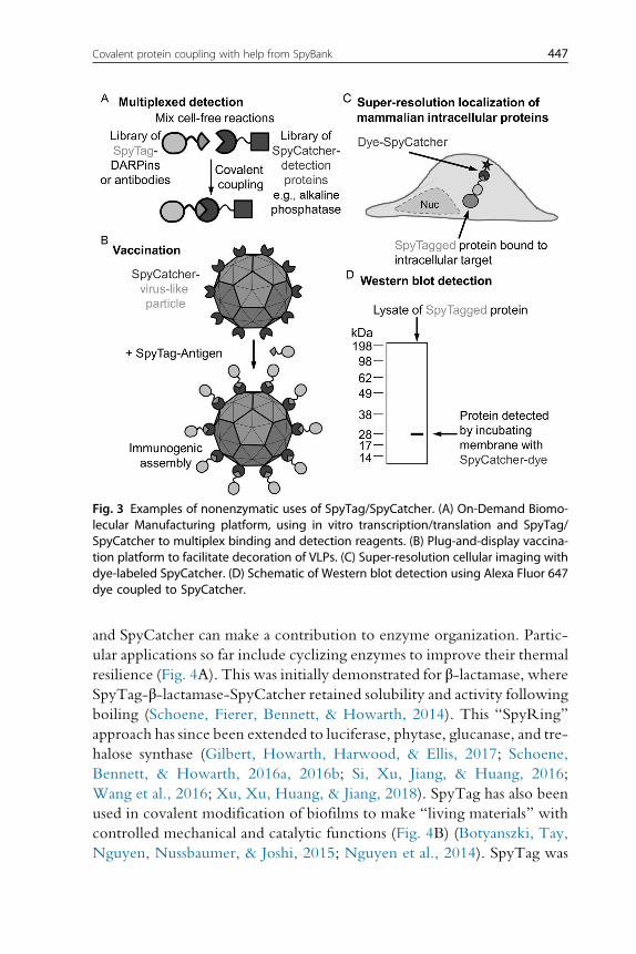
SpyTag/SpyCatcher reactions have a widespread use in assembling com-
plexes, enabling the combination of properties of one protein with the pro-
tein to which it is covalently coupled (Fig. 3). Libraries of antigen-specific
binding reagents (DARPins, nanobodies, etc.) fused to SpyTag and libraries
of detection proteins (fluorescent proteins, reporter enzymes, or toxins)
fused to SpyCatcher (Fig. 3A) have been created as part of a “Portable,
On-Demand Biomolecular Manufacturing” platform (Pardee et al., 2016).
Virus-like particles (VLPs) fused to SpyCatcher have been used to create
“Plug-and-display” immunogens by multivalently displaying SpyTagged
vaccine candidates, to accelerate vaccine generation (Fig. 3B) (Brune &
Howarth, 2018). Intracellular super-resolution imaging reagents for mam-
malian cell microscopy have been generated: Cys-SpyCatcher was conju-
gated with maleimide-Alexa Fluor 647 and added to fixed HEK293T
cells expressing cellular components fused to SpyTag (Fig. 3C) (Pessino,
Citron, Feng, & Huang, 2017). SpyTag fusions were also detected with
high sensitivity in Western blot by Alexa Fluor 647-SpyCatcher, since
the low affinity of antibodies detecting conventional peptide tags can often
be limiting (Fig. 3D) (Dovala, Sawyer, Rath, & Metzger, 2016).
Improving the coordination of enzyme function has been a long-
standing challenge in biotechnology and synthetic biology. Genetic fusions
linking together different enzymes can be successful but can often lead to
misfolding and are restricted in the relative spatial orientation of the active
sites (Pr€oschel, Detsch, Boccaccini, & Sonnewald, 2015). Many peptide:
protein interactions have been employed to connect enzymes, but stability
and specificity of these contacts have often been limiting (Pr€oschel et al.,2015). Therefore, the simple and irreversible linkage between SpyTag
446 Anthony H. Keeble and Mark Howarth
and SpyCatcher can make a contribution to enzyme organization. Partic-
ular applications so far include cyclizing enzymes to improve their thermal
resilience (Fig. 4A). This was initially demonstrated for β-lactamase, where
SpyTag-β-lactamase-SpyCatcher retained solubility and activity following
boiling (Schoene, Fierer, Bennett, & Howarth, 2014). This “SpyRing”
approach has since been extended to luciferase, phytase, glucanase, and tre-
halose synthase (Gilbert, Howarth, Harwood, & Ellis, 2017; Schoene,
Bennett, & Howarth, 2016a, 2016b; Si, Xu, Jiang, & Huang, 2016;
Wang et al., 2016; Xu, Xu, Huang, & Jiang, 2018). SpyTag has also been
used in covalent modification of biofilms to make “living materials” with
controlled mechanical and catalytic functions (Fig. 4B) (Botyanszki, Tay,
Nguyen, Nussbaumer, & Joshi, 2015; Nguyen et al., 2014). SpyTag was
Fig. 3 Examples of nonenzymatic uses of SpyTag/SpyCatcher. (A) On-Demand Biomo-lecular Manufacturing platform, using in vitro transcription/translation and SpyTag/SpyCatcher to multiplex binding and detection reagents. (B) Plug-and-display vaccina-tion platform to facilitate decoration of VLPs. (C) Super-resolution cellular imaging withdye-labeled SpyCatcher. (D) Schematic of Western blot detection using Alexa Fluor 647dye coupled to SpyCatcher.
447Covalent protein coupling with help from SpyBank

Fig. 4 Examples of enzymatic uses of SpyTag/SpyCatcher. (A) Cyclization of enzymes toimprove thermal resilience, showing a cartoon of SpyTag-β-lactamase-SpyCatcher.(B) Production of enzymatic bacterial biofilms. (C) Attachment of chemically synthesizedcell penetrating peptides to enzymes, promoting delivery of functional enzyme intra-cellularly. CCF2-AM is a membrane-permeable dye which is deesterified by esterasesin the cytosol to give CCF2. Cleavage by cytosolic β-lactamase changes CCF2’s fluores-cent spectrum. (D) Biological nanoreactors through formation of a covalently linkedenzyme network from a dimeric cytochrome P450 monooxygenase and a tetramericglucose dehydrogenase. Adapted from Yin, L., Guo, X., Liu, L., Zhang, Y., & Feng, Y.(2018). Self-assembled multimeric-enzyme nanoreactor for robust and efficient biocatalysis.ACS Biomaterials Science & Engineering, 4(6), 2095–2099.
448 Anthony H. Keeble and Mark Howarth

applied to the intracellular delivery of enzymes by modular linkage to cell
penetrating peptides (Fig. 4C) (Hoffmann et al., 2018; Stone et al., 2018).
SpyTag also facilitated construction ofmultimerized enzyme assemblies either
using VLPs (R€oder, Fischer, & Commandeur, 2017) (as in Fig. 3B) or other
biological nanoreactors (Fig. 4D) (Alves et al., 2017; Giessen & Silver, 2016;
Pr€oschel et al., 2015; Yin et al., 2018). Such complexes also enable a scaffold-
ing function, organizing proteases for amplification toward cancer diagnosis
(Stein, Nabi, & Alexandrov, 2017), as well as immobilizing enzymes for
robust nanopore DNA sequencing devices (Stranges et al., 2016).
We have assembled amore complete list of publications and patents using
SpyTag/SpyCatcher technology at the SpyInfo webpage (https://www.
bioch.ox.ac.uk/howarth/info.htm). The webpage also contains the papers
applying other spontaneous isopeptide bond-forming systems (Abe et al.,
2013; Zakeri et al., 2012). The first split pair was isopeptag/Pilin-C, but
pilin-C is much larger than SpyCatcher and reacts much slower (Zakeri &
Howarth, 2010). SnoopTag/SnoopCatcher results from the engineering
of a D4 domain of the RrgA protein from Streptococcus pneumoniae, reacting
orthogonally (with no cross-reactivity) to SpyTag/SpyCatcher (Veggiani
et al., 2016). Further engineering of the CnaB2 domain produced SpyLigase,
while SnoopLigase was generated from the RrgAD4 domain (Buldun, Jean,
Bedford, & Howarth, 2018; Fierer, Veggiani, & Howarth, 2014). These
ligases join two peptides (formed from parts of the original domains) together
via isopeptide bonds. However, the speed of reaction of these ligases and
their activity with low concentration of target protein is worse than for
SpyTag/SpyCatcher ligation.
The procedures outlined below should help to avoid potential pitfalls, as
well as integrating the insight that is now available from the validation of a
wide range of SpyTag and SpyCatcher fusions in different organisms, com-
partments, and protein contexts.
2. SpyDesign—Construction of successfulSpyTag/SpyCatcher reagents
2.1 SpyBank databaseSpyBank is an online database that we have compiled of amino acid sequences
of SpyTag or SpyCatcher fusions available from published papers or patents by
different academics or companies. Example entries are shown in Table 1. As of
September 2018, there aremore than 400 sequence entries, available for down-
load from our webpage (https://www.bioch.ox.ac.uk/howarth/info.htm).
449Covalent protein coupling with help from SpyBank
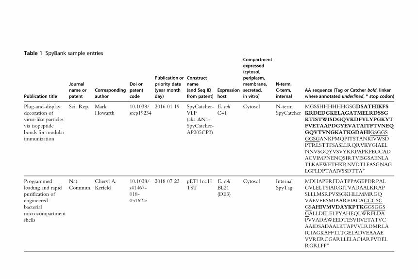
Table 1 SpyBank sample entries
Publication title
Journalname orpatent
Correspondingauthor
Doi orpatentcode
Publication orpriority date(year monthday)
Constructname(and Seq IDfrom patent)
Expressionhost
Compartmentexpressed(cytosol,periplasm,membrane,secreted,in vitro)
N-term,C-term,internal
AA sequence (Tag or Catcher bold, linkerwhere annotated underlined, * stop codon)
Plug-and-display:
decoration of
virus-like particles
via isopeptide
bonds for modular
immunization
Sci. Rep. Mark
Howarth
10.1038/
srep19234
2016 01 19 SpyCatcher-
VLP
(aka ΔN1-
SpyCatcher-
AP205CP3)
E. coli
C41
Cytosol N-term
SpyCatcher
MGSSHHHHHHGSGDSATHIKFS
KRDEDGKELAGATMELRDSSG
KTISTWISDGQVKDFYLYPGKYT
FVETAAPDGYEVATAITFTVNEQ
GQVTVNGKATKGDAHIGSGGS
GGSGANKPMQPITSTANKIVWSD
PTRLSTTFSASLLRQRVKVGIAEL
NNVSGQYVSVYKRPAPKPEGCAD
ACVIMPNENQSIRTVISGSAENLA
TLKAEWETHKRNVDTLFASGNAG
LGFLDPTAAIVSSDTTA*
Programmed
loading and rapid
purification of
engineered
bacterial
microcompartment
shells
Nat.
Commun.
Cheryl A.
Kerfeld
10.1038/
s41467-
018-
05162-z
2018 07 23 pET11n::H
TST
E. coli
BL21
(DE3)
Cytosol Internal
SpyTag
MDHAPERFDATPPAGEPDRPAL
GVLELTSIARGITVADAALKRAP
SLLLMSRPVSSGKHLLMMRGQ
VAEVEESMIAAREIAGAGGGSG
GSAHIVMVDAYKPTKGGSGGS
GALLDELELPYAHEQLWRFLDA
PVVADAWEEDTESVIIVETATVC
AAIDSADAALKTAPVVLRDMRLA
IGIAGKAFFTLTGELADVEAAAE
VVRERCGARLLELACIARPVDEL
RGRLFF*

In addition to listing corresponding author and citation, SpyBank contains
important experimental design information on the expression host, cellular
compartment, where the protein was fused, which version of SpyCatcher or
SpyTag was used, as well as the amino acid sequence of the constructs used
(where available). The plasmids for basic SpyCatcher and SpyTag constructs
are available from the Addgene plasmid repository (https://www.addgene.
org): SpyCatcher (#35044); ΔN1ΔC2SpyCatcher (#87376); SpyTag-
maltose binding protein (MBP) (#35050); AviTag-SpyCatcher (#72326);
SpyCatcher002 (#102827); and SpyTag002-MBP (#102831).
SpyBank reveals that Escherichia coli is by far the most common system for
expression of SpyTag/SpyCatcher fusions. However, a range of other spe-
cies have been successfully used. Other bacteria used for expression were
Bacillus subtilis, Lactococcus lactis, and Salmonella Typhimurium. Among eukary-
otes, expression has been performed in Saccharomyces cerevisiae, C. elegans,
insect cells (ExpresS2, Sf9, Trichoplusia ni), human cell lines (HeLa,
HEK293), and the plantNicotiana benthamiana. Expression has also been per-
formed in cell-free systems (Pardee et al., 2016). This diversity supports the
wide-spread biological compatibility of SpyCatcher and SpyTag.
In addition to cytosolic protein expression, SpyTagged proteins have been
secreted from HEK293T cells, using signal peptides at the N-terminus of
the construct to target for endoplasmic reticulum translocation. Some signal
peptides are useful for many proteins, but others are more specific (Kober,
Zehe, & Bode, 2013), so trials with different signal peptides may be useful
for the highest expression levels. These cells can also be used to express pro-
teins targeted to the membrane. Alternative signal peptides compatible with
membrane display of SpyTagged proteins are listed in SpyBank.
2.2 Construct designWhen designing constructs involving SpyTag and SpyCatcher variants, the
following considerations should be taken into account:
(i) Choice of SpyCatcher and SpyTag variant
For N- or C-terminal fusion of SpyCatcher, we would recommend the
ΔN1SpyCatcher construct (Fig. 2C) (Li et al., 2014), which gave us the best
results in the challenging situation of VLP fusion (Brune et al., 2016). If the
SpyCatcher is to be inserted in an internal protein loop, we suggest
ΔN1ΔC2SpyCatcher, which was successfully applied for bacterial micro-
compartments (Hagen, Sutter, Sloan, & Kerfeld, 2018). If the reaction speed
451Covalent protein coupling with help from SpyBank

is limiting, we recommend switching to SpyCatcher002 to accelerate the
reaction with SpyTag, especially at low (<100nM) protein concentrations
(Keeble et al., 2017). We have found that the N-terminal sequence of
SpyCatcher expression constructs starting with GAMVD results in a trace
amount of side reaction with the reactive lysine of another SpyCatcher mol-
ecule, since this sequence partially resembles the SpyTag sequence (Keeble
et al., 2017). During the development of the faster reacting SpyCatcher002,
we found that this off-pathway reaction was enhanced, requiring the muta-
tion of the sequence toGAMVT (Keeble et al., 2017). Thus, we recommend
also including this mutation in SpyCatcher constructs, as well as ensuring that
sequences like this are not used in linker regions.
SpyBank shows many examples of SpyTag being used at the N- or
C-terminus. SpyTag may also be used in exposed linker regions between
protein domains (Zakeri et al., 2012). There are fewer examples of SpyTag
being used in a loop within a folded domain (Hagen et al., 2018; Kasaraneni,
Chamoun-Emanuelli, Wright, & Chen, 2017; Moon, Bae, Kim, & Kang,
2016); optimization may sometimes be required, because SpyTag reacts
in an elongated conformation with SpyCatcher (Fig. 1B) (Li et al., 2014).
SpyTag and SpyTag002 can be used interchangeably, but the reaction with
any SpyCatcher variant will be faster with SpyTag002 (Keeble et al., 2017).
(ii) Choice of fusion site
SpyCatcher and SpyTag can be used equally well on the N- or C-terminus,
since the reaction is mediated through side chains. SpyBank also contains
several constructs with more than one SpyTag or more than one SpyCatcher
moiety (Wieduwild & Howarth, 2018; Zhang et al., 2013). One may prefer
a particular terminus because it is distant from a binding site or an active site,
because previous fusion to other tags has been successful (e.g., to a His6-tag
or fluorescent protein), or because there is lower sequence conservation at
that terminus (Chen, Zaro, & Shen, 2013). If an initial construct shows sub-
optimal activity, it is worth moving SpyCatcher or SpyTag to the opposite
terminus, to see if activity is improved (Si et al., 2016).
SpyCatcher and SpyTag must be sterically accessible to one another for
reaction to occur. This consideration is of particular importance when using
large multimeric proteins, where either the N- or C-terminus may point
into the core of the protein (Brune & Howarth, 2018; Hagen et al., 2018).
(iii) Choice of linker
We always insert a linker between SpyTag or SpyCatcher and the protein of
interest. Initially we usually try GSGESGSG (Veggiani et al., 2016). Gly/Ser
452 Anthony H. Keeble and Mark Howarth

linkers show a good balance of flexibility, solubility, and protease resistance
(Chen et al., 2013), and we often include a Glu in the spacer to increase
hydrophilicity. The linker increases the accessibility of SpyTag/SpyCatcher
for faster and higher yielding reaction. The linker also reduces the chance of
interference in the structure or function of the fused protein. If a linker has
been used on a similar protein in SpyBank, we would suggest copying that
linker. If we find slow or incomplete reaction, we will often move to a
10-residue linker, such as GGGGSGGGGS (Veggiani et al., 2016). When
using ΔN1ΔC2SpyCatcher in an internal loop, GGGSGGS was used on
each side (Hagen et al., 2018). With SpyTag in an internal loop, linkers such
as GGGS on both sides have been used successfully in SpyBank (Alves
et al., 2017).
We suggest avoiding linkers containing Asp or Asn close to the C-terminal
side of SpyCatcher. For example, a sequence at the C-terminus of SpyCatcher
like ...AHIGSGDG... could promote self-reaction of the Asp in bold with the
reactive lysine of SpyCatcher (C-terminus of SpyCatcher is underlined). Also,
we avoid negatively charged peptides (e.g., myc tag or C-tag) immediately
adjacent to SpyTag, where electrostatic complementarity may slow reaction
with SpyCatcher.
(iv) Codon usage
We have nearly always used the original codons for SpyTag and
SpyCatcher from S. pyogenes and this usage has not caused problems for
E. coli expression. Gene design companies or online servers are able to sug-
gest optimized codon usage for any species, but sometimes unpredicted
effects can lead to worse expression. For expression in other organisms,
it is worth looking in SpyBank to identify previous constructs expressed
in that species. For simplicity, SpyBank only shows amino acid sequences,
but SpyBank should help users to go to the primary source and find the
DNA sequence. An exception that we found is the expression of SpyTag
at the N-terminus in E. coli, where the issue is not codon frequency but
codon complementarity. Secondary structure near the Shine-Dalgarno
sequence can greatly reduce protein expression yield. Thus, the sequence
needs to be optimized using a Ribosome Binding Calculator (score 10,000
or above) to maximize expression levels (Salis, 2011). We give here
SpyTag DNA sequences for two common promoter systems for bacterial
expression:
T7 vector 50-gcacacatagtaatggtagacgcctacaagccgacgaag-30
T5 vector 50-gctcatatcgtcatggttgacgcgtataaaccgaccaaa-30
A H I V M V D A Y K P T K
453Covalent protein coupling with help from SpyBank

Our constructs will typically have a glycine immediately after the initiating
methionine/formyl methionine, which can help expression, consistent with
the N-end rule for protein stability (Varshavsky, 2011).
2.3 Cellular expression and purificationAfter creation of the desired expression construct, the plasmid is next trans-
formed into an expression strain. We will not go into detail here on proto-
cols that are standard for any protein but focus on issues specific to SpyTag/
SpyCatcher. Our standard Ni-NTA purification protocol is described in
detail previously (Howarth & Ting, 2008).
(i) Bacterial expression
SpyBank reveals that a wide-range of different E. coli strains can be used for
expression of SpyTag/SpyCatcher fusions. Hence, if you find low expres-
sion in one strain, you should try others. Two E. coli strains we recommend
are BL21 (DE3) RIPL (Agilent) (helping to express proteins with rare
codons) and C41 (DE3) (helping expression of toxic proteins) (Miroux &
Walker, 1996). E. coli BL21 (DE3) transformed with a plasmid encoding
Erv1p andDsbC allowed us to express efficiently proteins containing a disul-
fide bond in the cytosol (Veggiani et al., 2016). Supplementing LB media
with 0.8% glucose may reduce leaky expression before induction, which aids
growth for toxic proteins (Grossman, Kawasaki, Punreddy, & Osburne,
1998). Alternative growth media such as 2� TY or auto-induction media
can also be used with SpyTag/SpyCatcher constructs. After inducing with
IPTG at OD600 0.5, we typically grow the cells for a further 4h at 30°C. Formost constructs we can clearly see induction by SDS-PAGEwith Coomassie
staining on the whole cell lysate (Fig. 2A).
If expression has failed, we will first try an alternative induction temper-
ature (18°C for 16h) or a different E. coli strain. Purification of His6-tagged
fusions by Ni-NTA is carried out by standard methods (Zakeri et al., 2012)
ensuring protease inhibitors are present during cell disruption and initial
purification. After elution from affinity chromatography, proteins can
be dialyzed into the buffer of choice (typically phosphate-buffered saline,
PBS: 137mM NaCl, 2.7mM KCl, 10mM Na2HPO4, 1.8mM KH2PO4
pH 7.5). Dialysis is especially important after Ni-NTA purification, since
high concentrations of imidazole slow down the SpyTag/SpyCatcher
reaction. If there is substantial aggregation upon dialysis, we will try a
buffer with a pH more than 1 unit from the isoelectric point (pI) of the
fusion protein (e.g., 50mM Tris base with the pH adjusted using boric
454 Anthony H. Keeble and Mark Howarth

acid, or 50mMglycine with25mM sodiumcitrate with the pH adjusted
using HCl or NaOH).
A further round of purification such as gel filtration can be used if
required, but many SpyCatcher- or SpyTag-linked proteins are typically
pure enough to couple to the binding partner after one step purification
(Fig. 2A). C-tag purification is also efficient for SpyTag fusions and may
be preferred for vaccine studies (Bruun, Andersson, Draper, & Howarth,
2018). For the highest purity, we typically combine Ni-NTA purification
with gel filtration chromatography or C-tag purification (Brune et al., 2016).
Expression yields of 15–30mg/L of culture are typically achieved with
SpyCatcher or SpyTag fusions, although VLP fusion was more challenging
(Bruun et al., 2018). Proteins can be stored at 4°C overnight or indefinitely
in aliquots at �80°C. Certain fusions are likely to be sensitive to freeze–thaw, but where we have tested SpyCatcher fusions on vaccine platforms,
we found good resilience to freezing or lyophilization (Brune et al.,
2017; Bruun et al., 2018).
(ii) Mammalian secretion
Three related mammalian cell lines have been widely used in SpyBank—
HEK293T (adherent), HEK293 FreeStyle, and HEK293Expi. Although
several SpyTag-containing constructs are reported (with SpyTag at either
N- or C-terminus), there are fewer examples in SpyBank of secretion of
a SpyCatcher fusion. The dynamic structure of SpyCatcher may sometimes
reduces the efficiency of secretion. Successful published examples include
SpyCatcher at the N-terminus of the IgG Fc domain (Alam et al., 2017)
and SpyCatcher linked to a range of antigens for immunization (Thrane
et al., 2016). It appears more difficult to express SpyCatcher fusions than
SpyTag fusions in mammalian cells.
Expression constructs can be transiently transfected into the cells at mid-
confluency using polyethylene imine (PEI) (HEK293T and HEK293 Free-
Style) or ExpiFectamine 293 (HEK293Expi). After secretion of the protein
into the media for the desired time (typically 3–7 days), the media is
removed from the cells and protease inhibitors are added to prevent prote-
olysis. After a repeated centrifugation to decrease the amount of cell debris,
the supernatant should be syringe-filtered (0.45μm). Binding buffer should
now be added to the filtered supernatant as required for purification: 50mM
Tris�HCl, 300mM NaCl, pH 7.8 to 1/4 of total volume. This is important
for pH buffering, since the growth medium does not buffer effectively with-
out high CO2. Given the large volume of cell supernatant produced
455Covalent protein coupling with help from SpyBank
(100–1000mL typically), affinity purification (His6-tag or C-tag) is essential
to concentrate the protein and can now proceed as required for the standard
protocols.
3. Analysis of SpyTag/SpyCatcher covalent couplingreactions
3.1 Reaction of componentsSpyTag/SpyCatcher reaction readily occurs without requiring any specific
anions or cations, at a range of temperatures (4–37°C), at pH from 4 to 8,
in the presence of various detergents (Tween-20, Triton X-100, Nonidet
P-40, CHAPS, but not SDS), and even under chaotropic conditions such
as 4M urea (Keeble et al., 2017). This flexibility makes SpyTag/SpyCatcher
coupling suited to a wide range of coupling tasks. We typically mix the
SpyCatcher and SpyTag proteins in a buffer such as PBS pH 7.5 with each
partner at 10μM and incubate for 2h at room temperature.Reaction is slower,
for example, on the crowded surface of VLPs, where we usually incubate for
16h (Brune et al., 2016; Bruun et al., 2018). To ensure one partner is fully
reacted, we recommend incubating with one component in 1.5–3� excess.
Given that the reaction follows second-order kinetics (Keeble et al., 2017;
Zakeri et al., 2012), it is not just the ratio but also the absolute concentration
which is important for reaction speed. Given the broad tolerance of SpyTag/
SpyCatcher reaction described above, it is worth including additives (e.g., Ca2+,
glycerol, cofactors) likely to stabilize the proteins of interest.
When testing new constructs, we recommend using purified SpyCatcher
or SpyTag-MBP proteins as positive controls under the same conditions.
These positive controls are very helpful for troubleshooting, to determine
whether the buffer or protein constructs may be at fault, if the novel protein
partners do not react as desired. Negative control constructs are also helpful
to validate that any observed effects depend on covalent reaction. Such con-
trols bear a mutation in the reactive residue of SpyTag (SpyTag002
DA-MBP Addgene # 102832) or SpyCatcher (SpyCatcher EQ Addgene
# 35045 or SpyCatcher002 EQ Addgene # 102830) (Keeble et al., 2017;
Zakeri et al., 2012).
3.2 Analysis of reaction(i) SDS-PAGE
Formation of covalent SpyTag/SpyCatcher reaction products in vitro can
be observed and quantified by SDS-polyacrylamide gel electropho-
resis (Fig. 2A), since the isopeptide bond is stable to boiling in SDS
456 Anthony H. Keeble and Mark Howarth

(Keeble et al., 2017; Zakeri et al., 2012). This approach enables convenient
quantitation by densitometry, based on either formation of the SpyTag:
SpyCatcher conjugate or the depletion of SpyTag or SpyCatcher starting
material. Note that the mobility of the SpyTag:SpyCatcher conjugate
may be slightly different from the molecular weight because of its branched
nature (Schoene et al., 2016a, 2016b; Zakeri et al., 2012) and a second band
may be present on SpyCatcher fusions, as discussed in Section 3.2 (ii).
(ii) Mass spectrometry
Mass spectrometry is helpful to confirm the exact product of SpyTag/
SpyCatcher reaction. Isopeptide bond formation for this pair results in loss
of H2O (18Da). For analysis of the reaction of purified samples, we dialyze
into 10mM ammonium acetate, since the low ionic strength and volatile
buffer components assist in obtaining a clean spectrum. A common impurity
after E. coli expression is a gluconylated adduct (Geoghegan et al., 1999), but
we have never experienced functional consequences of this impurity.
(iii) Fluorescence microscopy
The specificity and efficiency of in vivo reaction of SpyTag/SpyCatcher can
be examined by microscopy. The first step is labeling with fluorescent pro-
teins fused to the SpyCatcher or SpyTag construct and using microscopy to
detect the cellular distribution of the target protein. This approach was used
to demonstrate that SpyTag-Channelrhodopsin-mCherry constructs can
react with SpyCatcher-GFP in C. elegans (Bedbrook et al., 2015).
(iv) Western blotting
Western blotting was performed on lysate from cells expressing SpyCatcher
or SpyTagged proteins, with reaction occurring upon staining of the trans-
ferred membrane (Dovala et al., 2016). Western blotting can also be used
to follow reaction occurring on living cells, such as to show that
biotinylated-AviTag-SpyTag002-MBP reacted specifically with the bacte-
rial outer membrane-displayed Intimin-myc tag-SpyCatcher002, using
Streptavidin-Horse Radish Peroxidase (to detect the biotinylated protein)
or an anti-myc antibody (followed by secondary antibody-HRP) to follow
the total pool of cell-surface Intimin (Keeble et al., 2017).
3.3 Conjugate purificationAfter the desired reaction time, unreacted SpyTag or SpyCatcher compo-
nents may be purified from the covalent complexes, using size-exclusion
457Covalent protein coupling with help from SpyBank

chromatography, spin-filtration, or dialysis (Brune et al., 2016). When there
is a large size difference between the two partners (e.g., SpyTag-antigen sep-
aration from antigen-decorated VLP), we found dialysis to be a simple and
efficient approach, leading to minimal dilution of the sample (Brune
et al., 2016).
4. Concluding remarks
We have described here guidelines on how to design and purify con-
structs to use SpyTag/SpyCatcher for covalent coupling of proteins, based
on our own experience and the results of many other groups, as compiled in
SpyBank. It is relatively simple to generate functional SpyTag or SpyCatcher
constructs in a range of cellular systems. Reaction is typically efficient and
selective either in vitro or at the surface of cells. There are few examples
of SpyTag/SpyCatcher reaction inside living cells, so it will be important
to validate this application in future work, perhaps with new iterations of
Tag/Catcher pairs. Future development should also seek to establish the
principles for efficient use of SpyTag/SpyCatcher in protein loops and effi-
cient secretion of SpyCatcher fusions from mammalian cells. Combining
SpyTag/SpyCatcher with related technologies such as SnoopCatcher
(Veggiani et al., 2016) and SnoopLigase (Buldun et al., 2018) will also pro-
vide further opportunities for creating novel protein architectures, signaling
teams (Veggiani et al., 2016) and immune stimulants (Brune et al., 2017).
AcknowledgmentFunding was provided by the European Research Council (ERC-2013-CoG 615945-
PeptidePadlock).
Conflicts of interestM.H. and A.H.K. are authors on a patent application covering sequences for enhanced
isopeptide bond formation (UK Intellectual Property Office 1706430.4). M.H. is an
author on a patent for isopeptide bond formation (EP2534484) and a SpyBiotech
cofounder, shareholder, and consultant.
ReferencesAbe, H., Wakabayashi, R., Yonemura, H., Yamada, S., Goto, M., & Kamiya, N. (2013).
Split Spy0128 as a potent scaffold for protein cross-linking and immobilization.Bioconjugate Chemistry, 24(2), 242–250.
Alam, M. K., Gonzalez, C., Hill, W., El-Sayed, A., Fonge, H., Barreto, K., et al. (2017).Synthetic modular antibody construction by using the SpyTag/SpyCatcher protein-ligase system. Chembiochem, 18(22), 2217–2221.
458 Anthony H. Keeble and Mark Howarth

Alves, N. J., Turner, K. B., Daniele, M. A., Oh, E., Medintz, I. L., & Walper, S. A. (2017).Bacterial nanobioreactors—Directing enzyme packaging into bacterial outer membranevesicles. ACS Applied Materials & Interfaces, 7(44), 24963–24972.
Banerjee, A., & Howarth, M. (2018). Nanoteamwork: Covalent protein assembly beyondduets towards protein ensembles and orchestras. Current Opinion in Biotechnology, 51,16–23.
Bedbrook, C. N., Kato, M., Ravindra Kumar, S., Lakshmanan, A., Nath, R. D., Sun, F.,et al. (2015). Genetically encoded spy peptide fusion system to detect plasmamembrane-localized proteins in vivo. Chemistry & Biology, 22(8), 1108–1121.
Botyanszki, Z., Tay, P. K., Nguyen, P. Q., Nussbaumer, M. G., & Joshi, N. S. (2015).Engineered catalytic biofilms: Site-specific enzyme immobilization onto E. coli curlinanofibers. Biotechnology and Bioengineering, 112(10), 2016–2024.
Brune, K. D., Buldun, C. M., Li, Y., Taylor, I. J., Brod, F., Biswas, S., et al. (2017). Dualplug-and-display synthetic assembly using orthogonal reactive proteins for twin antigenimmunization. Bioconjugate Chemistry, 28(5), 1544–1551.
Brune, K. D., & Howarth, M. (2018). New routes and opportunities for modular construc-tion of particulate vaccines: Stick, click, and glue. Frontiers in Immunology, 9, 1432.
Brune, K. D., Leneghan, D. B., Brian, I. J., Ishizuka, A. S., Bachmann, M. F., Draper, S. J.,et al. (2016). Plug-and-display: Decoration of virus-like particles via isopeptide bonds formodular immunization. Scientific Reports, 6, 19234.
Bruun, T. U. J., Andersson, A. C., Draper, S. J., & Howarth, M. (2018). Engineering a rug-ged nanoscaffold to enhance plug-and-display vaccination. ACS Nano, 12, 8855–8866.
Buldun, C. M., Jean, J. X., Bedford, M. R., & Howarth, M. (2018). SnoopLigase catalyzespeptide-peptide locking and enables solid-phase conjugate isolation. Journal of the Amer-ican Chemical Society, 140(8), 3008–3018.
Chen, X., Zaro, J. L., & Shen, W. C. (2013). Fusion protein linkers: Property, design andfunctionality. Advanced Drug Delivery Reviews, 65(10), 1357–1369.
Dovala, D., Sawyer, W. S., Rath, C. M., & Metzger, L. E., 4th. (2016). Rapid analysis ofprotein expression and solubility with the SpyTag-SpyCatcher system. Protein Expressionand Purification, 117, 44–51.
Fairhead, M., Veggiani, G., Lever, M., Yan, J., Mesner, D., Robinson, C. V., et al. (2014).SpyAvidin hubs enable precise and ultrastable orthogonal nanoassembly. Journal of theAmerican Chemical Society, 136(35), 12355–12363.
Fierer, J. O., Veggiani, G., & Howarth, M. (2014). SpyLigase peptide-peptide ligation poly-merizes affibodies to enhance magnetic cancer cell capture. Proceedings of the NationalAcademy of Sciences of the United States of America, 111(13), E1176–E1181.
Geoghegan, K. F., Dixon, H. B., Rosner, P. J., Hoth, L. R., Lanzetti, A. J., Borzilleri, K. A.,et al. (1999). Spontaneous alpha-N-6-phosphogluconoylation of a “His tag” in Escherichiacoli: The cause of extra mass of 258 or 178 Da in fusion proteins. Analytical Biochemistry,267(1), 169–184.
Giessen, T.W., & Silver, P. A. (2016). A catalytic nanoreactor based on in vivo encapsulationof multiple enzymes in an engineered protein nanocompartment. Chembiochem, 17(20),1931–1935.
Gilbert, C., Howarth, M., Harwood, C. R., & Ellis, T. (2017). Extracellular self-assembly offunctional and tunable protein conjugates from Bacillus subtilis. ACS Synthetic Biology,6(6), 957–967.
Grossman, T. H., Kawasaki, E. S., Punreddy, S. R., & Osburne, M. S. (1998). SpontaneouscAMP-dependent derepression of gene expression in stationary phase plays a role inrecombinant expression instability. Gene, 209(1–2), 95–103.
Hagan, R. M., Bj€ornsson, R., McMahon, S. A., Schomburg, B., Braithwaite, V., B€uhl, M.,et al. (2010). NMR spectroscopic and theoretical analysis of a spontaneously formed Lys-Asp isopeptide bond. Angewandte Chemie (International Ed. in English), 49(45), 8421–8425.
459Covalent protein coupling with help from SpyBank

Hagen, A., Sutter, M., Sloan, N., & Kerfeld, C. A. (2018). Programmed loading and rapidpurification of engineered bacterial microcompartment shells. Nature Communications,9(1), 2881.
Hinrichsen, M., Lenz, M., Edwards, J. M., Miller, O. K., Mochrie, S. G. J., Swain, P. S.,et al. (2017). A new method for post-translationally labeling proteins in live cells forfluorescence imaging and tracking. Protein Engineering, Design & Selection, 30(12),771–780.
Hoffmann, K., Milech, N., Juraja, S. M., Cunningham, P. T., Stone, S. R., Francis, R. W.,et al. (2018). A platform for discovery of functional cell-penetrating peptides for efficientmulti-cargo intracellular delivery. Scientific Reports, 8(1), 12538.
Howarth, M., & Ting, A. Y. (2008). Imaging proteins in live mammalian cells with biotinligase and monovalent streptavidin. Nature Protocols, 3(3), 534–545.
Kasaraneni, N., Chamoun-Emanuelli, A. M., Wright, G., & Chen, Z. (2017). Retargetinglentiviruses via SpyCatcher-SpyTag chemistry for gene delivery into specific cell types.MBio, 8(6), e01860-17.
Keeble, A.H., Banerjee, A., Ferla,M. P., Reddington, S. C., Anuar, I.N. A.K., &Howarth,M.(2017). Evolving accelerated amidation by SpyTag/SpyCatcher to analyze membranedynamics. Angewandte Chemie (International Ed. in English), 56(52), 16521–16525.
Kober, L., Zehe, C., & Bode, J. (2013). Optimized signal peptides for the development ofhigh expressing CHO cell lines. Biotechnology and Bioengineering, 110(4), 1164–1173.
Li, L., Fierer, J. O., Rapoport, T. A., & Howarth, M. (2014). Structural analysis and opti-mization of the covalent association between SpyCatcher and a peptide Tag. Journal ofMolecular Biology, 426(2), 309–317.
Miroux, B., & Walker, J. E. (1996). Over-production of proteins in Escherichia coli: Mutanthosts that allow synthesis of somemembrane proteins and globular proteins at high levels.Journal of Molecular Biology, 260, 289–298.
Moon, H., Bae, Y., Kim, H., & Kang, S. (2016). Plug-and-playable fluorescent cell imagingmodular toolkits using the bacterial superglue, SpyTag/SpyCatcher. Chemical Communi-cations (Cambridge, England), 52(97), 14051–14054.
Nguyen, P. Q., Botyanszki, Z., Tay, P. K., & Joshi, N. S. (2014). Programmable biofilm-based materials from engineered curli nanofibres. Nature Communications, 5, 4945.
Pardee, K., Slomovic, S., Nguyen, P. Q., Lee, J. W., Donghia, N., Burrill, D., et al. (2016).Portable, on-demand biomolecular manufacturing. Cell, 167(1), 248–259.
Peschke, T., Rabe, K. S., & Niemeyer, C. M. (2017). Orthogonal surface tags for whole-cellbiocatalysis. Angewandte Chemie (International Ed. in English), 56(8), 2183–2186.
Pessino, V., Citron, Y. R., Feng, S., & Huang, B. (2017). Covalent protein labeling bySpyTag-SpyCatcher in fixed cells for super-resolution microscopy. Chembiochem,18(15), 1492–1495.
Pr€oschel, M., Detsch, R., Boccaccini, A. R., & Sonnewald, U. (2015). Engineering of met-abolic pathways by artificial enzyme channels. Frontiers in Bioengineering and Biotechnology,3, 168.
R€oder, J., Fischer, R., & Commandeur, U. (2017). Engineering potato virus X particles for acovalent protein based attachment of enzymes. Small, 13(48), 1702151.
Salis, H. M. (2011). The ribosome binding site calculator. Methods in Enzymology, 498,19–42.
Schoene, C., Bennett, S. P., &Howarth, M. (2016a). SpyRing interrogation: Analyzing howenzyme resilience can be achieved with phytase and distinct cyclization chemistries. Sci-entific Reports, 6, 21151.
Schoene, C., Bennett, S. P., & Howarth, M. (2016b). SpyRings declassified: A blueprint forusing isopeptide-mediated cyclization to enhance enzyme thermal resilience.Methods inEnzymology, 580, 149–167.
460 Anthony H. Keeble and Mark Howarth

Schoene, C., Fierer, J. O., Bennett, S. P., &Howarth, M. (2014). SpyTag/SpyCatcher cycli-zation confers resilience to boiling on a mesophilic enzyme. Angewandte Chemie(International Ed. in English), 53(24), 6101–6104.
Si, M., Xu, Q., Jiang, L., & Huang, H. (2016). SpyTag/SpyCatcher cyclization enhances thethermostability of firefly luciferase. PLoS One, 11, e0162318.
Stein, V., Nabi, M., & Alexandrov, K. (2017). Ultrasensitive scaffold-dependent proteasesensors with large dynamic range. ACS Synthetic Biology, 6(7), 1337–1342.
Stone, S. R., Heinrich, T., Juraja, S. M., Satiaputra, J. N., Hall, C. M., Anastasas, M., et al.(2018). β-Lactamase tools for establishing cell internalization and cytosolic delivery ofcell penetrating peptides. Biomolecules, 8(3), 51.
Stranges, P. B., Palla, M., Kalachikov, S., Nivala, J., Dorwart, M., Trans, A., et al. (2016).Design and characterization of a nanopore-coupled polymerase for single-moleculeDNA sequencing by synthesis on an electrode array. Proceedings of the National Academyof Sciences of the United States of America, 113(44), E6749–E6756.
Sun, F., Zhang, W. B., Mahdavi, A., Arnold, F. H., & Tirrell, D. A. (2014). Synthesis ofbioactive protein hydrogels by genetically encoded SpyTag-SpyCatcher chemistry.Proceedings of the National Academy of Sciences of the United States of America, 111(31),11269–11274.
Thrane, S., Janitzek, C. M., Matondo, S., Resende, M., Gustavsson, T., de Jongh, W. A.,et al. (2016). Bacterial superglue enables easy development of efficient virus-like particlebased vaccines. Journal of Nanobiotechnology, 14, 30.
Varshavsky, A. (2011). The N-end rule pathway and regulation by proteolysis. Protein Science,20(8), 1298–1345.
Veggiani, G., Nakamura, T., Brenner, M. D., Gayet, R. V., Yan, J., Robinson, C. V., et al.(2016). Programmable polyproteams built using twin peptide superglues. Proceedings ofthe National Academy of Sciences of the United States of America, 113(5), 1202–1207.
Wang, J., Wang, Y., Wang, X., Zhang, D., Wu, S., & Zhang, G. (2016). Enhanced thermalstability of lichenase from Bacillus subtilis 168 by SpyTag/SpyCatcher-mediated sponta-neous cyclization. Biotechnology for Biofuels, 9, 79.
Wang, X. W., & Zhang, W. B. (2016). Cellular synthesis of protein catenanes. AngewandteChemie (International Ed. in English), 55(10), 3442–3446.
Wang, X. W., & Zhang, W. B. (2018). Chemical topology and complexity of protein archi-tectures. Trends in Biochemical Sciences, 43, 806–817.
Wieduwild, R., & Howarth, M. (2018). Assembling and decorating hyaluronan hydrogelswith twin protein superglues to mimic cell-cell interactions. Biomaterials, 180, 253–264.
Xu, C., Xu, Q., Huang, H., & Jiang, L. (2018). Enhancing the stability of trehalose synthasevia SpyTag/SpyCatcher cyclization to improve its performance in industrial biocatalysts.Bioscience, Biotechnology, and Biochemistry, 82(9), 1473–1479.
Yin, L., Guo, X., Liu, L., Zhang, Y., & Feng, Y. (2018). Self-assembled multimeric-enzymenanoreactor for robust and efficient biocatalysis. ACS Biomaterials Science & Engineering,4(6), 2095–2099.
Zakeri, B., Fierer, J. O., Celik, E., Chittock, E. C., Schwarz-Linek, U., Moy, V. T., et al.(2012). Peptide tag forming a rapid covalent bond to a protein, through engineering abacterial adhesin. Proceedings of the National Academy of Sciences of the United States ofAmerica, 109(12), E690–E697.
Zakeri, B., & Howarth, M. (2010). Spontaneous intermolecular amide bond formationbetween side chains for irreversible peptide targeting. Journal of the American ChemicalSociety, 132(13), 4526–4527.
Zhang, W. B., Sun, F., Tirrell, D. A., & Arnold, F. H. (2013). Controlling macromoleculartopology with genetically encoded SpyTag-SpyCatcher chemistry. Journal of the AmericanChemical Society, 135(37), 13988–13997.
461Covalent protein coupling with help from SpyBank