Insect Biochemistry and Molecular Biology · facilitating genetic sexing, male sterility, and sperm...
8
The b2-tubulin gene from three tephritid fruit fly species and use of its promoter for sperm marking Grazyna J. Zimowska, Xavier Nirmala, Alfred M. Handler * Center for Medical, Agricultural, and Veterinary Entomology, Agricultural Research Service, U.S. Department of Agriculture,1700 SW 23rd Drive, Gainesville, FL 32608, USA article info Article history: Received 11 August 2008 Received in revised form 6 May 2009 Accepted 29 May 2009 Keywords: beta2-Tubulin Tephritid fruit fly Insect transgenesis Spermatogenesis Sperm marker abstract To isolate testis-specific regulatory DNA that could be used in genetically transformed insect pest species to improve their biological control, b2-tubulin genes and their proximal genomic DNA were isolated from three economically important tephritid pest species, Anastrepha suspensa, Anastrepha ludens, and Bac- trocera dorsalis. Gene isolation was first attempted by degenerate PCR on an A. suspensa adult male testes cDNA library, which fortuitously isolated the 2.85 kb b1-tubulin gene that encodes a 447 amino acid polypeptide. Subsequent PCR using 5 0 and 3 0 RACE generated the 1.4 kb Asb2-tubulin gene that encodes a 446 amino acid polypeptide. Using primers to conserved sequences, the highly similar A. ludens and B. dorsalis b2-tubulin genes, encoding identical amino acid sequences, were then isolated. To verify Asb2- tubulin gene identification based on gene expression, qRT-PCR showed that Asb2-tubulin transcript was most abundant in pupal and adult males, and specific to the testes. This was further tested in trans- formants having the DsRed.T3 reporter gene regulated by the Asb2-tubulin 1.3 kb promoter region. Fluorescent protein was specifically expressed in testes from third instar larvae to adults, and fluorescent sperm could be detected in the spermathecae of non-transgenic females mated to transgenic males.To confirm these matings, a PCR protocol was developed specific to the transgenic sperm DNA. Published by Elsevier Ltd. 1. Introduction The ability to create transgenic strains of economically and medically important insect species has the potential to greatly enhance our ability to improve existing biological control methods and develop more novel means of control. An important facet of this technology is the use of sex- and tissue-specific regulatory systems for directed gene expression (Handler, 2002). In particular, a testis or spermatocyte-specific promoter that directs gene expression specifically in male gonadal tissue has been recognized as a means to improve the existing sterile insect technique (SIT) by facilitating genetic sexing, male sterility, and sperm marking. A visible fluorescent marker expressed in spermatocytes can be used for larval male selection, identification of mated females in the field, and for sperm precedence studies. Lethal gene expression similarly directed by a testis-specific promoter could confer male sterility that would provide a major advance over radiation- induced sterility. A primary candidate for testis-specific promoter regulation comes from the b2-tubulin gene. This gene was first identified in Drosophila melanogaster as functioning solely during spermatogenesis during larval development and continuing throughout male adulthood. The b2-tubulin isoform is first observed in early spermatocytes, as a switch from b1-tubulin iso- form production (Buttgereit and Renkawitz-Pohl, 1993), where it is specifically used in the axoneme for motile sperm development (Hoyle et al., 1995). This function has been elucidated, in part, by Drosophila b2-tubulin mutations that result in aberrant axonemal microtubules that disrupt sperm motility, with resultant sterility (Kemphues et al., 1982; Rudolph et al., 1987). Axoneme function is dependent, in particular, on a C-terminal tail amino acid motif specific to the b2-tubulin isoform whose variable nucleotide sequence distinguishes b2-tubulin from other members of the conserved tubulin family (Raff et al., 2008). After Drosophila, the b2-tubulin gene was first isolated from a testes cDNA library from the moth Heliothis virescens (Davis and Miller, 1988) and more recently identified in the mosquitoes Anopheles stephensi (Catteruccia et al., 2005) and Aedes aegypti (Smith et al., 2007), and the tephritid fly, Ceratitis capitata (Scolari et al., 2008). In the latter three species the 5 0 upstream promoter was linked to fluorescent protein genes that resulted in fluorescent sperm that could be detected in the testes, and for A. aegypti and C. capitata, in the spermathecae of females mated to transgenic males. The transparent nature of mosquito larvae allowed fluorescent testes to be identified in third instar males, with efficient sexing of * Corresponding author. Tel.: þ1 352 374 5793; fax: þ1 352 374 5794. E-mail address: [email protected] (A.M. Handler). Contents lists available at ScienceDirect Insect Biochemistry and Molecular Biology journal homepage: www.elsevier.com/locate/ibmb 0965-1748/$ – see front matter Published by Elsevier Ltd. doi:10.1016/j.ibmb.2009.05.004 Insect Biochemistry and Molecular Biology 39 (2009) 508–515
Transcript of Insect Biochemistry and Molecular Biology · facilitating genetic sexing, male sterility, and sperm...

lable at ScienceDirect
Insect Biochemistry and Molecular Biology 39 (2009) 508–515
Contents lists avai
Insect Biochemistry and Molecular Biology
journal homepage: www.elsevier .com/locate/ ibmb
The b2-tubulin gene from three tephritid fruit fly species and useof its promoter for sperm marking
Grazyna J. Zimowska, Xavier Nirmala, Alfred M. Handler*
Center for Medical, Agricultural, and Veterinary Entomology, Agricultural Research Service, U.S. Department of Agriculture, 1700 SW 23rd Drive, Gainesville, FL 32608, USA
a r t i c l e i n f o
Article history:Received 11 August 2008Received in revised form6 May 2009Accepted 29 May 2009
Keywords:beta2-TubulinTephritid fruit flyInsect transgenesisSpermatogenesisSperm marker
* Corresponding author. Tel.: þ1 352 374 5793; faxE-mail address: [email protected] (A.M. Han
0965-1748/$ – see front matter Published by Elsevierdoi:10.1016/j.ibmb.2009.05.004
a b s t r a c t
To isolate testis-specific regulatory DNA that could be used in genetically transformed insect pest speciesto improve their biological control, b2-tubulin genes and their proximal genomic DNA were isolated fromthree economically important tephritid pest species, Anastrepha suspensa, Anastrepha ludens, and Bac-trocera dorsalis. Gene isolation was first attempted by degenerate PCR on an A. suspensa adult male testescDNA library, which fortuitously isolated the 2.85 kb b1-tubulin gene that encodes a 447 amino acidpolypeptide. Subsequent PCR using 50 and 30 RACE generated the 1.4 kb Asb2-tubulin gene that encodesa 446 amino acid polypeptide. Using primers to conserved sequences, the highly similar A. ludens and B.dorsalis b2-tubulin genes, encoding identical amino acid sequences, were then isolated. To verify Asb2-tubulin gene identification based on gene expression, qRT-PCR showed that Asb2-tubulin transcript wasmost abundant in pupal and adult males, and specific to the testes. This was further tested in trans-formants having the DsRed.T3 reporter gene regulated by the Asb2-tubulin 1.3 kb promoter region.Fluorescent protein was specifically expressed in testes from third instar larvae to adults, and fluorescentsperm could be detected in the spermathecae of non-transgenic females mated to transgenic males.Toconfirm these matings, a PCR protocol was developed specific to the transgenic sperm DNA.
Published by Elsevier Ltd.
1. Introduction
The ability to create transgenic strains of economically andmedically important insect species has the potential to greatlyenhance our ability to improve existing biological control methodsand develop more novel means of control. An important facet ofthis technology is the use of sex- and tissue-specific regulatorysystems for directed gene expression (Handler, 2002). In particular,a testis or spermatocyte-specific promoter that directs geneexpression specifically in male gonadal tissue has been recognizedas a means to improve the existing sterile insect technique (SIT) byfacilitating genetic sexing, male sterility, and sperm marking. Avisible fluorescent marker expressed in spermatocytes can be usedfor larval male selection, identification of mated females in thefield, and for sperm precedence studies. Lethal gene expressionsimilarly directed by a testis-specific promoter could confer malesterility that would provide a major advance over radiation-induced sterility. A primary candidate for testis-specific promoterregulation comes from the b2-tubulin gene. This gene was firstidentified in Drosophila melanogaster as functioning solely during
: þ1 352 374 5794.dler).
Ltd.
spermatogenesis during larval development and continuingthroughout male adulthood. The b2-tubulin isoform is firstobserved in early spermatocytes, as a switch from b1-tubulin iso-form production (Buttgereit and Renkawitz-Pohl, 1993), where it isspecifically used in the axoneme for motile sperm development(Hoyle et al., 1995). This function has been elucidated, in part, byDrosophila b2-tubulin mutations that result in aberrant axonemalmicrotubules that disrupt sperm motility, with resultant sterility(Kemphues et al., 1982; Rudolph et al., 1987). Axoneme function isdependent, in particular, on a C-terminal tail amino acid motifspecific to the b2-tubulin isoform whose variable nucleotidesequence distinguishes b2-tubulin from other members of theconserved tubulin family (Raff et al., 2008).
After Drosophila, the b2-tubulin gene was first isolated froma testes cDNA library from the moth Heliothis virescens (Davis andMiller, 1988) and more recently identified in the mosquitoesAnopheles stephensi (Catteruccia et al., 2005) and Aedes aegypti(Smith et al., 2007), and the tephritid fly, Ceratitis capitata (Scolariet al., 2008). In the latter three species the 50 upstream promoterwas linked to fluorescent protein genes that resulted in fluorescentsperm that could be detected in the testes, and for A. aegypti and C.capitata, in the spermathecae of females mated to transgenic males.The transparent nature of mosquito larvae allowed fluorescenttestes to be identified in third instar males, with efficient sexing of
G.J. Zimowska et al. / Insect Biochemistry and Molecular Biology 39 (2009) 508–515 509
A. stephensi demonstrated using an automated fluorescent-sortingsystem (Catteruccia et al., 2005).
Here we describe the isolation of the b2-tubulin gene from threetephritid fruit fly species, the Caribbean fruit fly, Anastrepha sus-pensa, the Mexican fruit fly, Anastrepha ludens, and the oriental fruitfly, Bactrocera dorsalis. Linking the promoter of the A. suspensa b2-tubulin (Asb2tub) to the DsRed.T3 fluorescent protein (Bevis andGlick, 2002) allowed characterization of the gene by the tissue anddevelopmental specificity of its expression. Initial efforts to isolatethe b2-tubulin gene from A. suspensa resulted in the fortuitousisolation of the closely related b1-tubulin gene, allowing structuraland developmental comparisons.
2. Materials and methods
2.1. Fruit fly rearing and nucleic acid preparation
A wild-type laboratory colony of A. suspensa (Homestead, Flor-ida) and transgenic strains created from this colony were main-tained at 25 �C in a larval diet of wheat germ-yeast-glucose. Thirdinstar larvae were transferred to vermiculite with pupae main-tained under humid conditions until adult emergence, and adultsmaintained on a yeast-sucrose diet. Pupal samples were providedfrom wild type strains of A. ludens (Tapachula, Mexico), B. dorsalis(Kahuku, Hawaii), and Bactrocera papayae (Penang, Malaysia) andkept frozen at �80 �C until DNA extraction. Genomic DNA wasisolated from pupae using the DNAzol reagent (Molecular ResearchCenter) with the addition of RNase, and total RNA was isolated fromindicated tissues using Trizol (Invitrogen).
2.2. Isolation of the Asb2tub gene and regulatory sequences
Full length cDNA was generated from RNA isolated from testes of4–5 day old wild-type A. suspensa adult males using the SMARTcDNA synthesis kit (BD Biosciences). Asb2tub was isolated by firstperforming 30 RACE using a forward degenerate primer (AH307)based on b2-tubulin homology in the C-terminal amino acid motifconserved in several insect species and a CDS 30 adapter primer (seeTable 1 for primer sequences). The 50 region of the gene wasgenerated by 50 RACE using the 50 SMART IV adapter primer and theAH311 gene specific primer.
Genomic DNA sequences of Asb2tub were generated usingprimers from the 50 and 30 end of the full length cDNA. The 50 UTRand promoter of Asb2tub, and subsequently for the other b2-tubulingenes, were isolated by inverse PCR according to Handler andHarrell (2001a) using primers AH335 and AH333. All DNA
Table 1PCR primer sequences.
Primer Sequence
AH307 50-ATGTTYGAYGCNAAGAAYATGATGG-30
AH311 50-GGTACTGCTGATATTCCGATACCAAA-30
AH313 50-CGGTCCAAAGATAAGTTATCAATT-30
AH314 50-ATGCGCGAAATAGTACATATTCAA-30
AH323 50-ATGGATCCGCGCATATTGTAATGTTTCCGCTAAA-30
AH324 50-ATCTCGAGTCTGAAGCGTGCGTCAGTTT-30
AH333 50-GCTTGAATATGTACTATTTCGCGCAT-30
AH335 50-CCGTCCGGATAATTTTGTATT-30
AH358 50-CGGGCTTCTTGGCCATGTAGAT-30
QAsb2F2 50-TACGGCAATGTTTAGGCGCAAA-30
QAsb2R2 50-GTACTGCTGATATTCCGATACCA-30
QH3F 50-CGAATTTCACGCAAAGCCACTG-30
QH3R 50-GCTCGTAAATCTGCTCCTTCAACC-30
QDTS5F 50-CAATCGTTCCCTGAATATCAAGTG-30
QDTS5R 50-GCAATCACAACATAATCCTCACC-30
amplifications were performed using Expand Long Template DNApolymerase (Roche) with products cloned into the pCR4 TOPO TAvector (Invitrogen) and subsequently sequenced using vectorprimers. Gene amplification cycling conditions included initialdenaturation at 95 �C for 5 min followed by 40 cycles of 95 �C for30 s, 55 �C for 30 s and 68 �C for 5 min and a final extension at 68 �Cfor 10 min.
2.3. Sequence analysis and comparisons
Nucleotide and amino acid sequence analysis and comparisonswere performed using MegAlign (DNASTAR, Inc.) and GeneWorks2.5 (Oxford Molecular Group) software, and BLASTP (Altschul et al.,1997). Sequence pair distances were determined with MegAlignfrom ClustalW multiple sequence alignments (Higgins et al., 1994).
2.4. Plasmid construction
The 50 upstream genomic sequence of Asb2tub was amplifiedusing primers AH324 and AH323 having XhoI and BamHI restric-tion sites, respectively. After subcloning into TOPO vector, a 1.3 kbXhoI/BamHI fragment was isolated and ligated into the corre-sponding cloning sites upstream to DsRed.T3 within thepDsRed.T3-N1 vector (Bevis and Glick, 2002) to create pAsb2tub/DsRed.T3-N1. The pAsb2tub/DsRed.T3-N1 plasmid was digestedwith AflII, blunted and digested with BglII with the resulting2.1 kb Ab2tub/DsRed.T3 fragment ligated into the BglII and EcoRVsites within the piggyBac vector, pB[XLPUbEGFP] to createpB[XLPUbEGFP/Asb2tub-DsRed.T3].
2.5. Transformation and fluorescent marker detection
The pB[XLPUbEGFP/Asb2tub-DsRed.T3] vector was transformedinto wild type A. suspensa and C. capitata host strains with phspBachelper using standard methods (see Handler and Harrell, 2001a).Transformants expressing EGFP were selected using a Leica MZFLIIIfluorescence stereo-microscope with a FITC/RSGFP filter set(#HQ41001; Chroma), and testis-specific DsRed.T3 fluorescencewas observed using the Texas Red filter set (#HQ41004; Chroma).The same filter sets were used to observe fluorescent sperm usingan Olympus IX71 inverted fluorescence microscope, with spermnuclei detected after staining with 40-6-Diamidino-2-phenylindole(DAPI) using the UV blue excitation cube (Olympus U-MWB cube).Three independent G1 transgenic individuals were selected in A.suspensa with the number of vector integrations determined bySouthern blot hybridization as described previously (Handler andHarrell, 2001a). Lines F2B and M5A had single integrations, and lineF4B had two integrations (data not shown). Integration numberwas not determined for C. capitata transformants.
2.6. Quantitative real-time PCR
Total RNA was isolated from tissues with 1 mg used for cDNAsynthesis using the iScript� cDNA synthesis kit (BioRad) afterDNase I (Invitrogen) treatment. Quantitative real-time PCR (qRT-PCR) was performed on diluted cDNA (1:10) using the iQ SYBRGreen Supermix (BioRad) in a Chromo4� real-time PCR detector(BioRad). PCR cycling included an initial denaturation at 95 �C for3 min followed by 40 cycles of 95 �C for 15 s, 55 �C for 10 s and 72 �Cfor 10 s with a plate read at the end of each cycle. All reactions wereperformed in triplicate and three independent experiments wereperformed to estimate variation. Gene specific primers for Asb2tub(QAsb2F2 and QAsb2R2 yielding a 120 bp product) were designedmanually due to limited nucleotide variations between Asb1tub andAsb2tub. Primers for the A. suspensa histone 3 (AsHis3) reference

G.J. Zimowska et al. / Insect Biochemistry and Molecular Biology 39 (2009) 508–515510
gene (QH3F and QH3R yielding an 83 bp product) were designedusing Beacon Designer 7.0 software (BioRad). Amplified fragmentswere analyzed on a 2% gel and subsequently cloned into pCR4 TOPOvector and sequenced to confirm specificity of the amplifications.
Relative accumulation of Asb2tub normalized against AsHis3was determined from the Ct values as described by Pfaffl (2001).PCR efficiency of each reaction was calculated using the LinRegPCRsoftware (Ramakers et al., 2003). Genomic copy number of Asb2tubwas estimated by qRT-PCR by absolute quantification according toLee et al. (2008) using a known single copy gene (AsPros26) asreference (Nirmala et al., 2009). Asb2tub was amplified usingprimers QAsb2F2 and QAsb2R2 and AsPros26 was amplified usingprimers QDTS5F and QDTS5R. Reactions were performed in tripli-cate using the iQ SYBR green supermix. The copy concentration ofAsb2tub and AsPros26 in a known quantity of genomic DNA wasestimated from Ct values using standard curves. The Asb2tub copynumber was determined by dividing the copy concentration ofAsb2tub by AsPros26.
3. Results
3.1. b1-tubulin and b2-tubulin gene isolation from A. suspensa
Isolation of the b2-tubulin gene from A. suspensa was firstattempted by PCR on an adult male testes cDNA library usingdegenerate primers to conserved sequences from the b1-tubulinand b2-tubulin isoforms. It was thought that enrichment for the b2-tubulin transcript in male testes would result in this being thefavored amplified product. PCR-generated genomic sequences,however, indicated that the b1-tubulin isoform was first isolatedbased on the presence and position of a 1.5 kb 50 intron sequencethat is consistent with the 2.6 kb and 1.0 kb b1-tubulin introns of D.melanogaster (Michiels et al., 1987) and A. aegypti (Smith et al.,2007), respectively. This is in comparison to the 59 bp and 57 bpintrons found in the b2-tubulin gene from these species (Michielset al., 1987; Smith et al., 2007). The A. suspensa b1-tubulin gene(Asb1tub; GB accession no. EU980443) is 2852 bp with a 1511 bpintervening sequence at nts 58–1568, encoding a 447 amino acidpolypeptide.
To more specifically target the b2-tubulin gene, a degenerateforward primer (AH307) was designed to the internal MFDAKNMMamino acid sequence motif specific to b2-tubulins (Smith et al.,2007). This primer was used with a reverse 30 cDNA adapter primerthat yielded a 0.6 kb sequence including the 30 UTR. The conceptualtranslation of the 30 terminus of the reading frame yielded theEGEFDEDEEGGGDE C-terminal tail amino acid motif specific to theDrosophila b2-tubulin (Popodi et al., 2008). 50 RACE was then
Inr 5'UTR +1-394
+1-451
+1
As 2-tubulinβ
Al 2-tubulinβ
Bd 2-tubulinβ
2UE1/UE2β
2UE1/UE2β
2UE1/UE2β
Fig. 1. Schematic diagram (not to scale) of the b2-tubulin gene sequences from A. suspensa (boxes) comprising a 1338 bp coding sequence (starting at þ1) interrupted by a single intronpromoter (striped box), transcription initiation site (Inr), and the 50 UTR including putative b2a black bar. The Inr position for Asb2tub is based on 50 RACE, and inferred for Alb2tub basedupstream promoter is not designated). Based on homology, a 59 bp deletion in the Asb2tub
performed using an internal 30 reverse primer (AH311), justupstream to this sequence, with a 50 cDNA adapter primer thatgenerated a 1.8 kb sequence. Direct PCR of genomic DNA usingprimers to the 50 UTR and 30 UTR generated a complete genomicsequence, and inverse PCR yielded a 2.0 kb 50 upstream sequenceincluding the presumptive promoter region. The Asb2tub gene is1398 bp encoding a 446 amino acid polypeptide (Fig. 1; GenBankaccession no. EU938671). Comparisons of the genomic and cDNAsequences revealed a 60 bp intron sequence at genomic nucleotides392–451, that is consistent with the size and position of the intronin other insect b2-tubulin genes. Based on the 50 RACE sequence,a 394 bp 50 UTR is deduced. Unlike the C. capitata b2-tubulin 50 UTR,comparison to the genomic sequence does not reveal an intron inthis region. A qRT-PCR analysis indicated that Asb2tub exists asa single copy gene per haploid genome, consistent with otheridentified b2-tubulins.
3.2. b2-tubulin gene isolation from A. ludens and B. dorsalis
Primers known to amplify the Asb2tub sequence were then usedin direct (AH314 and AH311) and inverse (AH335F and AH333R)PCR to isolate putative genomic b2-tubulin sequences from theMexican fruit fly, A. ludens, and the oriental fruit fly, B. dorsalis, withintron sites determined by comparison to the A. suspensa cDNA andamino acid sequences. The A. ludens b2-tubulin (Alb2tub) gene is1396 bp with a putative 58 bp intron at nucleotides 394–451,encoding a 446 amino acid polypeptide (Fig. 1; GenBank accessionno. EU938672). By comparison to the highly similar A. suspensa 50
UTR, a 451 bp 50 UTR is deduced.The B. dorsalis b2-tubulin (Bdb2tub) gene is 1405 bp with
a putative 67 bp intron at nucleotides 394–460, encoding a 446amino acid polypeptide (Fig. 1; GenBank accession no. EU938673).Amplifications from the papayae fruit fly, B. papayae, genomic DNAyielded sequences identical to the Bdb2tub, that further suggestthat these sibling species within the B. dorsalis species complex areconspecific.
3.3. b2-tubulin sequence comparisons
Consistent with other insect b2-tubulin genes, the tephritid b2-tubulin genes presented here are nearly identical to one anotherand with the C. capitata gene reported previously (Scolari et al.,2008). All share a single small intron having varying positions andlength (58–67 bp), but based on conceptual translations they havethe same amino acid sequence identical to the b2-tubulins in C.capitata (Scolari et al., 2008) and D. melanogaster (Michiels et al.,1987). This sequence diverges approximately 5% from the b2-
intron 3'UTR391 452 1,398
1,396
1,405
60 bp
58 bp
67 bp
452 393
461 393
Asb2tub), A. ludens (Alb2tub), and B. dorsalis (Bdb2tub). Each gene has two exons (black. Designated upstream sequences include a partial region (w500 bp) of the presumedUE1/UE2 sites (see Fig. 3 for positions), with other non-coding sequences designated byon sequence homology to Asb2tub, and was not determined for Bdb2tub (and thus an50 UTR relative to Alb2tub is shown (open triangle).
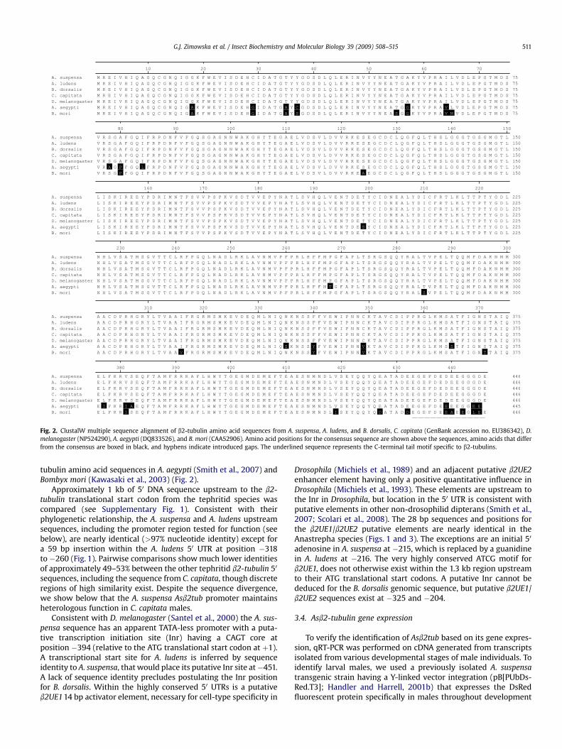
Fig. 2. ClustalW multiple sequence alignment of b2-tubulin amino acid sequences from A. suspensa, A. ludens, and B. dorsalis, C. capitata (GenBank accession no. EU386342), D.melanogaster (NP524290), A. aegypti (DQ833526), and B. mori (CAA52906). Amino acid positions for the consensus sequence are shown above the sequences, amino acids that differfrom the consensus are boxed in black, and hyphens indicate introduced gaps. The underlined sequence represents the C-terminal tail motif specific to b2-tubulins.
G.J. Zimowska et al. / Insect Biochemistry and Molecular Biology 39 (2009) 508–515 511
tubulin amino acid sequences in A. aegypti (Smith et al., 2007) andBombyx mori (Kawasaki et al., 2003) (Fig. 2).
Approximately 1 kb of 50 DNA sequence upstream to the b2-tubulin translational start codon from the tephritid species wascompared (see Supplementary Fig. 1). Consistent with theirphylogenetic relationship, the A. suspensa and A. ludens upstreamsequences, including the promoter region tested for function (seebelow), are nearly identical (>97% nucleotide identity) except fora 59 bp insertion within the A. ludens 50 UTR at position �318to �260 (Fig. 1). Pairwise comparisons show much lower identitiesof approximately 49–53% between the other tephritid b2-tubulin 50
sequences, including the sequence from C. capitata, though discreteregions of high similarity exist. Despite the sequence divergence,we show below that the A. suspensa Asb2tub promoter maintainsheterologous function in C. capitata males.
Consistent with D. melanogaster (Santel et al., 2000) the A. sus-pensa sequence has an apparent TATA-less promoter with a puta-tive transcription initiation site (Inr) having a CAGT core atposition �394 (relative to the ATG translational start codon at þ1).A transcriptional start site for A. ludens is inferred by sequenceidentity to A. suspensa, that would place its putative Inr site at�451.A lack of sequence identity precludes postulating the Inr positionfor B. dorsalis. Within the highly conserved 50 UTRs is a putativeb2UE1 14 bp activator element, necessary for cell-type specificity in
Drosophila (Michiels et al., 1989) and an adjacent putative b2UE2enhancer element having only a positive quantitative influence inDrosophila (Michiels et al., 1993). These elements are upstream tothe Inr in Drosophila, but location in the 50 UTR is consistent withputative elements in other non-drosophilid dipterans (Smith et al.,2007; Scolari et al., 2008). The 28 bp sequences and positions forthe b2UE1/b2UE2 putative elements are nearly identical in theAnastrepha species (Figs. 1 and 3). The exceptions are an initial 50
adenosine in A. suspensa at �215, which is replaced by a guanidinein A. ludens at �216. The very highly conserved ATCG motif forb2UE1, does not otherwise exist within the 1.3 kb region upstreamto their ATG translational start codons. A putative Inr cannot bededuced for the B. dorsalis genomic sequence, but putative b2UE1/b2UE2 sequences exist at �325 and �204.
3.4. Asb2-tubulin gene expression
To verify the identification of Asb2tub based on its gene expres-sion, qRT-PCR was performed on cDNA generated from transcriptsisolated from various developmental stages of male individuals. Toidentify larval males, we used a previously isolated A. suspensatransgenic strain having a Y-linked vector integration (pB[PUbDs-Red.T3]; Handler and Harrell, 2001b) that expresses the DsRedfluorescent protein specifically in males throughout development
β2UE1 β2UE2 position
Dm β2t ATCGT----AGTAGCCTATTTGTGAACATTC -216As β2t ATCGTCTGCACTAG-AAATTT-T-AATTTTC
Dm β2t ATCGT----AGTAGCCTATTTGTGAACATTC -215Al β2t GTCGTCTGCACTAG-AAATTT-T-AATTTTC
Dm β2t ATCGTAGTAGC-CTATTTGTGAACATTC -325Bd β2t _1 ATCGTTTTTTCAGAATTTTTTCACA--C
Dm β2t ATCGTAGTAGCCTATTTGTGAACAT-TC -204Bd β2t _2 ATCGAA--AGTGTA-CTAAGAAAACGTG
Fig. 3. Sequence alignments showing putative b2UE1/b2UE2 sequences in the 50 UTRof the tephritid b2-tubulin genes (A. suspensa, As b2t; A. ludens, Al b2t; and B. dorsalis,Bd b2t) based on similarity to the known sequences in D. melanogaster. Upstream 1.0 kbsequences for each tephritid gene were individually aligned to the Drosophila b2UE1/b2UE2 sequence (Dm b2t), with the highest identity sequences shown. Tephritidsequence positions are for the 50 nucleotide relative to the upstream ATG translationstart site (þ1). Hyphens indicate introduced gaps and asterisks indicate identities.
1
3
5
7
9
11
13
E 2L E3L L3L EP LP M3d M8d
Relative m
RN
A accu
mu
latio
n
1
51
101
151
201
251
301
351
401
451
501
F4B F F4B C F4B T F2B T M5A T
Relative m
RN
A accu
mu
latio
n
A
B
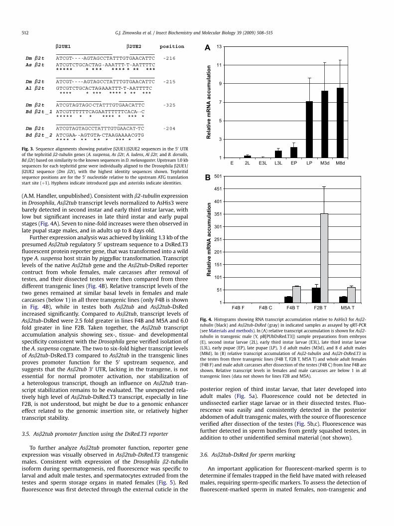
Fig. 4. Histograms showing RNA transcript accumulation relative to AsHis3 for Asb2-tubulin (black) and Asb2tub-DsRed (gray) in indicated samples as assayed by qRT-PCR(see Materials and methods). In (A) relative transcript accumulation is shown for Asb2-tubulin in transgenic male (Y, pB[PUbDsRed.T3]) sample preparations from embryos(E), second instar larvae (2L), early third instar larvae (E3L), late third instar larvae(L3L), early pupae (EP), late pupae (LP), 3 d adult males (M3d), and 8 d adult males(M8d). In (B) relative transcript accumulation of Asb2-tubulin and Asb2t-DsRed.T3 inthe testes from three transgenic lines (F4B T, F2B T, M5A T) and whole adult females(F4B F) and male adult carcasses after dissection of the testes (F4B C) from line F4B areshown. Relative transcript levels in females and male carcasses are below 1 in alltransgenic lines (data not shown for lines F2B and M5A).
G.J. Zimowska et al. / Insect Biochemistry and Molecular Biology 39 (2009) 508–515512
(A.M. Handler, unpublished). Consistent with b2-tubulin expressionin Drosophila, Asb2tub transcript levels normalized to AsHis3 werebarely detected in second instar and early third instar larvae, withlow but significant increases in late third instar and early pupalstages (Fig. 4A). Seven to nine-fold increases were then observed inlate pupal stage males, and in adults up to 8 days old.
Further expression analysis was achieved by linking 1.3 kb of thepresumed Asb2tub regulatory 50 upstream sequence to a DsRed.T3fluorescent protein reporter gene, that was transformed into a wildtype A. suspensa host strain by piggyBac transformation. Transcriptlevels of the native Asb2tub gene and the Asb2tub-DsRed reportercontruct from whole females, male carcasses after removal oftestes, and their dissected testes were then compared from threedifferent transgenic lines (Fig. 4B). Relative transcript levels of thetwo genes remained at similar basal levels in females and malecarcasses (below 1) in all three transgenic lines (only F4B is shownin Fig. 4B), while in testes both Asb2tub and Asb2tub-DsRedincreased significantly. Compared to Asb2tub, transcript levels ofAsb2tub-DsRed were 2.5 fold greater in lines F4B and M5A and 6.0fold greater in line F2B. Taken together, the Asb2tub transcriptaccumulation analysis showing sex-, tissue- and developmentalspecificity consistent with the Drosophila gene verified isolation ofthe A. suspensa cognate. The two to six-fold higher transcript levelsof Asb2tub-DsRed.T3 compared to Asb2tub in the transgenic linesproves promoter function for the 50 upstream sequence, andsuggests that the Asb2tub 30 UTR, lacking in the transgene, is notessential for normal promoter activation, nor stabilization ofa heterologous transcript, though an influence on Asb2tub tran-script stabilization remains to be evaluated. The unexpected rela-tively high level of Asb2tub-DsRed.T3 transcript, especially in lineF2B, is not understood, but might be due to a genomic enhancereffect related to the genomic insertion site, or relatively highertranscript stability.
3.5. Asb2tub promoter function using the DsRed.T3 reporter
To further analyze Asb2tub promoter function, reporter geneexpression was visually observed in Asb2tub-DsRed.T3 transgenicmales. Consistent with expression of the Drosophila b2-tubulinisoform during spermatogenesis, red fluorescence was specific tolarval and adult male testes, and spermatocytes extruded from thetestes and sperm storage organs in mated females (Fig. 5). Redfluorescence was first detected through the external cuticle in the
posterior region of third instar larvae, that later developed intoadult males (Fig. 5a). Fluorescence could not be detected inundissected earlier stage larvae or in their dissected testes. Fluo-rescence was easily and consistently detected in the posteriorabdomen of adult transgenic males, with the source of fluorescenceverified after dissection of the testes (Fig. 5b,c). Fluorescence wasfurther detected in sperm bundles from gently squashed testes, inaddition to other unidentified seminal material (not shown).
3.6. Asb2tub-DsRed for sperm marking
An important application for fluorescent-marked sperm is todetermine if females trapped in the field have mated with releasedmales, requiring sperm-specific markers. To assess the detection offluorescent-marked sperm in mated females, non-transgenic and

Fig. 5. Epifluorescent and brightfield images of testes from males transformed with pB[XLPUbEGFP/Asb2tub-DsRed.T3], and seminal material from spermathecae dissected fromnon-transgenic females mated to non-transgenic or transgenic males. The posterior region of a transgenic A. suspensa third instar larval male (a) showing epidermal EGFP fluo-rescence (left) and testis-specific DsRed fluorescence (right); (b) testes from a non-transgenic adult male (left) and a transgenic male (right) under brightfield, and (c) DsRedepifluorescence showing fluorescence limited to the transgenic testes; (d) seminal material extruded from a gently squashed spermatheca from a non-transgenic female mated toa non-transgenic male under brightfield (left), DAPI staining (center), and DsRed epifluorescence (right) showing a lack of fluorescence in sperm detected by DAPI; (e) seminalmaterial extruded from a spermatheca from a non-transgenic female mated to a transgenic male under brightfield with a region of exudate and sperm bundles (circled) observed athigher magnification (400�) in the right-hand panel (arrow); (f) the same images as in (e), but under DsRed epifluorescence showing fluorescence specific to the seminal materialexudate, and sperm bundles under 400� magnification in the right-hand panel; and (g) posterior/ventral view of a C. capitata adult male abdomen transformed with the Asb2tub-DsRed.T3 transgene under DsRed epifluorescence showing testis-specific fluorescence (verified after dissection; data not shown).
G.J. Zimowska et al. / Insect Biochemistry and Molecular Biology 39 (2009) 508–515 513
transgenic males were crossed to non-transgenic virgin femalesthat were then inspected by dissection after mating. In control testsof non-transgenic males mated to non-transgenic females, spermextruded from the spermathecae could be detected by DAPIstaining, but these did not exhibit DsRed fluorescence (Fig. 5d).When transgenic males were mated, however, red fluorescentsperm could be detected in the spermathecal duct (not shown), aswell as in sperm bundles and seminal material extruded fromspermathecae after gentle squashing (Fig. 5e,f). The spermathecaethemselves are highly autofluorescent from cuticular material, andthe presence of sperm could not be determined without extrusion.Heterologous function for the Asb2tub promoter was demonstratedby the strong expression of the DsRed.T3 reporter in the testes of C.capitata males transformed with the Asb2tub-DsRed.T3 reporterconstruct (Fig. 5g).
Since the spermathecae and stored sperm may degrade intrapped flies that have died, sperm-specific fluorescence may notbe detectable or distinguished from autofluorescence due tonecrosis. Thus a PCR assay for the Asb2tub-DsRed.T3 transgene wasdeveloped using primers AH313 and AH358, specific to the Asb2tubpromoter and DsRed.T3 sequences, respectively, which yield an850 bp product. Wild type virgin females were mated to malesheterozygous for the Asb2tub-DsRed.T3 transgene, with genomicDNA then extracted from individual abdomens of the females. PCRfor the Asb2tub-DsRed.T3 transgene yielded varying amounts of the850 bp product from each abdominal sample, though its presencewas unambiguous and not observable in negative controls (Fig. 6).
4. Discussion
The testis-specific b2-tubulin gene and its 50 upstream regula-tory sequence region have been isolated and sequenced fromthree economically important tephritid fruit fly species, and theconstitutive b1-tubulin gene has been isolated from A. suspensa.
Consistent with other b2-tubulin genes, there is a high level ofamino acid conservation among the tephritid species and withother insects, as well as some nucleotide sequence conservation inthe promoter region among the tephritids. Conservation of theamino acid sequence is quite strong, with identical polypeptides inall tephritid species, as well as D. melanogaster. Thus, the high b2-tubulin protein conservation originally observed among droso-philids extends to more distantly related dipterans (Michiels et al.,1987). Yet this does not extend to mosquitoes such as A. aegyptiwhose b2-tubulin (Smith et al., 2007) is 95% identical, and hasa greater similarity to the sequence from B. mori (Kawasaki et al.,2003).
Putative b2UE1/b2UE2 elements, shown to confer cell-typespecificity in Drosophila (Michiels et al., 1989, 1993), have beenidentified in the three tephritid species. These sequences all residein the 50 UTR regions, which is consistent for putative b2UE1/b2UE2elements identified in other dipteran species. An AT-rich b2DE1element, shown to confer b2-tubulin transcript stabilization inDrosophila (Santel et al., 2000), was not identified in any of tephritidgenes, though AT-rich regions do exist within the 50 UTRs.
For A. suspensa, the b2-tubulin gene was identified and charac-terized by the sex, stage and tissue specificities of its transcriptionusing qRT-PCR, which was limited to the testes in late larvae to adultmales. This is consistent with the cell-type specificity and timing ofthe switch from the b1-tubulin to the b2-tubulin isoform that occursin early spermatocytes in third larval instar Drosophila (Buttgereitand Renkawitz-Pohl, 1993; Hoyle et al., 1995). The specificity of theAsb2tub promoter function was further verified by linking it toa fluorescent-protein reporter, which showed testis fluorescencespecifically in the spermatocytes in transgenic males. The strongexpression of the reporter gene, lacking the Asb2tub 30 UTR, indi-cated a minimal if any influence of this domain on DsRed transcriptexpression or stabilization, which differs from its presumed role forAsb2tub transcript in Drosophila (Hoyle et al., 1995).
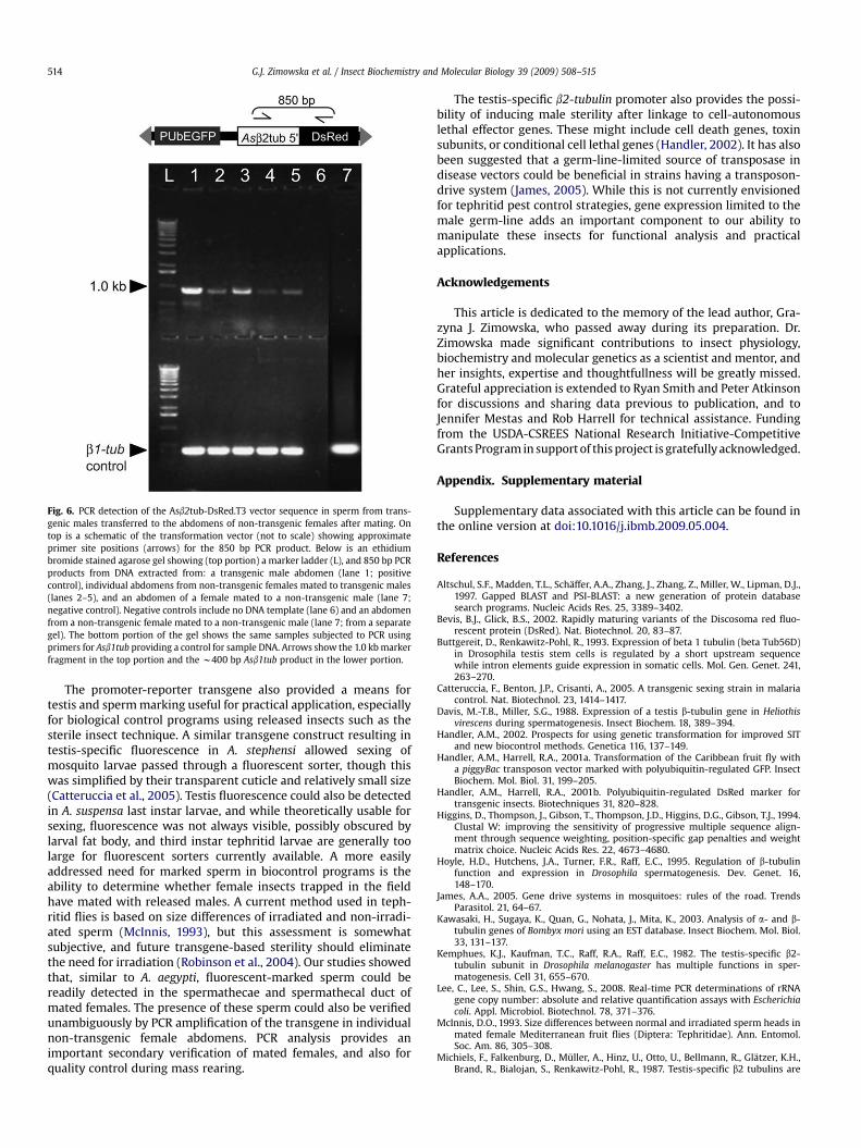
Fig. 6. PCR detection of the Asb2tub-DsRed.T3 vector sequence in sperm from trans-genic males transferred to the abdomens of non-transgenic females after mating. Ontop is a schematic of the transformation vector (not to scale) showing approximateprimer site positions (arrows) for the 850 bp PCR product. Below is an ethidiumbromide stained agarose gel showing (top portion) a marker ladder (L), and 850 bp PCRproducts from DNA extracted from: a transgenic male abdomen (lane 1; positivecontrol), individual abdomens from non-transgenic females mated to transgenic males(lanes 2–5), and an abdomen of a female mated to a non-transgenic male (lane 7;negative control). Negative controls include no DNA template (lane 6) and an abdomenfrom a non-transgenic female mated to a non-transgenic male (lane 7; from a separategel). The bottom portion of the gel shows the same samples subjected to PCR usingprimers for Asb1tub providing a control for sample DNA. Arrows show the 1.0 kb markerfragment in the top portion and the w400 bp Asb1tub product in the lower portion.
G.J. Zimowska et al. / Insect Biochemistry and Molecular Biology 39 (2009) 508–515514
The promoter-reporter transgene also provided a means fortestis and sperm marking useful for practical application, especiallyfor biological control programs using released insects such as thesterile insect technique. A similar transgene construct resulting intestis-specific fluorescence in A. stephensi allowed sexing ofmosquito larvae passed through a fluorescent sorter, though thiswas simplified by their transparent cuticle and relatively small size(Catteruccia et al., 2005). Testis fluorescence could also be detectedin A. suspensa last instar larvae, and while theoretically usable forsexing, fluorescence was not always visible, possibly obscured bylarval fat body, and third instar tephritid larvae are generally toolarge for fluorescent sorters currently available. A more easilyaddressed need for marked sperm in biocontrol programs is theability to determine whether female insects trapped in the fieldhave mated with released males. A current method used in teph-ritid flies is based on size differences of irradiated and non-irradi-ated sperm (McInnis, 1993), but this assessment is somewhatsubjective, and future transgene-based sterility should eliminatethe need for irradiation (Robinson et al., 2004). Our studies showedthat, similar to A. aegypti, fluorescent-marked sperm could bereadily detected in the spermathecae and spermathecal duct ofmated females. The presence of these sperm could also be verifiedunambiguously by PCR amplification of the transgene in individualnon-transgenic female abdomens. PCR analysis provides animportant secondary verification of mated females, and also forquality control during mass rearing.
The testis-specific b2-tubulin promoter also provides the possi-bility of inducing male sterility after linkage to cell-autonomouslethal effector genes. These might include cell death genes, toxinsubunits, or conditional cell lethal genes (Handler, 2002). It has alsobeen suggested that a germ-line-limited source of transposase indisease vectors could be beneficial in strains having a transposon-drive system (James, 2005). While this is not currently envisionedfor tephritid pest control strategies, gene expression limited to themale germ-line adds an important component to our ability tomanipulate these insects for functional analysis and practicalapplications.
Acknowledgements
This article is dedicated to the memory of the lead author, Gra-zyna J. Zimowska, who passed away during its preparation. Dr.Zimowska made significant contributions to insect physiology,biochemistry and molecular genetics as a scientist and mentor, andher insights, expertise and thoughtfullness will be greatly missed.Grateful appreciation is extended to Ryan Smith and Peter Atkinsonfor discussions and sharing data previous to publication, and toJennifer Mestas and Rob Harrell for technical assistance. Fundingfrom the USDA-CSREES National Research Initiative-CompetitiveGrants Program in support of this project is gratefully acknowledged.
Appendix. Supplementary material
Supplementary data associated with this article can be found inthe online version at doi:10.1016/j.ibmb.2009.05.004.
References
Altschul, S.F., Madden, T.L., Schaffer, A.A., Zhang, J., Zhang, Z., Miller, W., Lipman, D.J.,1997. Gapped BLAST and PSI-BLAST: a new generation of protein databasesearch programs. Nucleic Acids Res. 25, 3389–3402.
Bevis, B.J., Glick, B.S., 2002. Rapidly maturing variants of the Discosoma red fluo-rescent protein (DsRed). Nat. Biotechnol. 20, 83–87.
Buttgereit, D., Renkawitz-Pohl, R., 1993. Expression of beta 1 tubulin (beta Tub56D)in Drosophila testis stem cells is regulated by a short upstream sequencewhile intron elements guide expression in somatic cells. Mol. Gen. Genet. 241,263–270.
Catteruccia, F., Benton, J.P., Crisanti, A., 2005. A transgenic sexing strain in malariacontrol. Nat. Biotechnol. 23, 1414–1417.
Davis, M.-T.B., Miller, S.G., 1988. Expression of a testis b-tubulin gene in Heliothisvirescens during spermatogenesis. Insect Biochem. 18, 389–394.
Handler, A.M., 2002. Prospects for using genetic transformation for improved SITand new biocontrol methods. Genetica 116, 137–149.
Handler, A.M., Harrell, R.A., 2001a. Transformation of the Caribbean fruit fly witha piggyBac transposon vector marked with polyubiquitin-regulated GFP. InsectBiochem. Mol. Biol. 31, 199–205.
Handler, A.M., Harrell, R.A., 2001b. Polyubiquitin-regulated DsRed marker fortransgenic insects. Biotechniques 31, 820–828.
Higgins, D., Thompson, J., Gibson, T., Thompson, J.D., Higgins, D.G., Gibson, T.J., 1994.Clustal W: improving the sensitivity of progressive multiple sequence align-ment through sequence weighting, position-specific gap penalties and weightmatrix choice. Nucleic Acids Res. 22, 4673–4680.
Hoyle, H.D., Hutchens, J.A., Turner, F.R., Raff, E.C., 1995. Regulation of b-tubulinfunction and expression in Drosophila spermatogenesis. Dev. Genet. 16,148–170.
James, A.A., 2005. Gene drive systems in mosquitoes: rules of the road. TrendsParasitol. 21, 64–67.
Kawasaki, H., Sugaya, K., Quan, G., Nohata, J., Mita, K., 2003. Analysis of a- and b-tubulin genes of Bombyx mori using an EST database. Insect Biochem. Mol. Biol.33, 131–137.
Kemphues, K.J., Kaufman, T.C., Raff, R.A., Raff, E.C., 1982. The testis-specific b2-tubulin subunit in Drosophila melanogaster has multiple functions in sper-matogenesis. Cell 31, 655–670.
Lee, C., Lee, S., Shin, G.S., Hwang, S., 2008. Real-time PCR determinations of rRNAgene copy number: absolute and relative quantification assays with Escherichiacoli. Appl. Microbiol. Biotechnol. 78, 371–376.
McInnis, D.O., 1993. Size differences between normal and irradiated sperm heads inmated female Mediterranean fruit flies (Diptera: Tephritidae). Ann. Entomol.Soc. Am. 86, 305–308.
Michiels, F., Falkenburg, D., Muller, A., Hinz, U., Otto, U., Bellmann, R., Glatzer, K.H.,Brand, R., Bialojan, S., Renkawitz-Pohl, R., 1987. Testis-specific b2 tubulins are

G.J. Zimowska et al. / Insect Biochemistry and Molecular Biology 39 (2009) 508–515 515
identical in Drosophila melanogaster and D. hydei but differ from the ubiquitousb1 tubulin. Chromosoma 95, 387–395.
Michiels, F., Gasch, A., Kaltschmidt, B., Renkawitz-Pohl, R., 1989. A 14 bp promoterelement directs the testis specificity of the Drosophila b2 tubulin gene. EMBO J.8, 1559–1565.
Michiels, F., Buttgereit, D., Renkawitz-Pohl, R., 1993. An 18-bp element in the 50
untranslated region of the Drosophila b2 tubulin mRNA regulates the mRNAlevel postmeiotic stages of spermatogenesis. Eur. J. Cell Biol. 62, 66–74.
Nirmala, X., Zimowska, G.J., Handler, A.M., 2009. Characterization of the proteasomeb2 subunit gene and its mutant allele in the tephritid fruit fly pest, Anastrephasuspensa. Insect Mol. Biol. 18, 333–340.
Pfaffl, M.W., 2001. A new mathematical model for relative quantification in real-time RT-PCR. Nucleic Acids Res. 29, 2005–2007.
Popodi, E.M., Hoyle, H.D., Turner, F.R., Xu, K., Kruse, S., Raff, E.C., 2008. Axonemespecialization embedded in a ‘‘generalist’’ beta-tubulin. Cell Motil. Cytoskeleton65, 216–237.
Raff, E.C., Hoyle, H.D., Popodi, E.M., Turner, F.R., 2008. Axoneme beta-tubulinsequence determines attachment of outer dynein arms. Curr. Biol. 18, 911–914.
Ramakers, C., Ruijter, J.M., Lekanne Deprez, R.H., Moorman, A.F.M., 2003. Assump-tion-free analysis of quantitative real-time polymerase chain reaction (PCR)data. Neurosci. Lett. 339, 62–66.
Robinson, A.S., Franz, G., Atkinson, P.W., 2004. Insect transgenesis and itspotential role in agriculture and human health. Insect Biochem. Mol. Biol. 34,113–120.
Rudolph, J.E., Kimble, M., Hoyle, H.D., Subler, M.A., Raff, E.C., 1987. ThreeDrosophila beta-tubulin sequences: a developmentally regulated isoform (b3),the testis-specific isoform (b2), and an assembly-defective mutation of thetestis-specific (B2t8) reveal both an ancient divergence in metazoan isotypesand structural constraints for beta-tubulin function. Mol. Cell Biol. 7,2231–2242.
Santel, A., Kaufmann, J., Hyland, R., Renkawitz-Pohl, R., 2000. The initiator elementof the Drosophila b2 tubulin gene core promoter contributes to gene expressionin vivo but is not required for male germ-cell specific expression. Nucleic AcidsRes. 28, 1439–1446.
Scolari, F., Schetelig, M.F., Bertin, S., Malacrida, A.R., Gasperi, G., Wimmer, E.A.,2008. Fluorescent sperm marking to improve the fight against the pest insectCeratitis capitata (Wiedemann; Diptera: Tephritidae). New Biotechnol. 25,76–84.
Smith, R.C., Walter, M.F., Hice, R.H., O’Brochta, D.A., Atkinson, P.W., 2007.Testis-specific expression of the b2 tubulin promoter of Aedes aegypti and itsapplication as a genetic sex-separation marker. Insect Mol. Biol. 16,61–71.