
Innovations in studying in vivo cell behavior and pharmacology in complex tissues – microvascular...
23
REVIEW Innovations in studying in vivo cell behavior and pharmacology in complex tissues – microvascular endothelial cells in the spotlight Elise Langenkamp & Jan A. A. M. Kamps & Michal Mrug & Elisabeth Verpoorte & Yilmaz Niyaz & Peter Horvatovich & Rainer Bischoff & Harry Struijker-Boudier & Grietje Molema Received: 15 May 2013 /Accepted: 18 July 2013 /Published online: 27 September 2013 # Springer-Verlag Berlin Heidelberg 2013 Abstract Many studies on the molecular control underlying normal cell behavior and cellular responses to disease stimuli and pharmacological intervention are conducted in single-cell culture systems, while the read-out of cellular engagement in disease and responsiveness to drugs in vivo is often based on overall tissue responses. As the majority of drugs under de- velopment aim to specifically interact with molecular targets in subsets of cells in complex tissues, this approach poses a major experimental discrepancy that prevents successful de- velopment of new therapeutics. In this review, we address the shortcomings of the use of artificial (single) cell systems and of whole tissue analyses in creating a better understanding of cell engagement in disease and of the true effects of drugs. We focus on microvascular endothelial cells that actively engage in a wide range of physiological and pathological processes. We propose a new strategy in which in vivo molecular control of cells is studied directly in the diseased endothelium instead of at a (far) distance from the site where drugs have to act, thereby accounting for tissue-controlled cell responses. The strategy uses laser microdissection-based enrichment of mi- crovascular endothelium which, when combined with transcriptome and (phospho)proteome analyses, provides a factual view on their status in their complex microenviron- ment. Combining this with miniaturized sample handling using microfluidic devices enables handling the minute sam- ple input that results from this strategy. The multidisciplinary approach proposed will enable compartmentalized analysis of cell behavior and drug effects in complex tissue to become widely implemented in daily biomedical research and drug development practice. Keywords (Endothelial) cell behavior . Pharmacology . In vivo . Laser microdissection . Omics technology E. Langenkamp : J. A. A. M. Kamps : G. Molema University Medical Center Groningen, Department of Pathology and Medical Biology, Medical Biology section, University of Groningen, Groningen, The Netherlands M. Mrug Division of Nephrology, Department of Medicine, Nephrology Research and Training Center, University of Alabama at Birmingham, Birmingham, AL, USA M. Mrug Department of Veterans Affairs Medical Center, Birmingham, AL, USA E. Verpoorte Groningen Research Institute of Pharmacy, Pharmaceutical Analysis, University of Groningen, Groningen, The Netherlands Y. Niyaz Carl Zeiss Microscopy GmbH, Jena, Germany P. Horvatovich : R. Bischoff Groningen Research Institute of Pharmacy, Analytical Biochemistry, University of Groningen, Groningen, The Netherlands H. Struijker-Boudier Department of Pharmacology and Toxicology, Maastricht University, Maastricht, The Netherlands G. Molema (*) University Medical Center Groningen, Hanzeplein 1, Groningen NL-9713 GZ, The Netherlands e-mail: [email protected] Present Address: E. Langenkamp Department of Immunology, Genetics and Pathology, Uppsala University, Uppsala, Sweden Cell Tissue Res (2013) 354:647–669 DOI 10.1007/s00441-013-1714-7
Transcript of Innovations in studying in vivo cell behavior and pharmacology in complex tissues – microvascular...
REVIEW
Innovations in studying in vivo cell behaviorand pharmacology in complex tissues – microvascularendothelial cells in the spotlight
Elise Langenkamp & Jan A. A. M. Kamps & Michal Mrug &
Elisabeth Verpoorte & Yilmaz Niyaz & Peter Horvatovich &
Rainer Bischoff & Harry Struijker-Boudier & Grietje Molema
Received: 15 May 2013 /Accepted: 18 July 2013 /Published online: 27 September 2013# Springer-Verlag Berlin Heidelberg 2013
Abstract Many studies on the molecular control underlyingnormal cell behavior and cellular responses to disease stimuliand pharmacological intervention are conducted in single-cellculture systems, while the read-out of cellular engagement indisease and responsiveness to drugs in vivo is often based onoverall tissue responses. As the majority of drugs under de-velopment aim to specifically interact with molecular targetsin subsets of cells in complex tissues, this approach poses amajor experimental discrepancy that prevents successful de-velopment of new therapeutics. In this review, we address theshortcomings of the use of artificial (single) cell systems andof whole tissue analyses in creating a better understanding ofcell engagement in disease and of the true effects of drugs. Wefocus on microvascular endothelial cells that actively engagein a wide range of physiological and pathological processes.We propose a new strategy in which in vivo molecular controlof cells is studied directly in the diseased endothelium instead
of at a (far) distance from the site where drugs have to act,thereby accounting for tissue-controlled cell responses. Thestrategy uses laser microdissection-based enrichment of mi-crovascular endothelium which, when combined withtranscriptome and (phospho)proteome analyses, provides afactual view on their status in their complex microenviron-ment. Combining this with miniaturized sample handlingusing microfluidic devices enables handling the minute sam-ple input that results from this strategy. The multidisciplinaryapproach proposed will enable compartmentalized analysis ofcell behavior and drug effects in complex tissue to becomewidely implemented in daily biomedical research and drugdevelopment practice.
Keywords (Endothelial) cell behavior . Pharmacology . Invivo . Laser microdissection . Omics technology
E. Langenkamp : J. A. A. M. Kamps :G. MolemaUniversity Medical Center Groningen, Department of Pathology andMedical Biology, Medical Biology section, University of Groningen,Groningen, The Netherlands
M. MrugDivision of Nephrology, Department of Medicine,Nephrology Research and Training Center,University of Alabama at Birmingham, Birmingham, AL, USA
M. MrugDepartment of Veterans Affairs Medical Center, Birmingham, AL,USA
E. VerpoorteGroningen Research Institute of Pharmacy, Pharmaceutical Analysis,University of Groningen, Groningen, The Netherlands
Y. NiyazCarl Zeiss Microscopy GmbH, Jena, Germany
P. Horvatovich :R. BischoffGroningen Research Institute of Pharmacy,Analytical Biochemistry, University of Groningen,Groningen, The Netherlands
H. Struijker-BoudierDepartment of Pharmacology and Toxicology,Maastricht University,Maastricht, The Netherlands
G. Molema (*)University Medical Center Groningen, Hanzeplein 1,Groningen NL-9713 GZ, The Netherlandse-mail: [email protected]
Present Address:E. LangenkampDepartment of Immunology, Genetics and Pathology,Uppsala University, Uppsala, Sweden
Cell Tissue Res (2013) 354:647–669DOI 10.1007/s00441-013-1714-7

Abbreviations
ACE Angiotensin converting enzymeALK Activin receptor-like KinaseBAD B-cell lymphoma 2-associated death
promoterBBB Blood–brain barrierBcl2 B-cell lymphoma 2COX CyclooxygenaseECM Extracellular matrixERK Extracellular signal-regulated
kinaseFGF Fibroblast growth factorFT-MS Fourier transform mass spectrometryHIF Hypoxia-inducible factorHPLC High performance liquid
chromatographyICAM Intercellular adhesion moleculeIL-1 Interleukin-1LDA Low-density arrayLDH Lactase dehydrogenaseLFA Leukocyte function antigenlncRNA Long non-coding RNALoaC Lab on a chipLPS LipopolysaccharideMALDI-TOF (MS) Matrix-assisted laser desorption/
ionization-time of flight (massspectometry)
MAPK Mitogen activated protein kinaseNFκB Nuclear factor κ-BNGS Next generation sequencingNOS Nitric oxide synthasePAGE Polyacrylamide gel electrophoresisPDGF Platelet-derived growth factorPI3K Phosphoinositide 3-kinasePKB Protein kinase BPKC Protein kinase CPV-1 Plasmalemma vesicle-associated
protein-1RCC Renal cell carcinomaRT-PCR Reverse transcriptase-polymerase
chain reactionSELDI-TOF (MS) Surface-enhanced laser desorption/
ionization-time of flight (massspectometry)
SRM Single reaction monitoringTGF Transforming growth factorTNF(R) Tumor necrosis factor (receptor)tPA Tissue-type plasminogen activatoruPA Urokinase-type plasminogen
activatorVCAM Vascular cell adhesion moleculeVE-cadherin Vascular endothelial cadherin
VEGF(R) Vascular endothelial growth factor(receptor)
VE-PTP Vascular endothelial protein tyrosinephosphatase
VLA Very late antigenVVO Vesiculo-vacuolar organelleZO Zonal occludens
Introduction
Lack of efficacy and the occurrence of uncontrollable toxicityare major hurdles for the introduction of new drugs into dailyclinical practice. While lack of efficacy in patients accountsfor 30 % of attrition, toxicity makes up for an additional 20 %(Kola and Landis 2004). An early example of the former is themulti-million dollar trials on restenosis prevention followingpercutaneous transluminal coronary angioplasty using theangiotensin converting enzyme (ACE) inhibitor cilazapril.The drug failed to show any effect (MERCATOR study group1992; Faxon 1995) despite successful studies of neointimaformation prevention in rat models of arterial injury (Powellet al. 1989). A more recent example is the significant inhibitionof tumor outgrowth in pre-clinical xenograft and syngeneicanimal models using angiogenesis inhibitors, while the major-ity of clinical studies with anti-angiogenic drugs reported so farshow limited responses (Fury et al. 2007; Morabito et al. 2006;Sitohy et al. 2012). The unexpected doubling in heart attackand stroke rate in patients treated with COX-2 inhibitors lead-ing to drug withdrawal from the market in 2004 (Ortiz 2004),the increased risk of death associated with the use of the good-cholesterol-inducing-drug torcetrapib (Barter et al. 2007) andthe development of heart failure in renal cell and stomachcancer patients treated with the anti-angiogenic drug sunitinibreported in 2008 (Schmidinger et al. 2008) are more recentexamples of drug failures due to unforeseen drug toxicity.
From this, one has to conclude that our understanding ofcell behavior, endothelial or otherwise and drug effects onspecific cell types in the human body is inadequate and thatextrapolation of data obtained in cell culture to pre-clinicalanimal models and from animal models to the patient, is oflimited value. Reasons for the discrepancies between findingsin pre-clinical studies and clinical reality include (1) the use ofdifferent doses of drugs in cell and animal experiments, (2)differences in drug transport and metabolism between species,(3) the timing of drug administration in relation to the stage ofdisease, (4) poor validity of animal disease models in relationto human pathology and (5) lack of knowledge of the molec-ular control of disease and cellular responses to drug treatmentin complex in vivo systems. While the first two issues havebeen extensively addressed as part of the drug developmentprocess (Martignoni et al. 2006; van Montfoort et al. 2003),
648 Cell Tissue Res (2013) 354:647–669
the latter issues are muchmore difficult to tackle. They should,however, not be neglected, as they can lead to assignment ofsurrogate biomarkers for disease activity and to molecular andcellular models of drug efficacy and toxicity that have limitedvalue for the clinic.
In recent years, it has become increasingly clear that thebehavior of cells within an organ is intricately controlled bythe microenvironment. Biological factors, including extracel-lular matrix components, locally produced growth factors,interactions with neighboring cells and mechanical forces,influence general cell performance. For example, when inmice an external jugular vein is connected to the commoncarotid artery—representative of the interposition of a venoussegment into the arterial circulation as occurs during coronaryartery bypass surgery—endothelial cells covering the venularwall adapt to the change in the microenvironment and biome-chanics by phenotypically shifting toward arterial endothelialbehavior (Kwei et al. 2004). Furthermore, the microenviron-ment likely has important ramifications for the effects of drugson cells (Langenkamp and Molema 2009; Molema 2010a).
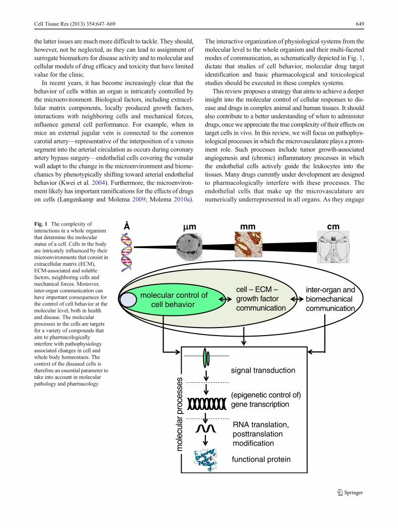
The interactive organization of physiological systems from themolecular level to the whole organism and their multi-facetedmodes of communication, as schematically depicted in Fig. 1,dictate that studies of cell behavior, molecular drug targetidentification and basic pharmacological and toxicologicalstudies should be executed in these complex systems.
This review proposes a strategy that aims to achieve a deeperinsight into the molecular control of cellular responses to dis-ease and drugs in complex animal and human tissues. It shouldalso contribute to a better understanding of when to administerdrugs, once we appreciate the true complexity of their effects ontarget cells in vivo. In this review, we will focus on pathophys-iological processes in which themicrovasculature plays a prom-inent role. Such processes include tumor growth-associatedangiogenesis and (chronic) inflammatory processes in whichthe endothelial cells actively guide the leukocytes into thetissues. Many drugs currently under development are designedto pharmacologically interfere with these processes. Theendothelial cells that make up the microvasculature arenumerically underrepresented in all organs. As they engage
Å m mm cm
molecular control ofcell behavior
cell – ECM –growth factor communication
inter-organ andbiomechanicalcommunication
signal transduction
(epigenetic control of)gene transcription
RNA translation,posttranslationmodification
functional protein
mol
ecul
ar p
roce
sses
Fig. 1 The complexity ofinteractions in a whole organismthat determine the molecularstatus of a cell. Cells in the bodyare intricately influenced by theirmicroenvironments that consist inextracellular matrix (ECM),ECM-associated and solublefactors, neighboring cells andmechanical forces. Moreover,inter-organ communication canhave important consequences forthe control of cell behavior at themolecular level, both in healthand disease. The molecularprocesses in the cells are targetsfor a variety of compounds thataim to pharmacologicallyinterfere with pathophysiologyassociated changes in cell andwhole body homeostasis. Thecontext of the diseased cells istherefore an essential parameter totake into account in molecularpathology and pharmacology
Cell Tissue Res (2013) 354:647–669 649
in disease processes by employing both endothelial-restricted and non-endothelial-restricted genes, analysis ofwhole organ samples will not reveal the endothelial contri-bution to the signal. In addition, these cells are functionallyand molecularly highly heterogenic depending on theirorgan niche and rapidly lose their in vivo features whencultured in vitro. These features dictate that endothelialbiomedicine be addressed in the complexity of the organsin which the diseases are present. Although we focus onmicrovascular endothelial cells, the concepts discussed inthis review are of similar importance for other cell typesinvolved in disease initiation and progression.
We start with a brief overview of what is known aboutendothelial cells in different microvascular beds in organs,their contribution to disease initiation and progression andthe types of drugs that are under development to interfere withendothelial engagement. Next, we describe the (experimental)hurdles that prevent effective translation of in vitro observa-tions to animal models and from animal models to the patientand put forward a new strategy to study microvascular endo-thelial cells in their complex microenvironment in vivo. Thetechnologies underlying this strategy encompass laser micro-dissection of cells of interest from tissue sections followed bytranscriptome and (phospho)proteome analysis. Technologi-cal advances and scientific progress in fields like proteomics,genetics and bioinformatics provide a foundation upon whichto build more sophisticated endothelial studies. Microfluidics-based miniaturization of sample handling will be addressed asan essential enabling technology when working with minuteamounts of biological materials. We discuss the first experi-mental studies that make use of this strategy and criticallyanalyze the technological challenges ahead before it can bewidely implemented in biomedical research and in the drugdevelopment process.
The vasculature and its function in the body
The vasculature consists of the large arteries and veins, thesmaller arterioles and venules and the smallest capillaries thatare situated in the organs. The vascular endothelium repre-sents a cell type that is common to all blood vessels andparticipates in a variety of physiological processes, includingthe development and remodeling of the vasculature, the con-trol of vascular tone and blood fluidity and the trafficking ofblood cells and nutrients (dela Paz and D'Amore 2009;Minami and Aird 2005). Endothelial cells differ widely inmorphology and function as one travels through the vasculartree, with each of the vascular segments fulfilling specificfunctions in the control of whole-body homeostasis and he-mostasis (Aird 2007a, b).
Arteries and veins both serve as conduit vessels, yet differin fundamental ways. The thicker walls, comprising multiplelayers of smooth muscle cells and elastic material and thetighter intercellular junctions in arteries of all calibers contrastwith the thinner walls, the absence of an internal elastic laminaand the loosely organized tight junctions in veins (dela Pazand D'Amore 2009; Aird 2007b; Simionescu et al. 1976,1975). The smaller arterioles control vasomotor tone, whilepostcapillary venules are the primary site of permeabilitycontrol and leukocyte recruitment. Postcapillary venule endo-thelium is rich in vesiculo-vacuolar organelles (VVOs), focalcollections of membrane-bound vesicles and vacuoles. Thisfeature together with lower flow rate, thinner walls and fewertight junctions, makes postcapillary venules most suited forpermeability control and leukocyte trafficking (dela Paz andD'Amore 2009; Aird 2007a, b).
Capillaries are the major exchange vessels in the circula-tion. The diameter of capillaries throughout the body is lessthan 10 μm and their wall is extraordinarily thin, thus mini-mizing the diffusional path length to and from cells in tissue(Aird 2007b). Also, transcytosis of plasma molecules occursprimarily at the level of capillaries, which is mediated bycaveolae and transendothelial channels and controlled inreceptor-dependent as well as receptor-independent ways(Simionescu et al. 2009). Capillaries are only supported bysparsely distributed pericytes that are located within the base-ment membrane and cover the endothelium with finger-likeprotrusions. The presence of pericytes is a prerequisite forvessel stability and proper vessel function (Gaengel et al.2009), yet the degree to which they cover the endotheliumvaries substantially from the capillary bed of one tissue to theother. Capillary endothelial cells are uniquely adapted to theunderlying tissue and the microvasculature in the major or-gans performing functions specific for each organ (Aird2007b).
Microvascular endothelial heterogeneity
Many of the pathological processes in the body are facilitatedby the capillaries and the first part of the postcapillary ve-nules. Their location in the organs dictates a substantialheterogeneity in function associated with the underlying mo-lecular heterogeneity. The brain microvasculature, for exam-ple, is an integral part of the blood–brain barrier (BBB)(Liebner et al. 2011), with capillary endothelial cells charac-terized by the presence of many tight junctions in addition toadherens junctions that protect the brain from fluctuations inblood composition. Endothelial cell-to-cell adhesion atadherens junctions is mainly mediated by vascular endothe-lial (VE)-cadherin. Tight junction proteins include claudin-5and occludin (Dejana 2004; Taddei et al. 2008), zonaloccludens (ZO)-1, ZO-2, ZO-3 and cingulin. Endothelial
650 Cell Tissue Res (2013) 354:647–669

cells in the brain are furthermore characterized by their flatpresentation, the small number of caveolae at the luminalsurface of the cell and the high number of mitochondria(Liebner et al. 2011).
In contrast to the tight and relatively impermeable brainvasculature, endothelial cells in specialized parts of the liverand kidney are characterized by fenestrations, transcellularpores that extend through the full thickness of the cell. Fen-estrations function as a size-selective filter for fluids, solutesand particles, thereby enabling passage of molecules to becleared from the body (Tse and Stan 2010). Liver sinusoidalendothelium is discontinuous and functions as a selectivesieve, with fenestrae of ∼175 nm in diameter that occupy 6–8 % of the surface (Wisse et al. 1996). The endothelium of therenal glomerular microcirculation is mostly continuous, withfenestrations that can reach up to 60 nm in diameter and cover20–50 % of the endothelial surface (Aird 2007b; Molema andAird 2012). While many capillaries in the different organsexpress plasmalemmal vesicle-associated protein (PV)-1, atype II transmembrane glycoprotein that is associated withbridging diaphragms, glomerular fenestrae lack the expressionof this molecule (Stan 2005; Ballermann and Stan 2007).Glomerular endothelial cells actively synthesize basementmembrane and, together with local podocytes and mesangialcells, they provide a filtration barrier with charge selectivity(Obeidat et al. 2012). In contrast, the endothelium of thedescending vasa recta is continuous and has no fenestrations.It expresses specific transporters for, e.g., urea (urea transport-er UT-B) and water (Aquaporin-1). Endothelial cells of theascending vasa recta, in contrast, have fenestrae containing adiaphragm with PV-1 and promote net movement of solutes(NaCl and urea) to the interstitium (Molema and Aird 2012;Aird 2007b).
The above represent only a few examples of what isknown about the different functionalities of the microvascularbeds in the body. Yet it clearly exemplifies the diversity invascular functions depending on the location of the micro-vasculature in the body. This diversity implies the existenceof an extensive variation in the molecular make-up of endo-thelial cells in the microvessels, the identity of which is stilllargely unknown.
Endothelial cell engagement in angiogenesis
The new formation of blood vessels during wound healingand solid tumor growth mainly takes place in the capillariesand postcapillary venules (Potente et al. 2011; Pober andSessa 2007). Although being studied mostly for its role intumor growth, angiogenesis is also a hallmark of ophthal-mological disorders, psoriasis and chronic inflammatorydiseases such as rheumatoid arthritis, atherosclerosis andinflammatory bowel disease. Angiogenesis involves the
activation, proliferation and migration of endothelial cells,assembly into vascular tubes and formation of a lumen,recruitment of vascular support cells, or pericytes and fi-nally perfusion of the newly formed vessel. The process ofangiogenesis is regulated by a large array of growth factors,adhesion molecules, proteases and signaling molecules, asschematically represented in Fig. 2.
One of the most extensively studied factors withangiogenesis-inducing capacity is vascular endothelial growthfactor (VEGF) (Dvorak 2006), which is a major target of anti-angiogenic tumor therapy to date. VEGF receptor (VEGFR)2belongs to the class of receptor tyrosine kinases and is theprincipal mediator of several physiological and pathologicaleffects of VEGF on endothelial cells. Binding of VEGF leadsto VEGFR2 dimerization, autophosphorylation of the receptorand subsequent activation of a multitude of intracellular sig-naling cascades via Tyr1175 phosphorylation (see, for a recentreview, Koch et al. 2011). These pathways include phospho-lipase Cγ, protein kinase C (PKC) and mitogen-activatedprotein kinase (MAPK)/extracellular signal-regulated kinase1/2 (ERK1/2) and lead to proliferation of endothelial cells.Tyr1175 phosphorylation also results in the activation ofphosphoinositide 3-kinase (PI3K), regulating endothelial cellsurvival via activation of Akt/protein kinase B (PKB), whichinduces an anti-apoptotic repertoire via deactivation of thepro-apoptotic protein BAD and induction of the anti-apoptotic proteins Bcl2 and A1. Akt/PKB also regulates nitricoxide production via endothelial NO synthase (eNOS). Phos-phorylation of VEGFR2 at Tyr1214 plays a role in vascularpermeability through the subsequent activation of p38 MAPKand actin remodeling. VEGF furthermore induces expressionand activation of proteases that are important in cellular inva-sion and tissue remodeling (Abdollahi et al. 2007; Witmeret al. 2004; Prager et al. 2004; van Hinsbergh and Koolwijk2008). In addition to VEGF, many other factors contribute tothis complex process. Destabilization of the vasculature byAngiopoietin-2, which signals via the receptor tyrosine kinaseTie2, is important for rendering the endothelium sensitive toangiogenic growth factors (Saharinen et al. 2010).
Recruitment of pericytes to support newly formed vesselsis mediated by platelet-derived growth factor (PDGF). Stableinteractions between endothelial cells and pericytes further-more require Angiopoietin-1-mediated activation of Tie2,TGF (transforming growth factor)-β/activin-like kinase(ALK)5 signaling and (lipid-signaling-based) activation ofthe adhesion molecule N-cadherin (Paik et al. 2004; Gaengelet al. 2009). Ligand/receptor systems, such as the family ofEphrins, the Slit/Robo system and the Notch/Delta-like ligand4/Jagged family, also play a role in neovascularization, eitherby shaping the vasculature or promoting vascular maturation(Legg et al. 2008; Liu et al. 2010; Lobov et al. 2007; Adamsand Eichmann 2010).
Cell Tissue Res (2013) 354:647–669 651

Tumors can also acquire their blood supply via othermechanisms, such as co-option of the pre-existing vascula-ture (Zeng et al. 2008; Leenders et al. 2004) and the recruit-ment of endothelial progenitor cells (Nolan et al. 2007),contributing to the diversity in mechanisms of neo-vascularization and regulation thereof and to the variationsin tumor vascular phenotypes. Heterogeneity in tumor vas-culatures has been described in various tumor types, intumors from the same origin growing in a different hostenvironment, in different stages of tumor outgrowth andeven within the vasculature of one tumor at a given moment(Fathers et al. 2005; Langenkamp and Molema 2009;Langenkamp et al. 2011; Sikkema et al. 2011; Ohga et al.2012). The clinical relevance of the occurrence of variations
in vascular behavior of tumors is illustrated by variableexpression of endothelial markers and angiogenesis-regulating genes in, e.g., human gliomas, head and necksquamous cell carcinomas and prostate cancer metastasis tobone, liver and lymph node (Bian et al. 2006; Hasina et al.2008; Morrissey et al. 2008). Spatiotemporal variations intumor vascular behavior can have major effects on the re-sponsiveness of endothelial cells to therapy (Bergers et al.1999; Helfrich et al. 2010; Wood et al. 2000). The identifi-cation of the molecular variation in vascular phenotypesthroughout the tumor is thus essential for the design of drugregimen to broadly interfere with the tumor vasculature andcreate relevant therapeutic efficacy (Langenkamp andMolema 2009).
Fig. 2 Schematic representation of the various levels in cell behaviorcontrol that can be therapeutically targeted by drugs. For many angio-genesis and inflammation related processes, the initiation of endothelialengagement in the disease starts with growth factor or cytokine binding toits transmembrane receptor. This initiates a cascade of (kinase based)signaling, which eventually leads to changes in gene transcription andprotein expression. A broad array of drugs has been developed to
counteract these processes and interfere at different levels, ranging fromgrowth factor/cytokine-receptor binding to kinase-based substrate phos-phorylation and de-phosphorylation pathways to epigenetic pathwaysinvolving DNA and histone modifications (depicted in red boxes). Thecomplexity of these pathways and their (micro)environmentally con-trolled traits justify detailed in vivo studies into the exact status and thefactual effects of drugs on these pathways
652 Cell Tissue Res (2013) 354:647–669

Endothelial cell engagement in inflammatory leukocyterecruitment
Inflammation is a response of the body to infectious oraseptic stimuli induced by, e.g., tissue injury. It involvesvasodilation, localized leakage of plasma protein-rich fluidinto the tissue and recruitment and activation of circulatingleukocytes into the infected or damaged tissues (Medzhitov2010). In response to a quick rise in Ca2+ level via G-proteincoupled receptors such as the histamine receptor, endothelialcells exocytose the content of Weibel-Palade bodies andrelease, among others, P-selectin, von Willebrand Factorand Angiopoietin-2. By this means, a rapid interaction be-tween the activated endothelium, platelets and neutrophils iscreated, facilitating leukocyte rolling (Rondaij et al. 2006;Zarbock et al. 2007). A more sustained inflammatory re-sponse is created by factors such as tumor necrosis factor(TNF)-α and interleukin (IL)-1, which are locally or system-ically produced during many inflammatory conditions.TNF-α exerts its effect by binding to TNF-receptor (TNFR)1 or TNFR2, the former one being in general more promi-nently expressed on the vascular compartment in vivo (Al-Lamki et al. 2005). Receptor activation initiates signaling viaNFκB, p38 MAPK, ERK1/ERK2, PI3K/Akt and the c-JunNH2-terminal kinase (JNK) pathway. IL-1α and IL-1β bothbind to IL-1 receptor type 1 (IL-1R1). Although the upstreamsignaling pathways activated by IL-1R1 deviate from thoseevoked by TNFα, both pathways overlap to a large extent.Similar to TNFα, IL-1 receptor activation leads to activationof NFκB, p38 MAPK, ERK1/ERK2, and PI3K/Akt (Pober2002; Kuldo et al. 2005a).
Activation by TNF-α and IL-1 results in the production of E-selectin, which interacts with the tetrasaccharide sialyl-Lewis Xexpressed on immune cells, leading to rolling adherence ofleukocytes to the endothelium. Firm arrest of leukocytes onthe endothelium is facilitated by endothelial expression of theadhesion molecules vascular cell adhesion molecule (VCAM)-1 and intercellular adhesion molecule (ICAM)-1, -2 and -3,which bind to the integrins very late antigen (VLA)-4 andleukocyte function antigen (LFA)-1 or macrophage-1 antigen,respectively, on leukocytes (Vestweber 2012). In addition, var-ious chemokines and cytokines are produced by the endothelialcells to properly guide the leukocytes to the required site(Wittchen 2009). The transmigration of leukocytes can occurvia junctions between adjacent endothelial cells, the paracellularroute, as well as through the body of endothelial cells, thetranscellular route (Nourshargh et al. 2010). Furthermore, pro-inflammatory endothelial cell activation induces leakage ofplasma proteins by stimulating venule endothelial cells to reor-ganize their actin and tubulin cytoskeletons, thereby creatinggaps between adjacent cells and by interfering with VE-cadherin localization (Petrache et al. 2003). Although being
mainly known for its function in endothelium-related vascularsmooth muscle relaxation processes in the larger vessels, eNOScan also contribute to microvascular permeability control(Duran et al. 2010).
Pro-inflammatory endothelial activation in vivo is also char-acterized by a heterogenic response that depends on the locationof the vasculature in the body. One example is the highlyinduced expression of E-selectin in the glomerular endothelialcompartment in response to systemic TNFα exposure and dur-ing hemorrhagic shock. At the same time, the other microvas-cular segments in the kidney are hardly affected (vanMeurs et al.2008; Asgeirsdottir et al. 2012). In contrast, VCAM-1 protein ismainly induced in the arteriolar, peritubular and postcapillaryvenule segments, with much less expression in the glomerularcompartment. This reduced level of expression could be relatedto higher levels of microRNA-126 that interfere with VCAM-1mRNA translation (Asgeirsdottir et al. 2012; Harris et al. 2008).Similarly, interorgan differences in microvascular responsive-ness to inflammatory stimuli have been reported, as reviewedin (Molema 2010b). The molecular basis of this heterogeneityhas only been marginally studied at present, though its conse-quences for pharmacological intervention are obvious.
Crosstalk between angiogenesis and inflammation
As mentioned above, chronic inflammation is often associ-ated with sustained angiogenesis. Pathologies characterizedby an interplay between inflammation and angiogenesis in-clude psoriasis, rheumatoid arthritis, diabetes and Crohn’sdisease (Costa et al. 2007). The two processes synergize witheach other, witnessed by the fact that inflammatory cells candirectly release pro-angiogenic factors, such as VEGF,angiopoietins, fibroblast growth factor (FGF)-2, PDGF,TGFβ and matrix metalloproteinases that can act on thevasculature. In addition, neovascularization sustains inflam-mation by providing oxygen and nutrients to meet the met-abolic needs of the cells present at the inflammation site. Theinflammatory response also increases capillary permeabilityand induces endothelial activation that can result in capillarysprouting (Arroyo and Iruela-Arispe 2010). A novel mecha-nism recently reported involves dissociation of vascular en-dothelial phosphatase VE-PTP from VE-cadherin, which istriggered in vivo by VEGF stimulation as well as lipopoly-saccharide (LPS) challenge. This dissociation controls bothLPS- and cytokine-induced leukocyte extravasation andVEGF- and LPS-induced permeability of the vasculature(Nottebaum et al. 2008; Broermann et al. 2011). Further-more, extensive overlap in endothelial responses to inflam-matory and angiogenic activation exists, in which activationof the NFκB-pathway upregulates the expression of matrixmetalloproteinases and urokinase-type plasminogen activator(uPA) and pro-inflammatory cytokines and chemokines
Cell Tissue Res (2013) 354:647–669 653

contribute to new vessel formation (Sakurai and Kudo 2011;Ribatti 2012). On the other hand, TNFα can attenuateVEGFR2 activation by employing SHP-1 phosphatase(Guo et al. 2000) and interfere with Notch and Jaggedsignaling in angiogenesis (Sainson et al. 2008), whilepro-angiogenic Angiopoietin-2 can sensitize endothelialcells to TNFα (Fiedler et al. 2006) and facilitateinflammation-related vascular remodeling (Thurston andDaly 2012).
Drugs that inhibit endothelial cell activation
The above describes in a nut shell what is presently knownabout the molecular control of endothelial engagement inangiogenic and pro-inflammatory conditions. Based on thisknowledge, an extensive number of molecular targets havebeen proposed for which new (inhibitory) drugs have beendesigned. These drugs range from antibodies that neutralizegrowth factors and/or receptors (Brown et al. 2010; Ellis andReardon 2009) to small molecular entities that inhibit thevarious signaling pathways (Fig. 2) (Keri et al. 2006; Tieand Desai 2012). For angiogenesis inhibition, VEGF-targeted therapies have been studied most extensively. Byneutralizing VEGF protein, antibodies prevent VEGF fromactivating its receptor. Small molecule tyrosine kinase inhibi-tors such as PTK787 and SU5416 with selectivity forVEGFRs prevent binding of ATP to the ATP-binding pocketof the receptor and thus autophosphorylation and downstreamkinase activation. Owing to their mode of action at the ATPbinding pocket, these tyrosine kinase inhibitors are con-sidered selective rather than specific, meaning that, inaddition to their main target receptor, they often haveaffinity for other growth factor receptors or for down-stream kinases (Morabito et al. 2006; Fedorov et al.2007; Ellis and Hicklin 2008). Some of these compoundshave found their way into the clinic (Mackey et al. 2012).In addition, antibodies and soluble receptors to neutralizecytokines and signaling receptors, as well as small chem-ical kinase inhibitors, have been developed for the treat-ment of inflammatory diseases and are in clinical trials orhave been approved for the market (Kuldo et al. 2005a, b;Keri et al. 2006; Danese 2012).
Though there has been undeniable progress in the lastdecades, the disappointing lack of effects of these drugs, aswell as their sometimes overwhelming toxicity seen in theclinic, justify reconsidering the basic concepts underlyingthe development in the first place. It is now clear thatchanges in cell behavior during the onset and progressionof tumor outgrowth and inflammatory processes are drivenby changes in kinase/phosphatase-controlled signaling cas-cades. The effects of biologicals and small chemical
inhibitors of receptor and kinase signaling are extensive,inhibiting the expression of a broad array of genes, whichin turn prevents a pro-angiogenic/pro-inflammatory pheno-type from being attained. Interestingly, detailed descriptionsof intracellular signaling pathways and the effects of drugson these pathways have been mainly generated in cellculture systems and as yet lack widespread in vivo valida-tion. This is likely due to the fact that methods to studysignaling cascades in complex tissues are not widely avail-able. Moreover, we lack knowledge regarding relevant bio-markers of drug effects in the target endothelial compart-ment in vivo, in part because present-day imaging methodscannot provide such detailed information. Another compli-cating factor is the occurrence of microvascular endothelialheterogeneity, which we postulate to have important rami-fications for the control of endothelial engagement in dis-ease and that prohibits extrapolation of observations fromone microvascular segment to the other (Langenkamp andMolema 2009; Molema 2010a). More than a decade ago,Bergers and colleagues published a landmark paper inwhich they showed that, at different stages of RIP-Tag2pancreatic islet carcinoma outgrowth in mice, anti-angiogenic drugs targeting specific processes of angiogen-esis had remarkably different effects (Bergers et al. 1999).Although the authors did not address the molecular basisof the observed differences in drug effects, it is highlylikely that tumor endothelial heterogeneity contributedsignificantly to this phenomenon. Many studies have beenpublished in the last decade on the effects of drugs ondisease initiation and progression in the fields of anti-angiogenic and anti-inflammatory therapy. However, fewof these studies have proven convincingly that the drugactually directly affected the endothelial cells, leading tothe observed beneficial effects on patho(physio)logy. Thisleaves us with unresolved questions as to how the dys-functional cells are molecularly controlled and how thedrugs pharmacologically affected the intended targetcell(s).
Recent studies have shown that gene expression rapidlydrifts upon isolating endothelial cells from an organ andputting them in culture (Liu et al. 2008; Lyck et al. 2009).Together with in vivo nurture-driven heterogeneity, thisimplies that the most relevant approach to accurately studyendothelial responsiveness to drugs and disease relatedstimuli is the in vivo approach. Heterogeneity in behaviordemands preservation of information with respect to the loca-tion of the microvessel under study, while the numerical un-derrepresentation of endothelial cells in tissues requires a strat-egy to enrich endothelial samples prior to analysis of theirmolecular content. Laser microdissection of (micro)vascularendothelial cells from tissue biopsies followed by genomic,transcriptomic and proteomic analyses provide an attractivestrategy for this purpose.
654 Cell Tissue Res (2013) 354:647–669

The potential of laser microdissection
In the last decade, new technologies applied to the field of(functional) genomics, proteomics as well as (epi)genetics,have demonstrated their value in providing insight at themolecular level into the complex traits of cells generally andendothelial cells specifically. A prerequisite for successfullymonitoring these traits in vivo in selected organs or tissuecompartments is, as mentioned above, that the architecturalinformation of the tissues be maintained. In addition, thepreservation of biomolecular integrity during sample han-dling is essential for enabling the study of biological pro-cesses as an integrated and interacting network of genes,proteins, (micro)RNAs and biochemical reactions responsi-ble for an organism’s form and function (Sauer et al. 2007;Kuehbacher et al. 2008; Rodriguez-Gonzalez et al. 2013).Laser-capture microdissection instrumentation permits theseparation of target areas from tissue sections by contactand non-contact isolation techniques (Murray 2007; Schutzeet al. 2007). For the latter, a laser is coupled to an invertedmicroscope and focused through the objective onto thesample plane, creating an energy-rich spot within whichthe energy transfer into the material is sufficient to cuthistological specimens (Vogel et al. 2007). Laser microdis-section requires histological sectioning of the tissue material,ideally onto slides with membranes that are biochemicallyinert and that accelerate the sample generation procedure(Niyaz et al. 2005; Vogel et al. 2007). Furthermore, thetechnology can also be used for archived patient sampleson standard glass slides (Lahr 2000; Rahimi et al. 2006).The microdissected specimen is transferred into a capturevessel without affecting biomolecular integrity and is thenamenable for processing for further downstream applications(Schutze et al. 2007).
Inevitably, tissue sections contain damaged cells, with fur-ther loss of material during processing. Formalin fixation ofthe tissues has a negative impact on protein integrity, thoughpeptides can be extracted from formalin-fixed, paraffin-embedded tissues (Hood et al. 2006; Wisniewski et al. 2011).High-quality RNA as well as proteins can generally begenerated from frozen tissue sections, as published bymany researchers. Histological staining methods andimmunohistochemistry/immunofluorescence staining priorto laser microdissection can also be employed on frozenmaterial without having a major effect on the yield andquality of RNA or proteins (Silasi et al. 2006; Porombkaet al. 2008; Malusecka et al. 2012).
Combining laser microdissection and -omics techniques
Progress in the -omics field has been catalyzed by projectsaimed at identifying all the genes and genomic sequences in
man and other species that are frequently used as experimentalmodels. Technological advances that stem from these discov-eries have changed our understanding of cell behavior inhealth and disease, as well as permitted high-throughputgenome- and proteome-wide screening, e.g., for potential drugtargets in drug discovery (Kingsmore 2006; Kramer and Co-hen 2004). In addition, recent advances in bioinformatics haveresulted in a number of approaches to automatically processthe large amount of data provided by -omics technologies(Box 1).
Box 1. Recent advances in bioinformatics to processand analyze large amounts of data provided by -omicstechnologies
Factorial design techniques have been employed to identify andstandardize experimental parameters that have a significantinfluence on the quality of acquired data (Szalowska et al. 2007).Accurate automatic data processing workflows of large amountsof data generated using microarrays (Armstrong and van de Wiel2004) or mass spectrometry-based profiling techniques(Nesvizhskii et al. 2007; Bantscheff et al. 2007; Mueller et al.2008; Suits et al. 2008; Christin et al. 2008, 2010; Hoekmanet al. 2012) comprise a variety of algorithms. These are used toobtain highly accurate aligned quantitative feature (mRNA, me-tabolite, or peptide peaks) matrices, which can serve as input forstatistical or pathway analyses if the quantitative feature matrix isannotated. A general problem with all -omics approaches, how-ever, is that the dimensions of the matrices are highly asymmet-ric, while the number of analyzed samples (~10–100) are muchsmaller than the number of observed features (~1,000–20,000).This leads to the high dimensionality - small sample size prob-lem, often resulting in a selection of variables that present dif-ferences that are only due to chance (so-called over-fitting)(Hilario et al. 2006). Several statistical techniques exist to reducethe occurrence of over-fitting, for example, feature dimensionalityreduction using independent (treating all variables independently)or in-context (taking the mutual interdependence between vari-ables into account) variable selection algorithms (Hilario et al.2006). The sensitivity of the statistical model with respect to differentsubsets of samples and the generalizability of the statistical modelcharacterized by the error produced by the model when applied todatasets that were not used for model building, need to be tested usingcross-validation techniques. In addition, permutation tests are requiredto define the significance of the obtained statistical model as com-pared to a model that results by pure chance (Smit et al. 2008).
The statistical analysis may be facilitated by the fact that lasermicrodissection allows comparative samples to be obtained (e.g.,healthy control tissue and diseased tissue) from the same patientand tissue section (Mustafa et al. 2012). This permits thestatistical analysis of paired data, which greatly reduces the effectof biological (patient-to-patient) variability on the obtainedstatistical models, thereby increasing the probability ofdiscovering disease- or treatment-relevant changes. Importantly,independent biological validation of the findings is a critical nextstep in the process and only further validation with a highernumber of samples using selective and sensitive analyticalmethods will reveal whether the primary discovery was accurate(Christin et al. 2010).
Cell Tissue Res (2013) 354:647–669 655
Laser microdissection enables the generation of minutesamples for -omics-based analysis. This includes highlysensitive mRNA determination, which can be carried outeven on a single-cell level to, for example, assist in eluci-dating cell-to-cell variation in gene expression in a complexorgan (Springer et al. 2003; Porombka et al. 2008; Joglekaret al. 2010). Laser microdissection also allows for the sen-sitive examination of DNA in the dissected samples, asdemonstrated by Patocs et al., who investigated whethercompartmentalization of tumor suppressor gene TP53 muta-tions could help determine if these mutations can be used asa prognostic tool for breast cancer. They showed that stro-mal TP53 mutations and stromal loss of heterozygosity inparticular were associated with nodal metastasis (Patocset al. 2007).
Understanding the nature and kinetics of complex pro-tein expression profiles during disease progression anddrug action at the cellular and tissue level are also ofgreat importance for drug development. Furthermore,many of the new drugs under development are designedto interfere with the kinase family of proteins that func-tion in disease-related intracellular signal transduction(see Box 2) (Lim 2005; Vieth et al. 2005; Fabian et al.2005; Folkman 2007; Sweeney and Firestein 2006; Zhanget al. 2007a). Changes in the phosphoproteome in cellsubsets are thus of major importance for the interpretationof the molecular effects of drugs in relation to diseaseactivity. Until recently, it was not possible to obtain anoverview of the proteome in a limited number of cells oran intact tissue with sufficient spatial resolution. Howev-er, the application of laser microdissection technology toisolate specific cells with high precision from complex tissueshas opened new possibilities (Murray 2007; Ostasiewicz et al.2010; Maitre et al. 2011). In a proof-of-concept study,Poznanović and colleagues identified 29 proteins that weredifferentially expressed in normal kidney versus renal cellcarcinoma (RCC). The laser microdissection procedureallowed controlled, high-resolution isolation of healthy cellsand cancerous cells from one and the same patient. From theapproximately 7.5-mm2 microdissected surface per sample,3.8 μg of protein was extracted and subjected to ProteoToperadioactive differential proteomics technology combined withimmobilized pH-gradient, isoelectric-focusing 2D-PAGEanalysis. By subsequent MALDI-TOF peptide mass finger-printing, several RCC upregulated proteins could be identi-fied, including annexin A4 and A5 and several proteins in-volved in energy and redox regulation, while the breast cancerassociated tumor suppressor protein mammary derivedgrowth inhibitor was absent in the tumor (Poznanovic et al.2005).
Box 2. Protein kinase activities in tissue compartments to aiddrug development
Kinases may be differentially expressed in one cell type or in subsets ofcells belonging to the same lineage and as many as one-third ofintracellular proteins may be phosphorylated at any given moment intime. The side effects observed with kinase inhibitors in clinical testingimply that these enzymes not only become activated by conditionalstimuli but are also important in controlling cell behavior in whole-body homeostasis (Keri et al. 2006; Kaminska 2005). Therefore, amajor step forward would be to understand the network of kinaseactivities and its differentiated responses to drug treatment in tissuecompartments in vivo. To create this extra dimension of networkinformation, protein isolation from microdissected samples should bestringent enough to preserve the phosphorylation status of the proteins.Subsequent screens for kinase activity can be done using differenttechnologies, including peptide phosphorylation platforms and reverse-phase protein microarray development with phospho-protein specificantibodies (Johnson and Hunter 2005; Wulfkuhle et al. 2006). Animportant caveat may be the loss of information on the geographicalclustering of the kinases and substrates via scaffolding proteins(Turjanski et al. 2007) when analyzing a heterogeneous cell popula-tion. Furthermore, the interpretation of peptide phosphorylation pat-terns is not as unambiguous as the interpretation of cDNA microarraydata due to the promiscuity of kinases in peptide substrate phosphor-ylation. Recently, it was shown that the analysis of a few thousandmicrodissected cells for hundreds of phosphorylated proteins candescribe the cellular ‘circuitry’ that controls cell function and dys-function (Wulfkuhle et al. 2006). Using reverse-phase protein micro-arrays, protein signaling profiles in non-dissected breast tissue lysateswere compared with microdissection-procured breast tumor epithelialcells. The study demonstrated statistically significant higher phos-phorylation levels in microdissected tissues for proteins such as epi-dermal growth factor receptor (at Y992 and Y1173), human epidermalgrowth factor receptor Her2 (at Y1248), serine/threonine-specific pro-tein kinase Akt and mammalian target of Rapamycin mTOR(Wulfkuhle et al. 2008). Analyses like these can become of majorsignificance for diagnostic purposes, rational therapy decision-makingand determination of cellular responsiveness to therapeutics in theclinic, as well as for preclinical research.
Advancement in transcriptomics and proteomics analysis
Genomics
Buckanovich and colleagues used immunohistochemistry-guided laser-microdissection combined with genome-widetranscriptional profiling to identify human ovarian cancervascular markers (Buckanovich et al. 2007). For this, theyused two rounds of amplification, as the yield of RNAobtained from microdissected sample collections is typicallytoo low for direct analysis with transcriptome profiling assaysand although oligoarray platforms have undergone a numberof miniaturization steps (Day 2006), commonly used systemsstill require 1–15 μg of total RNA input for mRNA or long
656 Cell Tissue Res (2013) 354:647–669

non-coding RNA (lncRNA) detection. Linear mRNA ampli-fication—the most widely used RNA amplification proto-col—allows the analysis of minute RNA amounts frommicrodissected samples (Van Gelder et al. 1990) but thismay be at the cost of the loss of low abundant transcriptsand 5’ truncation of amplified mRNA (McClintick et al.2003). In addition, transcriptome analyses of microdissectedsamples are limited by general conceptual problems that stemfrom the design of current profiling platforms, which maymean that such gene expression data may correlate poorlywith protein-based analyses (Tian et al. 2004). These includerestricted expression analyses mostly focused at protein-encoding transcripts and an inability to analyze individualalternatively spliced mRNA variants derived from the samegene (Gardina et al. 2006). These mRNAvariants may encodestructurally or functionally different proteins or even regulatethe rate of translation from other mRNA variants of the samegene (Modrek and Lee 2002).
Parts of these technological limitations have more recentlybeen addressed by expanding the number of profiled se-quences, e.g., in Affymetrix Exon or Tiling Arrays®. Analternative strategy to transcriptome profiling is the analysisof a smaller number of genes with more sensitive and reliabletechnologies. For example, Agilent microRNA (miRNA) mi-croarrays can provide biologically relevant data from 100 ngof total RNA derived frommicrodissected tissues (Wang et al.2010). Alternatively, real-time PCR (RT-PCR)-based technol-ogies such as TaqMan® Low Density Arrays (LDA) allowsimultaneous profiling of mRNA, miRNA, or lncRNA in upto 384 real-time PCR reactions. This hypothesis-driven ap-proach profoundly reduces throughput, yet it offers the flexi-bility to freely select desired genes or their individual exonsfor expression profiling (Vandenbroucke et al. 2001) and hasbeen successfully used for the effective validation of cDNAmicroarray-derived transcriptome data (Abruzzo et al. 2005;Guglielmelli et al. 2007). At first glance, low-density array-based analysis of a limited number of transcripts diminishesthe opportunities for conceptually novel findings. However,experience with this approach for analysis of microvascularendothelial cells laser-microdissected from different vascularbeds in mouse tumor specimens (Langenkamp et al. 2012) andof tumor cell islands microdissected from human cervicalcancer (Hagemann et al. 2007), prove otherwise. In the firststudy, a 48-gene low-density array-analysis was performed toinvestigate the molecular effects of treatment with theVEGFR2 inhibitor Vandetanib on the vasculature of subcuta-neously growing B16.F10 melanoma and Lewis Lung carci-noma in C57Bl/6 mice. Visualization of the vasculature duringlaser dissection was achieved either by hematoxylin stainingor by immunofluorescent labeling with a fluorophore-labeled
anti-collagen IV antibody. The study revealed a shift in vas-cular gene expression towards increased vascular stabiliza-tion, as demonstrated by upregulated levels of Tie2 and N-cadherin and downregulated levels of Angiopoietin-2 andintegrin β3. This latter change only occurred in Lewis LungCarcinoma vasculature and not in B16.F10 melanoma vascu-lature, indicative of two different molecular outcomesresulting from one and the same treatment of two differenttumor types (Langenkamp et al. 2012). The unmasking of thistumor-specific effect demonstrates a powerful feature of com-bining laser microdissection of tumor vessels and real-timeRT-PCR analysis of the isolated compartment. Similar RT-PCR-based technologies will likely continue to be used insituations when transcriptional analyses benefit from high-dynamic range and high sensitivity such as laser-capture-aided evaluation of endothelial cells (Demarest et al. 2012).
The advent of massively parallel sequencing (or next gen-eration sequencing; NGS) has provided an alternative to mi-croarray and RT-PCR-based transcription analyses ofmicrodissected samples. Compared to microarrays, NGS-based approaches offer a higher dynamic range with improvedoverall sensitivity (for example, the amount of total RNArequired for Illumina® TruSeq® analyses is 100–1,000 ng).However, the cost of NGS-based assays remains relativelyhigh when compared to microarrays and RT-PCR arrays.
Proteomics
It is no easy task to analyze the protein content of laser-microdissected cells and tissues by today’s methods and onecan easily identify a number of particular challenges. Forinstance, proteomic studies attempting to identify unknownproteins require larger pooled tissue areas with a higher num-ber of cells as input as compared to genomic studies, sincemolecular amplification is not feasible (von Eggeling et al.2007; Cooper 1999). In those cases where protein identity isknown, single chain Fv-antibody based protein microarraysare one of the emerging proteomic technologies. It enablesefficient global proteome analysis, reaching picomolar tofemtomolar detection limits and only requiring attomole toeven zeptomole analyte input (Wingren et al. 2005). Similarly,the rolling circle amplification technology provides a meansfor rapid protein profiling at high sensitivity. It employsprotein-specific antibodies modified with oligonucleotidesfor subsequent PCR-based amplification (Shao et al. 2003;Haab and Lizardi 2006). The identification of low-abundanceproteins in a limited number of cells using laser-inducedfluorescence (Michels et al. 2002) or proximity ligation(Gustafsdottir et al. 2005) is also feasible but not whensearching for unknown low-abundance proteins.
Cell Tissue Res (2013) 354:647–669 657
The challenge of performing proteomics at femtomole toattomole levels is not only a question of sensitivity of theultimate analytical detection step but also of handling ex-tremely small amounts of sample. Proteins are difficult tomaintain in solution at very low amounts throughout thesample preparation process. One way to tackle this problemis to directly digest all proteins after lysis of themicrodissected cells and to do so in very small volumes,for example by using microfluidics technology to keep theconcentration high, as discussed below. This ‘shotgun pro-teomics’ approach generates a myriad of peptides, whichare easier to handle than the original proteins but thatrequire separation technologies of exquisite resolution andefficiency. Shotgun proteomics in combination with nanohigh-performance liquid chromatography (nano HPLC) andFourier transform mass spectrometry (FT-MS) allow theidentification of approximately 2,000 proteins from 3,000cells microdissected from cancer tissue (Wisniewski et al.2011; Cha et al. 2010). The more recently developedselected reaction monitoring (SRM) approach for quantita-tive targeted protein analysis has also been combined withlaser microdissection (Guzel et al. 2011).
For validation purposes but also as a technique com-plementary to proteomics of microdissected cells, imagingmass spectrometry may be applied to localize and profileproteins and peptides in a tissue (Reyzer and Caprioli2007). Of specific importance for drug development isthe opportunity created by this technique to combineproteome profiling in pre-destined areas with the analysisof local drug accumulation (Fehniger et al. 2011; Marko-Varga et al. 2011).
Despite these challenges, laser microdissection improvessample quality as it enables the isolation of proteins originatingfrom more uniform cell populations, while avoiding samplingof any unwanted tissue. As a consequence, the actual amount ofsample that is needed for proteomic analysis is decreased. Cellnumbers as low as 125 cells for a MALDI-TOF MS/MSanalysis have been reported (de Groot et al. 2005), making itnow feasible to combine genomics and proteomics studies indiseased and healthy areas within the same tissue. For example,high-quality RNA and protein extracted from homogeneouscell populations of diseased versus neighboring normal cellsin early-stage prostatic carcinoma tissue were subjected inparallel to cDNA microarray and SELDI-TOF MS analysis.By this combined genomic and proteomic analysis of themicrodissected tissue, gene and protein expression could beassigned to the same selected tissue areas and three candidatemarkers for early prostate cancer were thus traced (Schlommet al. 2005; Schutze et al. 2008).
It is important to note that proteomics techniques basedon mass spectrometry that is boosted to highest sensitivityallow only the exploration of high abundant proteinspresent in the microdissected samples (Anderson and An-derson 2002). Investigation of wider dynamic concentra-tion ranges of complex samples with small sample vol-umes would require the combination with signal amplifi-cation, e.g., using rolling circle signal amplification tech-nology as referred to above.
Connecting microdissection and -omics technologiesby microfluidics
While the application of the laser microdissection tech-nique has enormous potential to advance our understand-ing of local cell behavior and effects of drugs on cellsin vivo, it is safe to say that the analytical challenges thataccompany this development are substantial. As cells areminute, a few thousand obtained by laser microdissectiongenerally yield no more than a few nanoliters of sample. Itis therefore crucial that the available sample is containedin such a way that no molecules are lost and that it isanalyzed in such a way that molecules are compressedinto as small a solution volume as possible throughout theanalytical procedure.
The fused-silica capillary does offer a suitable vessel foranalytical separations of nanoliter samples containing nucleicacids or proteins. High-resolution separation of sample com-ponents by nano-reversed-phase HPLC or capillary electro-phoresis, for example, is well established and used in con-junction with microdissection for tissue proteomics-basedbiomarker discovery (Guo et al. 2006). Despite this, initialsample preparation procedures such as cell lysis, nucleic acidextraction and amplification, and protein digestion and enrich-ment are still mostly performed on what could be considered a‘macroscopic’ scale when compared with initial lysate vol-umes. Steps such as protein digestion inflate picoliter andnanoliter volumes to microliters. Moreover, biomolecules,particularly proteins at low concentrations, tend to adsorb toplastic and glass (van Midwoud et al. 2007), leading to majorlosses of analyte to vial walls and similar surfaces duringconventional sample preparation procedures.
Micro- and nanotechnologies enable the development ofnew micrometer- and even nanometer-dimensioned toolsthat are compatible with (sub)-nanoliter liquid handling andultra-sensitive detection into the single-molecule regime.Microfluidic or lab-on-a-chip (LoaC) technologies in particu-lar represent a paradigm shift in ultra-small-volume liquidhandling, by integrating sample processing into planar devices
658 Cell Tissue Res (2013) 354:647–669

containing networks of micrometer-dimensioned channelswith lengths of micrometers tometers. Overall system volumesare in the order of nanoliters, with the capability of picolitersample analysis. During operation, fluids are shuttled from oneregion of the network to the other to undergo different pro-cessing steps. In so-called continuous flow microfluidic sys-tems, discrete samples are transported and manipulated inflowing solution streams confined to microchannels. Flowcharacteristics are very well defined at the micrometer scalebut both diffusion and convective effects will cause samplezones to broaden over time. This in turn limits the number ofsamples that can be sequentially analyzed per unit time, ifoverlap of samples in the stream is to be avoided. The pastdecade has seen the emergence of another form ofmicrofluidics, droplet microfluidics, in which samples or re-action mixtures are contained in single droplets that aretransported and handled in an immiscible, flowing carrier fluid(Teh et al. 2008). Droplets range in size from nanometers tomicrometers, with corresponding volumes in the order offemtoliters to nanoliters. The potential for vastly increasedthroughput using this concept is enormous. Brouzes et al.,for instance, reported a droplet-based single-cell cytotoxicityassay, with droplets containing single mammalian cells, whichcould be read out at a rate of 100 to 500 droplets per second(Brouzes et al. 2009). The use of LoaC technologies, whethercontinuous flow or droplet-based, has been strongly toutedover the past decade for high-throughput applications in drugdiscovery (Neuzi et al. 2012; Lombardi and Dittrich 2010;Sung et al. 2010; Weigl et al. 2003), proteomics (Xu et al.2012; Osiri et al. 2011; Lion et al. 2003) and medical diag-nostics (Chin et al. 2012; Bange et al. 2005; Petersen et al.1998). These systems offer unique advantages for automated,hands-off sample handling, enhanced speed of analysis andportability. They are thus promising for the development ofsingle analytical modules incorporating all the necessary stepsfor the comprehensive analysis of cell content.
Progress in the area of LoaC for nucleic acid analysis overthe past decade has been considerable. Size-based electropho-retic separations of DNA in gel-filled microchannels are nowwell established for sizing, sequencing and genotyping appli-cations (Verpoorte 2002; Ugaz et al. 2004). Moreover,microfluidic systems with increasing levels of integrationhave appeared for the complete handling and analysis of smallquantities of nucleic acid. One early stand-alone exampleincorporated integrated heaters and temperature sensors toperform fast PCR in nanoliter-sized chambers coupled directlyto matrix-filled microchannels for electrophoretic size separa-tion of PCR products (Lagally et al. 2001). An even moresophisticated system from the same group enabled nanoliter
scale Sanger sequencing, incorporating minute membranevalves to control solution flows from a thermal cyclingnanochamber through a purification channel to an electropho-retic separation channel. Starting with just 1 fmol of DNA, itwas possible to read 556 bases in 34 min (Blazej et al. 2006).Another fully integrated sample-to-answer genetic analysisdevice capable of rapidly screening for infectious pathogensin whole blood samples was described by Easley et al.(2006). An important step towards single-cell gene expres-sion analysis was the successful reverse transcription of 10 pgof mRNA, the amount present in a single cell, in 7 nL in aLoaC device. All mRNA was converted to cDNA in thedevice, whereas only short mRNA reacted under highly diluteconditions in a conventional, 10-μL tube (Bontoux et al.2005). Single-cell gene expression has since been demon-strated by Toriello et al., who constructed a microfluidicsystem capable of single-cell capture with subsequent tran-scription, amplification and analysis of the cell’s mRNA(Toriello et al. 2008). With the single-cell analysis barrierbroken for gene expression and the analysis of small DNAamounts facilitated generally, microfluidics has become aviable option for the efficient analysis of laser-microdissected cells and tissue.
In the area of protein analysis, LoaC devices containingelements for sample purification and enrichment have beenreported. In one case, a microchannel for solid-phase extrac-tion (SPE) was coupled to a second, packed, microchannelfor nano-LC. The separated peptides and proteins were fedinto a mass spectrometer for detection via an electrosprayinterface (Yin et al. 2005). Arrays of perforated siliconnanovials filled with bead-based SPE materials have alsobeen used to extract and enrich proteins for detection byMALDI−TOF (Ekstrom et al. 2006). More recently, theanalysis of single red blood cells using a microfluidic devicecoupled by electrospray ionization to a mass spectrometerwas reported (Mellors et al. 2010). Red blood cells werelysed one by one on the chip through application of high-voltage pulses. The content of each cell was then separatedelectrophoretically on the chip and the separated componentssubjected to electrospray ionization from the chip and massspectrometric analysis of cellular hemoglobin. Thus, thesingle-cell analysis threshold has also been crossed in proteinanalysis. These LoaC approaches have demonstrated superi-or performance in terms of detection sensitivities and areparticularly illustrative of the potential of microfluidics tech-nology for cell analysis after microdissection from tissuebiopsies.
The technologies described above face a number of par-ticular challenges before they can be integrated in such a
Cell Tissue Res (2013) 354:647–669 659

way that semi-automated sampling, handling and -omicsread-out can be achieved (Box 3). Once these issues havebeen successfully addressed, combining them will accelerateacceptance of laser microdissection facilitated cell and tissueselection in daily cell biological and biomedical preclinicalresearch, drug development research and clinical practice.The ongoing efforts aimed at miniaturizing read-out systemsfor application to small sample volumes and the steady risein studies applying laser microdissection to address biomed-ical and pharmacological research questions indicate that theapplication of combined technologies can be expected with-in the next decade.
Box 3. Technical challenges for studies in microdissected cells
- Automation of cell identification coupled to subsequent lasermicrodissection, e.g., by immunofluorescence labeling of cellscombined with image analysis;
- Handling of small numbers of biomolecules using advancedmicrofluidic liquid-handling systems to work reliably with sub–microliter volumes in an automated manner;
- Coupling protein enrichment to downstream proteomics techniquesusing miniaturized approaches such as microfluidics for sensitiveanalysis of sub-classes of proteins including phosphorylated orglycosylated proteins;
- Sample handling to stabilize/maintain posttranslational modifications;- Integration of enzymatic reactions such as trypsin digestion and de-
phosphorylation into microfluidic devices;- Improving sensitivity and dynamic concentration range of
downstream analysis methods, including signal amplification, moresensitive mass spectrometers and further downscaling of separationsystems to low nL/min flow-rates;
- Reducing the risk of data overfitting using strategies of paired dataanalysis and rigorous statistical validation strategies.
Proof-of-concept studies using combined technologies
Although not employing a fully integrated LMD–LoaC–omics strategy, a number of more recent studies have demon-strated how information on subsets of cells within the com-plexity of the tissue can be unmasked by separating tissuecompartments by laser microdissection prior to biomolecularanalysis. For example, a basic cell biological question wasaddressed by Pachter and colleagues, with the aim to betterunderstand the relative contribution of the different microvas-cular segments to the BBB function. In this study, lasermicrodissection of immuno-identified capillaries and venulesegments from mouse brain was combined with real-time RT-PCR analysis of 87 pre-selected genes. Using this approach,MacDonald et al. demonstrated that most BBB properties aremolecularly encoded by bothmicrovascular segments, yet that
capillaries preferentially express genes involved in solutetransport, while venules preferentially express genes involvedin inflammatory function (Macdonald et al. 2010). Using asimilar approach (though with pre-amplification of cDNA toprepare the samples for Taqman LowDensity Array analysis),the Pachter laboratory recently showed that in the choroidplexus of the mouse brain, stromal capillaries and choroidalepithelium mounted different changes in gene expression intime in response to immunization with an experimental auto-immune encephalomyelitis-inducing peptide (Murugesanet al. 2012). Using laser microdissection prior to real-timeRT-PCR, we showed that, in normal mouse kidneys, the ex-pression of receptor tyrosine kinase Tie2, the receptor for theangiopoietins that is involved in vascular stabilization andintegrity control (van Meurs et al. 2009b), was highest inglomeruli, intermediate in the upstream arterioles and lowestin downstream postcapillary venules. In contrast, VEGF and itsreceptor VEGFR2 were highly expressed in glomeruli andmore than 10-fold less in arterioles and postcapillary venules(van Meurs et al. 2009a). Dieterich et al. identified thetranscriptome of pleiomorphic, pathologically altered vesselsin human high-grade glioma by combining laser microdissec-tion of tumor and normal brain microvasculature withAffymetrix array transcriptional profiling. They revealed akey role for TGFβ signaling in regulating the vascular pheno-type, which could not be identified when glioma tissue wasanalyzed as a whole (Dieterich et al. 2012).
Several studies have aimed to create a more detailed viewof the effects of drugs on subsets of cells in a compartmental-ized manner. Wilson and colleagues combined laser microdis-section of tumor cells from primary tumor tissue from meta-static colorectal carcinoma patients treated first-line withFOLFOX4 (folinic acid, 5-FU, oxaliplatin) chemotherapyplus placebo or FOLFOX4 chemotherapy plus the smallchemical VEGFR kinase inhibitor PTK787 with real-timeRT-PCR analysis of HIF1α-related genes LDH (lactate dehy-drogenase), Glut-1 (glucose transporter-1), VEGFA andVEGFR1 and R2. They showed that elevated LDH andVEGFR1 mRNA levels were associated with improvedprogression-free survival in chemotherapy/PTK787-treatedpatients. This suggests that intratumoral mRNA levels ofhypoxia inducible factor (HIF)-1α-related genes may predictthe outcome of VEGFR2 inhibitor efficacy in these patients(Wilson et al. 2012).
Studies on the effects of drugs on the microvasculature ininflammatory processes using laser microdissection of thecompartment(s) under investigation prior to mRNA analysishave also been reported. For example, treatment with thecholesterol lowering drug atorvastatin plus tissue-type plas-minogen activator to counteract stroke was revealed to beassociatedwith induction of the zinc finger transcription factor
660 Cell Tissue Res (2013) 354:647–669

Egr-1 and VEGF expression in cerebral microvasculature(Zhang et al. 2007b). We have shown that the pharmacolog-ical effects of dexamethasone on microvascular subsets en-gaging in an inflammatory response in the mouse kidneycould only be properly mapped when the endothelial cellsin the microvascular segments were laser microdissectedprior to analysis (Box 4) (Asgeirsdottir et al. 2007, 2008).Lastly, as mentioned above, Langenkamp and colleaguesshowed that a shift in vascular gene expression towardsincreased vascular stabilization upon treatment with theVEGFR2 inhibitor, Vandetanib occurred only in Lewis LungCarcinoma and not in B16.F10 melanoma mouse tumors.This study was carried out using a 48-gene low-density arrayafter laser microdissection of tumor microvessels(Langenkamp et al. 2012). The molecules involved couldnot have been unmasked if laser microdissection had notbeen applied prior to gene expression analysis.
Box 4. In vivo drug effects on microvascular subsetsin the kidney
We recently provided a first piece of evidence for the added value ofstudying effects of drugs in separate cell compartments in vivo (Fig. 3).In the study we treated mice suffering from glomerulonephritis, a renalinflammatory disease localized in the glomeruli, intravenouslywith twodifferent formulations of the anti-inflammatory corticosteroid dexa-methasone (DEXA), i.c., freely administered DEXA and a targeteddrug delivery system containing the drug (DEXA-Immunoliposome(Everts et al. 2003)). The targeted system was designed to specificallydeliver dexamethasone into the inflamed glomerular endothelial cells.
Various analyses showed that both therapies improved renal function,which was associated with lower expression levels of severalinflammatory genes as assessed in whole kidney RNA isolates (see(Asgeirsdottir et al. 2007)). Interestingly, the inflammatoryendothelial adhesion molecule VCAM-1, known to assist leukocyterecruitment, was not affected by the liposomal formulation accordingto the analysis of whole kidneyRNA (Fig. 3a). Since renal expressionof this gene is not per se limited to cells in the diseased glomeruli, wehypothesized that the local effects of the drug onVCAM-1 expressionin glomeruli wasmasked by expression elsewhere in the organ. Usinglaser microdissection, we isolated glomeruli, as well as arteriole andvenule microvascular segments and subjected them to analysis ofVCAM-1 mRNA by real-time RT-PCR. This analysis revealed thatDEXA-immunoliposomes induced a strong inhibition of VCAM-1expression in the target vasculature, while no effects were observed inthe upstream arterioles and downstream venules. In contrast,untargeted dexamethasone affected all microvascular endothelial cellsto a similar extent (Fig. 3b).
We thus unmasked a local response of diseased cells to targetedintervention that was not noticeable in in vitro cell culture systemsnor in whole tissue analysis. A relationship between renal functionimprovement and inhibition of VCAM-1 became apparent in thisway. Without zooming in on the diseased compartment of thekidney, this information would not have been revealed, leading to anunjustified interpretation of the role of this gene in disease progres-sion and the observed therapeutic effects.
Concluding remarks
Non-linear and dynamic interactions among cellular constit-uents give rise not only to biochemical pathways in cells(Sauer et al. 2007) but also to the molecular effects ofdrugs. Molecular analysis of cells in complex tissues afterlaser microdissection is necessary to eliminate variationsdue to tissue composition, to allow for a closer look atcompartmentalization of cellular responses in a complextissue and to link pathology to the local pharmacologicaleffects of drugs on cells in vivo. Combining today’s tech-nologies (Fig. 4) will create new knowledge about the truenature of cellular dysfunction and pharmacological inter-vention. It should be kept in mind, nevertheless, that eachmultiple cell sample obtained by laser microdissection bydefinition represents a heterogeneous sample. As a conse-quence, the information on the molecular status of thesample will reflect an averaged status. In the above-described studies, the laser microdissected samples wereenriched for endothelial cells from a specific microvascularsegment. They also contained non-endothelial impuritiesand represented a heterogenic population of endothelialcells. Despite this, analysis of such samples can provideus with new, more detailed knowledge about the reactionsof (subsets of) cells in the complexity of their in vivomicroenvironment.
So far, the (separate) technologies described here haveshown added value in selected study cases dealing with themolecular profiles of different (microvascular) compart-ments within one organ, their engagement in disease activ-ity and the molecular effects of drugs on each of them.Examples of compartmentalization of drug effects in cellsor tissue compartments, which should aid in decision-making to either continue or abort further studies with thedrug under investigation during the drug development pro-cess, are at present lacking. One could, however, envisionthat studying whether vascular COX-2 related tissue factorrepression (Ghosh et al. 2007) is a generalized vascularphenomenon or specific only to specialized segments ofthe (micro)vasculature, with correlation to observed sideeffects, could assist in better defining the therapeutic win-dow of COX-2 inhibitor use. Furthermore, assigningchanges in phospho-kinase activities in different compart-ments of animal and human tumors biopsied after anti-angiogenic drug treatment may help to explain why thesedrugs are in general highly active in pre-clinical studies butless effective in cancer patients. Combining them withlaser microdissection-assisted identification of (vascular)compartment-specific gene expression for suitable biomark-er identification (Buckanovich et al. 2007) would provideimportant opportunities for experimental medicine.
Cell Tissue Res (2013) 354:647–669 661

Besides analyzing the cell with regard to gene andprotein expression profiles, one can also determine theDNA methylation status in microdissected (tumor endothe-lial) cells (Hellebrekers et al. 2006). It seems only amatter of time before more complicated assays, including
chromatin immunoprecipitation and ChIP-sequencing forgenome-wide analysis of transcription factor binding siteswill be applied as downstream readout systems in combi-nation with laser microdissection (O'Neill et al. 2006;Valouev et al. 2008). Still, several general technological
0
5
10
15
ctrl GN DEXA DEXA-IL
rel.
VC
AM
-1 m
RN
A l
evel *
arterioles venulesglomeruli
G
V
AG
DEXA DEXA-IL
0
20
40
60
80
100
120
140
- DEXA DEXA-IL
rel.
VC
AM
-1 m
RN
A le
vel
* *
0
20
40
60
80
100
120
- DEXAre
l. V
CA
M-1
mR
NA
leve
l *
0
20
40
60
80
100
120
140
- DEXA
rel.
VC
AM
-1 m
RN
A le
vel *
a
b
DEXA-IL DEXA-IL
Fig. 3 Unmasking the truth: how studying the pharmacological effects ofdrugs in compartmentalized areas of an organ can teach us the precisemolecular responses of cells to treatment. a Targeted drug delivery is aspecialized area in pharmacology that aims to induce a pharmacologicaleffect in diseased cells with less or no side effects elsewhere in the body.We developed immunoliposomes (IL) modified with antibodies specific forE-selectin, which is expressed by endothelial cells that engage in inflam-matory processes. These IL contained the glucocorticosteroid dexametha-sone-phosphate as an active compound. Mice suffering from glomerulone-phritis (GN) were i.v. treated at disease onset with free dexamethasone(DEXA) and dexamethasone-containing immunoliposomes (DEXA-IL).Analyzing whole kidney RNA isolates by real-time RT-PCR establishedthat free dexamethasone inhibited glomerulonephritis-induced VCAM-1
expression to approximately 70 % of the level in untreated diseased mice,while selective delivery of the drug into the diseased endothelial cells wasdevoid of any activity. b Upon analyzing VCAM-1 transcript levels in theglomerular, arteriolar and venule microvascular compartments of the kid-ney that were obtained by laser microdissection, the strong inhibitory effectof DEXA-immunoliposomes on VCAM-1 expression in the target micro-vasculature, the glomeruli, became apparent. At the same time, DEXA-immunoliposomes were devoid of an effect in the arterioles and venules,thereby underscoring cell selectivity of the targeted drug delivery approach.Freely administered dexamethasone affected VCAM-1 expression in allmicrovascular beds to a similar extent. – untreated group, *P<0.05. Dataadapted from Asgeirsdottir et al. (2007, 2008)
662 Cell Tissue Res (2013) 354:647–669

challenges need to be addressed before the added value ofthis strategy for drug development can be irrefutablyshown to its full potential, as summarized in Box 3. Inaddition, the application of this more time-consuming wayof studying cells in complex microenvironments to differ-ent steps in drug development requires a break in thetrend from high-throughput analysis toward intermediateor even low-throughput analysis, as well as additionalefforts to develop new reagents and protocols for valida-tion of findings in tissues, not isolated cell systems. Theintellectual challenges associated with this novel way ofstudying in vivo cell biological and pharmacological pro-cesses will be to understand the complex information thatwill be generated. Yet, the stakes are high, as diseases arecomplex and some of them present themselves at anearlier age due to changes in lifestyle. Moreover, majorchronic diseases persist for longer periods of time due toincreasing life expectancy of the population. The ultimategoal, therefore, will be to develop therapeutics that areeffective and safe, also when administered over aprolonged time span. We anticipate that progress towardsachieving both of these goals will be catalyzed by
integration of technological advances in molecular pathol-ogy and in vivo pharmacology as described here.
Acknowledgments We are indebted to Dr. Geny M.M. Groothuis forfruitful discussion on species difference and drug metabolism. Researchby G.M. and M.M. has in part been funded by the Genzyme RenalInnovations Program.
References
Abdollahi A, Schwager C, Kleeff J, Esposito I, Domhan S, Peschke P,Hauser K, Hahnfeldt P, Hlatky L, Debus J, Peters JM, Friess H,Folkman J, Huber PE (2007) Transcriptional network governing theangiogenic switch in human pancreatic cancer. Proc Natl Acad SciUSA 104:12890–12895
Abruzzo LV, Lee KY, Fuller A, Silverman A, Keating MJ, Medeiros LJ,Coombes KR (2005) Validation of oligonucleotide microarray datausing microfluidic low-density arrays: a new statistical method tonormalize real-time RT-PCR data. Biotechniques 38:785–792
Adams RH, Eichmann A (2010) Axon guidance molecules in vascularpatterning. Cold Spring Harb Perspect Biol 2:a001875
Aird WC (2007a) Phenotypic heterogeneity of the endothelium: I. Struc-ture, function, and mechanisms. Circ Res 100:158–173
human /animal biopsies
laser microdissection
• healthy • diseased• drug treated
target cells vs. bystander cells
Lab-on-a-Chip sample handling
(phospho)protein,DNA, RNA
(p)proteome transcriptomeepigenome
full spectrum of intracellular network of communication in health, disease, and in
response to drugs
conventional approach
Fig. 4 Dissecting life by constructing a molecular magnifying glass.Schematic presentation of how innovations in drug development can begenerated by connecting laser microdissection of subsets of cells from atissue specimen to genome, transcriptome, proteome and kinome analysisvia microfluidics facilitated nano-sample handling. By this means, amolecular magnifying glass is created that describes the status of cells
and cell subsets in a complex microenvironment in vivo in health, diseaseand in response to drug treatment. Conventional approaches, representedby the dotted line, examine the above mentioned parameters in samplesprepared from whole tissue, thereby neglecting compartmentalized,heterogenic behavior of different cells and cell types present in the tissue
Cell Tissue Res (2013) 354:647–669 663

Aird WC (2007b) Phenotypic heterogeneity of the endothelium: II.Representative vascular beds. Circ Res 100:174–190
Al-Lamki RS,Wang J, Vandenabeele P, Bradley JA, Thiru S, Luo D,MinW, Pober JS, Bradley JR (2005) TNFR1- and TNFR2-mediatedsignaling pathways in human kidney are cell type-specific anddifferentially contribute to renal injury. FASEB J 19:1637–1645
Anderson NL, Anderson NG (2002) The human plasma proteome: his-tory, character, and diagnostic prospects. Mol Cell Proteomics1:845–867
Armstrong NJ, van de Wiel MA (2004) Microarray data analysis: fromhypotheses to conclusions using gene expression data. Cell Oncol26:279–290
Arroyo AG, Iruela-ArispeML (2010) Extracellular matrix, inflammation,and the angiogenic response. Cardiovasc Res 86:226–235
Asgeirsdottir SA, Kamps JAAM, Bakker HI, Zwiers PJ, Heeringa P, vander Weide K, Van Goor H, Petersen AH, Morselt HW, Moorlag HE,Steenbergen E, Kallenberg CGM, Molema G (2007) Site-specificinhibition of glomerulonephritis progression by targeted delivery ofdexamethasone to glomerular endothelium.Mol Pharmacol 72:121–131
Asgeirsdottir SA, Zwiers PJ, Morselt HW, Moorlag HE, Bakker HI,Heeringa P, Kok JW, Kallenberg CGM, Molema G, Kamps JAAM(2008) Inhibition of inflammatory genes in anti-GBM glomerulo-nephritis by targeted dexamethasone-loaded AbEsel liposomes. AmJ Physiol Renal Physiol 294:F554–F561
Asgeirsdottir SA, van Solingen C, Kurniati NF, Zwiers PJ, Heeringa P,van Meurs M, Satchell SC, Saleem MA, Mathieson PW, Banas B,Kamps JA, Rabelink TJ, van Zonneveld AJ, Molema G (2012)MicroRNA-126 contributes to renal microvascular heterogeneityof VCAM-1 protein expression in acute inflammation. Am J PhysiolRenal Physiol 302:F1630–F1639
Ballermann BJ, Stan RV (2007) Resolved: capillary endothelium is amajor contributor to the glomerular filtration barrier. J Am SocNephrol 18:2432–2438
Bange A, Halsall HB, HeinemanWR (2005) Microfluidic immunosensorsystems. Biosens Bioelectron 20:2488–2503
Bantscheff M, Schirle M, Sweetman G, Rick J, Kuster B (2007) Quan-titative mass spectrometry in proteomics: a critical review. AnalBioanal Chem 389:1017–1031
Barter PJ, Caulfield M, Eriksson M, Grundy SM, Kastelein JJ, KomajdaM, Lopez-Sendon J, Mosca L, Tardif JC, Waters DD, Shear CL,Revkin JH, Buhr KA, Fisher MR, Tall AR, Brewer B (2007) Effectsof torcetrapib in patients at high risk for coronary events. N Engl JMed 357:2109–2122
Bergers G, Javaherian K, Lo KM, Folkman J, Hanahan D (1999) Effectsof angiogenesis inhibitors on multistage carcinogenesis in mice.Science 284:808–812
Bian XW, Wang QL, Xiao HL, Wang JM (2006) Tumor microvasculararchitecture phenotype (T-MAP) as a new concept for studies ofangiogenesis and oncology. J Neurooncol 80:211–213
Blazej RG, Kumaresan P, Mathies RA (2006) Microfabricatedbioprocessor for integrated nanoliter-scale Sanger DNA sequencing.Proc Natl Acad Sci U S A 103:7240–7245
Bontoux N, Dauphinot L, Studer V, Potier MC, Rossier J, Chen Y (2005)Single-cell-scale reverse transcription in a microfluidic PDMS device:Towardswhole transcriptome analysis. JensenKF, Han J, HarrisonDJVoldman J Micro Total Analysis Systems 2005: Proceedings of themTAS 2005 Conference; Boston, Massachusetts, U.S.A.; Oct. 9-13,2005. 2, 1516-1518. Transducer Research Foundation
Broermann A, Winderlich M, Block H, Frye M, Rossaint J, Zarbock A,Cagna G, Linnepe R, Schulte D, Nottebaum AF, Vestweber D(2011) Dissociation of VE-PTP from VE-cadherin is required for
leukocyte extravasation and for VEGF-induced vascular permeabil-ity in vivo. J Exp Med 208:2393–2401
Brouzes E, Medkova M, Savenelli N, Marran D, Twardowski M,Hutchison JB, Rothberg JM, Link DR, Perrimon N, SamuelsML (2009) Droplet microfluidic technology for single-cell high-throughput screening. Proc Natl Acad Sci USA 106:14195–14200
Brown JL, Cao ZA, Pinzon-Ortiz M, Kendrew J, Reimer C,Wen S, ZhouJQ, Tabrizi M, Emery S, McDermott B, Pablo L,McCoon P, BedianV, Blakey DC (2010) A human monoclonal anti-ANG2 antibodyleads to broad antitumor activity in combination with VEGF inhib-itors and chemotherapy agents in preclinical models. Mol CancerTher 9:145–156
Buckanovich RJ, Sasaroli D, O'Brien-Jenkins A, Botbyl J, Hammond R,Katsaros D, Sandaltzopoulos R, Liotta LA, Gimotty PA, Coukos G(2007) Tumor vascular proteins as biomarkers in ovarian cancer. JClin Oncol 25:852–861
Cha S, Imielinski MB, Rejtar T, Richardson EA, Thakur D, Sgroi DC,Karger BL (2010) In situ proteomic analysis of human breast cancerepithelial cells using laser capture microdissection: annotation byprotein set enrichment analysis and gene ontology. Mol Cell Prote-omics 9:2529–2544
Chin CD, Linder V, Sia SK (2012) Commercialization of microfluidicpoint-of-care diagnostic devices. Lab Chip 12:2118–2134
Christin C, Smilde AK, Hoefsloot HC, Suits F, Bischoff R, HorvatovichPL (2008) Optimized time alignment algorithm for LC-MS data:correlation optimized warping using component detection algorithm-selected mass chromatograms. Anal Chem 80:7012–7021
Christin C, Hoefsloot HC, Smilde AK, Suits F, Bischoff R, HorvatovichPL (2010) Time alignment algorithms based on selected mass tracesfor complex LC-MS data. J Proteome Res 9:1483–1495
Cooper J (1999) Laser microdissection for research into the molecularneuropathology of Batten Disease. Microsc Anal 9:11–12
Costa C, Incio J, Soares R (2007) Angiogenesis and chronic inflamma-tion: cause or consequence? Angiogenesis 10:149–166
Danese S (2012) New therapies for inflammatory bowel disease: from thebench to the bedside. Gut 61:918–932
Day PJ (2006) Miniaturization applied to analysis of nucleic acids inheterogeneous tissues. Expert Rev Mol Diagn 6:23–28
de Groot CJ, Steegers-Theunissen RP, Guzel C, Steegers EA, Luider TM(2005) Peptide patterns of laser dissected human trophoblasts ana-lyzed by matrix-assisted laser desorption/ionisation-time of flightmass spectrometry. Proteomics 5:597–607
Dejana E (2004) Endothelial cell-cell junctions: happy together. Nat RevMol Cell Biol 5:261–270
dela Paz NG, D'Amore PA (2009) Arterial versus venous endothelialcells. Cell Tissue Res 335:5–16
Demarest TG, Murugesan N, Shrestha B, Pachter JS (2012) Rapidexpression profiling of brain microvascular endothelial cells byimmuno-laser capture microdissection coupled to TaqMan((R))low density array. J Neurosci Methods 206:200–204
Dieterich LC,Mellberg S, Langenkamp E, Zhang L, Zieba A, Salomaki H,Teichert M, Huang H, Edqvist PH, Kraus T, Augustin HG, OlofssonT, Larsson E, Soderberg O, Molema G, Ponten F, Georgii-HemmingP, Alafuzoff I, Dimberg A (2012) Transcriptional profiling of humanglioblastoma vessels indicates a key role of VEGF-A and TGF-beta2in vascular abnormalization. J Pathol 228:378–390
Duran WN, Breslin JW, Sanchez FA (2010) The NO cascade, eNOSlocation, andmicrovascular permeability. Cardiovasc Res 87:254–261
Dvorak HF (2006) Discovery of vascular permeability factor (VPF). ExpCell Res 312:522–526
Easley CJ, Karlinsey JM, Bienvenue JM, Legendre LA, Roper MG,Feldman SH, Hughes MA, Hewlett EL, Merkel TJ, Ferrance JP,
664 Cell Tissue Res (2013) 354:647–669

Landers JP (2006) A fully integrated microfluidic genetic analysissystem with sample-in-answer-out capability. Proc Natl Acad SciUSA 103:19272–19277
Ekstrom S, Wallman L, Hok D, Marko-Varga G, Laurell T (2006)Miniaturized solid-phase extraction and sample preparation forMALDIMS using a microfabricated integrated selective enrichmenttarget. J Proteome Res 5:1071–1081
Ellis LM,Hicklin DJ (2008) VEGF-targeted therapy: mechanisms of anti-tumour activity. Nat Rev Cancer 8:579–591
Ellis LM, Reardon DA (2009) Cancer: the nuances of therapy. Nature458:290–292
Everts M, Koning GA, Kok RJ, Asgeirsdottir SA, Vestweber D, MeijerDKF, Storm G, Molema G (2003) In vitro cellular handling andin vivo targeting of E-selectin directed immunoconjugates andimmuno- liposomes used for drug delivery to inflamed endothelium.Pharm Res 20:64–72
Fabian MA, Biggs WH III, Treiber DK, Atteridge CE, Azimioara MD,Benedetti MG, Carter TA, Ciceri P, Edeen PT, Floyd M, Ford JM,Galvin M, Gerlach JL, Grotzfeld RM, Herrgard S, Insko DE, InskoMA, Lai AG, Lelias JM, Mehta SA, Milanov ZV, Velasco AM,Wodicka LM, Patel HK, Zarrinkar PP, Lockhart DJ (2005) A smallmolecule-kinase interaction map for clinical kinase inhibitors. NatBiotechnol 23:329–336
Fathers KE, Stone CM, Minhas K, Marriott JJ, Greenwood JD, DumontDJ, Coomber BL (2005) Heterogeneity of Tie2 expression in tumormicrocirculation: influence of cancer type, implantation site, andresponse to therapy. Am J Pathol 167:1753–1762
Faxon DP (1995) Effect of high dose angiotensin-converting enzymeinhibition on restenosis: final results of the MARCATOR Study, amulticenter, double-blind, placebo-controlled trial of cilazapril. TheMulticenter American Research Trial With Cilazapril After Angio-plasty to Prevent Transluminal Coronary Obstruction and Resteno-sis (MARCATOR) Study Group. J Am Coll Cardiol 25:362–369
Fedorov O, Marsden B, Pogacic V, Rellos P, Muller S, Bullock AN,Schwaller J, SundstromM, Knapp S (2007) A systematic interactionmap of validated kinase inhibitors with Ser/Thr kinases. Proc NatlAcad Sci USA 104:20523–20528
Fehniger TE, Vegvari A, Rezeli M, Prikk K, Ross P, Dahlback M, EdulaG, Sepper R, Marko-Varga G (2011) Direct demonstration of tissueuptake of an inhaled drug: proof-of-principle study using matrix-assisted laser desorption ionization mass spectrometry imaging.Anal Chem 83:8329–8336
Fiedler U, Reiss Y, Scharpfenecker M, Grunow V, Koidl S, Thurston G,Gale NW,WitzenrathM, Rosseau S, Suttorp N, Sobke A, HerrmannM, Preissner KT, Vajkoczy P, Augustin HG (2006) Angiopoietin-2sensitizes endothelial cells to TNF-alpha and has a crucial role in theinduction of inflammation. Nat Med 12:235–239
Folkman J (2007) Angiogenesis: an organizing principle for drug discov-ery? Nat Rev Drug Discov 6:273–286
Fury MG, Zahalsky A, Wong R, Venkatraman E, Lis E, Hann L, Aliff T,Gerald W, Fleisher M, Pfister DG (2007) A Phase II study ofSU5416 in patients with advanced or recurrent head and neckcancers. Invest New Drugs 25:165–172
Gaengel K, Genove G, Armulik A, Betsholtz C (2009) Endothelial-muralcell signaling in vascular development and angiogenesis. ArteriosclerThromb Vasc Biol 29:630–638
Gardina PJ, Clark TA, Shimada B, Staples MK, Yang Q, Veitch J,Schweitzer A, Awad T, Sugnet C, Dee S, Davies C, Williams A,Turpaz Y (2006) Alternative splicing and differential gene expres-sion in colon cancer detected by a whole genome exon array. BMCGenomics 7:325
Ghosh M, Wang H, Ai Y, Romeo E, Luyendyk JP, Peters JM, MackmanN, Dey SK, Hla T (2007) COX-2 suppresses tissue factor expression
via endocannabinoid-directed PPAR{delta} activation. J Exp Med204:2053–2061
Guglielmelli P, Zini R, Bogani C, Salati S, Pancrazzi A, Bianchi E,Mannelli F, Ferrari S, Bousse-Kerdiles MC, Bosi A, Barosi G,Migliaccio AR, Manfredini R, Vannucchi AM (2007) Molecularprofiling of CD34+ cells in idiopathic myelofibrosis identifies a setof disease-associated genes and reveals the clinical significance ofWilms' tumor gene 1 (WT1). Stem Cells 25:165–173
Guo DQ, Wu LW, Dunbar JD, Ozes ON, Mayo LD, Kessler KM, GustinJA, Baerwald MR, Jaffe EA, Warren RS, Donner DB (2000) Tumornecrosis factor employs a protein-tyrosine phosphatase to inhibitactivation of KDR and vascular endothelial cell growth factor-induced endothelial cell proliferation. J Biol Chem 275:11216–11221
Guo T, Lee CS, Wang WJ, DeVoe DL, Balgley BM (2006) Capillaryseparations enabling tissue proteomics-based biomarker discovery.Electrophoresis 27:3523–3532
Gustafsdottir SM, Schallmeiner E, Fredriksson S, Gullberg M, SoderbergO, Jarvius M, Jarvius J, Howell M, Landegren U (2005) Proximityligation assays for sensitive and specific protein analyses. AnalBiochem 345:2–9
Guzel C, UrsemNT, Dekker LJ, Derkx P, Joore J, van Dijk E, Ligtvoet G,Steegers EA, Luider TM (2011) Multiple reaction monitoring assayfor pre-eclampsia related calcyclin peptides in formalin fixed paraf-fin embedded placenta. J Proteome Res 10:3274–3282
Haab BB, Lizardi PM (2006) RCA-enhanced protein detection arrays.Methods Mol Biol 328:15–29
Hagemann T, Bozanovic T, Hooper S, Ljubic A, Slettenaar VI, Wilson JL,Singh N, Gayther SA, Shepherd JH, Van Trappen PO (2007) Molec-ular profiling of cervical cancer progression. Br J Cancer 96:321–328
Harris TA, Yamakuchi M, Ferlito M, Mendell JT, Lowenstein CJ (2008)MicroRNA-126 regulates endothelial expression of vascular celladhesion molecule 1. Proc Natl Acad Sci USA 105:1516–1521
Hasina R, Whipple ME, Martin LE, Kuo WP, Ohno-Machado L, LingenMW (2008) Angiogenic heterogeneity in head and neck squamouscell carcinoma: biological and therapeutic implications. Lab Invest88:342–353
Helfrich I, Scheffrahn I, Bartling S, Weis J, von Felbert V, Middleton M,KatoM, Ergun S, Schadendorf D (2010) Resistance to antiangiogenictherapy is directed by vascular phenotype, vessel stabilization, andmaturation in malignant melanoma. J Exp Med 207:491–503
Hellebrekers DMEI, Castermans K, Vire E, Dings RPM, Hoebers NTH,MayoKH, Oude EgbrinkMG,MolemaG, Fuks F, van EngelandM,Griffioen AW (2006) Epigenetic regulation of tumor endothelialanergy: silencing of intracellular adhesion molecule-1 by histonemodification. Cancer Res 66:10770–10777
Hilario M, Kalousis A, Pellegrini C, Muller M (2006) Processing andclassification of protein mass spectra. Mass Spectrom Rev 25:409–449
Hoekman B, Breitling R, Suits F, Bischoff R, Horvatovich P (2012)msCompare: a framework for quantitative analysis of label-freeLC-MS data for comparative candidate biomarker studies. Mol CellProteomics 11:M111
Hood BL, Conrads TP, Veenstra TD (2006) Unravelling the proteome offormalin-fixed paraffin-embedded tissue. Brief Funct Genomic Pro-teomic 5:169–175
Joglekar MV, Wei C, Hardikar AA (2010) Quantitative estimation ofmultiple miRNAs and mRNAs from a single cell. Cold Spring HarbProtoc 2010:pdb.prot5478
Johnson SA, Hunter T (2005) Kinomics: methods for deciphering thekinome. Nat Methods 2:17–25
Kaminska B (2005) MAPK signalling pathways as molecular targets foranti-inflammatory therapy-frommolecular mechanisms to therapeu-tic benefits. Biochim Biophys Acta 1754:253–262
Cell Tissue Res (2013) 354:647–669 665

Keri G, Orfi L, Eros D, Hegymegi-Barakonyi B, Szantai-Kis C, HorvathZ, Waczek F, Marosfalvi J, Szabadkai I, Pato J, Greft Z, HafenbradlD, Daub H, Muller G, Klebl B, Ullrich A (2006) Signal transductiontherapy with rationally designed kinase inhibitors. Current SignalTransduction Therapy 1:67–95
Kingsmore SF (2006) Multiplexed protein measurement: technologiesand applications of protein and antibody arrays. Nat Rev Drug Discov5:310–320
Koch S, Tugues S, Li X, Gualandi L, Claesson-Welsh L (2011) Signaltransduction by vascular endothelial growth factor receptors. BiochemJ 437:169–183
Kola I, Landis J (2004) Can the pharmaceutical industry reduce attritionrates? Nat Rev Drug Discov 3:711–715
Kramer R, Cohen D (2004) Functional genomics to new drug targets. NatRev Drug Discov 3:965–972
Kuehbacher A, Urbich C, Dimmeler S (2008) Targeting microRNAexpression to regulate angiogenesis. Trends Pharmacol Sci 29:12–15
Kuldo JM, Ogawara KI, Werner N, Asgeirsdottir SA, Kamps JAAM,Kok RJ, Molema G (2005a) Molecular pathways of endothelial cellactivation for (targeted) pharmacological intervention of chronicinflammatory diseases. Curr Vasc Pharmacol 3:11–39
Kuldo JM, Westra J, Asgeirsdottir SA, Kok RJ, Oosterhuis K, Rots MG,Schouten JP, Limburg PC,Molema G (2005b) Differential effects ofNFkB and p38MAPK inhibitors and combinations thereof on TNFaand IL-1b induced pro-inflammatory status of endothelial cellsin vitro. Am J Physiol Cell Physiol 289:C1229–C1239
Kwei S, Stavrakis G, Takahas M, Taylor G, FolkmanMJ, Gimbrone MAJr, Garcia-Cardena G (2004) Early adaptive responses of the vascularwall during venous arterialization in mice. Am J Pathol 164:81–89
Lagally ET, Emrich CA, Mathies RA (2001) Fully integrated PCR-capillary electrophoresis microsystem for DNA analysis. Lab Chip1:102–107
Lahr G (2000) RT-PCR from archival single cells is a suitable method toanalyze specific gene expression. Lab Invest 80:1477–1479
Langenkamp E, Molema G (2009) Microvascular endothelial cell hetero-geneity - General concepts and pharmacological consequences foranti-angiogenic therapy of cancer. Cell Tissue Res 335:205–222
Langenkamp E, vom Hagen FM, Zwiers PJ, Moorlag HE, Schouten JP,Hammes HP, Gouw AS, Molema G (2011) Tumor Vascular Mor-phology Undergoes Dramatic Changes during Outgrowth of B16Melanoma While Proangiogenic Gene Expression RemainsUnchanged. ISRN Oncol: Article ID 409308
Langenkamp E, Zwiers PJ,Moorlag HE, LeendersWP, St CB,MolemaG(2012) Vascular endothelial growth factor receptor 2 inhibition in-vivo affects tumor vasculature in a tumor type-dependent way anddownregulates vascular endothelial growth factor receptor 2 proteinwithout a prominent role for miR-296. Anticancer Drugs 23:161–172
Leenders WP, Kusters B, Verrijp K, Maass C, Wesseling P, Heerschap A,Ruiter D, Ryan A, De Waal R (2004) Antiangiogenic therapy ofcerebral melanoma metastases results in sustained tumor progres-sion via vessel co-option. Clin Cancer Res 10:6222–6230
Legg JA, Herbert JM, Clissold P, Bicknell R (2008) Slits and Round-abouts in cancer, tumour angiogenesis and endothelial cell migra-tion. Angiogenesis 11:13–21
Liebner S, Czupalla CJ, Wolburg H (2011) Current concepts of blood-brain barrier development. Int J Dev Biol 55:467–476
Lim YP (2005) Mining the tumor phosphoproteome for cancer markers.Clin Cancer Res 11:3163–3169
Lion N, Rohner TC, Dayon L, Arnaud IL, Damoc E, Youhnovski N, WuZY, Roussel C, Josserand J, Jensen H, Rossier JS, Przybylski M,Girault HH (2003) Microfluidic systems in proteomics. Electropho-resis 24:3533–3562
Liu M, Kluger MS, D'Alessio A, Garcia-Cardena G, Pober JS (2008)Regulation of Arterial-Venous Differences in Tumor Necrosis Fac-tor Responsiveness of Endothelial Cells by Anatomic Context. Am JPathol 172:1088–1099
Liu H, Zhang W, Kennard S, Caldwell RB, Lilly B (2010) Notch3 iscritical for proper angiogenesis and mural cell investment. Circ Res107:860–870
Lobov IB, Renard RA, Papadopoulos N, Gale NW, Thurston G,Yancopoulos GD, Wiegand SJ (2007) Delta-like ligand 4 (Dll4) isinduced by VEGF as a negative regulator of angiogenic sprouting.Proc Natl Acad Sci USA 104:3219–3224
Lombardi D, Dittrich PS (2010) Advances in microfluidics for drugdiscovery. Expert Opin Drug Discov 5:1081–1094
Lyck R, Ruderisch N, Moll AG, Steiner O, Cohen CD, Engelhardt B,Makrides V, Verrey F (2009) Culture-induced changes in blood-brain barrier transcriptome: implications for amino-acid transportersin vivo. J Cereb Blood Flow Metab 29:1491–1502
Macdonald JA, Murugesan N, Pachter JS (2010) Endothelial cell hetero-geneity of blood-brain barrier gene expression along the cerebralmicrovasculature. J Neurosci Res 88:1457–1474
Mackey JR, Kerbel RS, Gelmon KA, McLeod DM, Chia SK, Rayson D,Verma S, Collins LL, Paterson AH, Robidoux A, Pritchard KI(2012) Controlling angiogenesis in breast cancer: a systematic re-view of anti-angiogenic trials. Cancer Treat Rev 38:673–688
Maitre M, Roullot-Lacarriere V, Piazza PV, Revest JM (2011) Westernblot detection of brain phosphoproteins after performing Laser Mi-crodissection and Pressure Catapulting (LMPC). J NeurosciMethods 198:204–212
Malusecka E, Fiszer-Kierzkowska A, Herok R, Wronski S, Rembak-Szynkiewicz J (2012) Combining laser-assisted microdissectionwith/and immunohistochemistry - RNA qulaity of clinical LCM-derived samples. J Clin Expt Pathol 2
Marko-Varga G, Fehniger TE, Rezeli M, Dome B, Laurell T, Vegvari A(2011) Drug localization in different lung cancer phenotypes byMALDI mass spectrometry imaging. J Proteomics 74:982–992
Martignoni M, Groothuis GM, de Kanter R (2006) Species differencesbetween mouse, rat, dog, monkey and human CYP-mediated drugmetabolism, inhibition and induction. Expert Opin Drug MetabToxicol 2:875–894
McClintick JN, Jerome RE, Nicholson CR, Crabb DW, Edenberg HJ(2003) Reproducibility of oligonucleotide arrays using small sam-ples. BMC Genomics 4:4
Medzhitov R (2010) Inflammation 2010: new adventures of an old flame.Cell 140:771–776
Mellors JS, Jorabchi K, Smith LM, Ramsey JM (2010) Integratedmicrofluidic device for automated single cell analysis using electro-phoretic separation and electrospray ionization mass spectrometry.Anal Chem 82:967–973
MERCATOR study group (1992) Does the new angiotensin convertingenzyme inhibitor cilazapril prevent restenosis after percutaneoustransluminal coronary angioplasty? Results of the MERCATORstudy: a multicenter, randomized, double-blind placebo-controlledtrial. Multicenter European Research Trial with Cilazapril afterAngioplasty to Prevent Transluminal Coronary Obstruction andRestenosis (MERCATOR) Study Group. Circulation 86:100–110
Michels DA, Hu S, Schoenherr RM, Eggertson MJ, Dovichi NJ (2002)Fully automated two-dimensional capillary electrophoresis for highsensitivity protein analysis. Mol Cell Proteomics 1:69–74
Minami T, Aird WC (2005) Endothelial cell gene regulation. TrendsCardiovasc Med 15:174–184
Modrek B, Lee C (2002) A genomic view of alternative splicing. NatGenet 30:13–19
666 Cell Tissue Res (2013) 354:647–669

Molema G (2010a) Heterogeneity in endothelial responsiveness to cyto-kines, molecular causes and pharmacological consequences. SeminThromb Hemost 36:246–264
Molema G (2010b) Heterogeneity in responses of microvascular endo-thelial cells during inflammation. In: Dauphinee SM,Karsan A (eds)Endothelial dysfunction and inflammation. Progress in inflamma-tion research. Birkhauser, Basel
Molema G, Aird WC (2012) Vascular heterogeneity in the kidney. SeminNephrol 32:145–155
Morabito A, De Maio E, Di Maio M, Normanno N, Perrone F (2006)Tyrosine kinase inhibitors of vascular endothelial growth factor re-ceptors in clinical trials: current status and future directions. Oncolo-gist 11:753–764
Morrissey C, True LD, Roudier MP, Coleman IM, Hawley S, Nelson PS,Coleman R, Wang YC, Corey E, Lange PH, Higano CS, VessellaRL (2008) Differential expression of angiogenesis associated genesin prostate cancer bone, liver and lymph node metastases. Clin ExpMetastasis 25:377–388
Mueller LN, BrusniakMY,Mani DR, Aebersold R (2008) An assessmentof software solutions for the analysis of mass spectrometry basedquantitative proteomics data. J Proteome Res 7:51–61
Murray GI (2007) An overview of laser microdissection technologies.Acta Histochem 109:171–176
Murugesan N, Paul D, Lemire Y, Shrestha B, Ge S, Pachter JS (2012)Active induction of experimental autoimmune encephalomyelitis byMOG35-55 peptide immunization is associated with differentialresponses in separate compartments of the choroid plexus. FluidsBarriers CNS 9:15
Mustafa DA, Dekker LJ, Stingl C, Kremer A, StoopM, Sillevis Smitt PA,Kros JM, Luider TM (2012) A proteome comparison betweenphysiological angiogenesis and angiogenesis in glioblastoma. MolCell Proteomics 11:M111
Nesvizhskii AI, Vitek O, Aebersold R (2007) Analysis and validation ofproteomic data generated by tandemmass spectrometry. Nat Methods4:787–797
Neuzi P, Giselbrecht S, Lange K, Huang TJ, Manz A (2012) Revisitinglab-on-a-chip technology for drug discovery. Nat Rev Drug Discov11:620–632
Niyaz Y, StichM, Sagmuller B, Burgemeister R, Friedemann G, Sauer U,Gangnus R, Schutze K (2005) Noncontact laser microdissection andpressure catapulting: sample preparation for genomic, transcriptomic,and proteomic analysis. Methods Mol Med 114:1–24
Nolan DJ, Ciarrocchi A, Mellick AS, Jaggi JS, Bambino K, Gupta S,Heikamp E, McDevitt MR, Scheinberg DA, Benezra R, Mittal V(2007) Bone marrow-derived endothelial progenitor cells are a majordeterminant of nascent tumor neovascularization. Genes Dev 21:1546–1558
Nottebaum AF, Cagna G, Winderlich M, Gamp AC, Linnepe R,Polaschegg C, Filippova K, Lyck R, Engelhardt B, Kamenyeva O,Bixel MG, Butz S, Vestweber D (2008) VE-PTP maintains theendothelial barrier via plakoglobin and becomes dissociated fromVE-cadherin by leukocytes and by VEGF. J Exp Med 205:2929–2945
Nourshargh S, Hordijk PL, Sixt M (2010) Breaching multiple barriers:leukocyte motility through venular walls and the interstitium. NatRev Mol Cell Biol 11:366–378
Obeidat M, Obeidat M, Ballermann BJ (2012) Glomerular endothe-lium: a porous sieve and formidable barrier. Exp Cell Res 318:964–972
Ohga N, Ishikawa S, Maishi N, Akiyama K, Hida Y, Kawamoto T,Sadamoto Y, Osawa T, Yamamoto K, Kondoh M, Ohmura H,Shinohara N, Nonomura K, Shindoh M, Hida K (2012) Heteroge-neity of tumor endothelial cells: comparison between tumor
endothelial cells isolated from high- and low-metastatic tumors.Am J Pathol 180:1294–1307
O'Neill LP, VerMilyea MD, Turner BM (2006) Epigenetic characteriza-tion of the early embryo with a chromatin immunoprecipitationprotocol applicable to small cell populations. Nat Genet 38:835–841
Ortiz E (2004)Market withdrawal of Vioxx: is it time to rethink the use ofCOX-2 inhibitors? J Manag Care Pharm 10:551–554
Osiri JK, Shadpour H, Witek MA, Soper SA (2011) Integrated multi-functional microfluidics for automated proteome analyses. Top CurrChem 304:261–294
Ostasiewicz P, Zielinska DF, Mann M, Wisniewski JR (2010) Proteome,phosphoproteome, and N-glycoproteome are quantitatively pre-served in formalin-fixed paraffin-embedded tissue and analyzableby high-resolution mass spectrometry. J Proteome Res 9:3688–3700
Paik JH, Skoura A, Chae SS, Cowan AE, Han DK, Proia RL, Hla T(2004) Sphingosine 1-phosphate receptor regulation of N-cadherinmediates vascular stabilization. Genes Dev 18:2392–2403
Patocs A, Zhang L, XuY,Weber F, Caldes T, Mutter GL, Platzer P, Eng C(2007) Breast-cancer stromal cells with TP53 mutations and nodalmetastases. N Engl J Med 357:2543–2551
Petersen KE, McMillan WA, Kovacs GTA, Northrup MA, Christel LA,Pourahmadi F (1998) Toward next generation clinical diagnosticinstruments: Scaling and new processing paradigms. BiomedMicrodevices 1:71–79
Petrache I, Birukova A, Ramirez SI, Garcia JG, Verin AD (2003) The roleof the microtubules in tumor necrosis factor-alpha-induced endothe-lial cell permeability. Am J Respir Cell Mol Biol 28:574–581
Pober JS (2002) Endothelial activation: intracellular signaling pathways.Arthritis Res 4(Suppl 3):S109–S116
Pober JS, Sessa WC (2007) Evolving functions of endothelial cells ininflammation. Nat Rev Immunol 7:803–815
Porombka D, Baumgartner W, Herden C (2008) A rapid method for geneexpression analysis of Borna disease virus in neurons and astrocytesusing laser microdissection and real-time RT-PCR. J Virol Methods148:58–65
Potente M, Gerhardt H, Carmeliet P (2011) Basic and therapeutic aspectsof angiogenesis. Cell 146:873–887
Powell JS, Clozel JP, Muller RK, Kuhn H, Hefti F, Hosang M,Baumgartner HR (1989) Inhibitors of angiotensin-converting en-zyme prevent myointimal proliferation after vascular injury. Science245:186–188
Poznanovic S, Wozny W, Schwall GP, Sastri C, Hunzinger C, StegmannW, Schrattenholz A, Buchner A, Gangnus R, Burgemeister R, CahillMA (2005) Differential radioactive proteomic analysis of micro-dissected renal cell carcinoma tissue by 54 cm isoelectric focusing inserial immobilized pH gradient gels. J Proteome Res 4:2117–2125
Prager GW, Breuss JM, Steurer S, Mihaly J, Binder BR (2004) Vascularendothelial growth factor (VEGF) induces rapid prourokinase (pro-uPA) activation on the surface of endothelial cells. Blood 103:955–962
Rahimi F, Shepherd CE, Halliday GM, Geczy CL, Raftery MJ (2006)Antigen-epitope retrieval to facilitate proteomic analysis offormalin-fixed archival brain tissue. Anal Chem 78:7216–7221
Reyzer ML, Caprioli RM (2007) MALDI-MS-based imaging of smallmolecules and proteins in tissues. Curr Opin Chem Biol 11:29–35
Ribatti D (2012) Angiogenic activity of classical hematopoietic cyto-kines. Leuk Res 36:537–543
Rodriguez-Gonzalez FG, Mustafa DA, Mostert B, Sieuwerts AM (2013)The challenge of gene expression profiling in heterogeneous clinicalsamples. Methods 59:47–58
Rondaij MG, Bierings R, Kragt A, van Mourik JA, Voorberg J (2006)Dynamics and plasticity of Weibel-Palade bodies in endothelialcells. Arterioscler Thromb Vasc Biol 26:1002–1007
Cell Tissue Res (2013) 354:647–669 667

Saharinen P, Bry M, Alitalo K (2010) How do angiopoietins Tie in withvascular endothelial growth factors? Curr Opin Hematol 17:198–205
Sainson RC, Johnston DA, Chu HC, Holderfield MT, Nakatsu MN,Crampton SP, Davis J, Conn E, Hughes CC (2008) TNF primesendothelial cells for angiogenic sprouting by inducing a tip cellphenotype. Blood 111:4997–5007
Sakurai T, Kudo M (2011) Signaling pathways governing tumor angio-genesis. Oncology 81(Suppl 1):24–29
Sauer U, Heinemann M, Zamboni N (2007) Genetics. Getting closer tothe whole picture. Science 316:550–551
Schlomm T, Luebke AM, Sultmann H, Hellwinkel OJ, Sauer U, PoustkaA, David KA, Chun FK, Haese A, Graefen M, Erbersdobler A,Huland H (2005) Extraction and processing of high quality RNAfrom impalpable and macroscopically invisible prostate cancer formicroarray gene expression analysis. Int J Oncol 27:713–720
Schmidinger M, Zielinski CC, Vogl UM, Bojic A, Bojic M, Schukro C,Ruhsam M, Hejna M, Schmidinger H (2008) Cardiac toxicity ofsunitinib and sorafenib in patients with metastatic renal cell carci-noma. J Clin Oncol 26:5204–5212
Schutze K, Niyaz Y, Stich M, Buchstaller A (2007) Noncontact lasermicrodissection and catapulting for pure sample capture. MethodsCell Biol 82:649–673
Schutze K, Buchstaller A, Niyaz Y, Melle C, Ernst G, David K, SchlommF, von Eggeling F (2008) Sample preparation by laser microdissectionand catapulting for proteome analysis. Sample preparation for prote-omics. Wiley-VCH, Weinheim
Shao W, Zhou Z, Laroche I, Lu H, Zong Q, Patel DD, Kingsmore S,Piccoli SP (2003) Optimization of Rolling-Circle Amplified ProteinMicroarrays forMultiplexed Protein Profiling. J Biomed Biotechnol2003:299–307
Sikkema AH, de Bont ES, Molema G, Dimberg A, Zwiers PJ, Diks SH,Hoving EW, Kamps WA, Peppelenbosch MP, den Dunnen WF(2011) VEGFR-2 signalling activity in paediatric pilocytic astrocy-toma is restricted to tumour endothelial cells. Neuropathol ApplNeurobiol 37:538–548
Silasi DA, Alvero AB, Illuzzi J, Kelly M, Chen R, Fu HH, Schwartz P,Rutherford T, Azodi M, Mor G (2006) MyD88 predictschemoresistance to paclitaxel in epithelial ovarian cancer. Yale JBiol Med 79:153–163
Simionescu M, Simionescu N, Palade GE (1975) Segmental differentia-tions of cell junctions in the vascular endothelium. The microvas-culature. J Cell Biol 67:863–885
Simionescu M, Simionescu N, Palade GE (1976) Segmental differentia-tions of cell junctions in the vascular endothelium. Arteries andveins. J Cell Biol 68:705–723
Simionescu M, Popov D, Sima A (2009) Endothelial transcytosis inhealth and disease. Cell Tissue Res 335:27–40
Sitohy B, Nagy JA, Dvorak HF (2012) Anti-VEGF/VEGFR therapy forcancer: reassessing the target. Cancer Res 72:1909–1914
Smit S, Hoefsloot HC, Smilde AK (2008) Statistical data processing inclinical proteomics. J Chromatogr B 866:77–88
Springer J, McGregor GP, Fink L, Fischer A (2003) Alternative splicingin single cells dissected from complex tissues: separate expression ofprepro-tachykinin A mRNA splice variants in sensory neurones. JNeurochem 85:882–888
Stan RV (2005) Structure of caveolae. Biochim Biophys Acta 1746:334–348
Suits F, Lepre J, Du P, Bischoff R, Horvatovich P (2008) Two-Dimensional Method for Time Aligning Liquid Chromatography-Mass Spectrometry Data. Anal Chem 80:3095–3104
Sung JH, EschMB, Shuler ML (2010) Integration of in silico and in vitroplatforms for pharmacokinetic-pharmacodynamic modeling. ExpertOpin Drug Metab Toxicol 6:1063–1081
Sweeney SE, Firestein GS (2006) Mitogen activated protein kinaseinhibitors: where are we now and where are we going? Ann RheumDis 65(Suppl 3):iii83–iii88
Szalowska E, Hijum SA, Roelofsen H, Hoek A, Vonk RJ, Meerman GJ(2007) Fractional Factorial Design for Optimization of the SELDIProtocol for Human Adipose Tissue Culture Media. BiotechnolProg 23:217–224
Taddei A, Giampietro C, Conti A, Orsenigo F, Breviario F, PirazzoliV, Potente M, Daly C, Dimmeler S, DEJANA E (2008) Endo-thelial adherens junctions control tight junctions by VE-cadherin-mediated upregulation of claudin-5. Nat Cell Biol 10:923–934
Teh SY, Lin R, Hung LH, Lee AP (2008) Droplet microfluidics. Lab Chip8:198–220
Thurston G, Daly C (2012) The complex role of angiopoietin-2 in theangiopoietin-tie signaling pathway. Cold Spring Harb Perspect Med2:a006550
Tian Q, Stepaniants SB, Mao M, Weng L, Feetham MC, Doyle MJ,Yi EC, Dai H, Thorsson V, Eng J, Goodlett D, Berger JP,Gunter B, Linseley PS, Stoughton RB, Aebersold R, CollinsSJ, Hanlon WA, Hood LE (2004) Integrated genomic andproteomic analyses of gene expression in Mammalian cells.Mol Cell Proteomics 3:960–969
Tie J, Desai J (2012) Antiangiogenic therapies targeting the vascularendothelia growth factor signaling system. Crit Rev Oncog 17:51–67
Toriello NM, Douglas ES, Thaitrong N, Hsiao SC, Francis MB, BertozziCR, Mathies RA (2008) Integrated microfluidic bioprocessor forsingle-cell gene expression analysis. Proc Natl Acad Sci USA105:20173–20178
Tse D, Stan RV (2010) Morphological heterogeneity of endothelium.Semin Thromb Hemost 36:236–245
Turjanski AG, Vaque JP, Gutkind JS (2007) MAP kinases and the controlof nuclear events. Oncogene 26:3240–3253
Ugaz VM, Elms RD, Lo RC, Shaikh FA, Burns MA (2004)Microfabricated electrophoresis systems for DNA sequencing andgenotyping applications: current technology and future directions.Philos Trans R Soc Lond A 362:1105–1129
Valouev A, Johnson DS, Sundquist A, Medina C, Anton E, Batzoglou S,Myers RM, Sidow A (2008) Genome-wide analysis of transcriptionfactor binding sites based on ChIP-Seq data. Nat Methods 5:829–834
Van Gelder RN, von Zastrow ME, Yool A, Dement WC, Barchas JD,Eberwine JH (1990) Amplified RNA synthesized from limited quanti-ties of heterogeneous cDNA. Proc Natl Acad Sci USA 87:1663–1667
van Hinsbergh V, Koolwijk P (2008) Endothelial sprouting and angio-genesis: matrix metalloproteinases in the lead. Cardiovasc Res78:203–212
van Meurs M, Wulfert FM, Knol AJ, de Haes A, Houwertjes M,Aarts LPHJ, Molema G (2008) Early organ-specific endothelialactivation during hemorrhagic shock and resuscitation. Shock29:299
van Meurs M, Fisheri NK, Wulfert FM, Asgeirsdottir SA, de Graaf IA,Satchell SC, Mathieson PW, Jongman RM, Kumpers P, Zijlstra JG,Heeringa P, Molema G (2009a) Shock induced stress induces loss ofmicrovascular endothelial Tie2 in the kidney which is not associatedwith reduced glomerular barrier function. Am J Physiol RenalPhysiol 297:F272–F281
van Meurs M, Kumpers P, Ligtenberg JJ, Meertens JHJM, Molema G,Zijlstra JG (2009b) Bench-to-bedside-review: Angiopoietin signal-ling in critical illness; a future target? Crit Care 13:207
vanMidwoud PM, Rieux L, Bischoff R, Verpoorte E, Niederlander HAG(2007) Improvement of recovery and repeatability in liquidchromatography-mass spectrometry analysis of peptides. J Prote-ome Res 6:781–791
668 Cell Tissue Res (2013) 354:647–669

van Montfoort JE, Hagenbuch B, Groothuis GM, Koepsell H, Meier PJ,Meijer DK (2003) Drug uptake systems in liver and kidney. CurrDrug Metab 4:185–211
Vandenbroucke II, Vandesompele J, Paepe AD, Messiaen L (2001)Quantification of splice variants using real-time PCR. Nucleic AcidsRes 29:E68
Verpoorte E (2002) Microfluidic chips for clinical and forensic analysis.Electrophoresis 23:677–712
Vestweber D (2012) Novel insights into leukocyte extravasation. CurrOpin Hematol 19:212–217
Vieth M, Sutherland JJ, Robertson DH, Campbell RM (2005) Kinomics:characterizing the therapeutically validated kinase space. DrugDiscov Today 10:839–846
Vogel A, Lorenz K, Horneffer V, Huttmann G, von Smolinski D, GebertA (2007) Mechanisms of laser induced dissection and transport ofhistologic specimen. Biophys J 93:4481–4500
von Eggeling F, Melle C, Ernst G (2007) Microdissecting the proteome.Proteomics 7:2729–2737
Wang S, Wang L, Zhu T, Gao X, Li J, Wu Y, Zhu H (2010) Improvementof tissue preparation for laser capture microdissection: applicationfor cell type-specific miRNA expression profiling in colorectaltumors. BMC Genomics 11:163
Weigl BH, Bardell RL, Cabrera CR (2003) Lab-on-a-chip for drugdevelopment. Adv Drug Deliv Rev 55:349–377
Wilson PM, Yang D, Azuma M, Shi MM, Danenberg KD, Lebwohl D,Sherrod A, Ladner RD, Zhang W, Danenberg PV, Trarbach T,Folprecht G, Meinhardt G, Lenz HJ (2012) Intratumoral expressionprofiling of genes involved in angiogenesis in colorectal cancerpatients treated with chemotherapy plus the VEGFR inhibitorPTK787/ZK 222584 (vatalanib). Pharmacogenomics J
Wingren C, Steinhauer C, Ingvarsson J, Persson E, Larsson K,Borrebaeck CA (2005) Microarrays based on affinity-taggedsingle-chain Fv antibodies: sensitive detection of analyte in complexproteomes. Proteomics 5:1281–1291
Wisniewski JR, Ostasiewicz P, Mann M (2011) High recovery FASPapplied to the proteomic analysis of microdissected formalin fixedparaffin embedded cancer tissues retrieves known colon cancermarkers. J Proteome Res 10:3040–3049
Wisse E, Braet F, Luo D, De Zanger R, Jans D, Crabbe E, Vermoesen A(1996) Structure and function of sinusoidal lining cells in the liver.Toxicol Pathol 24:100–111
Witmer AN, van Blijswijk BC, van Noorden CJ, Vrensen GF,Schlingemann RO (2004) In vivo angiogenic phenotype of
endothelial cells and pericytes induced by vascular endothelialgrowth factor-A. J Histochem Cytochem 52:39–52
Wittchen ES (2009) Endothelial signaling in paracellular and transcellularleukocyte transmigration. Front Biosci 14:2522–2545
Wood JM, Bold G, Buchdunger E, Cozens R, Ferrari S, Frei J, HofmannF, Mestan J, Mett H, O'Reilly T, Persohn E, Rosel J, Schnell C,Stover D, Theuer A, Towbin H, Wenger F, Woods-Cook K, MenradA, Siemeister G, Schirner M, Thierauch KH, Schneider MR, DrevsJ, Martiny-Baron G, Totzke F (2000) PTK787/ZK 222584, a noveland potent inhibitor of vascular endothelial growth factor receptortyrosine kinases, impairs vascular endothelial growth factor-inducedresponses and tumor growth after oral administration. Cancer Res60:2178–2189
Wulfkuhle JD, Edmiston KH, Liotta LA, Petricoin EF III (2006) Tech-nology insight: pharmacoproteomics for cancer–promises of patient-tailored medicine using protein microarrays. Nat Clin Pract Oncol3:256–268
Wulfkuhle JD, Speer R, Pierobon M, Laird J, Espina V, Deng J,Mammano E, Yang SX, Swain SM, Nitti D, Esserman LJ, BellucoC, Liotta LA, Petricoin EF III (2008) Multiplexed cell signalinganalysis of human breast cancer applications for personalized ther-apy. J Proteome Res 7:1508–1517
Xu X, Liu K, Fan ZH (2012) Microscale 2D separation systems forproteomic analysis. Expert Rev Proteomics 9:135–147
Yin H, Killeen K, Brennen R, Sobek D,WerlichM, van de Goor T (2005)Microfluidic chip for peptide analysis with an integrated HPLCcolumn, sample enrichment column, and nanoelectrospray tip. AnalChem 77:527–533
Zarbock A, Polanowska-Grabowska RK, Ley K (2007) Platelet-neutrophil-interactions: linking hemostasis and inflammation. BloodRev 21:99–111
Zeng WJ, Gouw AS, van den Heuvel MC, Poppema S, Zhang N,Plateel I, de Jong KP, Molema G (2008) The angiogenic makeup of human hepatocellular carcinoma does not favour VEGF/Angiopoietin driven neovascularization. Hepatology 48:1517–1527
Zhang J, Shen B, Lin A (2007a) Novel strategies for inhibition of the p38MAPK pathway. Trends Pharmacol Sci 28:286–295
Zhang L, Zhang ZG, Liu XS, Hozeska-Solgot A, Chopp M(2007b) The PI3K/Akt pathway mediates the neuroprotectiveeffect of atorvastatin in extending thrombolytic therapy afterembolic stroke in the rat. Arterioscler Thromb Vasc Biol27:2470–2475
Cell Tissue Res (2013) 354:647–669 669