Innovations in EUS€¦ · Kahaleh M et al. Interventional Endoscopic Ultrasound Cholangiog raphy:...
36
Innovations in EUS Innovations in EUS Peter Vilmann, MD, DSci Department of surgical gastroenterology Gentofte University Hospital Denmark Peter Vilmann, MD, DSci Peter Vilmann, MD, DSci Department of surgical gastroenterology Department of surgical gastroenterology Gentofte University Hospital Gentofte University Hospital Denmark Denmark
Transcript of Innovations in EUS€¦ · Kahaleh M et al. Interventional Endoscopic Ultrasound Cholangiog raphy:...
Innovations in EUSInnovations in EUS
Peter Vilmann, MD, DSciDepartment of surgical gastroenterology
Gentofte University HospitalDenmark
Peter Vilmann, MD, DSciPeter Vilmann, MD, DSciDepartment of surgical gastroenterologyDepartment of surgical gastroenterology
Gentofte University HospitalGentofte University HospitalDenmarkDenmark
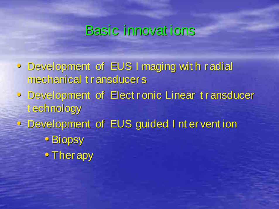
Basic innovationsBasic innovations
•• Development of EUS Imaging with radial Development of EUS Imaging with radial mechanical transducersmechanical transducers
•• Development of Electronic Linear transducer Development of Electronic Linear transducer technologytechnology
•• Development of EUS guided InterventionDevelopment of EUS guided Intervention•• BiopsyBiopsy•• TherapyTherapy
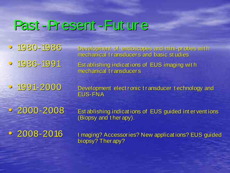
PastPast--PresentPresent--FutureFuture•• 19801980--19861986 Development of endoscopes and miniDevelopment of endoscopes and mini--probes with probes with
mechanical transducers and basic studiesmechanical transducers and basic studies
•• 19861986--19911991 Establishing indications of EUS imaging with Establishing indications of EUS imaging with mechanical transducersmechanical transducers
•• 19911991--20002000 Development electronic transducer technology and Development electronic transducer technology and EUSEUS--FNA FNA
•• 20002000--20082008 Establishing indications of EUS guided interventions Establishing indications of EUS guided interventions (Biopsy and therapy).(Biopsy and therapy).
•• 20082008--20162016 Imaging? Accessories? New applications? EUS guided Imaging? Accessories? New applications? EUS guided biopsy? Therapy?biopsy? Therapy?
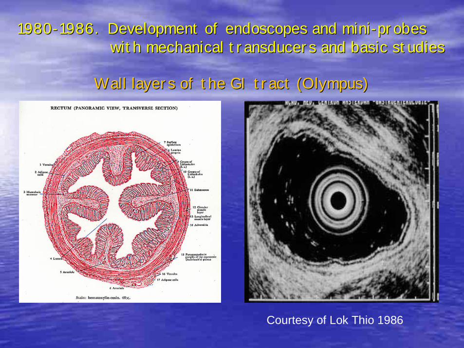
Wall layers of the GI tract (Olympus)Wall layers of the GI tract (Olympus)
Courtesy of Lok Thio 1986
19801980--1986. Development of endoscopes and mini1986. Development of endoscopes and mini--probes probes with mechanical transducers and basic studieswith mechanical transducers and basic studies
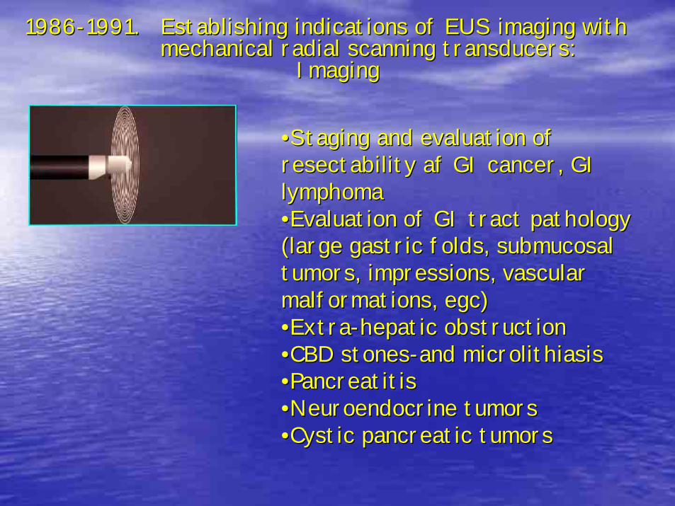
19861986--1991. 1991. Establishing indications of EUS imaging with Establishing indications of EUS imaging with mechanical radial scanning transducers: mechanical radial scanning transducers:
ImagingImaging
••Staging and evaluation of Staging and evaluation of resectability af GI cancer, GI resectability af GI cancer, GI lymphomalymphoma••Evaluation of GI tract pathology Evaluation of GI tract pathology (large gastric folds, submucosal (large gastric folds, submucosal tumors, impressions, vascular tumors, impressions, vascular malformations, egc)malformations, egc)••ExtraExtra--hepatic obstructionhepatic obstruction••CBD stonesCBD stones--and microlithiasisand microlithiasis••PancreatitisPancreatitis••Neuroendocrine tumorsNeuroendocrine tumors••Cystic pancreatic tumorsCystic pancreatic tumors
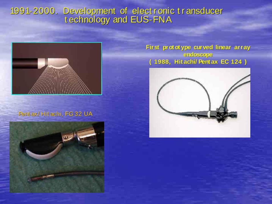
19911991--2000. Development of electronic transducer 2000. Development of electronic transducer technology and EUStechnology and EUS--FNAFNA
First prototype curved linear array First prototype curved linear array endoscope endoscope
( 1988, Hitachi/Pentax EC 124 )( 1988, Hitachi/Pentax EC 124 )
Pentax/Hitachi, FG 32 UAPentax/Hitachi, FG 32 UA
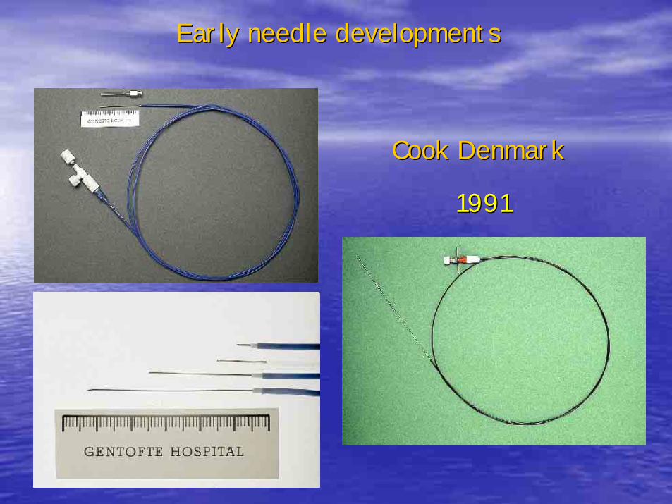
Early needle developmentsEarly needle developments
Cook DenmarkCook Denmark
19911991
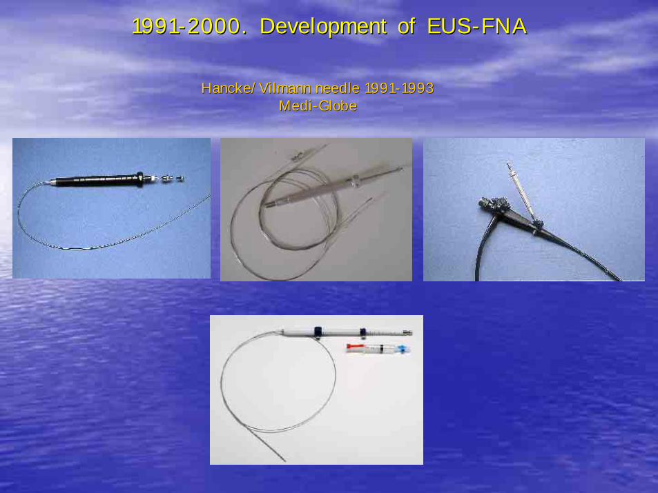
19911991--2000. Development of EUS2000. Development of EUS--FNAFNA
Hancke/Vilmann needle 1991Hancke/Vilmann needle 1991--19931993MediMedi--GlobeGlobe
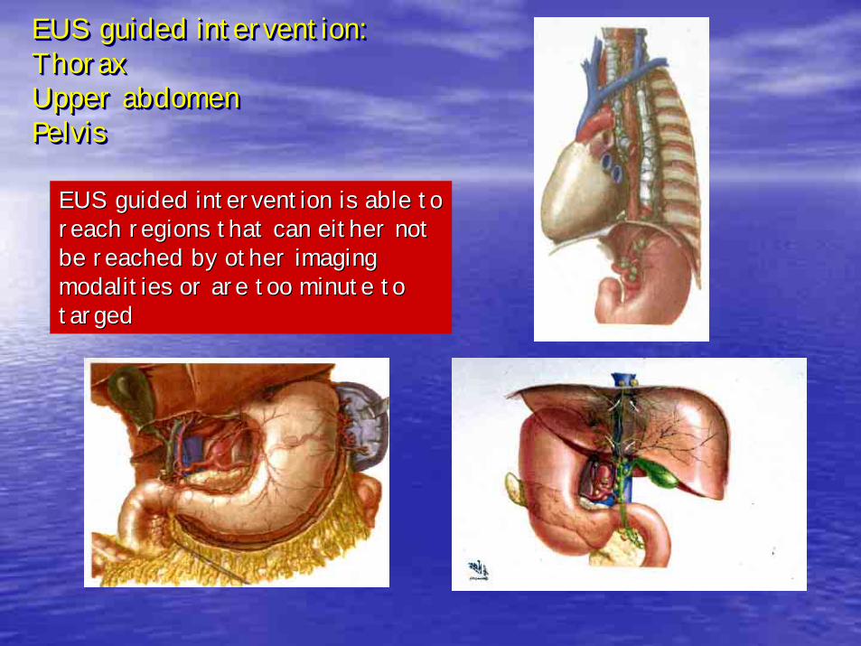
EUS guided intervention:ThoraxUpper abdomenPelvis
EUS guided intervention:EUS guided intervention:ThoraxThoraxUpper abdomenUpper abdomenPelvisPelvis
EUS guided intervention is able to EUS guided intervention is able to reach regions that can either not reach regions that can either not be reached by other imaging be reached by other imaging modalities or are too minute to modalities or are too minute to targedtarged
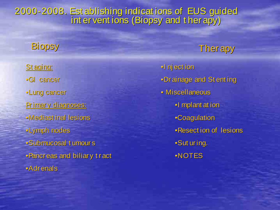
20002000--2008. Establishing indications of EUS guided 2008. Establishing indications of EUS guided interventions (Biopsy and therapy)interventions (Biopsy and therapy)
BiopsyBiopsy TherapyTherapy
Staging:Staging:
••GI cancerGI cancer
••Lung cancerLung cancer
Primary diagnoses:Primary diagnoses:
••Mediastinal lesionsMediastinal lesions
••Lymph nodesLymph nodes
••Submucosal tumoursSubmucosal tumours
••Pancreas and biliary tractPancreas and biliary tract
••AdrenalsAdrenals
••InjectionInjection
••Drainage and StentingDrainage and Stenting
•• MiscellaneousMiscellaneous
••ImplantationImplantation
••CoagulationCoagulation
••Resection of lesionsResection of lesions
••Suturing.Suturing.
••NOTESNOTES
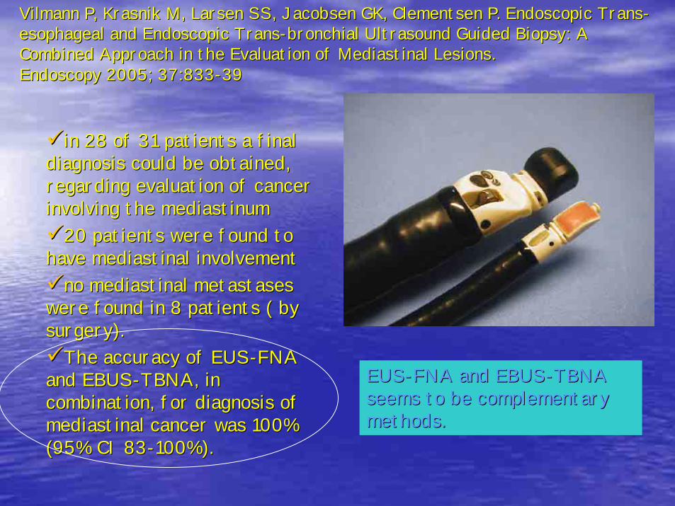
Vilmann P, Krasnik M, Larsen SS, Jacobsen GK, Clementsen P. EndoVilmann P, Krasnik M, Larsen SS, Jacobsen GK, Clementsen P. Endoscopic Transscopic Trans--esophageal and Endoscopic Transesophageal and Endoscopic Trans--bronchial Ultrasound Guided Biopsy: A bronchial Ultrasound Guided Biopsy: A Combined Approach in the Evaluation of Mediastinal Lesions. Combined Approach in the Evaluation of Mediastinal Lesions. Endoscopy 2005; 37:833Endoscopy 2005; 37:833--3939
in 28 of 31 patients a final in 28 of 31 patients a final diagnosis could be obtained, diagnosis could be obtained, regarding evaluation of cancer regarding evaluation of cancer involving the mediastinuminvolving the mediastinum
20 patients were found to 20 patients were found to have mediastinal involvement have mediastinal involvement
no mediastinal metastases no mediastinal metastases were found in 8 patients ( by were found in 8 patients ( by surgery). surgery).
The accuracy of EUSThe accuracy of EUS--FNA FNA and EBUSand EBUS--TBNA, in TBNA, in combination, for diagnosis of combination, for diagnosis of mediastinal cancer was 100% mediastinal cancer was 100% (95% CI 83(95% CI 83--100%).100%).
EUSEUS--FNA and EBUSFNA and EBUS--TBNA TBNA seems to be complementary seems to be complementary methods.methods.
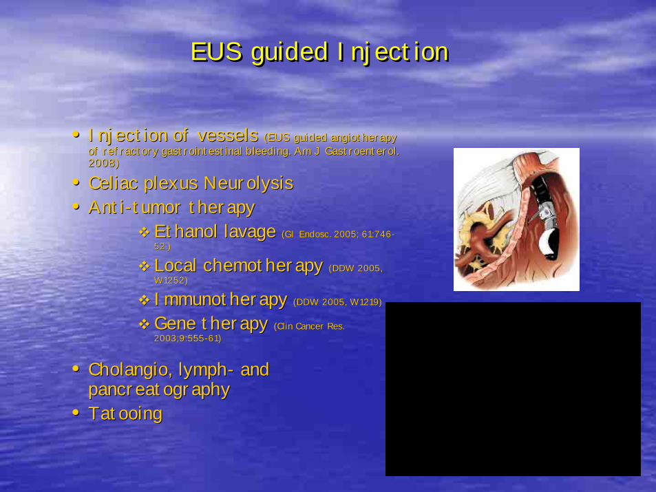
EUS guided InjectionEUS guided InjectionEUS guided Injection
•• Injection of vessels Injection of vessels ((EUS guided angiotherapy EUS guided angiotherapy of refractory gastrointestinal bleeding.of refractory gastrointestinal bleeding. Am J Gastroenterol. Am J Gastroenterol. 2008)2008)
•• Celiac plexus NeurolysisCeliac plexus Neurolysis•• AntiAnti--tumor therapytumor therapy
Ethanol lavage Ethanol lavage ((GI Endosc. 2005; 61:746GI Endosc. 2005; 61:746--52.)52.)
Local chemotherapy Local chemotherapy ((DDW 2005, DDW 2005, W1252)W1252)
Immunotherapy Immunotherapy ((DDW 2005, W1219DDW 2005, W1219))
Gene therapy Gene therapy ((Clin Cancer Res. Clin Cancer Res. 2003;9:5552003;9:555--61)61)
•• Cholangio, lymphCholangio, lymph-- and and pancreatographypancreatography
•• TatooingTatooing
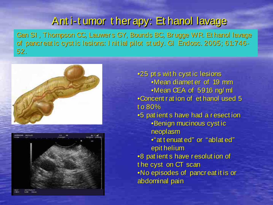
Anti-tumor therapy: Ethanol lavageAntiAnti--tumor therapy: Ethanol lavagetumor therapy: Ethanol lavageGan SI, Thompson CC, Lauwers GY, Bounds BC, Brugge WR. Ethanol lGan SI, Thompson CC, Lauwers GY, Bounds BC, Brugge WR. Ethanol lavage avage of pancreatic cystic lesions: Initial pilot study. GI Endosc. 20of pancreatic cystic lesions: Initial pilot study. GI Endosc. 2005; 61:74605; 61:746--52.52.
••25 pts with cystic lesions25 pts with cystic lesions••Mean diameter of 19 mmMean diameter of 19 mm••Mean CEA of 5916 ng/mlMean CEA of 5916 ng/ml
••Concentration of ethanol used 5 Concentration of ethanol used 5 to 80%to 80%••5 patients have had a resection5 patients have had a resection
••Benign mucinous cystic Benign mucinous cystic neoplasmneoplasm••““attenuatedattenuated”” or or ““ablatedablated””epitheliumepithelium
••8 patients have resolution of 8 patients have resolution of the cyst on CT scanthe cyst on CT scan••No episodes of pancreatitis or No episodes of pancreatitis or abdominal painabdominal pain
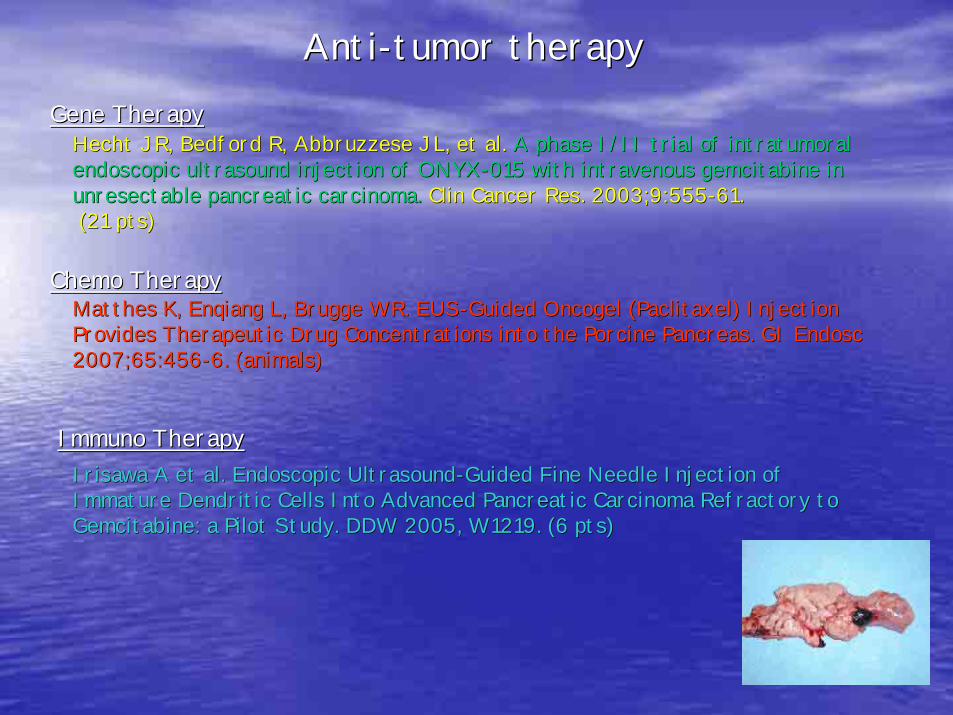
Hecht JR, Bedford R, Abbruzzese JL, et al. Hecht JR, Bedford R, Abbruzzese JL, et al. A phase I/II trial of intratumoral A phase I/II trial of intratumoral endoscopic ultrasound injection of ONYXendoscopic ultrasound injection of ONYX--015 with intravenous gemcitabine in 015 with intravenous gemcitabine in unresectable pancreatic carcinoma.unresectable pancreatic carcinoma. Clin Cancer Res. 2003;9:555Clin Cancer Res. 2003;9:555--61.61.(21 pts)(21 pts)
AntiAnti--tumor therapytumor therapy
Chemo TherapyChemo Therapy
Gene TherapyGene Therapy
Matthes K, Enqiang L, Brugge WR. EUSMatthes K, Enqiang L, Brugge WR. EUS--Guided Oncogel (Paclitaxel) Injection Guided Oncogel (Paclitaxel) Injection Provides Therapeutic Drug Concentrations into the Porcine PancreProvides Therapeutic Drug Concentrations into the Porcine Pancreas. GI Endosc as. GI Endosc 2007;65:4562007;65:456--6. (animals)6. (animals)
Irisawa A et al. Endoscopic UltrasoundIrisawa A et al. Endoscopic Ultrasound--Guided Fine Needle Injection of Guided Fine Needle Injection of Immature Dendritic Cells Into Advanced Pancreatic Carcinoma RefrImmature Dendritic Cells Into Advanced Pancreatic Carcinoma Refractory to actory to Gemcitabine: a Pilot Study. DDW 2005, W1219. (6 pts)Gemcitabine: a Pilot Study. DDW 2005, W1219. (6 pts)
Immuno TherapyImmuno Therapy
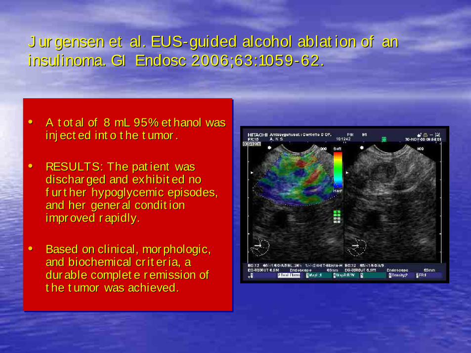
Jurgensen et al. EUSJurgensen et al. EUS--guided alcohol ablation of an guided alcohol ablation of an insulinoma. GI Endosc 2006;63:1059insulinoma. GI Endosc 2006;63:1059--62.62.
• A total of 8 mL 95% ethanol was injected into the tumor.
• RESULTS: The patient was discharged and exhibited no further hypoglycemic episodes, and her general condition improved rapidly.
• Based on clinical, morphologic, and biochemical criteria, a durable complete remission of the tumor was achieved.
•• A total of 8 mL 95% ethanol was A total of 8 mL 95% ethanol was injected into the tumor. injected into the tumor.
•• RESULTS: The patient was RESULTS: The patient was discharged and exhibited no discharged and exhibited no further hypoglycemic episodes, further hypoglycemic episodes, and her general condition and her general condition improved rapidly. improved rapidly.
•• Based on clinical, morphologic, Based on clinical, morphologic, and biochemical criteria, a and biochemical criteria, a durable complete remission of durable complete remission of the tumorthe tumor was achieved.was achieved.
EUS guided injection: cholangio, lymphEUS guided injection: cholangio, lymph-- and pancreatographyand pancreatographyWith or without RendezWith or without Rendez--vousvous
Mallery S, Matlock J, Freeman ML. EUSMallery S, Matlock J, Freeman ML. EUS--guided rendezvous drainage of obstructed biliary and guided rendezvous drainage of obstructed biliary and pancreatic ducts: Report of 6 cases. pancreatic ducts: Report of 6 cases. Gastrointest Endosc. 2004;59:100Gastrointest Endosc. 2004;59:100--77
Kahaleh M, Yoshida C, Kane L, Yeaton P. Interventional EUS cholaKahaleh M, Yoshida C, Kane L, Yeaton P. Interventional EUS cholangiography: A report of 5 cases. ngiography: A report of 5 cases. Gastrointest Endosc. 2004;60:138Gastrointest Endosc. 2004;60:138--4242
Kahaleh M, Wang P, Shami VM, Tokar J, Yeaton P. EUSKahaleh M, Wang P, Shami VM, Tokar J, Yeaton P. EUS--guided transhepatic cholangiography: report guided transhepatic cholangiography: report of 6 cases. of 6 cases. Gastrointest Endosc. 2005;61:307Gastrointest Endosc. 2005;61:307--13.13.
Kahaleh M et al. Interventional Endoscopic Ultrasound CholangiogKahaleh M et al. Interventional Endoscopic Ultrasound Cholangiography: Midraphy: Mid--Term FollowTerm Follow--Up of 18 Up of 18 Cases. DDW 2005, W1226.Cases. DDW 2005, W1226.
Poley JW et al. EUSPoley JW et al. EUS--guided transbulbar rendezvous after unsuccessful ERCP. A report guided transbulbar rendezvous after unsuccessful ERCP. A report of 2 cases. of 2 cases. DDW 2005, W1271DDW 2005, W1271..
Parasher VK, Hernandez LV, Leveen RF, Mladinich CR, Nonabur V, BParasher VK, Hernandez LV, Leveen RF, Mladinich CR, Nonabur V, Bhutani MS. hutani MS. Lymph sampling and Lymph sampling and lymphangiography via EUSlymphangiography via EUS--guided transesophageal thoracic duct puncture in a swine model. guided transesophageal thoracic duct puncture in a swine model. Gastrointest Endosc. 2004;59:564Gastrointest Endosc. 2004;59:564--7.7.
Dewitt J, McHenry L, Fogel E, Leblanc J, McGreevy K, Sherman S. Dewitt J, McHenry L, Fogel E, Leblanc J, McGreevy K, Sherman S. EUSEUS--guided methylene blue guided methylene blue pancreatography for minor papilla localization after unsuccessfupancreatography for minor papilla localization after unsuccessful ERCP. l ERCP. Gastrointest Endosc. Gastrointest Endosc. 2004;59:1332004;59:133--6.6.
Will U, Meyer F, Manger T, Wanzar I. Endoscopic ultrasoundWill U, Meyer F, Manger T, Wanzar I. Endoscopic ultrasound--assisted rendezvous maneuver to assisted rendezvous maneuver to achieve pancreatic duct drainage in obstructive chronic pancreatachieve pancreatic duct drainage in obstructive chronic pancreatitis. itis. Endoscopy. 2005;37:171Endoscopy. 2005;37:171--3.3.
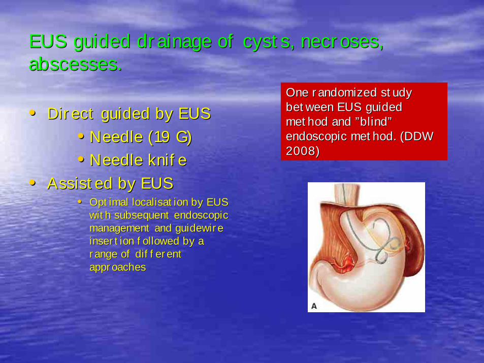
EUS guided drainage of cysts, necroses, EUS guided drainage of cysts, necroses, abscesses.abscesses.
•• Direct guided by EUSDirect guided by EUS•• Needle (19 G)Needle (19 G)•• Needle knifeNeedle knife
•• Assisted by EUSAssisted by EUS•• Optimal localisation by EUS Optimal localisation by EUS
with subsequent endoscopic with subsequent endoscopic management and guidewire management and guidewire insertion followed by a insertion followed by a range of different range of different approachesapproaches
One randomized study One randomized study between EUS guided between EUS guided method and method and ””blindblind””endoscopic method. (DDW endoscopic method. (DDW 2008)2008)
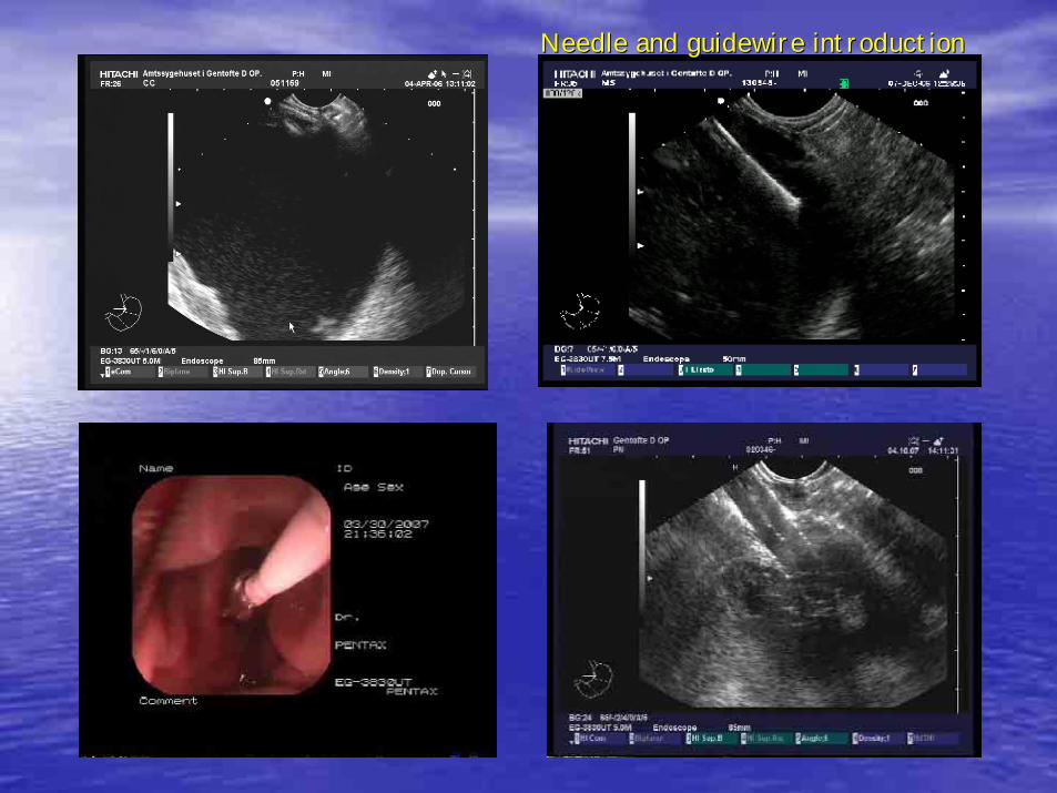
Needle and guidewire introductionNeedle and guidewire introduction
EUS guided drainageEUS guided drainageEUS guided drainage
•• Abscess drainageAbscess drainage–– Giovannini M, Bories E, Moutardier V, Pesenti C, Guillemin A, LeGiovannini M, Bories E, Moutardier V, Pesenti C, Guillemin A, Lelong B, Delpero long B, Delpero
JR. Drainage of deep pelvic abscesses using therapeutic echo endJR. Drainage of deep pelvic abscesses using therapeutic echo endoscopy. oscopy. EEndoscopy. 2003;35:511ndoscopy. 2003;35:511-- 4.4.
–– KKahaleh M. EUS drainage of a mediastinal abscess. ahaleh M. EUS drainage of a mediastinal abscess. Gastrointest Endosc. 2004 Gastrointest Endosc. 2004 ;60:158;60:158--6060
–– Seewald S. EUSSeewald S. EUS--guided drainage of hepatic abscess. guided drainage of hepatic abscess. Gastrointest Endosc. 2005 Gastrointest Endosc. 2005 ;61:495;61:495--8.8.
–– Wehrmann T. Endoscopic debridement of paraesophageal, mediastinaWehrmann T. Endoscopic debridement of paraesophageal, mediastinal l abscesses: a prospective case series. GI Endosc. 2005;62:344abscesses: a prospective case series. GI Endosc. 2005;62:344--99
•• necrosectomynecrosectomy–– Matthes H.Matthes H. Endoscopic transgastric EUS guided drainage and necrosectomy of Endoscopic transgastric EUS guided drainage and necrosectomy of
infected pancreatic pseudocysts. DDW 2005, infected pancreatic pseudocysts. DDW 2005, •• Gall bladder fossa fluid drainageGall bladder fossa fluid drainage
–– Kahaleh. Drainage of gallbladder fossa fluid collections with enKahaleh. Drainage of gallbladder fossa fluid collections with endoprosthesis doprosthesis placement under endoscopic ultrasound guidance: a preliminary replacement under endoscopic ultrasound guidance: a preliminary report of two port of two cases. cases. Endoscopy. 2005;37:393Endoscopy. 2005;37:393--6.6.
•• Hematoma drainageHematoma drainage–– Piraka C. Evolving role of interventional endoscopic ultrasound.Piraka C. Evolving role of interventional endoscopic ultrasound. DDW, W1270DDW, W1270
EUSEUS--guided transmural cholecystostomyguided transmural cholecystostomy
Lee SS et al. EUSLee SS et al. EUS--guided transmural cholecystostomy as rescue management guided transmural cholecystostomy as rescue management for acute cholecystitis in elderly or highfor acute cholecystitis in elderly or high--risk patients: a prospective risk patients: a prospective feasibility study. feasibility study. Gastrointest Endosc. 2007;66:1008Gastrointest Endosc. 2007;66:1008--12.12.
PATIENTS:PATIENTS: Nine elderly or highNine elderly or high--risk patients risk patients diagnosed with acute cholecystitis. diagnosed with acute cholecystitis.
INTERVENTIONSINTERVENTIONS: All inflamed gallbladders were : All inflamed gallbladders were drained by EUSdrained by EUS--guided transmural cholecystostomy. guided transmural cholecystostomy. MAIN OUTCOMEMAIN OUTCOME: Clinical resolution of acute : Clinical resolution of acute cholecystitis. cholecystitis.
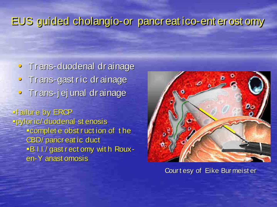
EUS guided cholangio-or pancreatico-enterostomyEUS guided cholangioEUS guided cholangio--or pancreaticoor pancreatico--enterostomyenterostomy
•• TransTrans--duodenal drainageduodenal drainage•• TransTrans--gastric drainagegastric drainage•• TransTrans--jejunal drainagejejunal drainage
failure by ERCPfailure by ERCPpyloric/duodenal stenosispyloric/duodenal stenosis
complete obstruction of the complete obstruction of the CBD/pancreatic ductCBD/pancreatic ductB II/gastrectomy with RouxB II/gastrectomy with Roux--
enen--Y anastomosisY anastomosisCourtesy of Eike BurmeisterCourtesy of Eike Burmeister
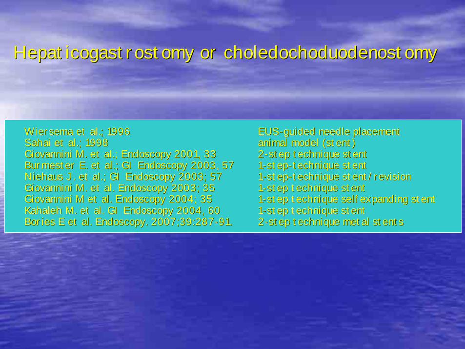
Hepaticogastrostomy or choledochoduodenostomyHepaticogastrostomy or choledochoduodenostomy
Wiersema et al.; 1996Wiersema et al.; 1996 EUSEUS--guided needle placementguided needle placementSahai et al.; 1998Sahai et al.; 1998 animal model (stent)animal model (stent)Giovannini M. et al.; Endoscopy 2001, 33Giovannini M. et al.; Endoscopy 2001, 33 22--step technique stentstep technique stentBurmester E. et al.; GI Endoscopy 2003, 57Burmester E. et al.; GI Endoscopy 2003, 57 11--stepstep--technique stenttechnique stentNiehaus J. et al.; GI Endoscopy 2003; 57 Niehaus J. et al.; GI Endoscopy 2003; 57 11--stepstep--technique stent/revisiontechnique stent/revisionGiovannini M. et al. Endoscopy 2003; 35Giovannini M. et al. Endoscopy 2003; 35 11--step technique stentstep technique stentGiovannini M et al. Endoscopy 2004; 35Giovannini M et al. Endoscopy 2004; 35 11--step technique selfexpanding stentstep technique selfexpanding stentKahaleh M. et al. GI Endoscopy 2004, 60Kahaleh M. et al. GI Endoscopy 2004, 60 11--step technique stentstep technique stentBories E et al. Bories E et al. Endoscopy. 2007;39:287Endoscopy. 2007;39:287--91.91. 22--step technique metal stentsstep technique metal stents
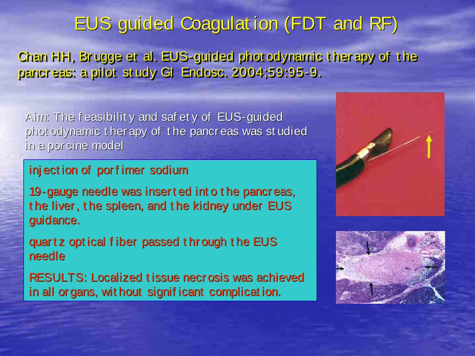
Chan HH, Brugge et al. EUS-guided photodynamic therapy of the pancreas: a pilot study GI Endosc. 2004;59:95-9.Chan HH, Brugge et al. EUSChan HH, Brugge et al. EUS--guided photodynamic therapy of the guided photodynamic therapy of the pancreas: a pilot study GI Endosc. 2004;59:95pancreas: a pilot study GI Endosc. 2004;59:95--9.9.
injection of porfimer sodium injection of porfimer sodium
1919--gauge needle was inserted into the pancreas, gauge needle was inserted into the pancreas, the liver, the spleen, and the kidney under EUS the liver, the spleen, and the kidney under EUS guidance. guidance.
quartz optical fiber passed through the EUS quartz optical fiber passed through the EUS needle needle
RESULTS: Localized tissue necrosis was achieved RESULTS: Localized tissue necrosis was achieved in all organs, without significant complication. in all organs, without significant complication.
Aim: The feasibility and safety of EUSAim: The feasibility and safety of EUS--guided guided photodynamic therapy of the pancreas was studied photodynamic therapy of the pancreas was studied in a porcine modelin a porcine model
EUS guided Coagulation (FDT and RF)EUS guided Coagulation (FDT and RF)
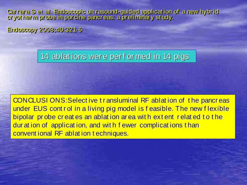
Carrara S et al. Endoscopic ultrasound-guided application of a new hybrid cryotherm probe in porcine pancreas: a preliminary study.
Endoscopy 2008;40:321-6
Carrara S et al. Endoscopic ultrasoundCarrara S et al. Endoscopic ultrasound--guided application of a new hybrid guided application of a new hybrid cryotherm probe in porcine pancreas: a preliminary study. cryotherm probe in porcine pancreas: a preliminary study.
Endoscopy 2008;40:321Endoscopy 2008;40:321--66
14 ablations were performed in 14 pigs14 ablations were performed in 14 pigs
CONCLUSIONS:CONCLUSIONS:Selective transluminal RF ablation of the pancreas Selective transluminal RF ablation of the pancreas under EUS control in a living pig model is feasible. The new fleunder EUS control in a living pig model is feasible. The new flexible xible bipolar probe creates an ablation area with extent related to thbipolar probe creates an ablation area with extent related to the e duration of application, and with fewer complications than duration of application, and with fewer complications than conventional RF ablation techniques.conventional RF ablation techniques.
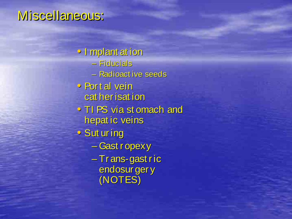
Miscellaneous: Miscellaneous: Miscellaneous:
•• ImplantationImplantation–– FiducialsFiducials–– Radioactive seedsRadioactive seeds
•• Portal vein Portal vein catherisationcatherisation
•• TIPS via stomach and TIPS via stomach and hepatic veinshepatic veins
•• SuturingSuturing–– GastropexyGastropexy–– TransTrans--gastric gastric
endosurgery endosurgery (NOTES)(NOTES)
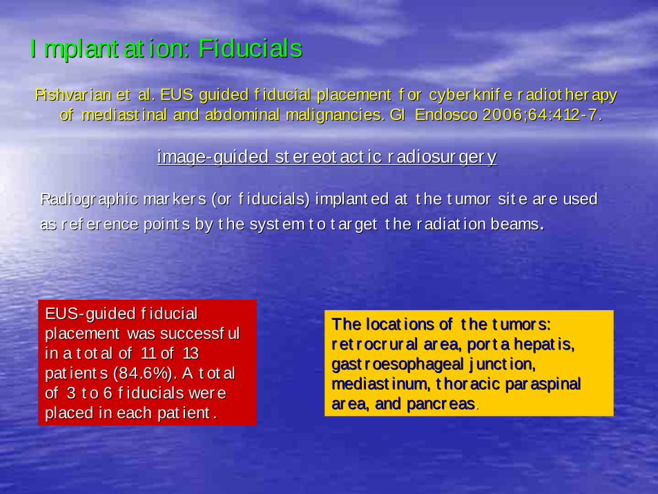
Implantation: FiducialsImplantation: FiducialsPishvarian et al. EUS guided fiducial placement for cyberknife rPishvarian et al. EUS guided fiducial placement for cyberknife radiotherapy adiotherapy
of mediastinal and abdominal malignancies. GI Endosco 2006;64:41of mediastinal and abdominal malignancies. GI Endosco 2006;64:4122--7.7.
imageimage--guided stereotactic radiosurgeryguided stereotactic radiosurgery
Radiographic markers (or fiducials) implanted at the tumor site Radiographic markers (or fiducials) implanted at the tumor site are used are used as reference points by the system to target the radiation beamsas reference points by the system to target the radiation beams. .
EUSEUS--guided fiducial guided fiducial placement was successful placement was successful in a total of 11 of 13 in a total of 11 of 13 patients (84.6%). A total patients (84.6%). A total of 3 to 6 fiducials were of 3 to 6 fiducials were placed in each patient. placed in each patient.
The locations of the tumors: The locations of the tumors: retrocrural area, porta hepatis, retrocrural area, porta hepatis, gastroesophageal junction, gastroesophageal junction, mediastinum, thoracic paraspinal mediastinum, thoracic paraspinal area, and pancreasarea, and pancreas. .
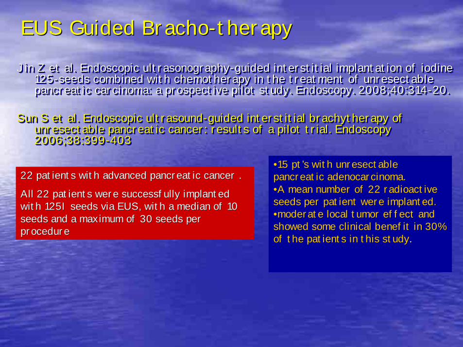
EUS Guided BrachoEUS Guided Bracho--therapytherapy
Jin Z et al. Endoscopic ultrasonography-guided interstitial implantation of iodine 125-seeds combined with chemotherapy in the treatment of unresectable pancreatic carcinoma: a prospective pilot study. Endoscopy. 2008;40:314-20.
Sun S et al. Endoscopic ultrasound-guided interstitial brachytherapy of unresectable pancreatic cancer: results of a pilot trial. Endoscopy 2006;38:399-403
Jin Z et al. Endoscopic ultrasonographyJin Z et al. Endoscopic ultrasonography--guided interstitial implantation of iodine guided interstitial implantation of iodine 125125--seeds combined with chemotherapy in the treatment of unresectablseeds combined with chemotherapy in the treatment of unresectable e pancreatic carcinoma: a prospective pilot study. pancreatic carcinoma: a prospective pilot study. Endoscopy. 2008;40:314Endoscopy. 2008;40:314--20. 20.
Sun S et al. Endoscopic ultrasoundSun S et al. Endoscopic ultrasound--guided interstitial brachytherapy of guided interstitial brachytherapy of unresectable pancreatic cancer: results of a pilot trial. Endoscunresectable pancreatic cancer: results of a pilot trial. Endoscopy opy 2006;38:3992006;38:399--403403
22 patients with advanced pancreatic cancer . 22 patients with advanced pancreatic cancer .
All 22 patients were successfully implanted All 22 patients were successfully implanted with 125I seeds via EUS, with a median of 10 with 125I seeds via EUS, with a median of 10 seeds and a maximum of 30 seeds per seeds and a maximum of 30 seeds per procedureprocedure
••15 pt15 pt’’s with unresectable s with unresectable pancreatic adenocarcinoma. pancreatic adenocarcinoma. ••A mean number of 22 radioactive A mean number of 22 radioactive seeds per patient were implanted.seeds per patient were implanted.••moderate local tumor effect and moderate local tumor effect and showed some clinical benefit in 30% showed some clinical benefit in 30% of the patients in this studyof the patients in this study. .
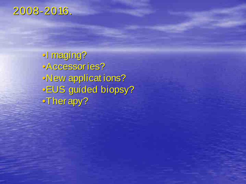
20082008--2016.2016.
••Imaging? Imaging? ••Accessories? Accessories? ••New applications? New applications? ••EUS guided biopsy? EUS guided biopsy? ••Therapy?Therapy?
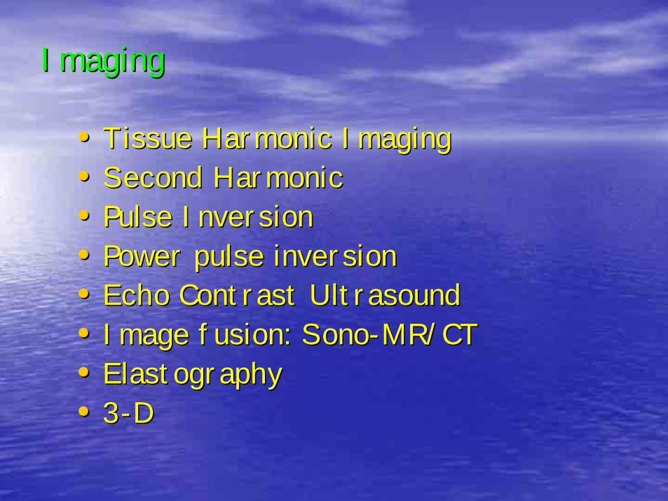
ImagingImaging
•• Tissue Harmonic ImagingTissue Harmonic Imaging•• Second HarmonicSecond Harmonic•• Pulse InversionPulse Inversion•• Power pulse inversionPower pulse inversion•• Echo Contrast UltrasoundEcho Contrast Ultrasound•• Image fusion: SonoImage fusion: Sono--MR/CTMR/CT•• ElastographyElastography•• 33--DD
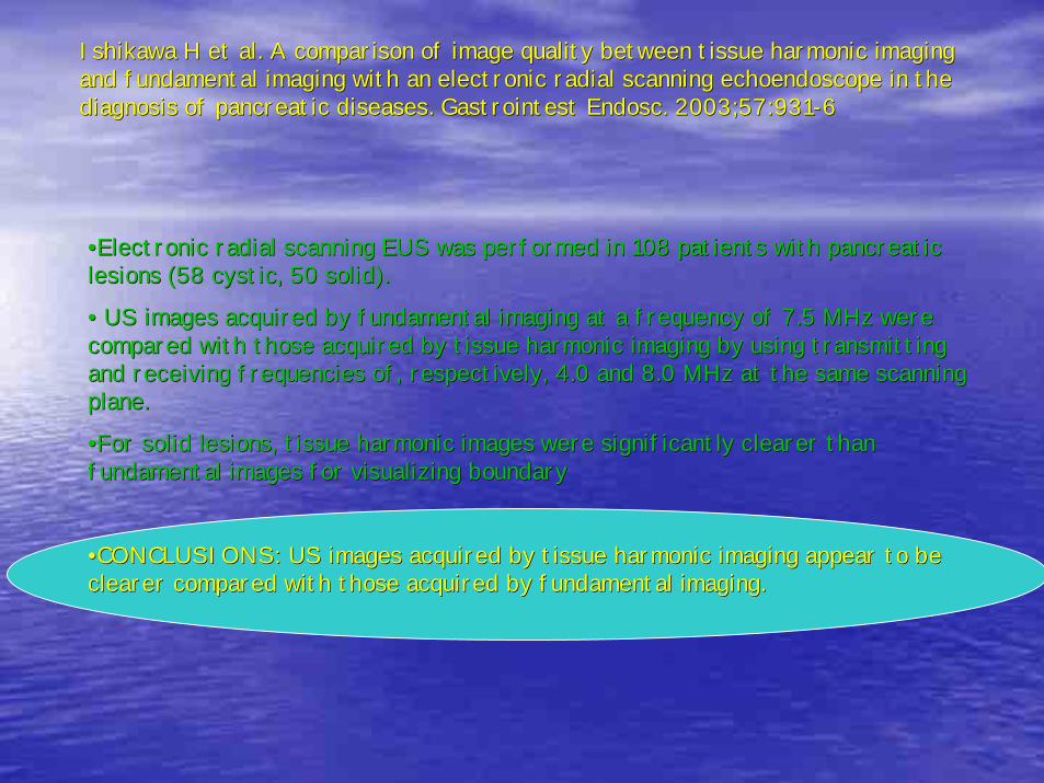
Ishikawa H et al. A comparison of image quality between tissue hIshikawa H et al. A comparison of image quality between tissue harmonic imaging armonic imaging and fundamental imaging with an electronic radial scanning echoeand fundamental imaging with an electronic radial scanning echoendoscope in the ndoscope in the diagnosis of pancreatic diseases. Gastrointest Endosc. 2003;57:9diagnosis of pancreatic diseases. Gastrointest Endosc. 2003;57:93131--6 6
••Electronic radial scanning EUS was performed in 108 patients witElectronic radial scanning EUS was performed in 108 patients with pancreatic h pancreatic lesions (58 cystic, 50 solid).lesions (58 cystic, 50 solid).
•• US images acquired by fundamental imaging at a frequency of 7.5US images acquired by fundamental imaging at a frequency of 7.5 MHz were MHz were compared with those acquired by tissue harmonic imaging by usingcompared with those acquired by tissue harmonic imaging by using transmitting transmitting and receiving frequencies of, respectively, 4.0 and 8.0 MHz at tand receiving frequencies of, respectively, 4.0 and 8.0 MHz at the same scanning he same scanning plane. plane.
••For solid lesions, tissue harmonic images were significantly cleFor solid lesions, tissue harmonic images were significantly clearer than arer than fundamental images for visualizing boundary fundamental images for visualizing boundary
••CONCLUSIONS: US images acquired by tissue harmonic imaging appeaCONCLUSIONS: US images acquired by tissue harmonic imaging appear to be r to be clearer compared with those acquired by fundamental imaging.clearer compared with those acquired by fundamental imaging.
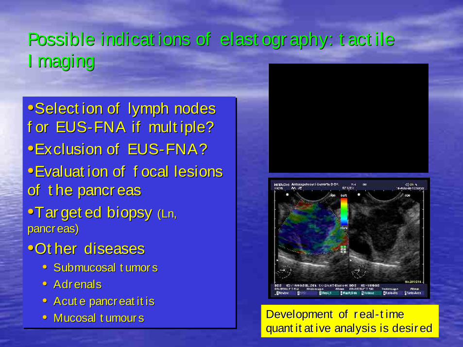
Possible indications of elastography: tactile Possible indications of elastography: tactile ImagingImaging
•Selection of lymph nodes for EUS-FNA if multiple?•Exclusion of EUS-FNA?•Evaluation of focal lesions of the pancreas•Targeted biopsy (Ln, pancreas)
•Other diseases• Submucosal tumors• Adrenals• Acute pancreatitis• Mucosal tumours
••Selection of lymph nodes Selection of lymph nodes for EUSfor EUS--FNA if multiple?FNA if multiple?••Exclusion of EUSExclusion of EUS--FNA?FNA?••Evaluation of focal lesions Evaluation of focal lesions of the pancreasof the pancreas••Targeted biopsy Targeted biopsy (Ln, (Ln, pancreas)pancreas)
••Other diseasesOther diseases•• Submucosal tumorsSubmucosal tumors•• AdrenalsAdrenals•• Acute pancreatitisAcute pancreatitis•• Mucosal tumoursMucosal tumours Development of realDevelopment of real--time time
quantitative analysis is desiredquantitative analysis is desired
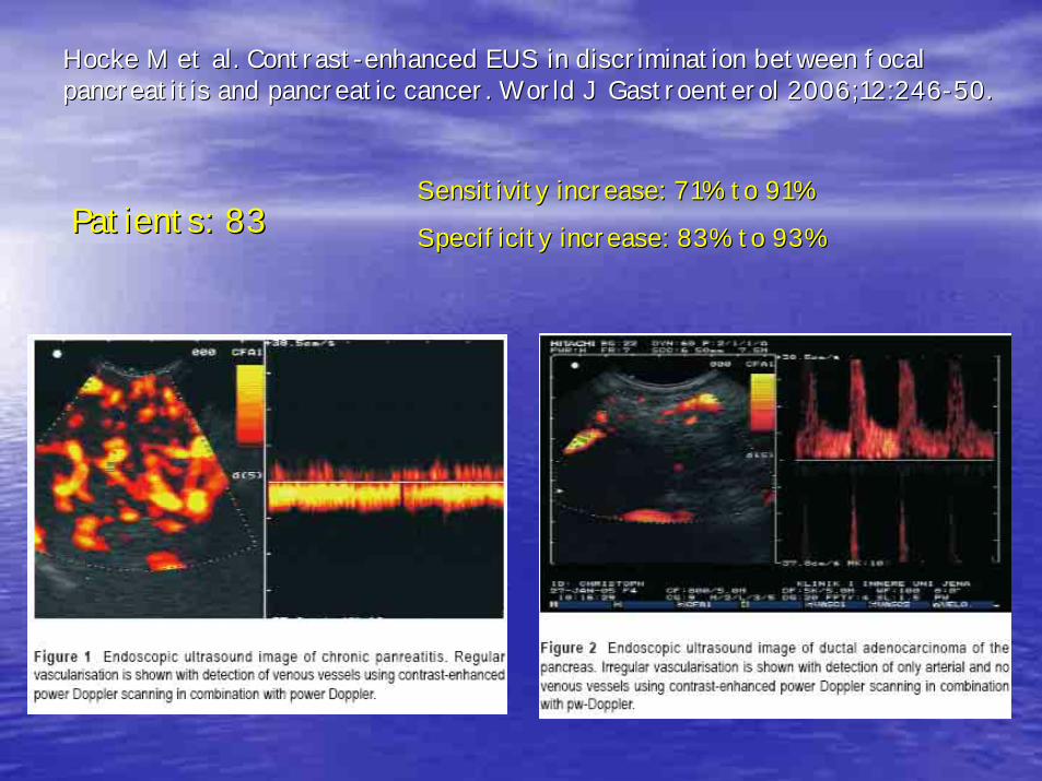
Hocke M et al. ContrastHocke M et al. Contrast--enhanced EUS in discrimination between focal enhanced EUS in discrimination between focal pancreatitis and pancreatic cancer. World J Gastroenterol 2006;1pancreatitis and pancreatic cancer. World J Gastroenterol 2006;12:2462:246--50.50.
Sensitivity increase: 71% to 91%Sensitivity increase: 71% to 91%
Specificity increase: 83% to 93%Specificity increase: 83% to 93%Patients: 83Patients: 83
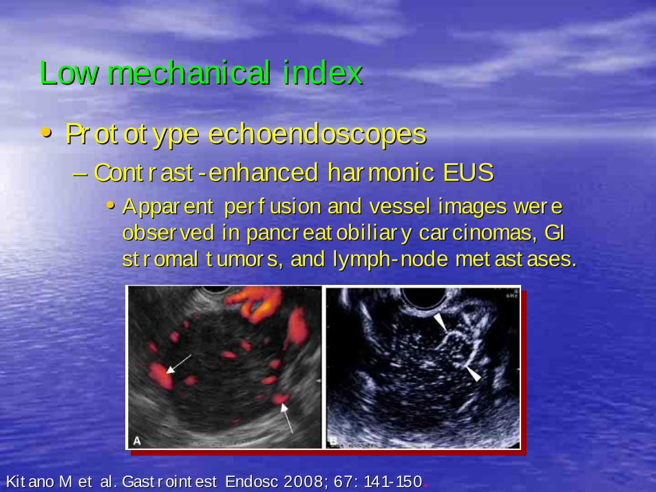
Low mechanical indexLow mechanical index
•• Prototype echoendoscopesPrototype echoendoscopes–– ContrastContrast--enhanced harmonic EUSenhanced harmonic EUS
•• Apparent perfusion and vessel images were Apparent perfusion and vessel images were observed in pancreatobiliary observed in pancreatobiliary carcinomas, GI carcinomas, GI stromal tumors, and lymphstromal tumors, and lymph--node metastases.node metastases.
Kitano M et al. Gastrointest Endosc 2008; 67: 141Kitano M et al. Gastrointest Endosc 2008; 67: 141--150150.
EUS Scope
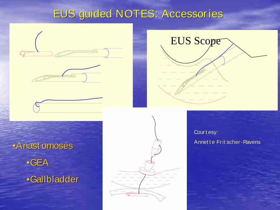
EUS guided NOTES: AccessoriesEUS guided NOTES: Accessories
••AnastomosesAnastomoses
••GEAGEA
••GallbladderGallbladder
Courtesy: Courtesy:
Annette FritscherAnnette Fritscher--RavensRavens
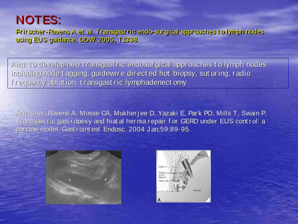
NOTES:Fritscher-Ravens A et al. Transgastric endo-surgical approaches to lymph nodes using EUS guidance. DDW 2005, T1338
NOTES:NOTES:FritscherFritscher--Ravens A et al. Ravens A et al. Transgastric endoTransgastric endo--surgical approaches to lymph nodes surgical approaches to lymph nodes using EUS guidance. DDW 2005, T1338using EUS guidance. DDW 2005, T1338
Aim: to develop new transgastric endosurgical approaches to lympAim: to develop new transgastric endosurgical approaches to lymph nodes h nodes including node tagging, guidewire directed hot biopsy, suturing,including node tagging, guidewire directed hot biopsy, suturing, radio radio frequency ablation, transgastric lymphadenectomyfrequency ablation, transgastric lymphadenectomy
FritscherFritscher--Ravens A, Mosse CA, Mukherjee D, Yazaki E, Park PO, Mills T, SwaRavens A, Mosse CA, Mukherjee D, Yazaki E, Park PO, Mills T, Swain P. in P. Transgastric gastropexy and hiatal hernia repair for GERD under Transgastric gastropexy and hiatal hernia repair for GERD under EUS control: a EUS control: a porcine model. porcine model. Gastrointest Endosc. 2004 Jan;59:89Gastrointest Endosc. 2004 Jan;59:89--95.95.
In conclusionIn conclusionIn conclusion
•Innovations in EUS have mainly been driven from improvements in
Imaging, development of electronic linear transducer technology and
of EUS guided interventions
•The combination of high resolution imaging and precise targeting of
minute lesions makes EUS a true minimal invasive modality.
•Only a few randomised comparisons with established therapies
•A huge potential for new developments in EUS
•EUS will possibly challenge other more invasive methods in the near
future.
••Innovations in EUS have mainly been driven from improvements in Innovations in EUS have mainly been driven from improvements in
Imaging, development of electronic linear transducer technology Imaging, development of electronic linear transducer technology and and
of EUS guided interventionsof EUS guided interventions
••The combination of high resolution imaging and precise targetingThe combination of high resolution imaging and precise targeting of of
minute lesions makes EUS a true minimal invasive modality.minute lesions makes EUS a true minimal invasive modality.
••Only a few randomised comparisons with established therapiesOnly a few randomised comparisons with established therapies
••A huge potential for new developments in EUSA huge potential for new developments in EUS
••EUS will possibly challenge other more invasive methods in the nEUS will possibly challenge other more invasive methods in the near ear
future.future.