Injuries of the Clavicle, Acromioclavicular Joint and Sternoclavicular Joint Andrew H. Schmidt, M.D....
121
Injuries of the Clavicle, Acromioclavicular Joint and Sternoclavicular Joint Andrew H. Schmidt, M.D. Revised October 2010 Andrew H. Schmidt, MD & T. J. McElroy, MD; Created March 2004; Revised January 2007 & October 2010
-
Upload
gustavo-seymour -
Category
Documents
-
view
219 -
download
0
Transcript of Injuries of the Clavicle, Acromioclavicular Joint and Sternoclavicular Joint Andrew H. Schmidt, M.D....
Injuries of the Clavicle, Acromioclavicular Joint and
Sternoclavicular Joint
Andrew H. Schmidt, M.D.Revised October 2010
Andrew H. Schmidt, MD & T. J. McElroy, MD; Created March 2004; Revised January 2007 & October 2010
Goals
1) Review anatomy of clavicle, AC joint, and sternoclavicular joint
2) Review imaging of these areas.
3) Clavicle FracturesNonoperative RX
Surgical Repair
Nonunions and Malunions
4) AC Joint Injuries
5) Sternoclavicular joint injuries
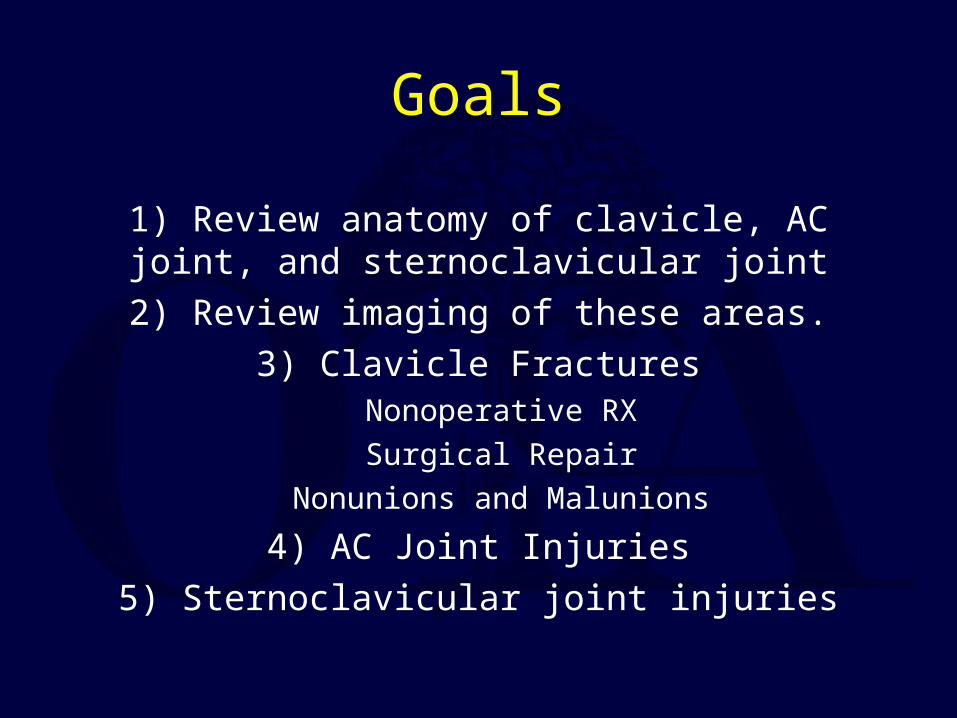
Clavicle
“S”-shaped bone Medial - sternoclavicular joint
Lateral - acromioclavicular joint and coracoclavicular ligaments
Muscle attachments:– Medial: sternocleidomastoid
– Lateral: Trapezius, pectoralis major
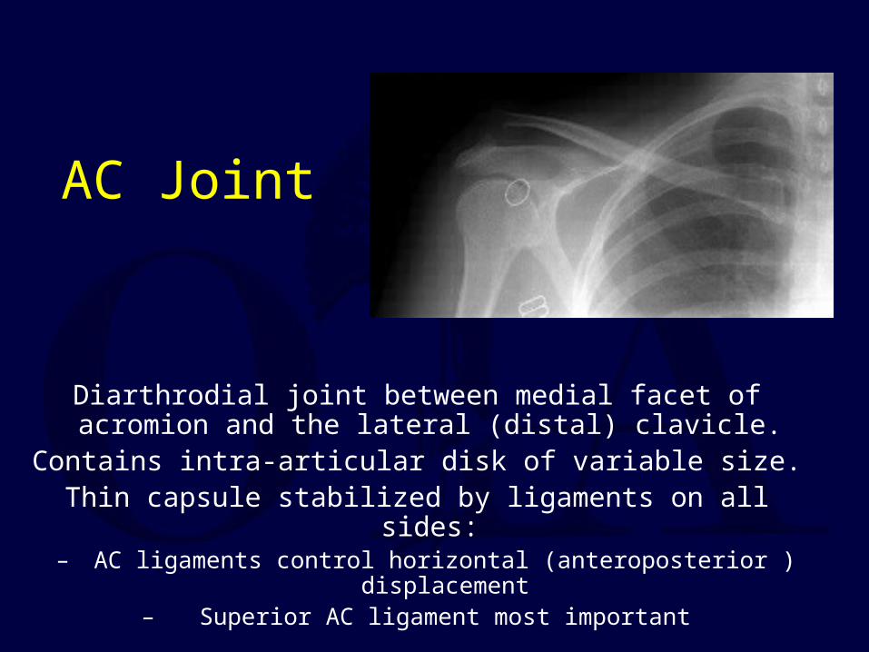
AC Joint
Diarthrodial joint between medial facet of acromion and the lateral (distal) clavicle.
Contains intra-articular disk of variable size.Thin capsule stabilized by ligaments on all sides:
– AC ligaments control horizontal (anteroposterior ) displacement– Superior AC ligament most important
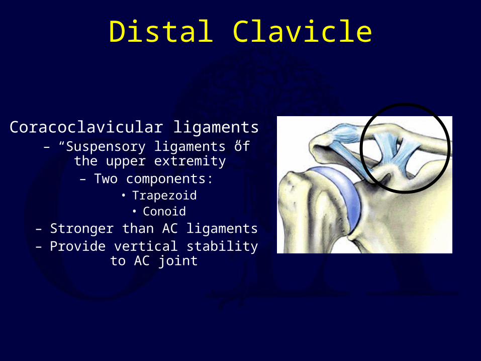
Distal Clavicle
Coracoclavicular ligaments– “Suspensory ligaments of the upper
extremity”– Two components:
• Trapezoid• Conoid
– Stronger than AC ligaments– Provide vertical stability to AC joint
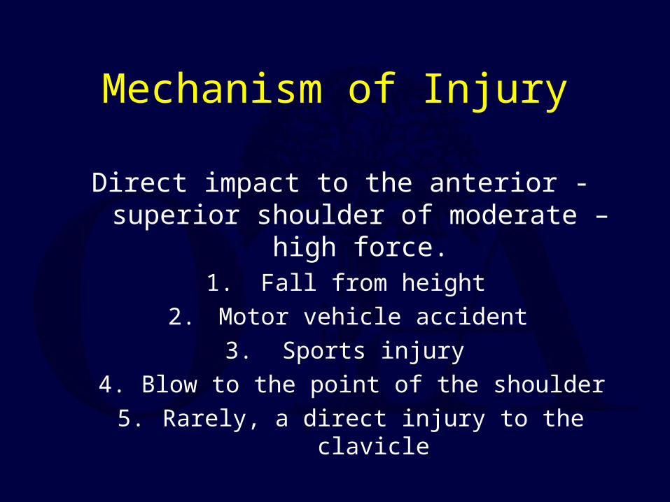
Mechanism of Injury
Direct impact to the anterior - superior shoulder of moderate – high force.
1. Fall from height
2. Motor vehicle accident
3. Sports injury
4. Blow to the point of the shoulder
5. Rarely, a direct injury to the clavicle
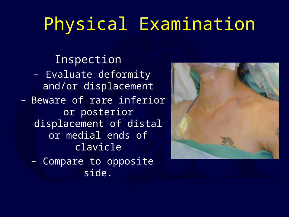
Physical Examination
Inspection– Evaluate deformity and/or
displacement– Beware of rare inferior or
posterior displacement of distal or medial ends of
clavicle– Compare to opposite side.
Physical Examination
PalpationEvaluate pain
Look for instability with stress
Physical Examination
Neurovascular examination– Must be done thoroughly and documented!
Evaluate upper extremity motor and sensation
Measure shoulder range-of-motion
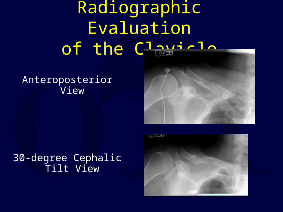
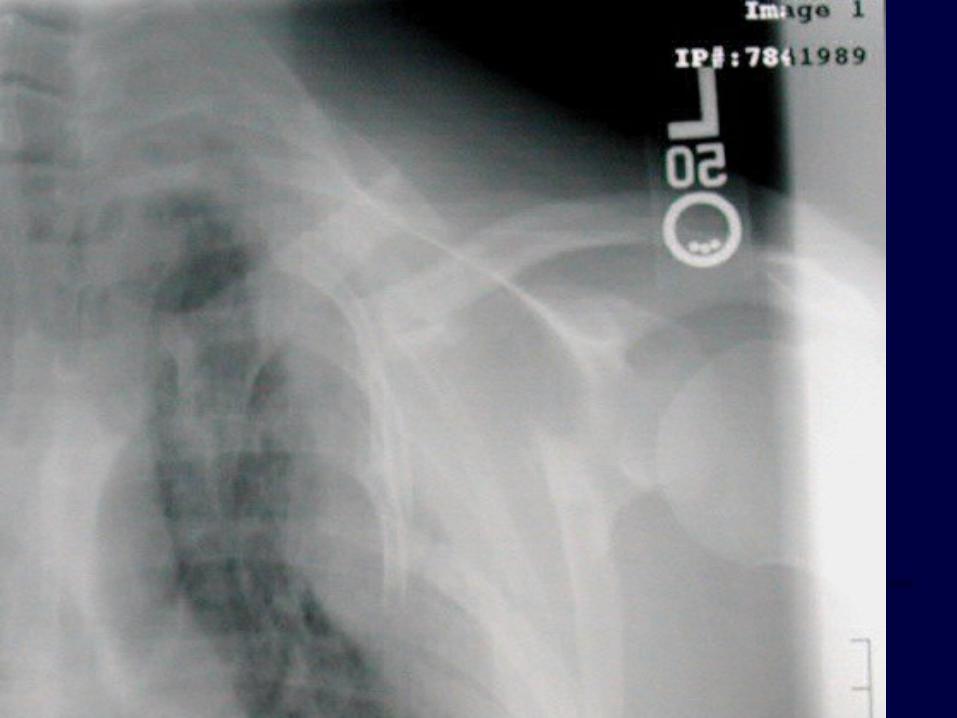
Radiographic Evaluationof the Clavicle
Anteroposterior View
30-degree Cephalic Tilt View
Radiographic Evaluation of the Clavicle
Quesana View– 45-degree angle superiorly and a 45-degree
angle inferiorly – Provide better assessment of the extent of
displacement
Radiographic Evaluation of the AC Joint
Zanca View– AP view centered at AC joint with 10
degree cephalic tilt– Less voltage than used for AP shoulder
Stress Views of the Distal Clavicle & AC Joint
Rationale: demonstrate instability and differentiate grade III AC separations from partial Grade I-II injuries.
Performed by having patient hold 10# weight with injured arm.
Rarely used today, since most Grade I-III AC joint injuries are treated the same anyway, and management of distal clavicle
fractures depends on initial displacement and location of fracture.
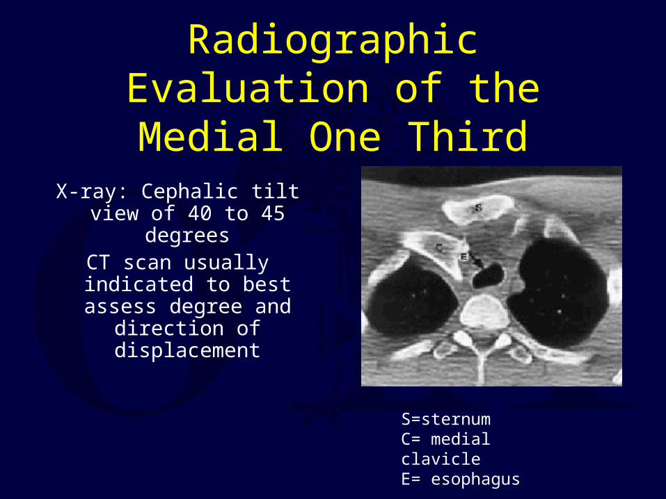
Radiographic Evaluation of the Medial One Third
X-ray: Cephalic tilt view of 40 to 45 degrees
CT scan usually indicated to best assess degree and
direction of displacement
S=sternumC= medial clavicleE= esophagus
Clavicle Fractures
Classification of Clavicle Fractures
Group I : Middle third– Most common (80% of clavicle fractures)
Group II: Distal third– 10-15% of clavicle injuries
Group III: Medial third– Least common (approx. 5%)
Treatment Options
Nonoperative– Sling– Brace
Surgical– Plate Fixation
– Screw or Pin Fixation– Titanium elastic nails (usually
inserted medial to lateral)
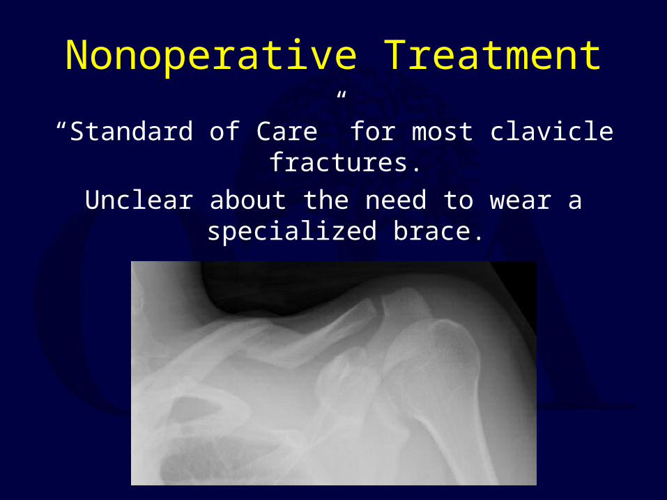
Nonoperative Treatment
“Standard of Care” for most clavicle fractures.
Unclear about the need to wear a specialized brace.
Simple Sling vs. Figure-of-8 Bandage
Prospective randomized trial of 61 patients
Simple sling– Less discomfort
Functional and cosmetic results identical
Alignment of healed fractures unchanged from the initial displacement in both groups
Andersen et al., Acta Orthop Scand 58: 71-4, 1987.

Nonoperative Treatment
It is difficult to reduce clavicle fractures by closed means.
Most clavicle fractures unite rapidly despite displacement.
Significantly displaced mid-shaft and distal-third injuries have a higher incidence of nonunion.
Nonoperative Treatment
There is new evidence that the outcome of nonoperative management of displaced
middle-third clavicle fractures is not as good as traditionally thought, with many patients
having significant functional problems.
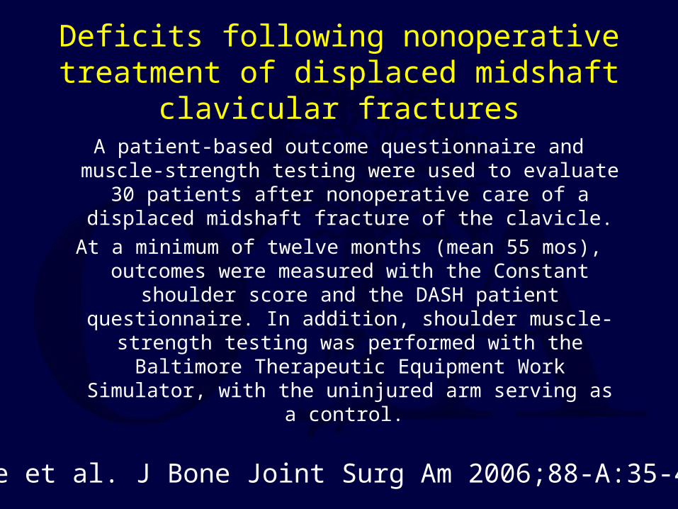
Deficits following nonoperative treatment of displaced midshaft clavicular fractures
A patient-based outcome questionnaire and muscle-strength testing were used to evaluate 30 patients after nonoperative
care of a displaced midshaft fracture of the clavicle.
At a minimum of twelve months (mean 55 mos), outcomes were measured with the Constant shoulder score and the
DASH patient questionnaire. In addition, shoulder muscle-strength testing was performed with the Baltimore Therapeutic Equipment Work Simulator, with the
uninjured arm serving as a control.
McKee et al. J Bone Joint Surg Am 2006;88-A:35-40.
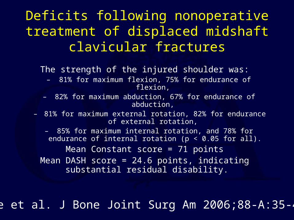
Deficits following nonoperative treatment of displaced midshaft clavicular fractures
The strength of the injured shoulder was:– 81% for maximum flexion, 75% for endurance of flexion,
– 82% for maximum abduction, 67% for endurance of abduction, – 81% for maximum external rotation, 82% for endurance of
external rotation, – 85% for maximum internal rotation, and 78% for endurance of
internal rotation (p < 0.05 for all).
Mean Constant score = 71 pointsMean DASH score = 24.6 points, indicating substantial
residual disability.
McKee et al. J Bone Joint Surg Am 2006;88-A:35-40.
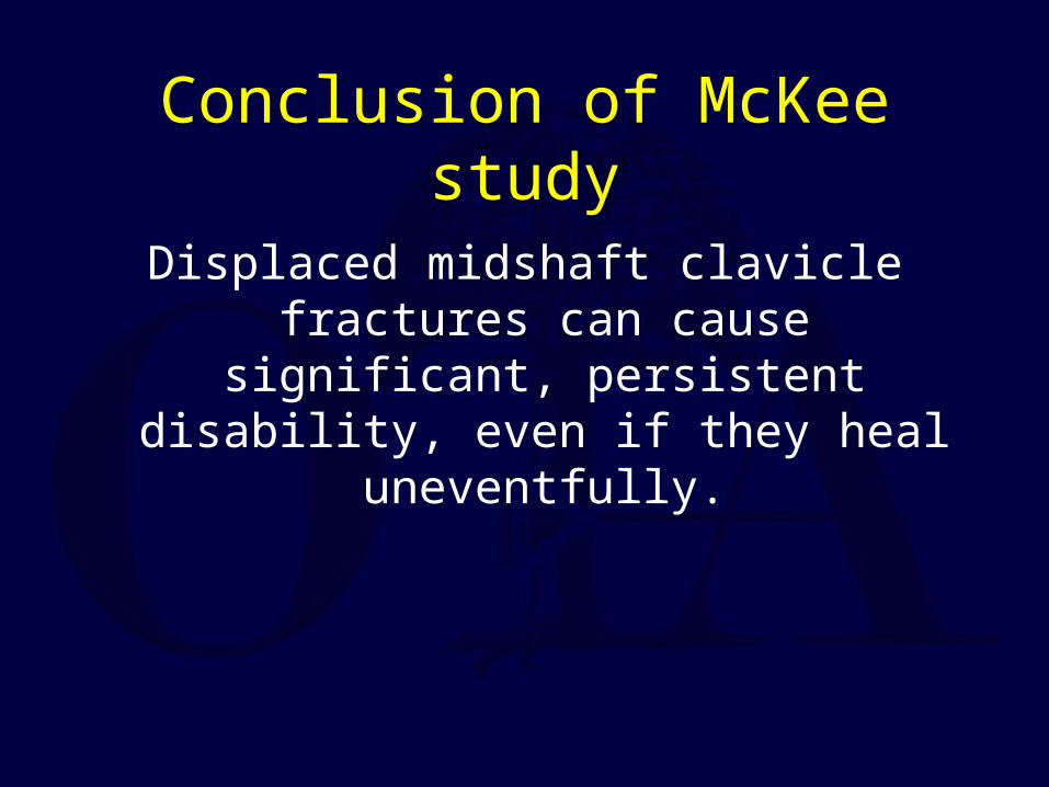
Conclusion of McKee study
Displaced midshaft clavicle fractures can cause significant, persistent disability, even
if they heal uneventfully.
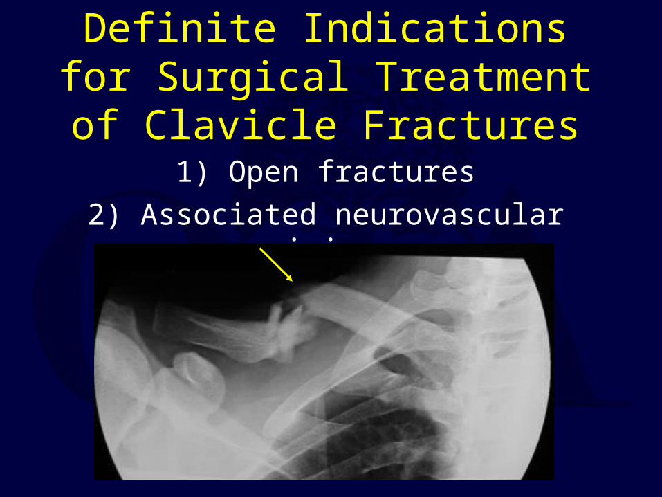
Definite Indications for Surgical Treatment of Clavicle Fractures
1) Open fractures
2) Associated neurovascular injury

Relative Indications for Acute Treatment of Clavicle Fractures
1) Widely displaced fractures2) Multiple trauma
3) Displaced distal-third fractures
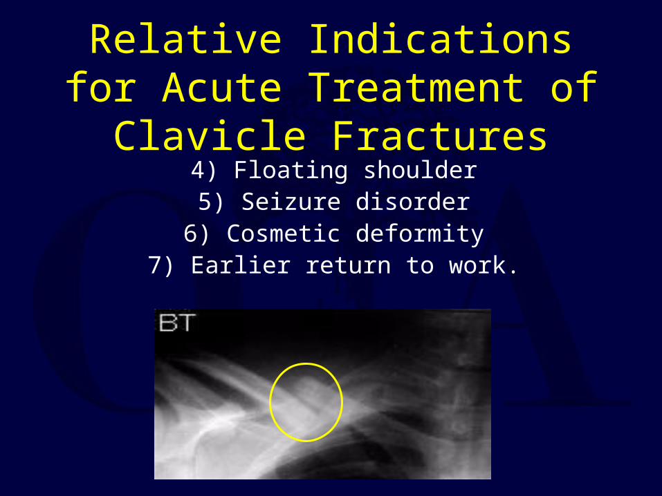
Relative Indications for Acute Treatment of Clavicle Fractures
4) Floating shoulder5) Seizure disorder
6) Cosmetic deformity7) Earlier return to work.
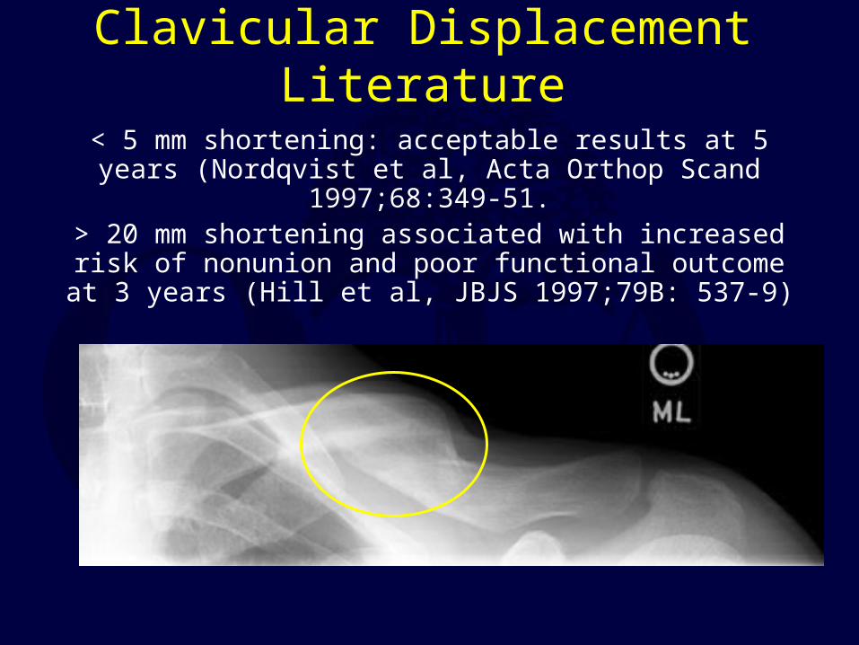
Clavicular Displacement Literature
< 5 mm shortening: acceptable results at 5 years (Nordqvist et al, Acta Orthop Scand 1997;68:349-51.
> 20 mm shortening associated with increased risk of nonunion and poor functional outcome at 3 years (Hill et al, JBJS
1997;79B: 537-9)
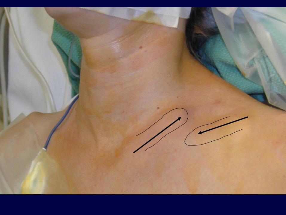
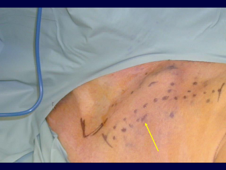
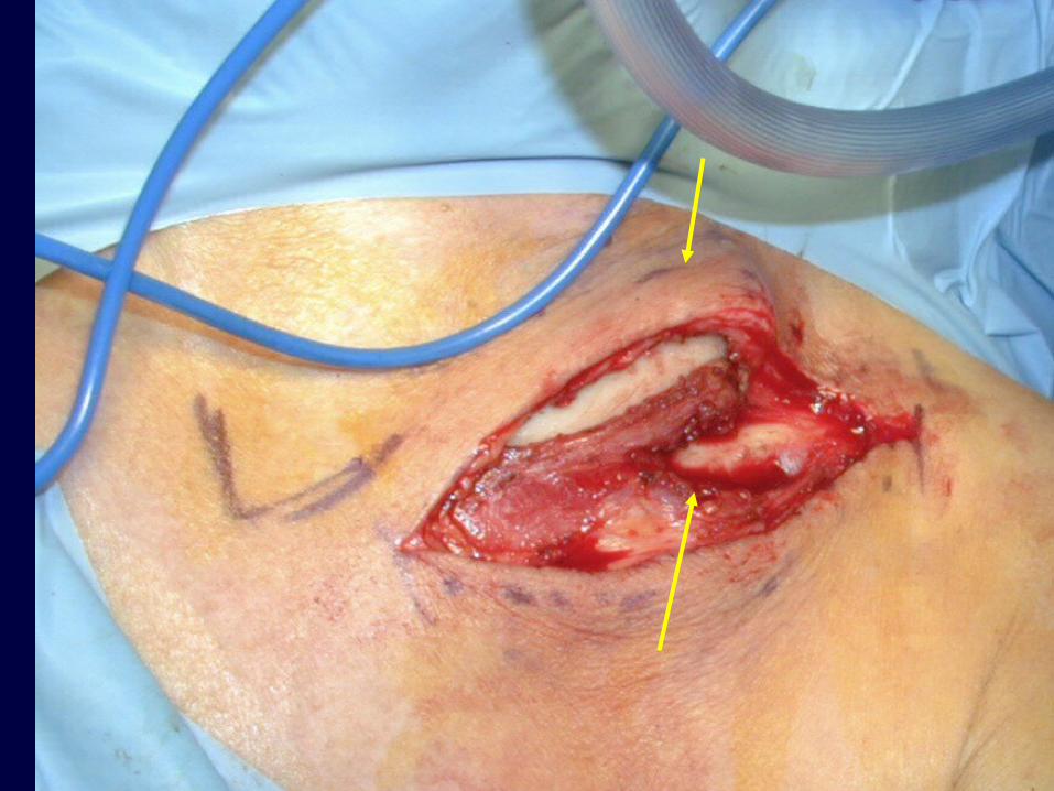
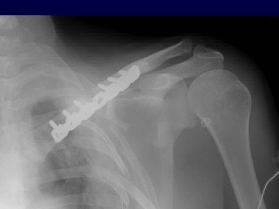
Plate Fixation
Traditional means of ORIF
Plate applied superiorly or inferiorlyInferior plating associated with lower risk of
hardware prominence.
Used for acute displaced fractures and nonunions.
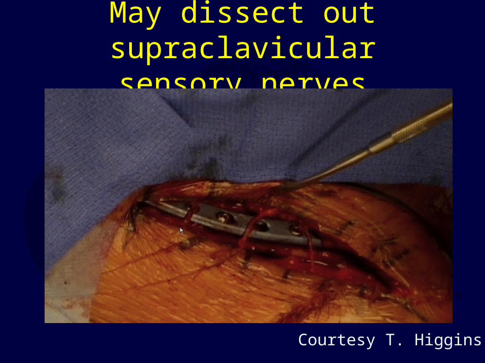
May dissect out supraclavicular sensory nerves
Courtesy T. Higgins

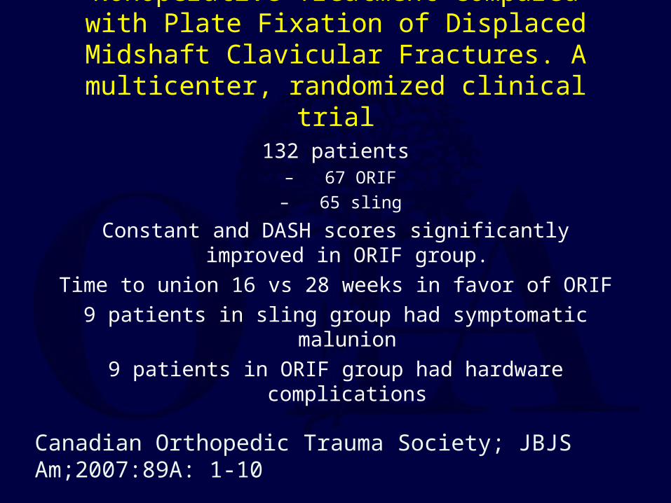
Nonoperative Treatment Compared with Plate Fixation of Displaced Midshaft Clavicular Fractures.
A multicenter, randomized clinical trial
132 patients– 67 ORIF
– 65 sling
Constant and DASH scores significantly improved in ORIF group.
Time to union 16 vs 28 weeks in favor of ORIF
9 patients in sling group had symptomatic malunion
9 patients in ORIF group had hardware complications
Canadian Orthopedic Trauma Society; JBJS Am;2007:89A: 1-10
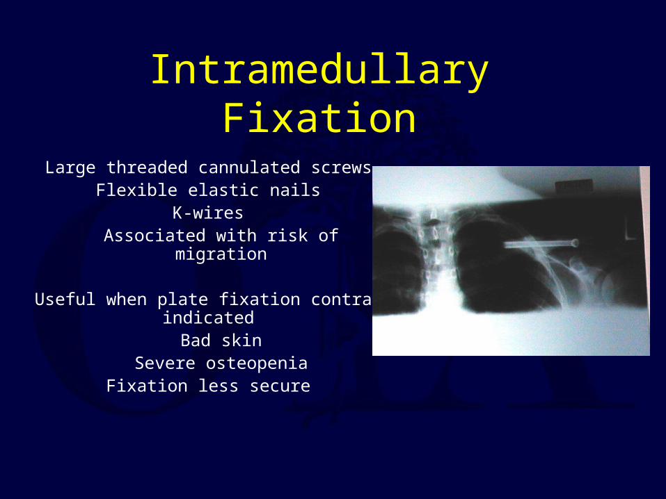
Intramedullary Fixation
Large threaded cannulated screwsFlexible elastic nails
K-wiresAssociated with risk of migration
Useful when plate fixation contra-indicatedBad skin
Severe osteopeniaFixation less secure
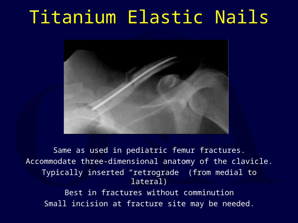
Titanium Elastic Nails
Same as used in pediatric femur fractures.
Accommodate three-dimensional anatomy of the clavicle.
Typically inserted “retrograde” (from medial to lateral)
Best in fractures without comminution
Small incision at fracture site may be needed.
Minimally Invasive Intramedullary Nailing of MidshaftClavicular Fractures Using Titanium Elastic Nails
31 cases evaluated 26 mos avg (6-46 mos)
Three groups:Isolated, n=9
Additional injuries, n=15
Multiple injuries, n=7
Mueller M, et al. J Trauma 2008;64:1528-1534
Minimally Invasive Intramedullary Nailing of MidshaftClavicular Fractures Using Titanium Elastic Nails
No nonunions or refractures in any group.
7 cases medial migration; 1 case lateral perforation in 1 case req’d shortening of nail.
No differences in outcome between groups in subjective outcome and objective scores
(DASH, Constant and Murley).
Mueller M, et al. J Trauma 2008;64:1528-1534
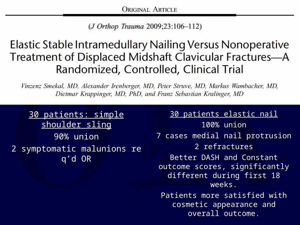
30 patients: simple shoulder sling
90% union
2 symptomatic malunions req’d OR
30 patients elastic nail
100% union
7 cases medial nail protrusion
2 refractures
Better DASH and Constant outcome scores, significantly different during
first 18 weeks.
Patients more satisfied with cosmetic appearance and overall outcome.
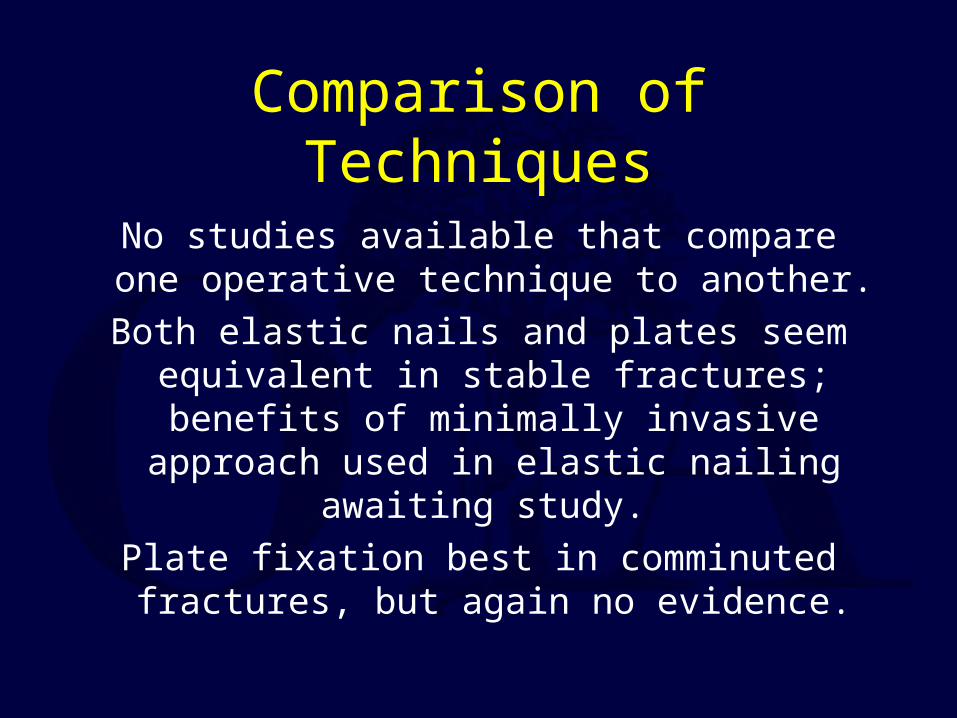
Comparison of Techniques
No studies available that compare one operative technique to another.
Both elastic nails and plates seem equivalent in stable fractures; benefits of minimally invasive approach used in elastic nailing
awaiting study.
Plate fixation best in comminuted fractures, but again no evidence.

Does Timing of Surgery Matter?
Matched group comparison of 15 patients who underwent early compression plate
fixation to 15 other patients who had operative repair of a malunion/nonunion at
avg of 63 months.
Potter JM, et al. J Shoulder Elbow Surg 2007;16:514-8
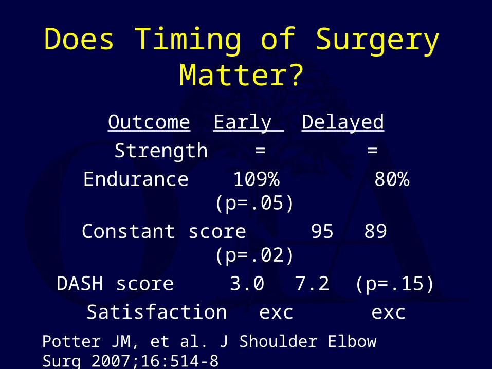
Does Timing of Surgery Matter?
Outcome Early Delayed
Strength = =
Endurance 109% 80% (p=.05)
Constant score 95 89 (p=.02)
DASH score 3.0 7.2 (p=.15)
Satisfaction exc exc
Potter JM, et al. J Shoulder Elbow Surg 2007;16:514-8
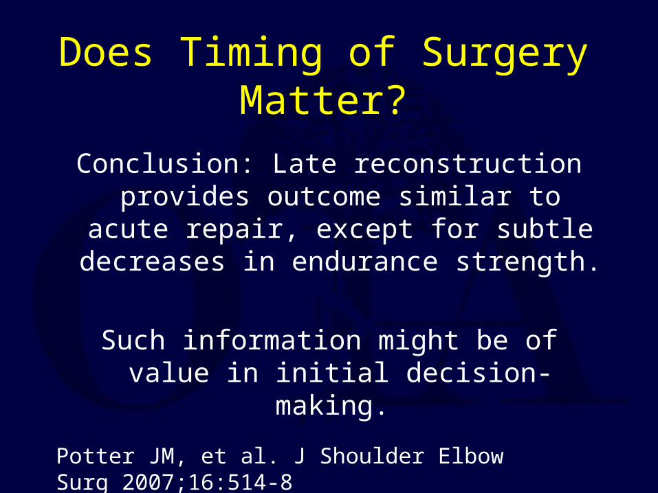
Does Timing of Surgery Matter?
Conclusion: Late reconstruction provides outcome similar to acute repair, except for
subtle decreases in endurance strength.
Such information might be of value in initial decision-making.
Potter JM, et al. J Shoulder Elbow Surg 2007;16:514-8
Complications of Clavicular Fractures and its Treatment
Nonunion
Malunion
Neurovascular Sequelae
Post-Traumatic Arthritis
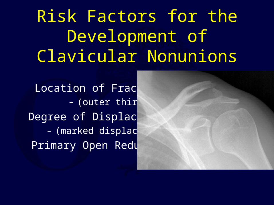
Risk Factors for the Development of Clavicular Nonunions
Location of Fracture – (outer third)
Degree of Displacement – (marked displacement)
Primary Open Reduction
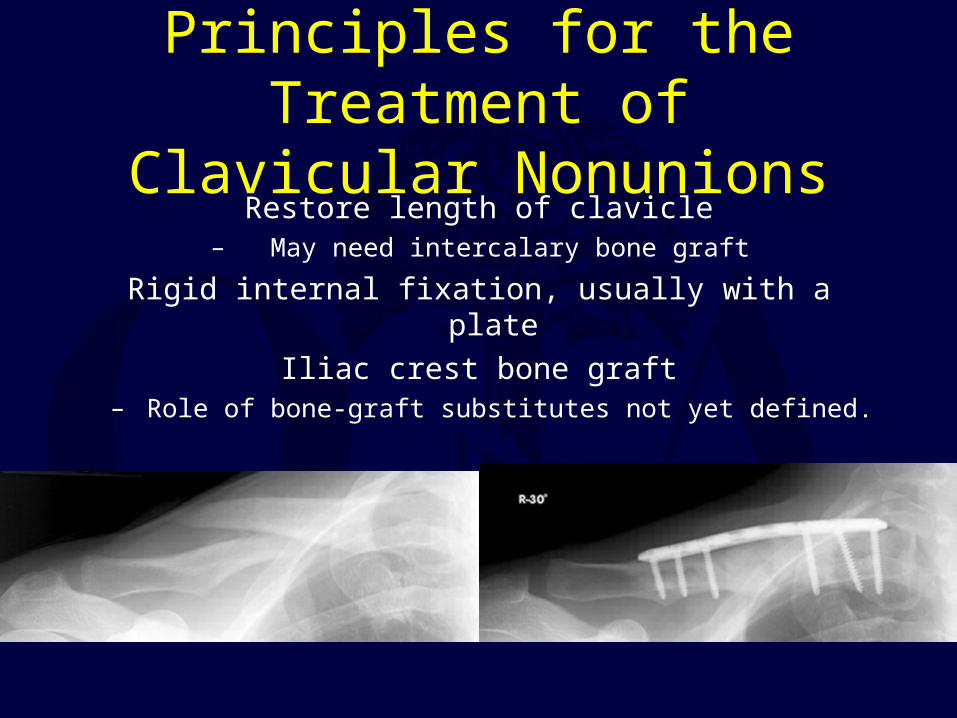
Principles for the Treatment of Clavicular Nonunions
Restore length of clavicle– May need intercalary bone graft
Rigid internal fixation, usually with a plate
Iliac crest bone graft– Role of bone-graft substitutes not yet defined.
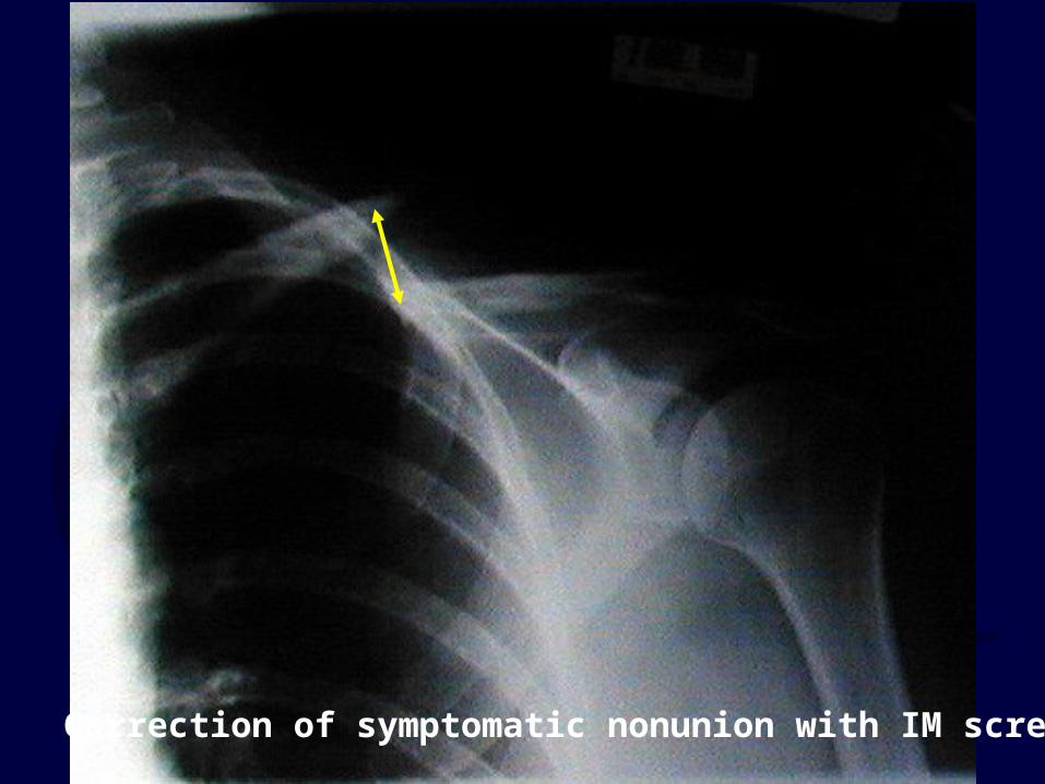
Correction of symptomatic nonunion with IM screw


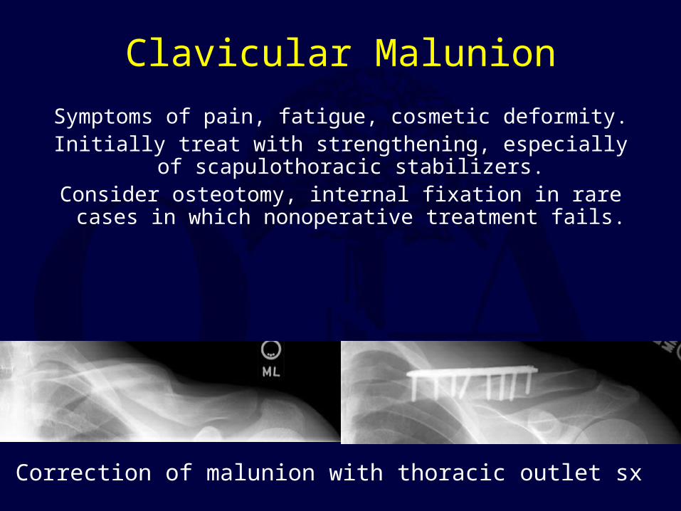
Clavicular Malunion
Symptoms of pain, fatigue, cosmetic deformity.Initially treat with strengthening, especially of
scapulothoracic stabilizers.Consider osteotomy, internal fixation in rare cases in
which nonoperative treatment fails.
Correction of malunion with thoracic outlet sx
Neurologic Sequelae
Occasionally, fracture fragments or abundant callus can cause brachial plexus symptoms.
Treatment is reduction and fixation of the fracture, or resection of callus with or
without osteotomy and fixation for malunions.
McKee MD, et al. J Bone Joint Surg Am 2003;85-A(5):790-7
Osteotomy for Clavicular Malunion
15 patients with malunion after nonoperative treatment of a displaced midshaft clavicle fracture of the clavicle.
Average clavicular shortening was 2.9 cm (range, 1.6 to 4.0 cm).
Mean time from the injury to presentation was three years (range, 1 to 15 years).
Outcome scores revealed major functional deficits.
All patients underwent corrective osteotomy of the malunion through the original fracture line and internal fixation.
McKee MD, et al. J Bone Joint Surg Am 2003;85-A(5):790-7
Osteotomy for Clavicular Malunion
At follow-up (mean 20 months postoperatively) the osteotomy site had united in 14 of 15 patients.
All 14 patients satisfied with the result.Mean DASH score for all 15 patients improved from 32 points preoperatively to 12 points at the time of follow-up
(p = 0.001).Mean shortening of the clavicle improved from 2.9 to 0.4 cm
(p = 0.01).There was 1 nonunion, and 2 patients had elective removal of
the plate.
Rosenberg N, et al. J Shoul Elbow Surg 2007;16:510-513
Functional Outcome of Surgical Treatment of Symptomatic Nonunion and Malunion of Midshaft
Clavicle Fractures
13 cases plate fixation / autogenous grafting of a clavicle nonunion / malunion, followed mean 41 months.
All united46% returned to previous job and sport
Constant scores remained lower than opposite arm<25% free of pain.
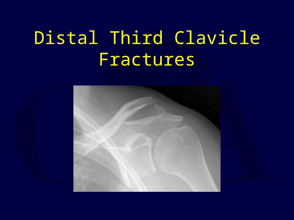
Distal Third Clavicle Fractures
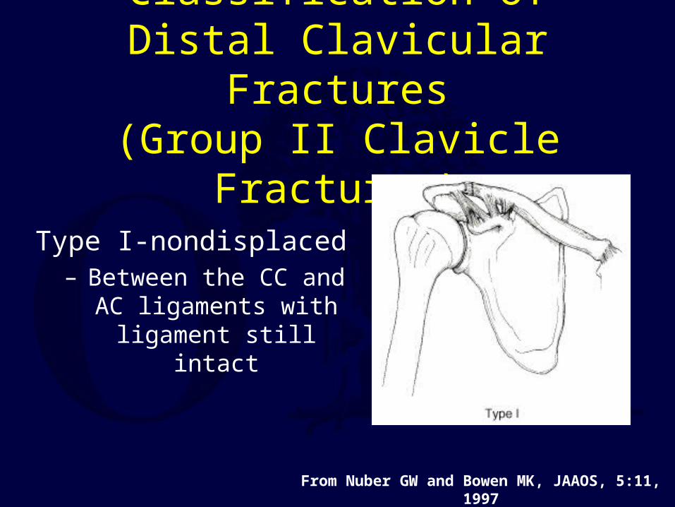
Classification of Distal Clavicular Fractures
(Group II Clavicle Fractures)
Type I-nondisplaced – Between the CC and
AC ligaments with ligament still intact
From Nuber GW and Bowen MK, JAAOS, 5:11, 1997
Classification of Distal Clavicular Fractures
Type II– Typically displaced secondary to a fracture
medial to the coracoclavicular ligaments, keeping the distal fragment reduced while allowing the medial fragmetn to displace
superiorly– Highest rate of nonunion (up to 30%)
– Two Types
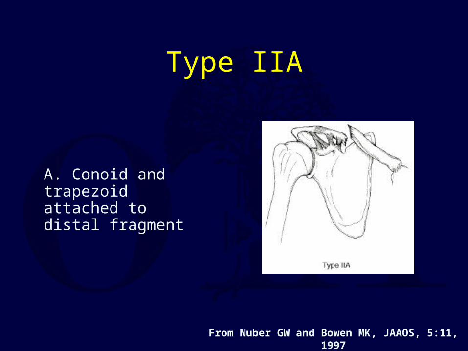
Type IIA
A. Conoid and trapezoid attached to distal fragment
From Nuber GW and Bowen MK, JAAOS, 5:11, 1997
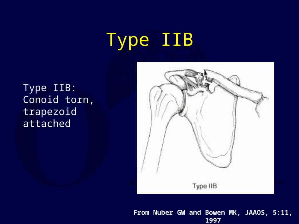
Type IIB
Type IIB: Conoid torn, trapezoid attached
From Nuber GW and Bowen MK, JAAOS, 5:11, 1997
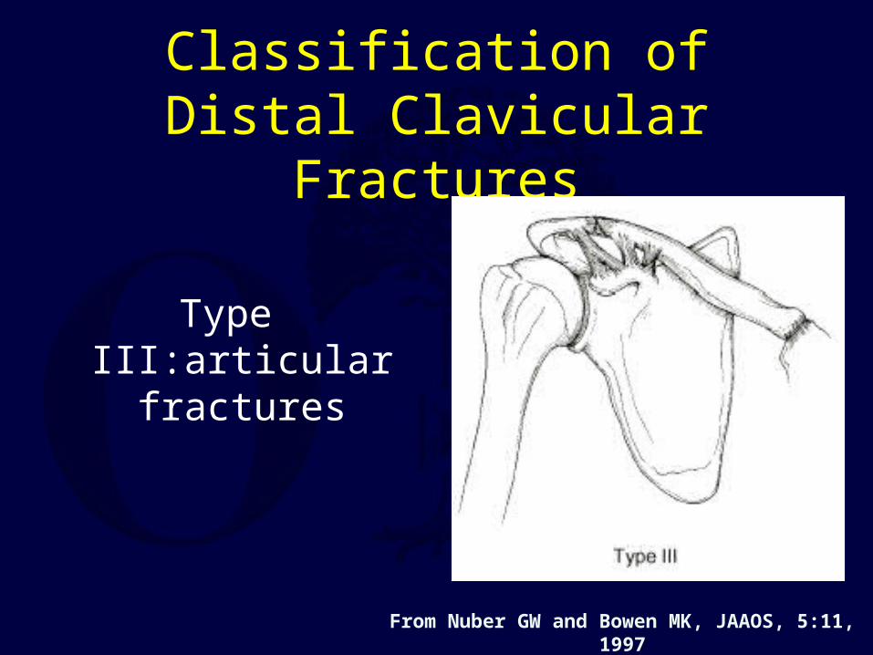
Classification of Distal Clavicular Fractures
Type III:articular fractures
From Nuber GW and Bowen MK, JAAOS, 5:11, 1997
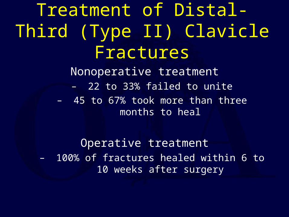
Treatment of Distal-Third (Type II) Clavicle Fractures
Nonoperative treatment– 22 to 33% failed to unite
– 45 to 67% took more than three months to heal
Operative treatment– 100% of fractures healed within 6 to 10 weeks after
surgery
Displaced Type II fractures of the distal clavicle are often treated more aggressively because of the increased risk of nonunion
with nonoperative treatment
Techniques for Acute Operative Treatment of Distal Clavicle Fractures
Kirschner wires inserted into the distal fragmentDorsal plate fixationCC screw fixation
Tension-band wire or sutureTransfer of coracoid process to the clavicle
Clavicular Hook Plate

For most techniques of clavicular fixation, coracoclavicular fixation is also needed to
prevent redisplacement of the medial clavicle.
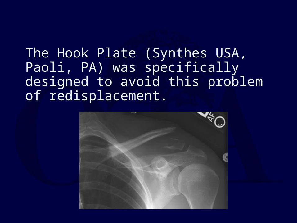
The Hook Plate (Synthes USA, Paoli, PA) was specifically designed to avoid this problem of redisplacement.

Hook Plate - Results
Recent series of distal clavicle fractuers treated with the Hook Plate document high union rates of 88% - 100%. Complications
are rare but potentially significant, including new fracture about the implant, rotator cuff tear, and frequent subacromial
impingement.
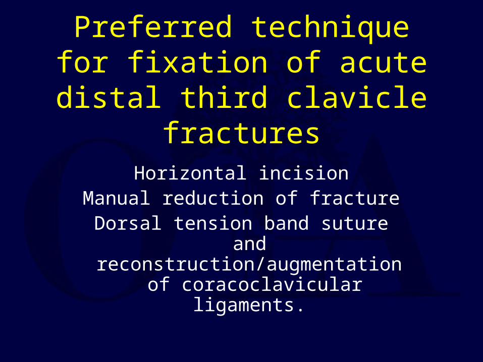
Preferred technique for fixation of acute distal third clavicle
fractures
Horizontal incisionManual reduction of fracture
Dorsal tension band suture and reconstruction/augmentation of
coracoclavicular ligaments.
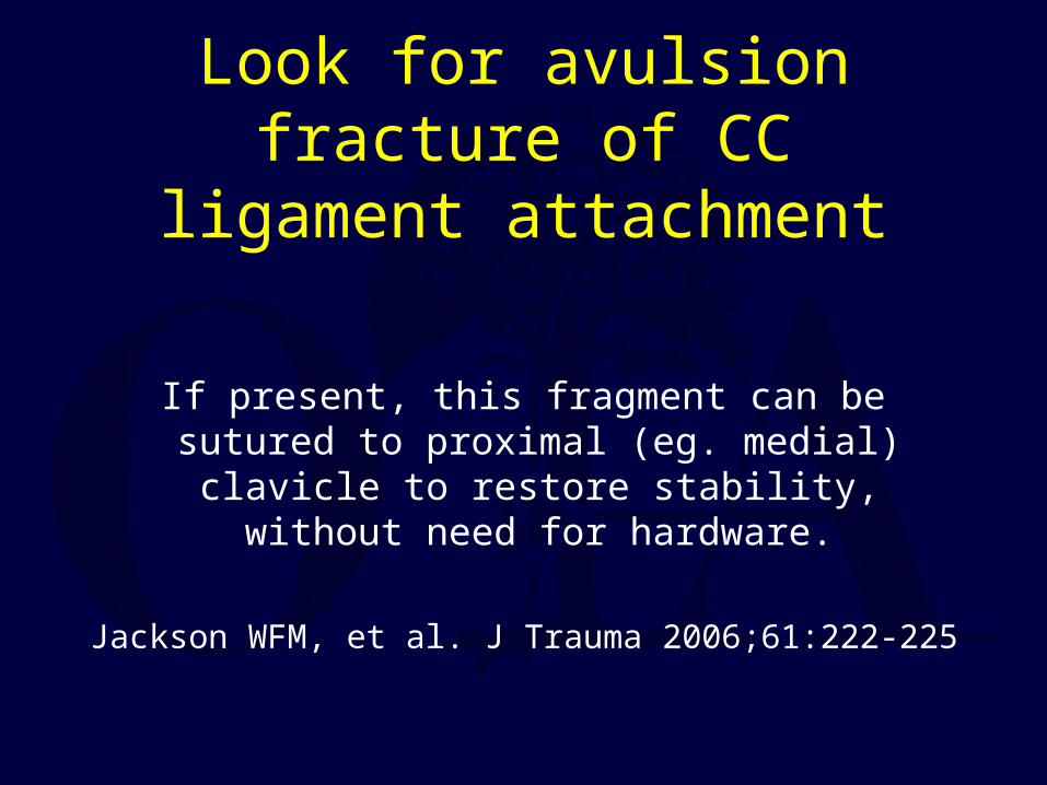
Look for avulsion fracture of CC ligament attachment
If present, this fragment can be sutured to proximal (eg. medial) clavicle to restore
stability, without need for hardware.
Jackson WFM, et al. J Trauma 2006;61:222-225
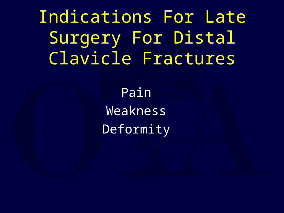
Indications For Late Surgery For Distal Clavicle Fractures
Pain
Weakness
Deformity
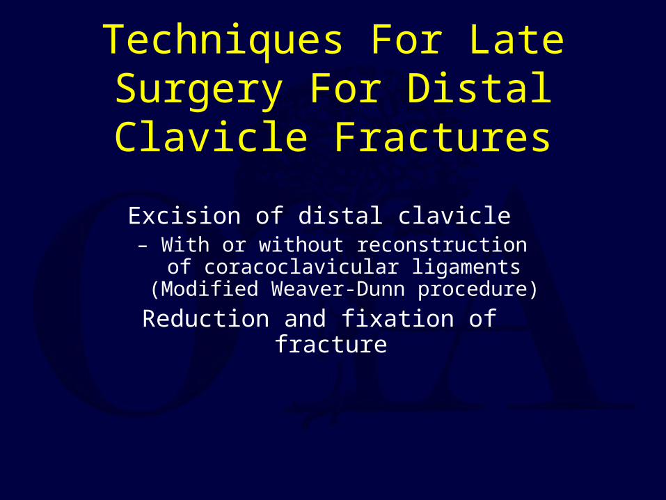
Techniques For Late Surgery For Distal Clavicle Fractures
Excision of distal clavicle– With or without reconstruction of coracoclavicular ligaments (Modified
Weaver-Dunn procedure)
Reduction and fixation of fracture
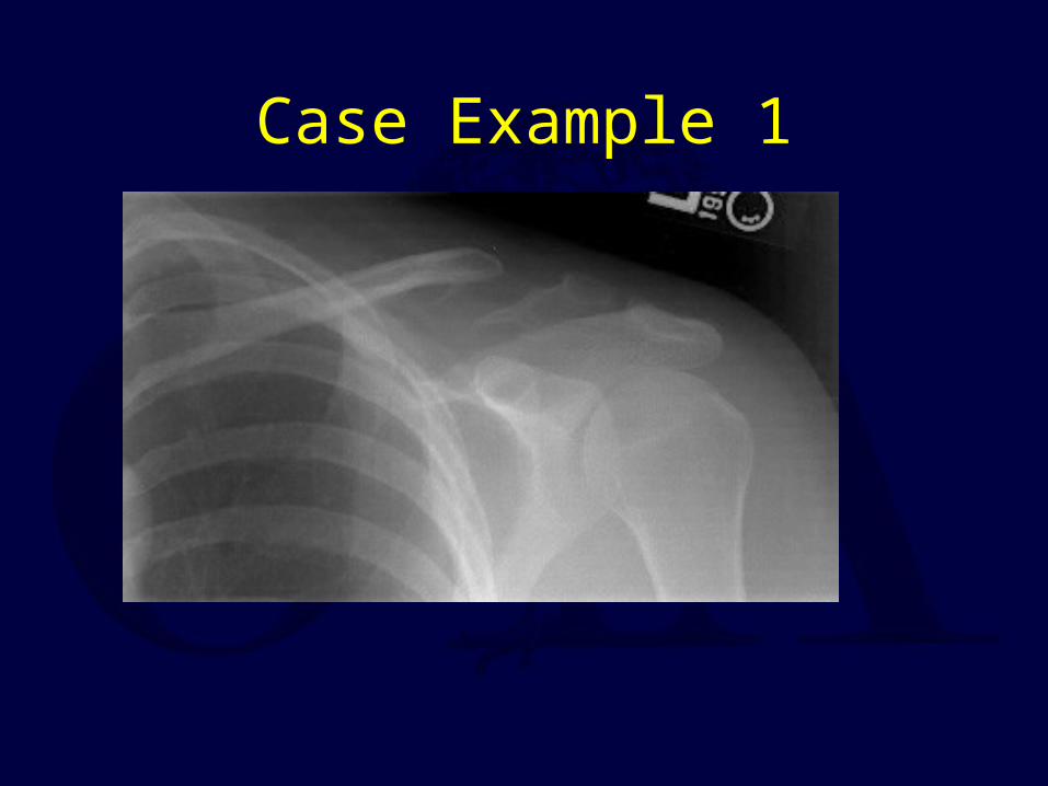
Case Example 1
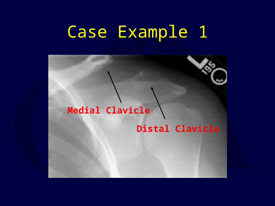
Case Example 1
Distal Clavicle
Medial Clavicle

Case Example 1
Coracoclavicular fixation not visible
Fixation to Acromion

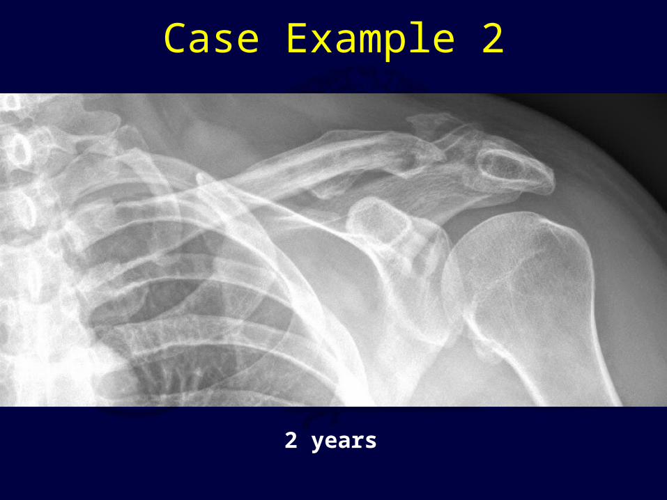
Case Example 2
This fragment likely has CC ligament attached; need to reduce and hold clavicle shaft to this piece.
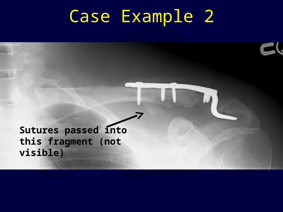
Case Example 2
This fragment likely has CC ligament attached; need to reduce and hold clavicle shaft to this piece.
Sutures passed into this fragment (not visible)
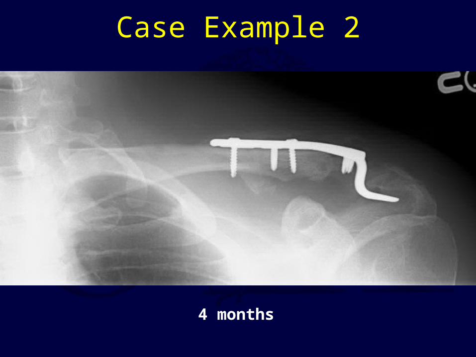
Case Example 2
This fragment likely has CC ligament attached; need to reduce and hold clavicle shaft to this piece.
Sutures passed into this fragment (not visible)
4 months
Case Example 2
This fragment likely has CC ligament attached; need to reduce and hold clavicle shaft to this piece.
Sutures passed into this fragment (not visible)
2 years
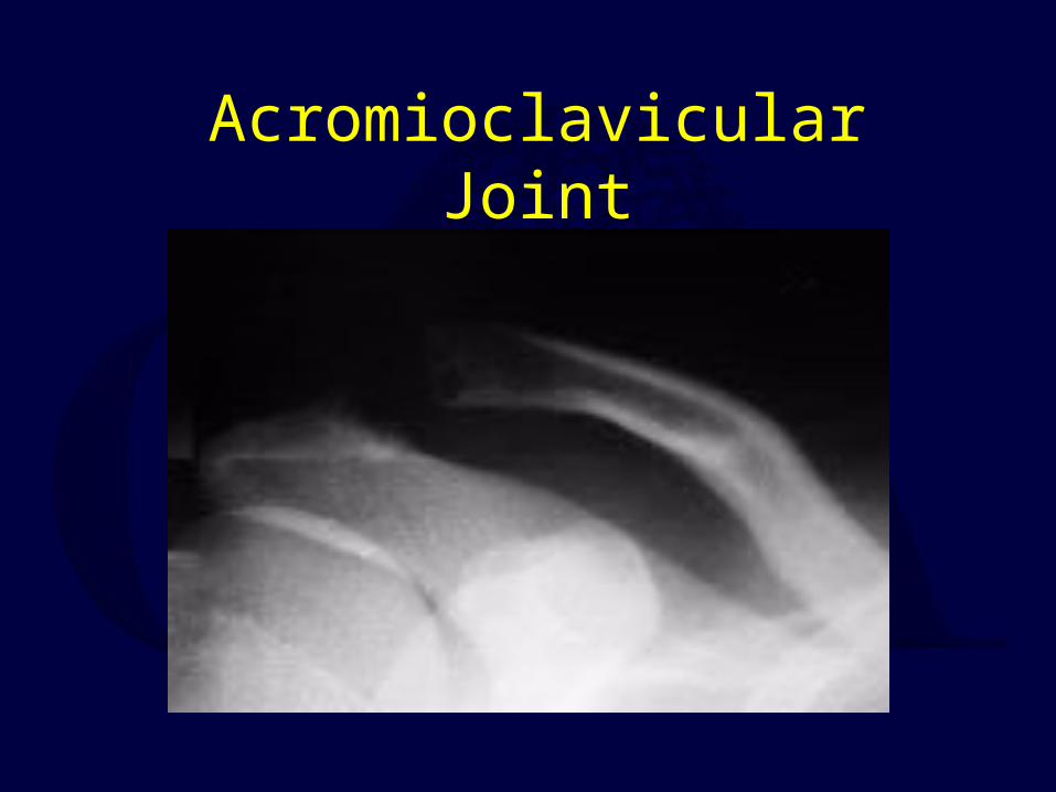
Acromioclavicular Joint

Mechanism
Sports injury or trauma.
Impact to superior acromion, driving the arm down and rupturing the AC joint capsule (first) and then the the coracoclavicular
ligaments (second).
Physical Findings
Pain over lateral clavicle / AC joint
May have prominent distal clavicle
May have skin abrasions
Unwilling to lift arm.
Should have full passive ROM of the shoulder.
Radiographic Evaluation of the Acromioclavicular Joint
Proper exposure of the AC joint requires one-third to one-half the x-ray penetration of routine shoulder
viewsInitial Views:
– Anteroposterior view– Zanca view (15 degree cephalic tilt)
Other views:– Axillary: demonstrates anterior-posterior displacement
– Stress views: not generally relevant for treatment decisions.
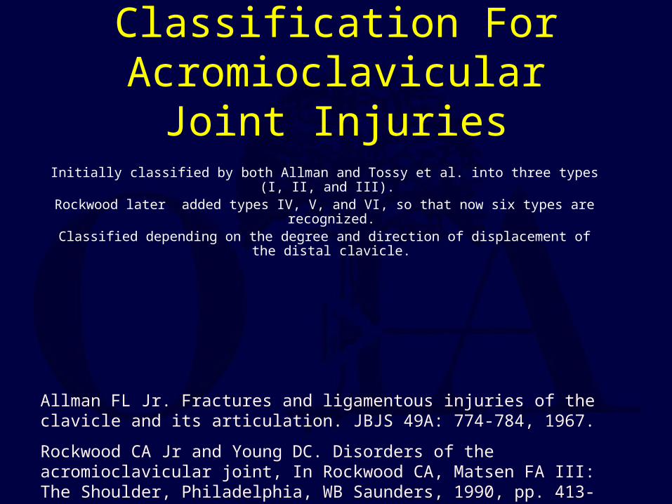
Classification For Acromioclavicular Joint Injuries
Initially classified by both Allman and Tossy et al. into three types (I, II, and III). Rockwood later added types IV, V, and VI, so that now six types are recognized.
Classified depending on the degree and direction of displacement of the distal clavicle.
Allman FL Jr. Fractures and ligamentous injuries of the clavicle and its articulation. JBJS 49A: 774-784, 1967.
Rockwood CA Jr and Young DC. Disorders of the acromioclavicular joint, In Rockwood CA, Matsen FA III: The Shoulder, Philadelphia, WB Saunders, 1990, pp. 413-476.
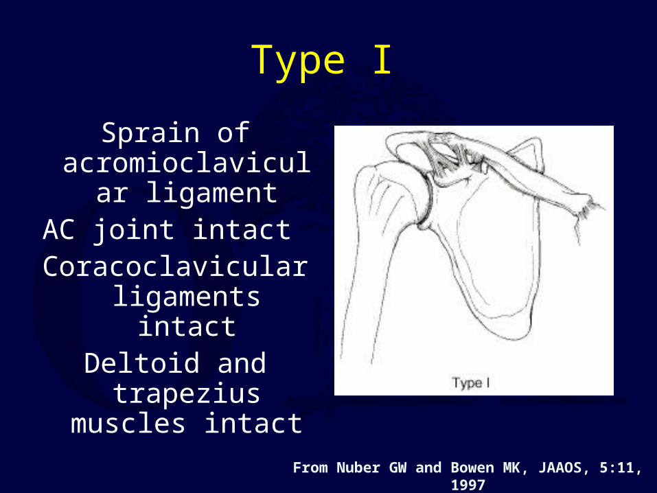
Type I
Sprain of acromioclavicular
ligamentAC joint intact
Coracoclavicular ligaments intact
Deltoid and trapezius muscles intact
From Nuber GW and Bowen MK, JAAOS, 5:11, 1997
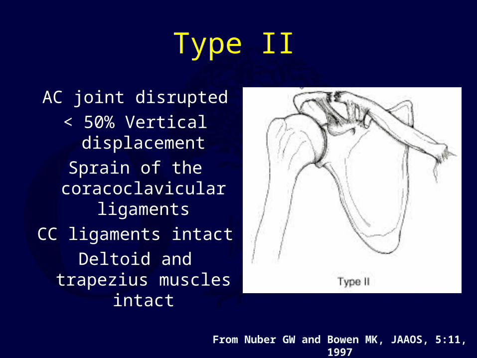
AC joint disrupted
< 50% Vertical displacement
Sprain of the coracoclavicular
ligaments
CC ligaments intact
Deltoid and trapezius muscles intact
Type II
From Nuber GW and Bowen MK, JAAOS, 5:11, 1997

Type III
AC ligaments and CC ligaments all disrupted
AC joint dislocated and the shoulder complex
displaced inferiorlyCC interspace greater than
the normal shoulder(25-100%)
Deltoid and trapezius muscles usually detached from the distal clavicle
From Nuber GW and Bowen MK, JAAOS, 5:11, 1997
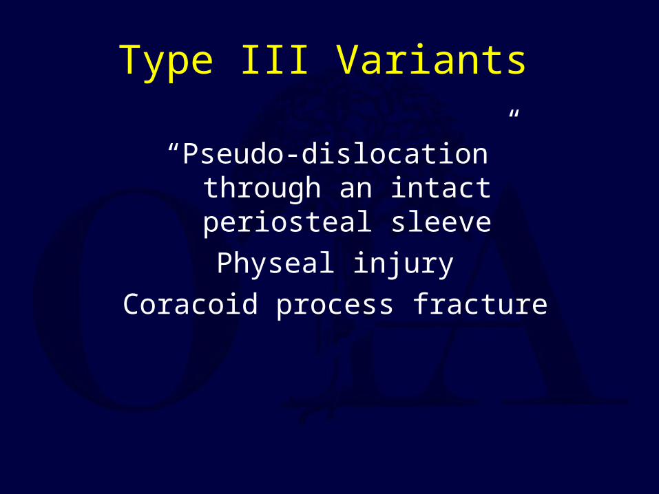
Type III Variants
“Pseudo-dislocation” through an intact periosteal sleeve
Physeal injury
Coracoid process fracture
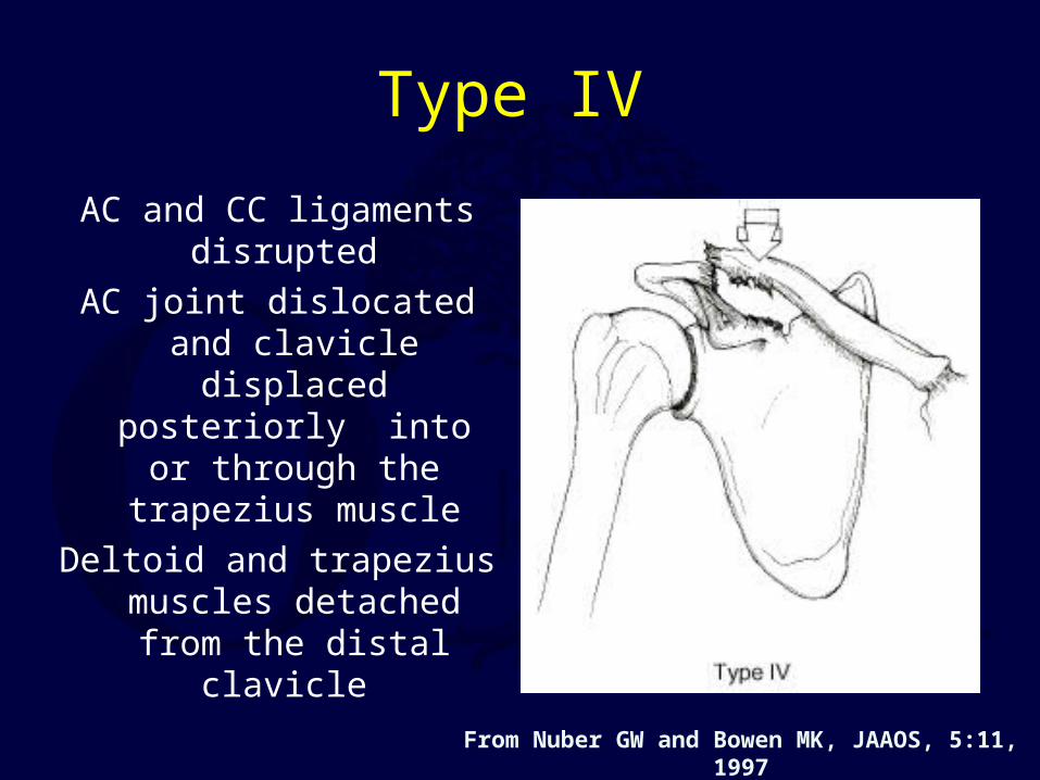
Type IV
AC and CC ligaments disrupted
AC joint dislocated and clavicle displaced posteriorly into or
through the trapezius muscle
Deltoid and trapezius muscles detached from
the distal clavicle
From Nuber GW and Bowen MK, JAAOS, 5:11, 1997
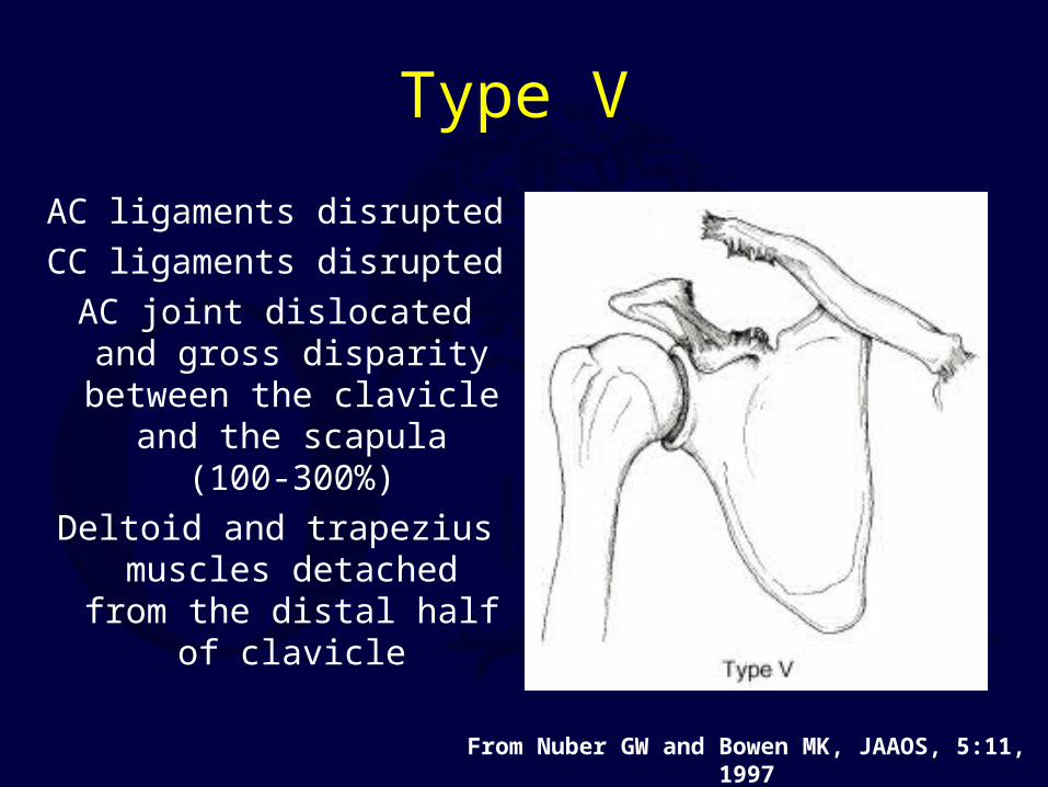
Type V
AC ligaments disrupted
CC ligaments disrupted
AC joint dislocated and gross disparity between
the clavicle and the scapula (100-300%)
Deltoid and trapezius muscles detached from
the distal half of clavicle
From Nuber GW and Bowen MK, JAAOS, 5:11, 1997

Type VI
AC joint dislocated and clavicle displaced inferior
to the acromion or the coracoid process
AC and CC ligaments disrupted
Deltoid and trapezius muscles detached from the
distal clavicle
From Nuber GW and Bowen MK, JAAOS, 5:11, 1997
Treatment Options For Types I - II Acromioclavicular Joint Injuries
Nonoperative: Ice and protection until pain subsides (7 to 10 days).
Return to sports as pain allows (1-2 weeks)
No apparent benefit to the use of specialized braces.
Type II operative treatment– Generally reserved only for the patient with
chronic pain.– Treatment is resection of the distal clavicle and
reconstruction of the coracoclavicular ligaments.
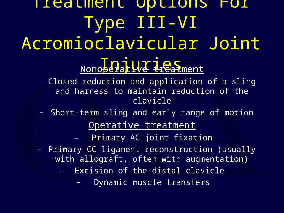
Treatment Options For Type III-VI Acromioclavicular Joint Injuries
Nonoperative treatment– Closed reduction and application of a sling and harness to
maintain reduction of the clavicle– Short-term sling and early range of motion
Operative treatment– Primary AC joint fixation
– Primary CC ligament reconstruction (usually with allograft, often with augmentation)
– Excision of the distal clavicle – Dynamic muscle transfers
Type III Injuries: Need for acute surgical treatment remains very controversial.
Most surgeons recommend conservative treatment except in the throwing athlete or
overhead worker.
Repair generally avoided in contact athletes because of the risk of reinjury.
Literature unable to support operative or nonoperative treatment as superior
Functional outcomes appear similar.
Cosmesis not different (scar vs bump)
Only 50% of surgical cases reduced at follow-up.
10% complications after surgery.
Ceccarelli et al. J Orthopaed Traumatol 2008;9:105-108.
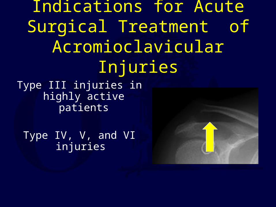
Indications for Acute Surgical Treatment of Acromioclavicular
Injuries
Type III injuries in highly active patients
Type IV, V, and VI injuries
Surgical Options for AC Joint Instability
Coracoid process transfer to distal transfer (Dynamic muscle transfer)
Primary AC joint fixation
Primary Coracoclavicular Fixation
CC ligament reconstruction +/- distal clavicle excision.
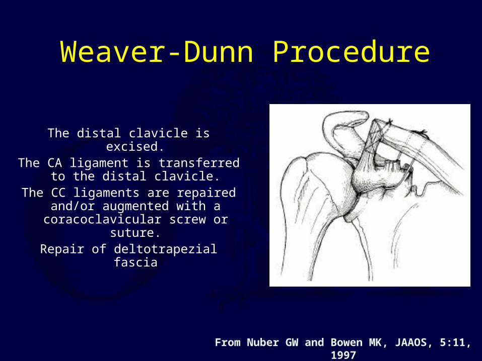
Weaver-Dunn Procedure
The distal clavicle is excised.The CA ligament is transferred to
the distal clavicle.The CC ligaments are repaired
and/or augmented with a coracoclavicular screw or
suture.Repair of deltotrapezial fascia
From Nuber GW and Bowen MK, JAAOS, 5:11, 1997
Indications for Late Surgical Treatment of Acromioclavicular
Injuries
Pain
Weakness
Deformity
Techniques for Late Surgical Treatment of Acromioclavicular
Injuries
Reduction of AC joint and repair of AC and CC ligaments
Resection of distal clavicle and reconstruction of CC ligaments (Weaver-Dunn Procedure)
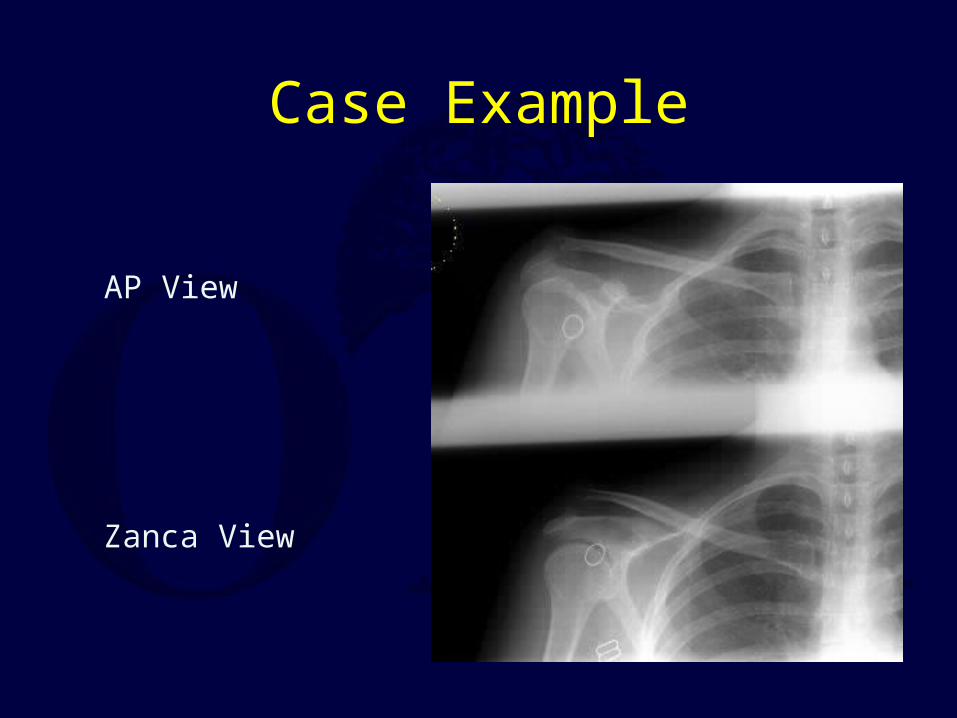
Case Example
AP View
Zanca View
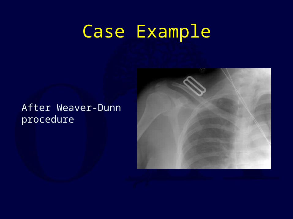
Case Example
After Weaver-Dunnprocedure
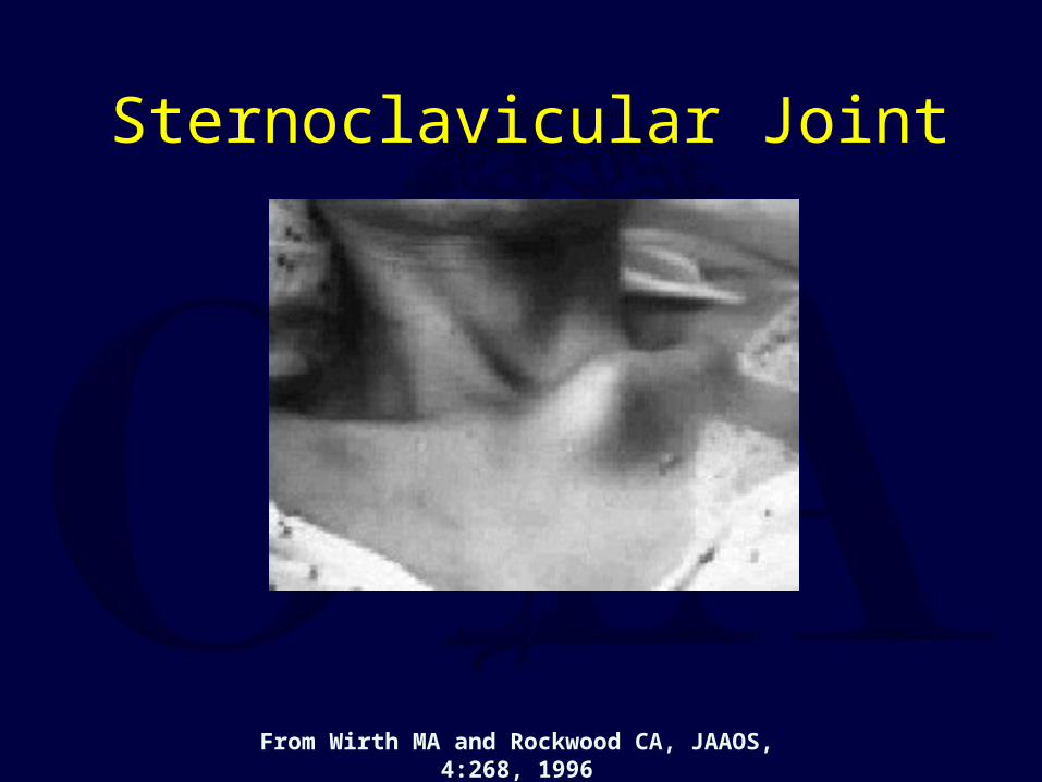
Sternoclavicular Joint
From Wirth MA and Rockwood CA, JAAOS, 4:268, 1996
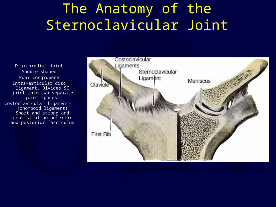
The Anatomy of the Sternoclavicular Joint
Diarthrodial Joint“Saddle shaped”Poor congruence
Intra-articular disc ligament. Divides SC joint into two
separate joint spaces.Costoclavicular ligament- (rhomboid ligament) Short and strong and consist of an anterior
and posterior fasciculus
Interclavicular ligament- Connects the superomedial aspects of each clavicle with the
capsular ligaments and the upper sternum
Capsular ligament- Covers the anterior and posterior aspects of the joint and represents
thickenings of the joint capsule. The anterior portion of the ligament is heavier and stronger
than the posterior portion.
Epiphysis of the Medial Clavicle
Medial Physis- Last of the ossification centers to appear in the body and the last
epiphysis to close.
Does not ossify until 18th to 20th year
Does not unite with the clavicle until the 23rd to 25th year
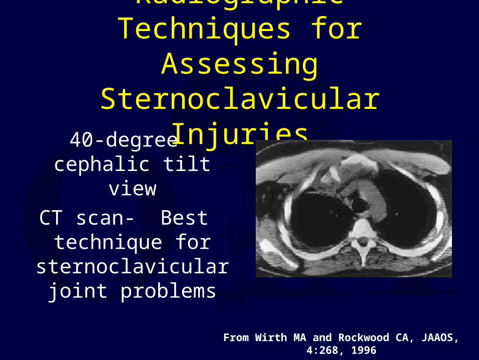
Radiographic Techniques for Assessing Sternoclavicular
Injuries40-degree cephalic tilt
view
CT scan- Best technique for
sternoclavicular joint problems
From Wirth MA and Rockwood CA, JAAOS, 4:268, 1996
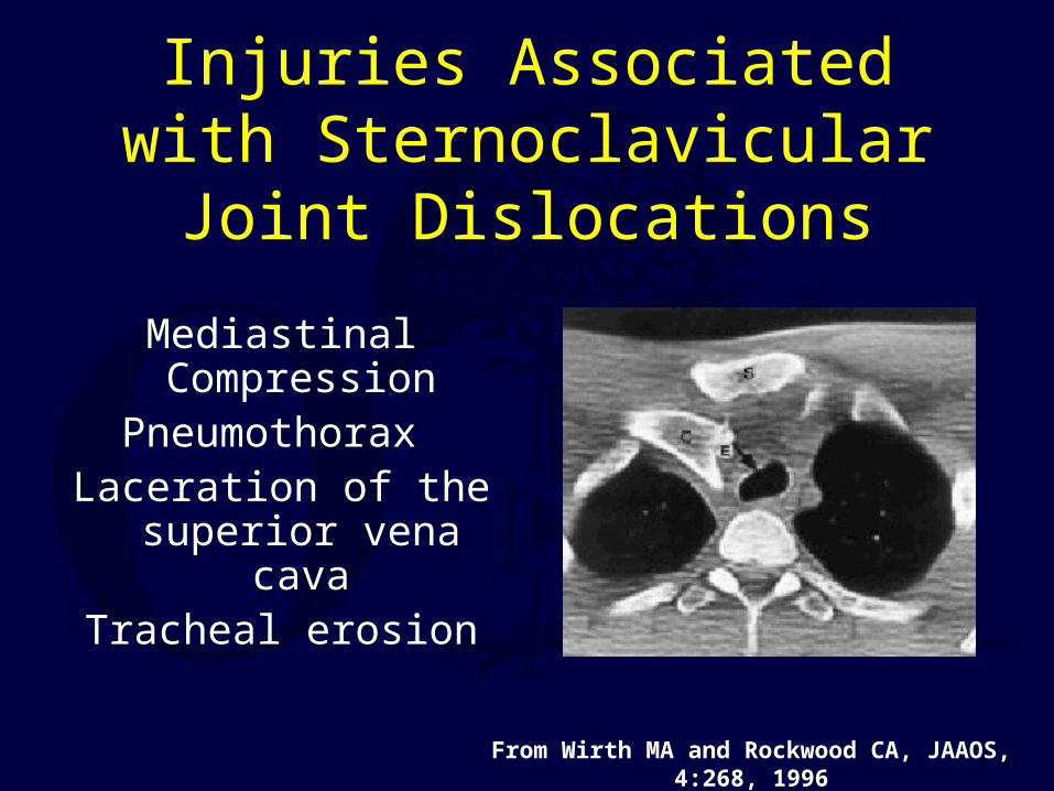
Injuries Associated with Sternoclavicular Joint
Dislocations
Mediastinal CompressionPneumothorax
Laceration of the superior vena cava
Tracheal erosion
From Wirth MA and Rockwood CA, JAAOS, 4:268, 1996
Treatment of Anterior Sternoclavicular Dislocations
Nonoperative treatment
• Analgesics and immobilization
• Functional outcome usually good
Closed reduction
• Often not successful
• Direct pressure over the medial end of the clavicle may reduce the joint
Treatment of Posterior Sternoclavicular Dislocations
Careful examination of the patient is extremely important to rule out vascular
compromise.Consider CT to rule out mediastinal
compressionAttempt closed reduction - it is often
successful and remains stable.
Closed Reduction Techniques
Abduction traction
Adduction traction
“Towel Clip” - anterior force applied to clavicle by percutaneously applied towel
clip
Operative techniques
Resection arthroplasty
– May result in instability of remaining clavicle unless stabilization is done.
– Suggest minimal resection of bone and fixation of medial clavicle to first rib.
Sternoclavicular reconstruction with suture, tendon graft.
Literature – For those interested in further review.
Clavicle Fractures
Andersen K; Jensen PO; Lauritzen J. Treatment of clavicular fractures. Figure-of-eight bandage versus a simple sling. Acta Orthop Scand 1987 Feb;58(1): p71-4.
Canadian Orthopaedic Trauma Society. Nonoperative Treatment Compared with Plate Fixation of Displaced Midshaft Clavicular Fractures. A multicenter, randomized clinical trial. J Bone Joint Surg
2007;89-A:1-10.
McKee MD, Pedersen EM, Jones C, et al. Deficits following nonoperative treatment of displaced midshaft clavicular fractures. J Bone Joint Surg 2006;88-A:35-40.
Mueller M, Rangger C, Striepens N, Burger C. Minimally Invasive Intramedullary Nailing of Midshaft Clavicular Fractures Using Titanium Elastic Nails. J Trauma 2008;1528-1534.
Nowak J, Holgersson M, Larsson S. Can we predict long-term sequelae after fractures of the clavicle based on initial findings? A prospective study with nine to ten years of follow up. J Shoudler Elbow
Surg 2004;13:479-486.
Potter JM, Jones C, Wild LM, Schemitsch EH, McKee MD. Does delay matter? The restoration of objectively measured shoulder strength and patient-oriented outcome after immediate fixation versus
delayed reconstruction of displaced midshaft fractures of the clavicle. J Shoulder Elbow Surg 2007;16:514-518.
Robinson CM, Court-Brown CM, McQueen MM, Wakefield AE. Estimating the risk of nonunion following nonoperative treatment of a clavicular fracture. J Bone Joint Surg Am 2004;86-A:1359-
1365.
Smekal V, Irenberger A, Struve P, Wambacher M, Krappinger D, Kralinger FS. Elastic Stable Intramedullary Nailing Versus Nonoperative Treatment of Displaced Midshaft Clavicular Fractures
—A Randomized, Controlled, Clinical Trial. J Orthop Trauma 2009;23:106-112.
Literature – For those interested in further review.
Distal – Third (lateral) Clavicle Fractures
Jackson WF, Bayne G, Gregg-Smith SJ. Fractures of the lateral third of the clavicle: an anatomic approach to treatment. J Trauma;61(1):222-225.
Meda PV, Machani B, Sinopidis C, et al. Clavicular hook plate for lateral end fractures:- a prospective study. Injury;2006:37(3):277-283.
Literature – For those interested in further review.
Acromioclavicular Joint Injuries
Calvo E, Lopez-Franco M, Arribas IM. Clinical and radiologic outcomes of surgical and conservative treatment of type III acromioclavicular joint injury. J Shoulder Elbow
Surg. 2006;15(3):300-305.
Ceccarelli E, Bondi R, Alviti F, et al. Treatment of acute grade III acromioclavicular dislocation: a lack of evidence. J Orthopaed Traumatol 2008;9:105-108.
Lizaur A, Marco L, Cebrian R. Acute dislocation of the acromioclavicular joint. Traumatic anatomy and the importance of deltoid and trapezius. JBJS 1994;76B:
602-606.
Mikek M. Long-term shoulder function after type I and II acromioclavicular joint disruption. Am J Sports Med. 2008;36:2147-2150.
Nadarajah R, Mahaluxmivala J, Amin A, Goodier DW. Clavicular hook—plate: complications of retaining the implant. Injury 2005;36:681-683.
Spencer EE. Treatment of grade III acromioclavicular joint injuries: a systematic review. Clin Orthop Relat Res. 2007;455:38-44.

Return to Upper Extremity
Index
E-mail OTA about
Questions/Comments
If you would like to volunteer as an author for the Resident Slide Project or recommend updates to any of the following slides, please send an e-mail to [email protected]