Initial Imaging Analysis of Budd-Chiari Syndrome in Henan ...
6
Initial Imaging Analysis of Budd-Chiari Syndrome in Henan Province of China: Most Cases Have Combined Inferior Vena Cava and Hepatic Veins Involvement Pengli Zhou . , Jianzhuang Ren . , Xinwei Han*, Gang Wu, Wenguang Zhang, Pengxu Ding, Yonghua Bi Department of Interventional Radiology, The First Affiliated Hospital, Zhengzhou University, Zhengzhou, Henan Province, China Abstract Aim: To evaluate the type of venous involvement in Chinese Budd-Chiari syndrome (BCS) patients and the relative diagnostic accuracy of the different imaging modalities. Methods: Using digital subtraction angiography (DSA) as a reference standard, color Doppler ultrasound (CDUS), computed tomography angiography (CTA), and magnetic resonance angiography (MRA) were performed on 338 patients with BCS. We analyzed the course of the main and any accessory hepatic veins (HVs) and the inferior vena cava (IVC) to assess the etiology of obstructed segments and diagnostic accuracy of CDUS, CTA and MRA. Results: Among the 338 cases, there were 8 cases (2.4%) of isolated IVC membranous obstruction, 45 cases (13.3%) of isolated HV occlusion, and 285 cases (84.3%) with both IVC membranous obstruction and HV occlusion. Comparing with DSA, CDUS, CTA had a diagnostic accuracy of 89.3% and 80.2% in detecting BCS, and 83.4% of cases correctly correlated by MRA. Conclusion: In Henan Province, most patients with BCS have complex lesions combining IVC and HV involvement. The combination of CDUS and CTA or MRI is useful for diagnosis of BCS and guiding therapy. Citation: Zhou P, Ren J, Han X, Wu G, Zhang W, et al. (2014) Initial Imaging Analysis of Budd-Chiari Syndrome in Henan Province of China: Most Cases Have Combined Inferior Vena Cava and Hepatic Veins Involvement. PLoS ONE 9(1): e85135. doi:10.1371/journal.pone.0085135 Editor: Sang Hoon Ahn, Yonsei University College of Medicine, Republic of Korea Received August 26, 2013; Accepted November 24, 2013; Published January 8, 2014 Copyright: ß 2014 Zhou et al. This is an open-access article distributed under the terms of the Creative Commons Attribution License, which permits unrestricted use, distribution, and reproduction in any medium, provided the original author and source are credited. Funding: The authors have no support or funding to report. Competing Interests: The authors have declared that no competing interests exist. * E-mail: [email protected] . These authors contributed equally to this work. Introduction Budd-Chiari syndrome (BCS) is a rare disease in Western countries. The prevalence is approximately 1:100,000, [1] and thrombotic obstruction of hepatic veins (HVs) is the most important cause. [2,3,4] One predisposing thrombophilic factor, such as Myeloproliferative neoplasms (MPNs), JAK2 V617F mutation and factor V G1691A mutation (FLVM), can be found in at least 90% of BCS patients, of which, MPNs are the most common cause and account for about 41% of cases. [5,6,7] However, the prevalence of BCS is higher in less developed countries, such as China, South Africa, India and Nepal. For example, BCS is more common in areas along the Yellow River and Huaihe River Basin, especially in Henan and Shandong Provinces of China. Unlike the Western countries, it is reported that membranous obstruction of inferior vena cava (IVC) represents the most common etiological factor, and only a few cases have an underlying thrombotic factor. [5,8,9] These differences excite wildly interest and need further investigation. The Henan Province has a population of 130 million, accounting for one-thirteenth of the total population of China, which is also the region with considerably high BCS incidence rate. Unfortunately, there is a lack of knowledge about BCS etiology and its imaging features. Nowadays, research on BCS has been growing, especially with the availability of Doppler ultrasound (US), CT angiography (CTA), magnetic resonance angiography (MRA), and digital subtraction angiography (DSA). [10,11,12] This study aims to evaluate the type of venous involvement in Chinese Budd-Chiari syndrome (BCS) patients and the relative diagnostic accuracy of the different imaging modalities. Materials and Methods Ethics This study has been approved by The First Affiliated Hospital of Zhengzhou University Ethics Review Committee and the National Ethics Review Committee. Written informed consents were obtained from all patients and the guardians, caretakers, or the next of kin on the behalf of the participants involved in this study. Patients All patients came from the regions along the Yellow and Huaihe Rivers, and most of them lived in rural areas under adverse socioeconomic and public health conditions. From August 2006 to PLOS ONE | www.plosone.org 1 January 2014 | Volume 9 | Issue 1 | e85135
Transcript of Initial Imaging Analysis of Budd-Chiari Syndrome in Henan ...

Initial Imaging Analysis of Budd-Chiari Syndrome inHenan Province of China: Most Cases Have CombinedInferior Vena Cava and Hepatic Veins InvolvementPengli Zhou., Jianzhuang Ren., Xinwei Han*, Gang Wu, Wenguang Zhang, Pengxu Ding, Yonghua Bi
Department of Interventional Radiology, The First Affiliated Hospital, Zhengzhou University, Zhengzhou, Henan Province, China
Abstract
Aim: To evaluate the type of venous involvement in Chinese Budd-Chiari syndrome (BCS) patients and the relativediagnostic accuracy of the different imaging modalities.
Methods: Using digital subtraction angiography (DSA) as a reference standard, color Doppler ultrasound (CDUS), computedtomography angiography (CTA), and magnetic resonance angiography (MRA) were performed on 338 patients with BCS. Weanalyzed the course of the main and any accessory hepatic veins (HVs) and the inferior vena cava (IVC) to assess the etiologyof obstructed segments and diagnostic accuracy of CDUS, CTA and MRA.
Results: Among the 338 cases, there were 8 cases (2.4%) of isolated IVC membranous obstruction, 45 cases (13.3%) ofisolated HV occlusion, and 285 cases (84.3%) with both IVC membranous obstruction and HV occlusion. Comparing withDSA, CDUS, CTA had a diagnostic accuracy of 89.3% and 80.2% in detecting BCS, and 83.4% of cases correctly correlated byMRA.
Conclusion: In Henan Province, most patients with BCS have complex lesions combining IVC and HV involvement. Thecombination of CDUS and CTA or MRI is useful for diagnosis of BCS and guiding therapy.
Citation: Zhou P, Ren J, Han X, Wu G, Zhang W, et al. (2014) Initial Imaging Analysis of Budd-Chiari Syndrome in Henan Province of China: Most Cases HaveCombined Inferior Vena Cava and Hepatic Veins Involvement. PLoS ONE 9(1): e85135. doi:10.1371/journal.pone.0085135
Editor: Sang Hoon Ahn, Yonsei University College of Medicine, Republic of Korea
Received August 26, 2013; Accepted November 24, 2013; Published January 8, 2014
Copyright: � 2014 Zhou et al. This is an open-access article distributed under the terms of the Creative Commons Attribution License, which permitsunrestricted use, distribution, and reproduction in any medium, provided the original author and source are credited.
Funding: The authors have no support or funding to report.
Competing Interests: The authors have declared that no competing interests exist.
* E-mail: [email protected]
. These authors contributed equally to this work.
Introduction
Budd-Chiari syndrome (BCS) is a rare disease in Western
countries. The prevalence is approximately 1:100,000, [1] and
thrombotic obstruction of hepatic veins (HVs) is the most
important cause. [2,3,4] One predisposing thrombophilic factor,
such as Myeloproliferative neoplasms (MPNs), JAK2 V617F
mutation and factor V G1691A mutation (FLVM), can be found
in at least 90% of BCS patients, of which, MPNs are the most
common cause and account for about 41% of cases. [5,6,7]
However, the prevalence of BCS is higher in less developed
countries, such as China, South Africa, India and Nepal. For
example, BCS is more common in areas along the Yellow River
and Huaihe River Basin, especially in Henan and Shandong
Provinces of China. Unlike the Western countries, it is reported
that membranous obstruction of inferior vena cava (IVC)
represents the most common etiological factor, and only a few
cases have an underlying thrombotic factor. [5,8,9] These
differences excite wildly interest and need further investigation.
The Henan Province has a population of 130 million,
accounting for one-thirteenth of the total population of China,
which is also the region with considerably high BCS incidence
rate. Unfortunately, there is a lack of knowledge about BCS
etiology and its imaging features. Nowadays, research on BCS has
been growing, especially with the availability of Doppler
ultrasound (US), CT angiography (CTA), magnetic resonance
angiography (MRA), and digital subtraction angiography (DSA).
[10,11,12] This study aims to evaluate the type of venous
involvement in Chinese Budd-Chiari syndrome (BCS) patients
and the relative diagnostic accuracy of the different imaging
modalities.
Materials and Methods
EthicsThis study has been approved by The First Affiliated Hospital of
Zhengzhou University Ethics Review Committee and the National
Ethics Review Committee. Written informed consents were
obtained from all patients and the guardians, caretakers, or the
next of kin on the behalf of the participants involved in this study.
PatientsAll patients came from the regions along the Yellow and Huaihe
Rivers, and most of them lived in rural areas under adverse
socioeconomic and public health conditions. From August 2006 to
PLOS ONE | www.plosone.org 1 January 2014 | Volume 9 | Issue 1 | e85135

October 2010, 338 BCS patients were admitted to the interven-
tional wards of No.1 Affiliated Hospital of Zhengzhou University.
Procedures of Doppler US, MRI, CT and DSADoppler US was performed on a seven-color apparatus
(General Electric, USA), examining the HV and IVC using
acoustic windows under the xiphoid and costal margin. A 6-slice
spiral CT (Brilliance, Philips, Holland), a 16-slice CT (Lightspeed,
General Electric, USA), and 64 rows of CT machines (Lightspeed,
General Electric, USA) were used. After acquisition of baseline
unenhanced images, a bolus of iodinated contrast material was
administered and images of the liver were acquired in the arterial
phase (25–30 s after injection), portal venous phase (60 s after
injection), and IVC phase (180 s after injection). The IVC phase
was timed in order to enable a stenotic IVC to fill with contrast
medium. [13] Images were formatted in the axial, sagittal, and
coronal planes. MRI with a dynamic enhanced angiography
sequence was performed on a 1.5 Tesla or 3.0 Tesla Signa (HDxt
3.0T, General Electronic, USA). Enhanced scanning was com-
menced 15 s after injecting of contrast agent (0.2–0.4 ml/kg body
weight) to obtain dynamic 3D images. By using Shimadzu Digitex
(Shimadzu, Tokyo, Japan) equipment, DSA was performed to
opacify the IVC via femoral vein injection. Hepatic venous
opacification was achieved by retrograde injection or, if necessary,
percutaneous transhepatic catheterization. [14].
Image EvaluationDoppler US examinations were evaluated by two experienced
operators (Ren JZ and Wu G) together, with a third physician
(Han XW) required in case of different opinions. Diagnosis was
not made until two physicians agreed. All the scanning images
were viewed separately by two experienced associate chief
physicians (Ren JZ and Wu G), with a third physician (Han
XW) required in case of different opinions. All the radiologists
diagnosed randomly and respectively, they were blinded to patient
information and did not know the results of the DSA images when
they are reading the ultrasound, CTA or MRA images.
Statistical AnalysisAll qualitative data were expressed as proportions. The chi-
square test was for study the categorical variables (SPSS Inc.,
Chicago, IL, USA). Differences were considered statistically
significant when P was less than 0.05.
Results
There were 209 males and 129 females, ranging from 12 to 72
years old, with an average age of 41.7610.1 years. The imaging
examinations were completed within one week for patients
suspected by clinical symptoms. All patients were firstly scanned
by CDUS, CTA and/or MRA were further performed if
necessary. All BCS cases underwent DSA to confirm the diagnosis
of BCS during radiological intervention. Fifty-five cases were
confirmed by CDUS, 126 cases by CDUS and CTA, and 157
cases by CDUS and MRA.
Evaluation of CDUS, MRI, CT and DSAComparing with DSA, CDUS had a diagnostic accuracy of
89.3% (302/338) in detecting BCS, with a 3.6% (12/338) of false-
positive diagnoses of BCS and 7.1% (14/338) of false-negative.
Among 126 cases confirmed by Doppler US and CTA, CTA has
an overall accuracy of 80.2% (101/126), with a 6.3% (8/126) of
false-positive and 13.5% (17/126) of cases indeterminate when
compared with DSA findings. Among 157 patients studied by
MRA, 83.4% (131/157) of cases correctly correlated with DSA
findings. The false-positive and false-negative were 11.5% (18/
157) and 5.1% (8/157), respectively.
CDUS is sensitive for membranous obstruction of IVC. HVs
and IVC were devoid of flow signal once completely obstructed.
HV may compensatory enlarge (Figure 1). Turbulent, reversed or
stagnant flow in HVs can be easily depicted by CDUS.
Heterogeneous patchy enhancement, nonvisualization of HVs,
intrahepatic collaterals and obstruction of the IVC are important
findings of CT or MRI. MRI allows good anatomic orientation of
HVs or IVC due to its multiplanar capacity, and IVC can be
better visualized on coronal images (Figure 2). DSA are useful for
showing the level and extent of obstruction (Figure 3). The CT
examination results were consistent with those of DSA (Figure 4).
Focal Liver Abnormality68.6% (232/338) of cases were of hepatic cirrhosis, and ascites
were often found during image assessment. Intrahepatic collateral
circulation and collateral circulation in the body were identified in
62.1% (210/338) and 65.1% (220/338) of cases due to portal
hypertension. 48.2% (163/338) of those patients had wider
accessory HVs or large HV collaterals. About half of cases
showed caudate lobe enlargement, the caudate lobe hypertrophied
and reached a mean diameter of 33.163.2 mm. Benign regener-
ative nodules were found in 23.4% (79/338) of cases, which were
typically small and multiple, with hypervascularity during the
arterial and portal venous phases. Hepatocellular carcinoma was
found in 7.7% (26/338) of patients. For identifying parenchymal
abnormalities, CT and MRI had a higher sensitive modality than
CDUS (90.5%, 91.3% versus 76.8%, P,0.05).
Type Analysis of BCSOut of 338 patients with BCS, 293 cases (86.7%) showed IVC
involvement, in which, 285 cases had an associated HV
obstruction (84.3%), and 8 cases had isolated IVC involvement
(2.4%). Of 330 cases with HV involvement (97.6%), 45 cases
(13.3%) had isolated HV obstruction with a normal IVC. Among
285 patients with venous lesions involving both the IVC and HV,
there were 186 cases (55.0%) with obstruction of the left, middle
and right HV branches and 82 cases (24.3%) with obstruction of
two branches (77 cases were obstructed in the left and middle
branches; 2 cases in the left and right branches; and 3 cases in the
middle and right branches), the remaining 17 cases (5.0%)
displayed obstruction of one HV branch, of which 11 cases had
obstruction of the right HV, 3 cases of the middle HV, and 3 cases
of the left HV. IVC lesions included 200 cases (59.2%) of complete
occlusion and 93 cases (27.5%) of stenosis (Table 1). Of the 200
occlusive cases, there were 163 cases (48.2%) of membranous
obstruction and 37 cases (10.9%) of thrombotic obstruction of the
IVC. 209 cases (61.8%) were membranous obstruction if adds the
stenotic cases caused by membrane in IVC.
Among 45 patients with isolated HV obstructive lesions
(Table 1), there were 44 cases of involving the left, middle, and
right HVs, and one case with the obstruction of only the right HV.
Among 8 patients with isolated IVC obstructive lesions, 6 cases
were occlusive and 2 cases were stenotic. All of 8 cases were of the
membranous type. There was no significant difference among the
findings of CDUS, CTA, MRA, or DSA according to the chi-
square test (P.0.05).
Discussion
Thrombotic obstruction of HVs is considered as the most
important cause of BCS in European regions. [2,3,4] The
Types of Budd-Chiari Syndrome in Henan
PLOS ONE | www.plosone.org 2 January 2014 | Volume 9 | Issue 1 | e85135

predisposing thrombophilic factors, particularly thrombosis due to
the hypercoagulable state, or thrombophlebitis resulted from long-
term oral contraceptives, cause the secondary onset of thrombus in
HV and obstruction of HV. [15] In the Eastern countries, Africa
and other less developed countries, BCS was considered to be due
to IVC membranous obstruction in the rear segment of the liver,
rarely due to thrombosis, and there was no intravasculitis,
hypercoagulability or thrombosis, [16] which was once called
IVC diaphragm obstructive syndrome. In 1962, Kimura initially
conducted the IVC diaphragm intervention through the heart.
[17] More cases were then reported in Japan, India, Asia, and
Africa. China is one of the areas suffering from a high incidence of
BCS, and Chinese BCS is generally considered as one of the
subtypes. The type of BCS in the West is quite different from that
of less developed regions in the East. Among 338 cases of BCS
confirmed by various imaging modalities, the most common type
had involved both the IVC and HV, accounting for 84.3% of all
cases. This result suggests that IVC and HV involvement is the
predominant pattern of Chinese BCS, or at least in Henan
Province, which is consistent with the relevant reports in recent
years. Similarly, Cheng D et al. reported that 61% of cases are
membranous obstruction, only 5% of cases have MPNs with a
JAK2 V617F mutation, and most of BCS patients (63%) suffered
from both HVs and IVC obstruction. [9].
As for the reason, inherited predisposition and acquired
thrombogenic stimulus may converge in the pathogenesis of
BCS. [18,19] FLVM is quite common in Caucasian populationsbe
and is found in at least 90% of BCS patient, interestingly, this
Figure 1. The preoperative color Doppler flow imaging. CDUS shows a membranous obstruction of IVC wiht thin beam-like flow (Figure 1 a),and the right HV is completely obstructed (Figure 1 b); There is a obliteration after the confluence of left HVs without blood flows (Figure 1 c), theright HV has compensatory enlargement, with blood flow draining into IVC (Figure 1 d).doi:10.1371/journal.pone.0085135.g001
Figure 2. MRI axial and 3D reconstruction images for showingHVs and IVC. The right HV is completely obstructed and the middleand left HVs are obstructed after their confluence (Figure 2 a). The IVCshows a membranous stenosis, and the right HV is compensatoryenlarged (Figure 2 b).doi:10.1371/journal.pone.0085135.g002
Figure 3. The inferior venocavography through the femoralaccess. The IVC is obstructed at the secondary porta of liver, and theadopted contrast agent is thin and beam-like (Figure 3 a), the right andrear HVs are remarkably dilated (Figure 3 b).doi:10.1371/journal.pone.0085135.g003
Types of Budd-Chiari Syndrome in Henan
PLOS ONE | www.plosone.org 3 January 2014 | Volume 9 | Issue 1 | e85135
mutated allele is not determined in Eastern populations, which
indicates that genetics differences between Western and Eastern
populations should account for the discordance of BCS. [20] The
most patients are chornic BCS and the course of disease is quite
long in China and Japan. [9,21] The situation in the East is
different from that in the West where most patients are acute BCS,
and the type of lesion may vary according to geography. [9].
BCS is distributed regionally in China, and there is the highest
prevalence in the Huanghuai region. [22] The most prevalent
etiological factor of BCS is also membranous obstruction in China.
Females of childbearing age are more likely to develop BCS in
Western countries, however, males with the median age of 46
years account for the majority of cases, and most of cases are
chronic BCS. [9] The HVs obstruction is lower, but the IVC
obstruction rate in Chinese patients is far higher when compared
with those in Western countries, indicating that BCS may vary
from region to region. In Henan Province, more than 80% of BCS
cases are peasant, and people in countryside are more likely to
develop BCS than oppidan in city. These indicate that BCS is
related to environmental conditions, diet and economic factors to
some extent in Henan Province, or even in China.
Various imaging modalities are available for dignosing BCS in
the clinical setting, including CDUS, CT, MRI and conventional
venography. The CDUS should be performed as the first imaging
method of choice in BCS due to its high diagnostic sensitivity,
inexpensiveness and convince, without contrast material. [23,24]
The CDUS has a diagnostic sensitivity of 75.0% to 90%.
[23,24,25] HVs and IVC devoid of flow signal and collateral
hepatic venous circulation can be indicative of BCS. [26]
Echogenic membrane can be easily depicted by sonography.
However, sonography is operator-dependent and interfered with
intestinal gas and excessive ascites. [27].
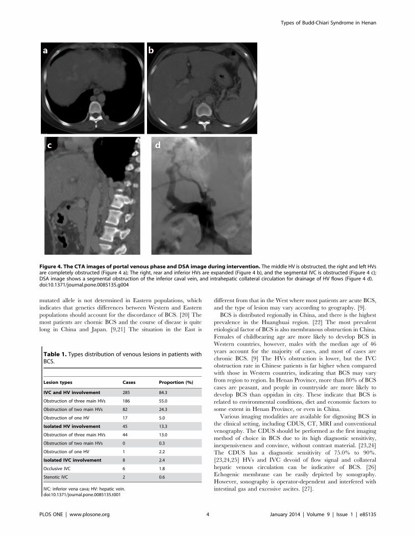
Figure 4. The CTA images of portal venous phase and DSA image during intervention. The middle HV is obstructed, the right and left HVsare completely obstructed (Figure 4 a); The right, rear and inferior HVs are expanded (Figure 4 b), and the segmental IVC is obstructed (Figure 4 c);DSA image shows a segmental obstruction of the inferior caval vein, and intrahepatic collateral circulation for drainage of HV flows (Figure 4 d).doi:10.1371/journal.pone.0085135.g004
Table 1. Types distribution of venous lesions in patients withBCS.
Lesion types Cases Proportion (%)
IVC and HV involvement 285 84.3
Obstruction of three main HVs 186 55.0
Obstruction of two main HVs 82 24.3
Obstruction of one HV 17 5.0
Isolated HV involvement 45 13.3
Obstruction of three main HVs 44 13.0
Obstruction of two main HVs 0 0.3
Obstruction of one HV 1 2.2
Isolated IVC involvement 8 2.4
Occlusive IVC 6 1.8
Stenotic IVC 2 0.6
IVC: inferior vena cava; HV: hepatic vein.doi:10.1371/journal.pone.0085135.t001
Types of Budd-Chiari Syndrome in Henan
PLOS ONE | www.plosone.org 4 January 2014 | Volume 9 | Issue 1 | e85135

The second line of investigation should be CT or MRI.
Heterogeneous patchy enhancement, nonvisualization of hepatic
veins, intrahepatic collaterals and obstruction of the IVC are their
important findings. For identifying the status of hepatic venous
thrombosis, CT has an overall accuracy of 50%. [28,29] CT has a
higher sensitive modality than sonography (90.0% versus 70.0%)
in showing parenchymal abnormalities. [28] Our results con-
firmed those findings. Moreover, CTA is useful in investigating the
patency of TIPS shunt and vascular involvement. [27] The
limitations of CT are the possible allergic reaction and nephro-
toxicity due to the use of contrast material, and CT is often unable
to show web in IVC.
MRI allows good anatomic orientation of HVs or IVC due to its
multiplanar capacity. The status of 80.0% of cases correctly
correlated with pathology when using MRI. [28] The advantages
of MRA over CDUS include better anatomic orientation, short
examination time without operator-dependency or restriction
from intestinal gas. [27] MRI is also advantageous over CT
imaging in some aspects. [27] The combination of CDUS and CT
or MRI is useful for guiding therapy due to the optimal delineation
of HV and IVC obstruction. [26].
Hepatic venography and inferior venocavography are conven-
tional diagnostic method for BCS, which currently serve as a third
line of investigation. Venography combined with venous pressure
measurements should be performed when radiological interven-
tion or surgical shunting is considered. [25,26] However,
venography often requires cannulation of HVs and large amounts
of contrast medium. [26].
Most of Chinese patients are chronic, ascites, caudate lobe
enlargement and abnormal hepatic configuration are present in
vast majority of the BCS cases. [27] Half of patients have wider
accessory HVs or large HV collaterals, which can help to drain off
the blocked HVs blood to relieve portal hypertension. [9] Caudate
lobe may hypertrophy due to its separate drainage into the IVC.
[30] Contrast-enhanced CT reveals inhomogeneous enhancement
of liver, such as peripheral regions with low attenuation due to
portal hypoperfusion and focal enhancing area owning to arterio-
portal shunting. [30] Benign regenerative nodules, intrahepatic
collateral circulation, body collateral circulation and hepatic
cirrhosis can be seen in chronic BCS cases. [31] Hepatocellular
carcinoma often develops in chronic end-stage liver disease,
[27,31] sometimes it will be shown in BCS cases.
Knowledge of the morphologic type of BCS is useful for guiding
the therapeutic approach and assessing operation risk. Shunt
surgery is no longer the standard care for BCS, with advances in
radiological interventions and a good midterm outcome. [32]
Interventional techniques can aid in treating and stabilising the
patient for possible orthotopic liver transplantation when liver
dysfunction progressive. [33].
Percutaneous transluminal angioplasty (PTA) can be used for
patients with post-hepatic IVC membranous-type obstruction,
balloon dilatation and percutaneous stent placement have been
found to be the techniques for those with segmental IVC
obstruction [34] or IVC web [35]. PTA is seldom adopted in
Western countries, however, and stenting are effective and safe for
treating BCS, with a good long-term outcome, [36] because most
chronic BCS are caused by membranous obstruction. According
to our experiences, large balloon dilation with a diameter of 25–
30 mm ballon catheter is safe and effective for most IVC
obstruction cases, and only few cases need further stenting after
large balloon dilation. The blood backflow of HV and liver
function can be basically compensated, as long as there was a
widely patent HV, regardless of which branch of the main HVs
was or whether it was the accessory HV. PTA, mainly balloon
dilation, is suitable for this kind of BCS. TIPS and orthotopic liver
transplantation are usually adopted in Western countries, although
the survival rate is poor. [37].
In case of short segment occlusion or stenosis of HV and/or
IVC, the obstruction between remnant HV and IVC should be
reopened by means of balloon angioplasty with or without stent
placement. [38] TIPS and surgical options are reserved for
patients with failure of interventions due to complete occlusion of
HV. [32] Patients with both the and segmental IVC occlusion
require opening of both occlusive segments, these may be difficult
and challenging and the several multiple approaches should be
required. [32] The IVC occlusion should be reopened as long as
they have unobstructed HV blood flow. [34] Besides, additional
therapies with anticoagulation, pharmacological or endoscopic
treatment for variceal bleeding and diuretics for ascites should be
considered. [26].
Author Contributions
Conceived and designed the experiments: PZ XH JR. Performed the
experiments: PZ JR GW WZ PD. Analyzed the data: GW WZ PD.
Contributed reagents/materials/analysis tools: PZ XH JR. Wrote the
paper: PZ JR YB. Revised the manuscript: YB.
References
1. Darwish Murad S, Valla DC, de Groen PC, Zeitoun G, Hopmans JA, et al.
(2004) Determinants of survival and the effect of portosystemic shunting inpatients with Budd-Chiari syndrome. Hepatology 39: 500–508.
2. Rav-Acha M, Gur C, Ilan Y, Verstandig A, Eid A (2004) [Budd-Chiari
syndrome: updated treatment modalities]. Harefuah 143: 372–376, 389.
3. Okuda K (2002) Obliterative hepatocavopathy-inferior vena cava thrombosis atits hepatic portion. Hepatobiliary Pancreat Dis Int 1: 499–509.
4. Valla D, Hadengue A, el Younsi M, Azar N, Zeitoun G, et al. (1997) Hepatic
venous outflow block caused by short-length hepatic vein stenoses. Hepatology25: 814–819.
5. Plessier A, Rautou PE, Valla DC (2012) Management of hepatic vasculardiseases. J Hepatol 56 Suppl 1: S25–38.
6. Smalberg JH, Arends LR, Valla DC, Kiladjian JJ, Janssen HL, et al. (2012)
Myeloproliferative neoplasms in Budd-Chiari syndrome and portal veinthrombosis: a meta-analysis. Blood 120: 4921–4928.
7. Pemmaraju N, Hamilton JP, Cameron AM, Sisson S, Moliterno AR (2012)
Abdominal venous thrombosis presenting in myeloproliferative neoplasm withJAK2 V617F mutation: a case report. J Med Case Rep 6: 102.
8. Singh V, Sinha SK, Nain CK, Bambery P, Kaur U, et al. (2000) Budd-Chiari
syndrome: our experience of 71 patients. J Gastroenterol Hepatol 15: 550–554.
9. Cheng D, Xu H, Lu ZJ, Hua R, Qiu H, et al. (2013) Clinical features and
etiology of Budd-Chiari syndrome in Chinese patients: a single-center study.
J Gastroenterol Hepatol 28: 1061–1067.
10. Yanaga K, Matsumata T, Hayashi H, Shimada M, Urata K, et al. (1994)
Isolated hepatic caudate lobectomy. Surgery 115: 757–761.
11. Sato TJ, Hirai I, Murakami G, Kanamura T, Hata F, et al. (2002) Ananatomical study of short hepatic veins, with special reference to delineation of
the caudate lobe for hanging maneuver of the liver without the usualmobilization. J Hepatobiliary Pancreat Surg 9: 55–60.
12. Goldsmith NA, Woodburne RT (1957) The surgical anatomy pertaining to liver
resection. Surg Gynecol Obstet 105: 310–318.
13. Meng WC, Shao CX, Mak KL, Lau PY, Yeung YP, et al. (2003) Anatomicaljustification of Belghiti’s ‘liver hanging manoeuvre’ in right hepatectomy with
anterior approach. ANZ J Surg 73: 407–409.
14. Elias H, Petty D (1952) Gross anatomy of the blood vessels and ducts within the
human liver. Am J Anat 90: 59–111.
15. Deltenre P, Denninger MH, Hillaire S, Guillin MC, Casadevall N, et al. (2001)Factor V Leiden related Budd-Chiari syndrome. Gut 48: 264–268.
16. Nakamura S, Tsuzuki T (1981) Surgical anatomy of the hepatic veins and the
inferior vena cava. Surg Gynecol Obstet 152: 43–50.
17. Kimura C (1964) Surgical Treatment of Portal Hypertension of Post-Hepatic
Origin, with Special Reference to Trans-Cardiac Membranotomy for Mem-
branous Obstruction of the Hepatic Portion of the Inferior Vena Cava. JpnCirc J 28: 181–183.
18. Janssen HL, Meinardi JR, Vleggaar FP, van Uum SH, Haagsma EB, et al.
(2000) Factor V Leiden mutation, prothrombin gene mutation, and deficiencies
Types of Budd-Chiari Syndrome in Henan
PLOS ONE | www.plosone.org 5 January 2014 | Volume 9 | Issue 1 | e85135

in coagulation inhibitors associated with Budd-Chiari syndrome and portal vein
thrombosis: results of a case-control study. Blood 96: 2364–2368.19. Mahmoud AE, Mendoza A, Meshikhes AN, Olliff S, West R, et al. (1996)
Clinical spectrum, investigations and treatment of Budd-Chiari syndrome. QJM
89: 37–43.20. Rees DC, Cox M, Clegg JB (1995) World distribution of factor V Leiden. Lancet
346: 1133–1134.21. Okuda H, Yamagata H, Obata H, Iwata H, Sasaki R, et al. (1995)
Epidemiological and clinical features of Budd-Chiari syndrome in Japan.
J Hepatol 22: 1–9.22. Wang ZG, Zhang FJ, Yi MQ, Qiang LX (2005) Evolution of management for
Budd-Chiari syndrome: a team’s view from 2564 patients. ANZ J Surg 75: 55–63.
23. Bolondi L, Gaiani S, Li Bassi S, Zironi G, Bonino F, et al. (1991) Diagnosis ofBudd-Chiari syndrome by pulsed Doppler ultrasound. Gastroenterology 100:
1324–1331.
24. Chawla Y, Kumar S, Dhiman RK, Suri S, Dilawari JB (1999) Duplex Dopplersonography in patients with Budd-Chiari syndrome. J Gastroenterol Hepatol 14:
904–907.25. Kamath PS (2006) Budd-Chiari syndrome: Radiologic findings. Liver Transpl
12: S21–22.
26. Janssen HL, Garcia-Pagan JC, Elias E, Mentha G, Hadengue A, et al. (2003)Budd-Chiari syndrome: a review by an expert panel. J Hepatol 38: 364–371.
27. Erden A (2007) Budd-Chiari syndrome: a review of imaging findings.Eur J Radiol 61: 44–56.
28. Miller WJ, Federle MP, Straub WH, Davis PL (1993) Budd-Chiari syndrome:imaging with pathologic correlation. Abdom Imaging 18: 329–335.
29. Noone TC, Semelka RC, Siegelman ES, Balci NC, Hussain SM, et al. (2000)
Budd-Chiari syndrome: spectrum of appearances of acute, subacute, and
chronic disease with magnetic resonance imaging. J Magn Reson Imaging 11:
44–50.
30. Bargallo X, Gilabert R, Nicolau C, Garcia-Pagan JC, Bosch J, et al. (2003)
Sonography of the caudate vein: value in diagnosing Budd-Chiari syndrome.
AJR Am J Roentgenol 181: 1641–1645.
31. Maetani Y, Itoh K, Egawa H, Haga H, Sakurai T, et al. (2002) Benign hepatic
nodules in Budd-Chiari syndrome: radiologic-pathologic correlation with
emphasis on the central scar. AJR Am J Roentgenol 178: 869–875.
32. Mukund A, Gamanagatti S (2011) Imaging and interventions in Budd-Chiari
syndrome. World J Radiol 3: 169–177.
33. Buckley O, O’Brien J, Snow A, Stunell H, Lyburn I, et al. (2007) Imaging of
Budd-Chiari syndrome. Eur Radiol 17: 2071–2078.
34. Xue H, Li YC, Shakya P, Palikhe M, Jha RK (2010) The role of intravascular
intervention in the management of Budd-Chiari syndrome. Dig Dis Sci 55:
2659–2663.
35. Khuroo MS, Al-Suhabani H, Al-Sebayel M, Al Ashgar H, Dahab S, et al. (2005)
Budd-Chiari syndrome: long-term effect on outcome with transjugular
intrahepatic portosystemic shunt. J Gastroenterol Hepatol 20: 1494–1502.
36. Qiao T, Liu CJ, Liu C, Chen K, Zhang XB, et al. (2005) Interventional
endovascular treatment for Budd-Chiari syndrome with long-term follow-up.
Swiss Med Wkly 135: 318–326.
37. Plessier A, Valla DC (2008) Budd-Chiari syndrome. Semin Liver Dis 28: 259–
269.
38. Eapen CE, Velissaris D, Heydtmann M, Gunson B, Olliff S, et al. (2006)
Favourable medium term outcome following hepatic vein recanalisation and/or
transjugular intrahepatic portosystemic shunt for Budd Chiari syndrome. Gut
55: 878–884.
Types of Budd-Chiari Syndrome in Henan
PLOS ONE | www.plosone.org 6 January 2014 | Volume 9 | Issue 1 | e85135