Inhibition of Wnt signaling by the osteoblast-specific ...
6
Inhibition of Wnt signaling by the osteoblast-specific transcription factor Osterix Chi Zhang*, Kyucheol Cho † , Yehong Huang ‡ , Jon P. Lyons † , Xin Zhou*, Krishna Sinha*, Pierre D. McCrea † , and Benoit de Crombrugghe* § Departments of *Molecular Genetics and † Biochemistry and Molecular Biology, University of Texas M. D. Anderson Cancer Center, Houston, TX 77030; and ‡ Department of Molecular Biology and Microbiology, Case Western Reserve University, Cleveland, OH 44106 Edited by Eric N. Olson, University of Texas Southwestern Medical Center, Dallas, TX, and approved March 18, 2008 (received for review November 14, 2007) The recent identification of the genes responsible for several human genetic diseases affecting bone homeostasis and the char- acterization of mouse models for these diseases indicated that canonical Wnt signaling plays a critical role in the control of bone mass. Here, we report that the osteoblast-specific transcription factor Osterix (Osx), which is required for osteoblast differentia- tion, inhibits Wnt pathway activity. First, in calvarial cells of embryonic day (E)18.5 Osx-null embryos, expression of the Wnt antagonist Dkk1 was abolished, and that of Wnt target genes c-Myc and cyclin D1 was increased. Moreover, our studies demon- strated that Osx bound to and activated the Dkk1 promoter. In addition, Osx inhibited -catenin-induced Topflash reporter activ- ity and -catenin-induced secondary axis formation in Xenopus embryos. Importantly, in calvaria of E18.5 Osx-null embryos har- boring the TOPGAL reporter transgene, -galactosidase activity was increased, suggesting that Osx inhibited the Wnt pathway in osteoblasts in vivo. Our data further showed that Osx disrupted binding of Tcf to DNA, providing a likely mechanism for the inhibition by Osx of -catenin transcriptional activity. We also showed that Osx decreased osteoblast proliferation. Indeed, E18.5 Osx-null calvaria showed greater BrdU incorporation than wild- type calvaria and that Osx overexpression in C2C12 mesenchymal cells inhibited cell growth. Because Wnt signaling has a major role in stimulating osteoblast proliferation, we speculate that Osx- mediated inhibition of osteoblast proliferation is a consequence of the Osx-mediated control of Wnt/-catenin activity. Our results add a layer of control to Wnt/-catenin signaling in bone. B one formation takes place through two distinct processes. Most of the skeleton is formed by endochondral ossification involving a cartilage model. A small number of skeletal elements, mainly craniofacial bones, are formed by intramembranous ossification, by which bones form directly from condensations of mesenchymal cells without a cartilage intermediate. The bone- forming osteoblasts and the cartilage-forming chondrocytes are derived from a common mesenchymal progenitor (1). Osteoblast differentiation occurs through a multistep molecular pathway regulated by different transcription factors and signaling pro- teins. Indian hedgehog (Ihh), a member of the Hedgehog family, which is required for endochondral but not for intramembranous bone formation (2), is needed for the establishment of the osteogenic portion of the perichondrium/periosteum and for the initial activation of the gene for Runx2, which is required for the differentiation of mesenchymal cells into preosteoblasts. The transcription factor Runx2 is needed for the formation of both endochondral and membranous skeletal elements. Indeed, in Runx2-null mutants, no endochondral and no membranous bones form, and osteoblast differentiation is arrested at an early step of bone development (3). Heterozygous mutations in Runx2 are the cause of the human genetic disease cleidocranial dys- plasia (4). Runx2 is also needed for the expression of another transcription factor Osterix (Osx), which is required for the differentiation of preosteoblasts into functional osteoblasts. Osx is specifically expressed in all osteoblasts and at low levels in prehypertrophic chondrocytes. In Osx-null embryos, a full car- tilage skeleton is formed, but the embryos completely lack bone formation. In these embryos, osteoblast differentiation markers, such as bone sialoprotein, osteonectin, and osteocalcin, are not expressed (5). The DNA-binding domain of Osx is located at its C terminus and consists of three C2H2-type zinc fingers that share a high degree of identity with a similar motif in Sp1, Sp3, and Sp4. Osx binds to specific GC-rich sequences and contains a proline-rich transcription activation domain. The canonical Wnt pathway is controlled both outside and inside the cell. Wnt polypeptides bind to frizzled receptors and LRP5/6 coreceptors (6). Several antagonists, including Dkks, sclerostin, and soluble frizzled receptors, inhibit Wnt signaling (7). The essential event in Wnt signaling is the accumulation and nuclear translocation of -catenin, which then interacts with members of the Lef/Tcf family of transcription factors to activate target genes (7). Distinct Wnts control early events during skeletal development such as limb patterning and joint forma- tion (8). Wnt/-catenin signaling, in addition to the transcription fac- tors Runx2 and Osx, is essential to osteoblast differentiation during embryonic development. Conditional inactivation of -catenin in either skeletal progenitor cells or at a later stage of osteoblast development in mouse embryos blocks osteoblast differentiation (9–12). When -catenin is inactivated in early skeletal progenitors, low levels of Runx2, but not Osx, expression are detected in these cells (11). At a later step, however, the inactivation of -catenin does not prevent the expression of Osx, indicating that Wnt signaling is not needed for Osx expression at this step (12). Several lines of evidence indicate that canonical Wnt signaling is also required for normal osteoblast proliferation. First, if -catenin is stabilized in osteoblasts during mouse embryonic development, a marked increase in osteoblast proliferation occurs (12). Moreover, Lrp5-null mice, which phenocopy the osteoporosis-pseudoglioma syndrome in humans (13), develop a phenotype with low bone mass because of decreased osteoblast proliferation (14). In contrast, gain-of-function mutants of Lrp5 cause high bone mass syndrome in patients (15) and in mice (16). In addition, the Wnt signaling antagonist Dkk1 prevents the activation of Wnt signaling by binding to LRP5/6. The bone formation and bone mass of heterozygous Dkk1 mutant mice increase with an increased number of osteoblasts (17). In contrast, the overexpression of Dkk1 in osteoblasts leads to severe osteopenia with decreased osteoblast numbers (18). Thus, Wnt/-catenin signaling stimulates osteoblast proliferation. Author contributions: C.Z., P.D.M., and B.d.C. designed research; C.Z., K.C., Y.H., J.P.L., and X.Z. performed research; K.S. contributed new reagents/analytic tools; C.Z., P.D.M., and B.d.C. analyzed data; and C.Z. and B.d.C. wrote the paper. The authors declare no conflict of interest. This article is a PNAS Direct Submission. § To whom correspondence should be addressed. E-mail: [email protected]. This article contains supporting information online at www.pnas.org/cgi/content/full/ 0710831105/DCSupplemental. © 2008 by The National Academy of Sciences of the USA 6936 – 6941 PNAS May 13, 2008 vol. 105 no. 19 www.pnas.orgcgidoi10.1073pnas.0710831105 Downloaded by guest on December 8, 2021
Transcript of Inhibition of Wnt signaling by the osteoblast-specific ...

Inhibition of Wnt signaling by the osteoblast-specifictranscription factor OsterixChi Zhang*, Kyucheol Cho†, Yehong Huang‡, Jon P. Lyons†, Xin Zhou*, Krishna Sinha*, Pierre D. McCrea†,and Benoit de Crombrugghe*§
Departments of *Molecular Genetics and †Biochemistry and Molecular Biology, University of Texas M. D. Anderson Cancer Center, Houston, TX 77030;and ‡Department of Molecular Biology and Microbiology, Case Western Reserve University, Cleveland, OH 44106
Edited by Eric N. Olson, University of Texas Southwestern Medical Center, Dallas, TX, and approved March 18, 2008 (received for review November 14, 2007)
The recent identification of the genes responsible for severalhuman genetic diseases affecting bone homeostasis and the char-acterization of mouse models for these diseases indicated thatcanonical Wnt signaling plays a critical role in the control of bonemass. Here, we report that the osteoblast-specific transcriptionfactor Osterix (Osx), which is required for osteoblast differentia-tion, inhibits Wnt pathway activity. First, in calvarial cells ofembryonic day (E)18.5 Osx-null embryos, expression of the Wntantagonist Dkk1 was abolished, and that of Wnt target genesc-Myc and cyclin D1 was increased. Moreover, our studies demon-strated that Osx bound to and activated the Dkk1 promoter. Inaddition, Osx inhibited �-catenin-induced Topflash reporter activ-ity and �-catenin-induced secondary axis formation in Xenopusembryos. Importantly, in calvaria of E18.5 Osx-null embryos har-boring the TOPGAL reporter transgene, �-galactosidase activitywas increased, suggesting that Osx inhibited the Wnt pathway inosteoblasts in vivo. Our data further showed that Osx disruptedbinding of Tcf to DNA, providing a likely mechanism for theinhibition by Osx of �-catenin transcriptional activity. We alsoshowed that Osx decreased osteoblast proliferation. Indeed, E18.5Osx-null calvaria showed greater BrdU incorporation than wild-type calvaria and that Osx overexpression in C2C12 mesenchymalcells inhibited cell growth. Because Wnt signaling has a major rolein stimulating osteoblast proliferation, we speculate that Osx-mediated inhibition of osteoblast proliferation is a consequence ofthe Osx-mediated control of Wnt/�-catenin activity. Our resultsadd a layer of control to Wnt/�-catenin signaling in bone.
Bone formation takes place through two distinct processes.Most of the skeleton is formed by endochondral ossification
involving a cartilage model. A small number of skeletal elements,mainly craniofacial bones, are formed by intramembranousossification, by which bones form directly from condensations ofmesenchymal cells without a cartilage intermediate. The bone-forming osteoblasts and the cartilage-forming chondrocytes arederived from a common mesenchymal progenitor (1). Osteoblastdifferentiation occurs through a multistep molecular pathwayregulated by different transcription factors and signaling pro-teins. Indian hedgehog (Ihh), a member of the Hedgehog family,which is required for endochondral but not for intramembranousbone formation (2), is needed for the establishment of theosteogenic portion of the perichondrium/periosteum and for theinitial activation of the gene for Runx2, which is required forthe differentiation of mesenchymal cells into preosteoblasts. Thetranscription factor Runx2 is needed for the formation of bothendochondral and membranous skeletal elements. Indeed, inRunx2-null mutants, no endochondral and no membranousbones form, and osteoblast differentiation is arrested at an earlystep of bone development (3). Heterozygous mutations in Runx2are the cause of the human genetic disease cleidocranial dys-plasia (4). Runx2 is also needed for the expression of anothertranscription factor Osterix (Osx), which is required for thedifferentiation of preosteoblasts into functional osteoblasts. Osxis specifically expressed in all osteoblasts and at low levels inprehypertrophic chondrocytes. In Osx-null embryos, a full car-
tilage skeleton is formed, but the embryos completely lack boneformation. In these embryos, osteoblast differentiation markers,such as bone sialoprotein, osteonectin, and osteocalcin, are notexpressed (5). The DNA-binding domain of Osx is located at itsC terminus and consists of three C2H2-type zinc fingers thatshare a high degree of identity with a similar motif in Sp1, Sp3,and Sp4. Osx binds to specific GC-rich sequences and containsa proline-rich transcription activation domain.
The canonical Wnt pathway is controlled both outside andinside the cell. Wnt polypeptides bind to frizzled receptors andLRP5/6 coreceptors (6). Several antagonists, including Dkks,sclerostin, and soluble frizzled receptors, inhibit Wnt signaling(7). The essential event in Wnt signaling is the accumulation andnuclear translocation of �-catenin, which then interacts withmembers of the Lef/Tcf family of transcription factors to activatetarget genes (7). Distinct Wnts control early events duringskeletal development such as limb patterning and joint forma-tion (8).
Wnt/�-catenin signaling, in addition to the transcription fac-tors Runx2 and Osx, is essential to osteoblast differentiationduring embryonic development. Conditional inactivation of�-catenin in either skeletal progenitor cells or at a later stage ofosteoblast development in mouse embryos blocks osteoblastdifferentiation (9–12). When �-catenin is inactivated in earlyskeletal progenitors, low levels of Runx2, but not Osx, expressionare detected in these cells (11). At a later step, however, theinactivation of �-catenin does not prevent the expression of Osx,indicating that Wnt signaling is not needed for Osx expression atthis step (12).
Several lines of evidence indicate that canonical Wnt signalingis also required for normal osteoblast proliferation. First, if�-catenin is stabilized in osteoblasts during mouse embryonicdevelopment, a marked increase in osteoblast proliferationoccurs (12). Moreover, Lrp5-null mice, which phenocopy theosteoporosis-pseudoglioma syndrome in humans (13), develop aphenotype with low bone mass because of decreased osteoblastproliferation (14). In contrast, gain-of-function mutants of Lrp5cause high bone mass syndrome in patients (15) and in mice (16).In addition, the Wnt signaling antagonist Dkk1 prevents theactivation of Wnt signaling by binding to LRP5/6. The boneformation and bone mass of heterozygous Dkk1 mutant miceincrease with an increased number of osteoblasts (17). Incontrast, the overexpression of Dkk1 in osteoblasts leads tosevere osteopenia with decreased osteoblast numbers (18). Thus,Wnt/�-catenin signaling stimulates osteoblast proliferation.
Author contributions: C.Z., P.D.M., and B.d.C. designed research; C.Z., K.C., Y.H., J.P.L., andX.Z. performed research; K.S. contributed new reagents/analytic tools; C.Z., P.D.M., andB.d.C. analyzed data; and C.Z. and B.d.C. wrote the paper.
The authors declare no conflict of interest.
This article is a PNAS Direct Submission.
§To whom correspondence should be addressed. E-mail: [email protected].
This article contains supporting information online at www.pnas.org/cgi/content/full/0710831105/DCSupplemental.
© 2008 by The National Academy of Sciences of the USA
6936–6941 � PNAS � May 13, 2008 � vol. 105 � no. 19 www.pnas.org�cgi�doi�10.1073�pnas.0710831105
Dow
nloa
ded
by g
uest
on
Dec
embe
r 8,
202
1
In other genetic experiments using mice in which a stabilized�-catenin was expressed in mature osteoblasts, the expression ofosteoprotegerin, a decoy receptor for RANK ligand, was in-creased. This resulted in a decrease in osteoclast differentiationand function, an inhibition of bone degradation, and increasedbone mass (19, 20). In contrast, in mice in which the �-cateningene was ablated in mature osteoblasts, an osteopenic phenotypeand lower level of osteoprotegerin were observed (19, 20). Theseexperiments indicate that activation of �-catenin in osteoblastsnoncell autonomously inhibits osteoclast differentiation andfunction.
The experiments reported in this article strongly support thehypothesis that, in addition to its essential role in osteoblastdifferentiation, the osteoblast-specific transcription factor Osxnegatively regulates Wnt/�-catenin signaling and osteoblastproliferation.
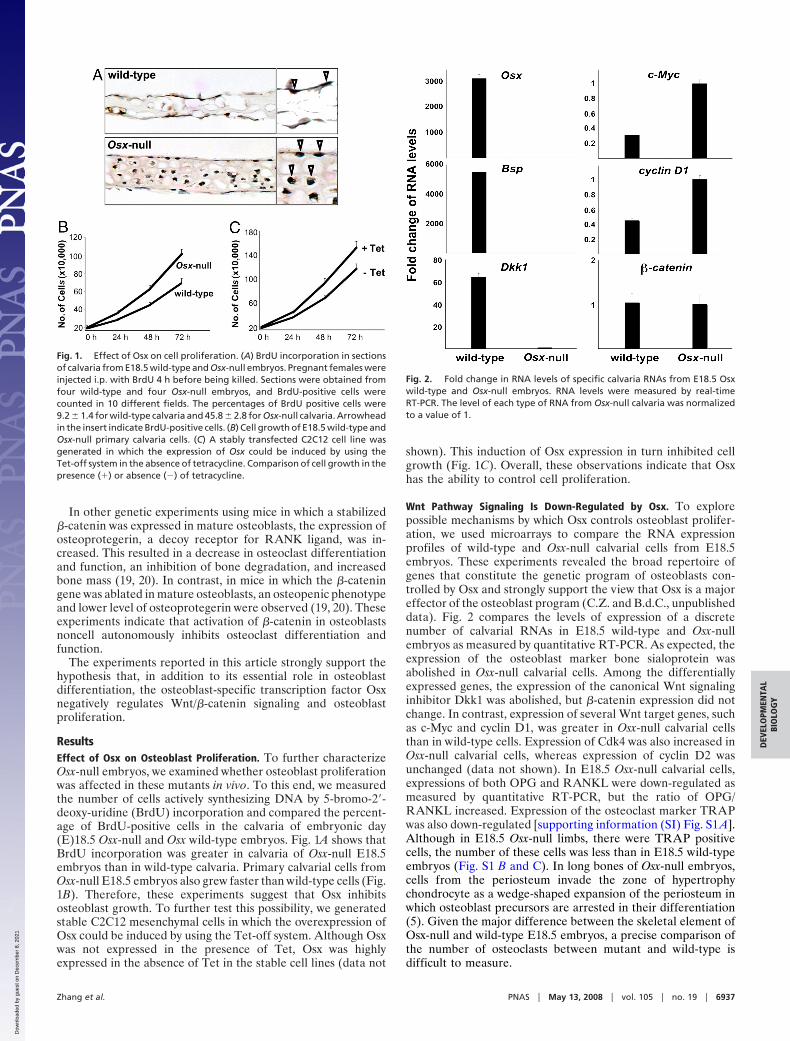
ResultsEffect of Osx on Osteoblast Proliferation. To further characterizeOsx-null embryos, we examined whether osteoblast proliferationwas affected in these mutants in vivo. To this end, we measuredthe number of cells actively synthesizing DNA by 5-bromo-2�-deoxy-uridine (BrdU) incorporation and compared the percent-age of BrdU-positive cells in the calvaria of embryonic day(E)18.5 Osx-null and Osx wild-type embryos. Fig. 1A shows thatBrdU incorporation was greater in calvaria of Osx-null E18.5embryos than in wild-type calvaria. Primary calvarial cells fromOsx-null E18.5 embryos also grew faster than wild-type cells (Fig.1B). Therefore, these experiments suggest that Osx inhibitsosteoblast growth. To further test this possibility, we generatedstable C2C12 mesenchymal cells in which the overexpression ofOsx could be induced by using the Tet-off system. Although Osxwas not expressed in the presence of Tet, Osx was highlyexpressed in the absence of Tet in the stable cell lines (data not
shown). This induction of Osx expression in turn inhibited cellgrowth (Fig. 1C). Overall, these observations indicate that Osxhas the ability to control cell proliferation.
Wnt Pathway Signaling Is Down-Regulated by Osx. To explorepossible mechanisms by which Osx controls osteoblast prolifer-ation, we used microarrays to compare the RNA expressionprofiles of wild-type and Osx-null calvarial cells from E18.5embryos. These experiments revealed the broad repertoire ofgenes that constitute the genetic program of osteoblasts con-trolled by Osx and strongly support the view that Osx is a majoreffector of the osteoblast program (C.Z. and B.d.C., unpublisheddata). Fig. 2 compares the levels of expression of a discretenumber of calvarial RNAs in E18.5 wild-type and Osx-nullembryos as measured by quantitative RT-PCR. As expected, theexpression of the osteoblast marker bone sialoprotein wasabolished in Osx-null calvarial cells. Among the differentiallyexpressed genes, the expression of the canonical Wnt signalinginhibitor Dkk1 was abolished, but �-catenin expression did notchange. In contrast, expression of several Wnt target genes, suchas c-Myc and cyclin D1, was greater in Osx-null calvarial cellsthan in wild-type cells. Expression of Cdk4 was also increased inOsx-null calvarial cells, whereas expression of cyclin D2 wasunchanged (data not shown). In E18.5 Osx-null calvarial cells,expressions of both OPG and RANKL were down-regulated asmeasured by quantitative RT-PCR, but the ratio of OPG/RANKL increased. Expression of the osteoclast marker TRAPwas also down-regulated [supporting information (SI) Fig. S1 A].Although in E18.5 Osx-null limbs, there were TRAP positivecells, the number of these cells was less than in E18.5 wild-typeembryos (Fig. S1 B and C). In long bones of Osx-null embryos,cells from the periosteum invade the zone of hypertrophychondrocyte as a wedge-shaped expansion of the periosteum inwhich osteoblast precursors are arrested in their differentiation(5). Given the major difference between the skeletal element ofOsx-null and wild-type E18.5 embryos, a precise comparison ofthe number of osteoclasts between mutant and wild-type isdifficult to measure.
Fig. 1. Effect of Osx on cell proliferation. (A) BrdU incorporation in sectionsof calvaria from E18.5 wild-type and Osx-null embryos. Pregnant females wereinjected i.p. with BrdU 4 h before being killed. Sections were obtained fromfour wild-type and four Osx-null embryos, and BrdU-positive cells werecounted in 10 different fields. The percentages of BrdU positive cells were9.2 � 1.4 for wild-type calvaria and 45.8 � 2.8 for Osx-null calvaria. Arrowheadin the insert indicate BrdU-positive cells. (B) Cell growth of E18.5 wild-type andOsx-null primary calvaria cells. (C) A stably transfected C2C12 cell line wasgenerated in which the expression of Osx could be induced by using theTet-off system in the absence of tetracycline. Comparison of cell growth in thepresence (�) or absence (�) of tetracycline.
Fig. 2. Fold change in RNA levels of specific calvaria RNAs from E18.5 Osxwild-type and Osx-null embryos. RNA levels were measured by real-timeRT-PCR. The level of each type of RNA from Osx-null calvaria was normalizedto a value of 1.
Zhang et al. PNAS � May 13, 2008 � vol. 105 � no. 19 � 6937
DEV
ELO
PMEN
TAL
BIO
LOG
Y
Dow
nloa
ded
by g
uest
on
Dec
embe
r 8,
202
1

Osx Stimulates Dkk1 Promoter Activity. Because both microarrayand real-time RT-PCR results suggested that Osx is required forDkk1 expression in osteoblasts, we asked whether Osx stimulatesDkk1 promoter activity. In transfection experiments of HEK293cells, Osx activated a 1-kb Dkk1 promoter luciferase reporter ina dose-dependent manner (Fig. 3A), suggesting that Osx mighttranscriptionally activate the Dkk1 gene in osteoblasts. DNAsequence analysis revealed three potential Osx binding sites in a1-kb Dkk1 promoter. In EMSA purified recombinant, Osx boundto sites of S1 and S2 (but not to S3) and anti-Osx antibodysupershifted Osx-DNA complexes. In addition, S1 mutationS1-M and S2 mutation S2-M inhibited Osx binding, indicatingthat Osx specifically binds to sites of S1 and S2 (Fig. 3B). Inprimary calvarial cells from new-born wild-type mice, ChIPanalysis showed that Osx was associated with the chromatin ofa segment of the Dkk1 promoter covering S1, S2, and S3 sites,whereas no Osx was associated with the chromatin of a more 5�segment of the Dkk1 gene (Fig. 3C). Mutation in either S1 or S2inhibited Osx activation of the 1-kb Dkk1 promoter 50–60%,whereas mutations in both sites almost abolished activation ofthe promoter (Fig. 3D).
Topflash is a reporter construct activated by Wnt signalingand, hence, by Wnt3A-conditioned medium. As shown in Fig. 4,the addition of recombinant Dkk1 inhibited the activity ofWnt3A-induced Topflash reporter in a dose-dependent manner.
The transfection of Osx also inhibited Wnt3A-induced Topflashreporter activity. The transfection of Osx together with theaddition of recombinant Dkk1 further inhibited the Wnt3A-induced Topflash reporter, suggesting that Osx and Dkk1 havean additive effect in the inhibition of Topflash reporter activity.These results raised the possibility that Osx might target addi-tional components of the Wnt pathway.
Effect of Osx on Wnt Signaling Activity. To begin to identify otherpossible Wnt pathway components targeted by Osx, we tookadvantage of the established model of �-catenin-induced Top-flash activation. Osx strongly inhibited �-catenin-induced Top-
Fig. 5. Inhibition of �-catenin transcription activity by Osx. (A) Osx inhibits�-catenin-induced Wnt reporter activity. HEK293 cells were transfected withthe Topflash or Fopflash reporter along with Myc-�-catenin, a plasmid ex-pressing a stabilized �-catenin, and increasing amounts of pEX-Osx DNA. (B)Osx inhibits �-catenin-induced secondary axis formation in Xenopus embryos.Embryos were microinjected with RNA for stabilized �-catenin (100 pg) andincreasing amounts of Osx RNA at the four-cell cleavage stage into theequatorial region of a single vegetal-ventral blastomere. Microinjection of�-catenin RNA in the ventral side of four-cell Xenopus embryos induces theformation of a secondary body axis. The phenotypes were evaluated by usinga binocular dissecting microscope at the tadpole stage. (C) Dose-dependentinhibition of �-catenin-induced secondary axis formation in Xenopus em-bryos by Osx. (D) �-galactosidase activity in calvarial extracts of E18.5Osx�/�;TOPGAL and Osx�/�;TOPGAL embryos. Results were expressed as theratio of �- galactosidase over DNA.
Fig. 3. Osx activates a Dkk1 promoter. (A) HEK293 cells were transfectedwith a 1-kb Dkk1 promoter luciferase reporter without or with an increasingamount of pEX-Osx as indicated. Cell extracts were subjected to Western blotanalysis to monitor the expression of transfected pEX-Osx. (B) Purified recom-binant Osx binds to two specific sites in the Dkk1 promoter in EMSA. Threepotential Osx binding sites in the 1-kb Dkk1 promoter, S1, S2, and S3, wereused as probes. S1-M is S1 site mutation and S2-M is S2 site mutation. (C) Osxassociates with the chromatin of a segment of the Dkk1 promoter in ChIPassay. Calvarial cells were isolated and cultured from wild-type new born mice.p1 primer set corresponds to a segment of the Dkk1 promoter covering S1, S2,and S3 sites within the proximal 500 bp. As a negative control, p2 primer setcovers a distal region of the Dkk1 promoter, which does not contain an Osxbinding site. Anti-acetylated histone H3 antibody (a-H3-a) was used as apositive control. (D) Mutations in sites S1 and S2 inhibit activation of the Dkk1promoter by Osx. Mutations in the 1-kb Dkk1 reporter were constructed.Mutant M1 contains the S1 site mutation, mutant M2 the S2 site mutation, anddouble mutant M12 (both S1 and S2 mutations) in the 1-kb Dkk1 promoter.HEK293 cells were transfected with wild-type and mutant Dkk1 promoterreporters without or with pEX-Osx, as indicated.
Fig. 4. Cooperation between Osx and Dkk1 in the inhibition of Wnt3A-induced Topflash activity. Topflash and Fopflash reporters were transfected inHEK293 cells with pEX-Osx as indicated. Wnt3A-conditioned medium wasused to activate the Topflash reporter. Recombinant Dkk1 protein was addedto the medium at the different concentrations as indicated. Results wereexpressed as the ratio of Topflash over Fopflash activity.
6938 � www.pnas.org�cgi�doi�10.1073�pnas.0710831105 Zhang et al.
Dow
nloa
ded
by g
uest
on
Dec
embe
r 8,
202
1

f lash reporter expression in a dose-dependent manner (Fig. 5A).This was further confirmed by in vivo results demonstrating thatOsx inhibited �-catenin-induced secondary axis formation inXenopus embryos in a dose-dependent manner (Fig. 5 B and C).To examine whether Wnt signaling in E18.5 mouse skeletalelements was increased in vivo in the absence of Osx, we used theWnt indicator TOPGAL transgenic mice. The TOPGAL trans-gene consists of a �-galactosidase gene driven by a Tcf/�-catenin-responsive promoter, which enables canonical Wnt activity to bemeasured by �-galactosidase activity in extracts of intact em-bryonic tissues. In E18.5 Osx�/� embryos harboring the TOP-GAL transgene, the calvarial �-galactosidase activity per unit ofDNA was increased approximately twofold compared with thatin Osx�/�;TOPGAL embryos (Fig. 5D). These experimentsprovide evidence that Osx inhibits the transcriptional activity of�-catenin. Furthermore, the increase in TOPGAL activity in thecalvaria of Osx-null embryos implies that the inhibition ofWnt/�-catenin signaling occurs in osteoblasts in vivo. Overall,our experiments suggest that Osx has the ability to activateexpression of the canonical Wnt signaling antagonist Dkk1 todirectly inhibit the transcriptional activity of �-catenin.
Because our results showed that Osx inhibited �-catenin-induced Topflash activity, we examined possible interactions ofOsx with nuclear components of the canonical Wnt pathway. Wefirst tested whether Osx and Tcf could associate with each otherin the same complex. Among TCF family members, both Tcf1and Tcf4 are expressed in osteoblasts. Tcf1 is detectable inosteoblasts from E14.5, whereas Tcf4 is detectable after E16.5.In cotransfections of HEK293 cells, Osx and Tcf1 were shown tocoimmunoprecipitate (Fig. 6A). Osx was also shown to coim-munoprecipitate with Tcf4 (data not shown). In GST pull-downexperiments, GST-�-catenin did not pull-down 35S-labeled Osx,but, as expected, it did pull down 35S-Tcf1. Bacculovirus-expressed Osx failed to disrupt the pull-down of 35S-Tcf1 byGST-�-catenin (Fig. 6B).
Tcfs bind to the promoter region of Wnt target genes toregulate their expression. Because Osx did not disrupt �-catenin/Tcf complex formation, we assessed whether Osx might disruptTcf binding to DNA. Based on promoter sequence analysis, thereis no Osx binding site overlapping the consensus Tcf bindingsequences; however, it is possible that, by interacting with Tcf,Osx disrupts Tcf binding to DNA. In EMSA, Tcf1 bound to a32P-labeled double-strand oligo containing a consensus Tcfbinding site, whereas Osx generated by in vitro transcription andtranslation (IVTT) did not bind to this DNA. When increasingamounts of Osx were added, Tcf1 binding to DNA was disruptedin a dose-dependent manner, whereas the control IVTT productfailed to affect Tcf1 binding to DNA (Fig. 6C).
To determine which domain of Osx was responsible for thedisruption of Tcf binding to DNA, a series of deletions wasgenerated (Fig. 7A). Osx contains a proline-rich region (PRR)transactivation domain toward the N-terminal part of the proteinand a three-zinc-finger DNA-binding motif in the C-terminalregion. As shown in Fig. 7B, mutants containing PRR, such asM1 and M4, inhibited Tcf binding to DNA, whereas mutantslacking part or all of the PRR, such as M2, M3, M5, and M6,failed to disrupt Tcf binding to DNA. These results suggest thatthe PRR domain is responsible for the inhibition of Tcf bindingto DNA. Next, we evaluated whether the PRR region wasrequired for the Osx inhibitory effect on Wnt signaling activity.To address this question, Osx mutants were cotransfected with�-catenin into HEK293 cells along with the Topflash reporter.Fig. 7C shows that the Osx mutants with the PRR region, M1 andM4, were able to inhibit �-catenin-induced Topflash reporter,whereas the Osx mutants without a complete PRR region, M2,M3, M5 and M6, were not. Therefore, we conclude that the PRRregion mediates both the Osx inhibition of �-catenin-inducedTopflash reporter activation and the disruption of Tcf binding toits target DNA.
DiscussionHere, we have presented evidence for two properties of theosteoblast-specific transcription factor Osx: that it inhibits Wnt
Fig. 6. Interaction between Osx and Tcf1. (A) Coimmunoprecipitation (CoIP)of Osx and Tcf1 in transfected HEK293 cells. Here, 1 �g of pEX-HA-Osx andpCDNA-Tcf1 were cotransfected or transfected alone into HEK293 cells.Whole-cell lysates (WCL) were immunoprecipitated with anti-HA (10 �l) andthe precipitate was immunoblotted with anti-Tcf1. (B) No disruption of �-catenin interaction with Tcf1 by Osx. (Upper) GST-�-catenin was used to pulldown 35S labeled Osx. Ten percent of the synthesized Osx was used as an input,and GST was used as a control. (Lower) GST-�-catenin was used to pull down35S-labeled Tcf1. Baculovirus-expressed Osx was used as Osx protein source. (C)Disruption of Tcf1 binding to DNA by Osx. Osx and Tcf1 proteins weresynthesized by TNT IVTT systems. Tcf1 bound to Tcf 1 binding probe in EMSA.TNT lysates containing pEX-Osx and control TNT lysates with pEX were addedto the DNA binding reaction.
Fig. 7. PRR region of Osx is needed for disruption of Tcf1 binding to DNA andfor inhibition of �-catenin transcriptional activity. (A) Schematic representa-tion of the Osx deletion mutants. PRR, proline-rich region; Z, zinc-fingerdomain. (B) Osx and Tcf1 proteins were synthesized by TNT IVTT systems. M1and M4 are mutants with full PRR region. M2, M3, M5, and M6 are mutantswithout full PRR region. (Lower) [35S]methionine-labeled Osx wild-type andmutants synthesized by IVTT system were detected by autoradiograph. (C) OsxPRR domain is required for Osx inhibitory effect on Wnt pathway. Vectorsexpressing Osx wild-type and mutants were transfected in HEK293 cells withor without vectors expressing activated �-catenin (�-cat) with the Topflashreporter.
Zhang et al. PNAS � May 13, 2008 � vol. 105 � no. 19 � 6939
DEV
ELO
PMEN
TAL
BIO
LOG
Y
Dow
nloa
ded
by g
uest
on
Dec
embe
r 8,
202
1

signaling and decreases cell proliferation (Fig. 8). In Osx-nullcalvarial cells, the expression of the gene for the Wnt antagonistDkk1 was abolished, suggesting that Osx positively controls theexpression of Dkk1. This is also supported by the Osx-mediatedactivation of a 1-kb Dkk1 promoter fragment, the existence oftwo functional Osx binding sites in this promoter and thepresence of Osx in the chromatin of this promoter in primaryosteoblasts. In addition, both DNA transfection experiments andthe inhibition of �-catenin-induced secondary axis formation inXenopus embryos strongly suggested that Osx inhibits the tran-scriptional activity of �-catenin. In the transfection experiments,the proline-rich transcription activation domain of Osx wasrequired for the inhibitory activity. Importantly, in vivo exper-iments showing that the Wnt-dependent TOPGAL promoter wasmore active in E18.5 Osx-null calvaria than in wild-type embry-onic calvaria strongly suggested that Osx inhibits Wnt signalingin osteoblasts in vivo.
In further transfection experiments, Osx and Tcf1, a DNAbinding partner of �-catenin, were coimmunoprecipitated, whichsuggested that they are part of the same complex. In addition, thedisruption of Tcf1 binding to DNA by Osx provided a likelymechanism for the inhibition of �-catenin transcriptional activityby Osx. Indeed, the same Osx mutants that failed to inhibit thetranscriptional activity of �-catenin also lacked the ability todisrupt Tcf1 binding to DNA. Thus, our findings indicate thatOsx negatively controls the activity of �-catenin in two ways:first, by being needed for the expression of a major Wntantagonist, and second, by inhibiting the transcriptional activityof �-catenin/Tcf. Note that our results do not rule out thepossibility that Osx might also target other components of Wntsignaling as part of its inhibitory effect.
The evidence for the ability of Osx to inhibit cell proliferationincludes the markedly enhanced proliferation of E18.5 Osx-nullcalvarial cells compared to wild-type embryonic calvarial cellsand the decrease in C2C12 cell proliferation after the inductionof Osx expression. Other studies (12–18) provide strong geneticevidence that Wnt signaling positively regulates osteoblast pro-liferation. Therefore, we speculate that the Osx-mediated inhi-bition of osteoblast proliferation is a consequence of the inhi-bition of Wnt signaling by Osx. In C2C12 mesenchymal cellstreated with amounts of Dkk1, which maximally inhibited cellproliferation, induction of Osx caused a further albeit modestreduction of cell growth (Fig. S2). Although even in the presenceof saturating amounts of Dkk1 a residual activity of Wntsignaling may persist that could be further inhibited by disrup-tion of Tcf binding to target genes by Osx, one cannot excludethat Osx might also inhibit other proproliferative pathways.
Our observation that Dkk1 expression is absent in Osx-nullcalvaria of E18.5 embryos parallels the finding reported in ref.12 that Dkk1 expression was absent in tibia of E18.5 embryos in
which �-catenin was inactivated by using an Osx-Cre transgene.This suggests that both Osx and �-catenin are required for Dkk1expression in osteoblasts and that the Dkk1 gene might be acommon target of these transcription factors in these cells.Furthermore, Osx is required for Dkk1 expression, which shouldfurther reinforce the feedback control effect that Dkk1 exerts onWnt/�-catenin signaling.
Reports indicate that Wnt/�-catenin signaling can controlbone mass by several mechanisms (9–12, 20). Indeed it hasessential functions in (i) in osteoblast differentiation, (ii) properosteoblast proliferation, and (iii) inhibiting bone resorptionnoncell autonomously by controlling osteoclast differentiation(9–12, 20). Mutations in LRP5, which result in either a high orlow bone mass phenotype affect mainly osteoblast proliferationbut do not produce an inhibition of osteoclast differentiation (14,16). In contrast, either expression of a stabilized �-catenin orinactivation of �-catenin, specifically in mature osteoblasts inmice, which also cause high or low bone mass respectively, affectmainly osteoclast differentiation and function by controlling theexpression in osteoblasts of osteoprotegrin, a decoy receptor forRANK ligand, without causing significant changes in osteoblastproliferation (19, 20). In the skeletal elements of E18.5 Osx-nullembryos, the number of TRAP-positive cells appear to bereduced compared to wild-type embryos. This observation issupported by the increased OPG/RANKL ratio and the de-creased TRAP mRNA in calvarial cell of E18.5 Osx-null em-bryos. Thus, it is possible that the inhibition of Wnt signaling byOsx also reduces osteoclast differentiation and function. Wespeculate that the inhibition of Wnt/�-catenin signaling by Osx,which itself has an essential role in osteoblast differentiation,ensures an optimal bone formation rate.
The extensive studies by many laboratories to understand thecontrol of the Wnt signaling pathway in osteoblasts stems fromthe realization that this pathway has an essential role in bonemass determination in the adult skeleton. There is also anexpectation that efforts to pharmacologically target this pathwayshould yield promising agents to treat bone diseases, such asosteoporosis. Our results showing that Osx inhibits Wnt/�-catenin signaling add an important new layer of control to thecomplex regulation of the Wnt pathway in osteoblasts (Fig. 8).
Materials and MethodsPlasmids, Protein Purification, Cell Culture, and Transient Transfections. Osxdeletion mutants were generated by PCR and subcloned into the pEX vectorfor transfection studies or into the pCS2� vector for Xenopus experiments.Dkk1 reporter mutants were made with the QuikChange site-directed mu-tagenesis kit (Stratagene). All constructs were verified by DNA sequencing.GST-�-catenin was expressed and purified by glutathione-agarose affinitychromatography, and the in vitro protein interaction assay was performed asdescribed in ref. 21. Osx cDNA was subcloned into the pBac vector, andrecombinant baculovirus-expressed Osx was purified by nickel affinity chro-matography (21). [35S]methionine-labeled protein was synthesized in a TNT T7coupled in vitro transcription-translation system, using rabbit reticulocytelysate (Promega). HEK293 cells were cultured in DMEM supplemented with10% FBS and transfected by FuGENE 6 (Roche). For Topflash reporter assays,250 ng of reporters and 50 ng of pSV2-�gal were cotransfected with theindicated expression plasmids. Luciferase activities were normalized for trans-fection efficiency to �-galactosidase activity. Conditioned media were fromWnt-3A-expressing mouse and parental L cells (American Type CultureCollection) (22).
RNA Isolation and Quantitative RT-PCR. Total RNA was isolated with TRIzolreagent (Invitrogen). RNA was subjected to quantitative RT-PCR, using theTaqMan One-Step RT-PCR Master Mix reagent (Applied Biosystems). Relativetranscript levels were measured by real-time PCR in a 50-�l reaction volume on96-well plates, using an ABI PRISM 7000 sequence detection system (AppliedBiosystems). Transcript levels were normalized to glyceraldehyde-3-phosphatedehydrogenase levels, using primers from Applied Biosystems.
Fig. 8. Proposed model of coordinated regulation of osteoblast differenti-ation and proliferation by Osx and Wnt/�-catenin signaling. Wnt/�-cateninsignaling has an essential role in osteoblast differentiation during embryonicdevelopment and has a major role in stimulating osteoblast proliferation bothduring embryonic development and postnatally. Osx is an osteoblast-specifictranscription factor, required for osteoblast differentiation. The inhibition ofWnt/�-catenin signaling activity by Osx also constitutes a possible mechanismfor the inhibition by Osx of osteoblast proliferation. Not shown is the inhib-itory role of Wnt/�-catenin in osteoclast differentiation and function.
6940 � www.pnas.org�cgi�doi�10.1073�pnas.0710831105 Zhang et al.
Dow
nloa
ded
by g
uest
on
Dec
embe
r 8,
202
1

Histological Analysis and Xenopus laevis Secondary-Axis Assays. To examine cellproliferation, pregnant female mice were injected i.p. with 100 �l of 100 �MBrdU and killed 4 h later. Calvaria from E18.5 embryos were isolated, fixed, andprocessed. Sections of 5-�m were prepared and stained with the BrdU stainingkit (Zymed). TRAP staining was performed in limb sections, using a leucocyteacid phosphatase kit (Sigma). X. laevis embryo microinjections were per-formed as described in ref. 22. Briefly, pCS2�-Osx and pCS2�-�-catenin werelinearized and transcribed in vitro into capped mRNA, using the SP6 mMA-CHINE kit (Ambion). Xenopus females were injected with 800 units of humanCG 10–16 h before manual squeezing to isolate eggs. The eggs were fertilizedin vitro. Embryos were microinjected at the four-cell cleavage stage into theequatorial region of a single vegetal-ventral blastomere and placed in asolution of 5% Ficoll in 1� Marc’s Modified Ringers (MMR) for 60–90 minbefore transfer to 0.1� MMR. mRNAs were injected at a volume of 10 nl perblastomere. Embryonic phenotypes were evaluated by using a standard bin-ocular dissecting microscope (Nikon; SMZ-U) and captured as digital images(Nikon; CoolPix 995) at the tadpole stage.
Chromatin Immunoprecipitation (ChIP) Assay. ChIP assays were performed asdescribed in ref. 23 with some modifications. Briefly, Calvarial cells wereisolated from wild-type newborn mice and were cultured in DMEM supple-mented with 10% FBS. Formaldehyde was used to cross-link the cells for 10min, and cross-linking was quenched with glycine. Cells were harvested andrinsed with PBS, and cell pellets were resuspended in 1 ml of lysis buffer. After
sonication, 100 �l of sheared chromatin was diluted to 1 ml with IP dilutionbuffer for each immunoprecipitation. The chromatin solution was preclearedwith 60 �l of protein G-agarose beads at 4°C for 1 h. The precleared chromatinwas collected and incubated at 4°C overnight with 5 �g of anti-Osx antibody,anti-acetylated histone H3 antibody (Upstate Biotechnology) or Rabbit IgG(Sigma) as a negative control. The immune complexes were precipitated with60 �l of protein G-agarose beads at 4°C for 1 h. After washes, the antibody-protein-DNA immunocomplexes were eluted twice with 100 �l of elutionbuffer. Formaldehyde cross-linking was reversed by heating at 65°C overnightwith the addition of 5 M NaCl. All of the samples were digested with RNase Aand proteinase K. The DNA was purified by using spin columns, and analyzedby PCR. The p1 primer sequences for the Dkk1 promoter were 5�-CTAGT-GCTCTAGTGACCCACACTC-3� and 5�-TGGACTGCGGAACCTCAACTTC-3�. Thep2 sequences were 5�-CACTCCATTGCCTGGCTGCATG-3� and 5�-CATGGTAT-CAGGTCACAGAGGGAG-3�.
Supporting Information. Details of immunoprecipitation, western blot analy-sis, the in vitro protein interaction assay, and EMSA may be found in SI Text.
ACKNOWLEDGMENTS. We thank Hui Dai, Lingna Zhang, and Dr. FrancoiseCoustry for technical assistance and Dr. Hans Clevers (Hubrecht Laboratory,Utrecht, The Netherlands) for providing the plasmids pcDNA-Tcf1 and pcDNA-Tcf4. This work was supported by National Institutes of Health Grant R01AR49072 (to B.d.C.), an Arthritis Foundation Postdoctoral Fellowship (to C.Z.),and National Institutes of Health Grant CA16672 (for DNA sequence analysis).
1. Akiyama H, et al. (2005) Osteo-chondroprogenitor cells are derived from Sox9 express-ing precursors. Proc Natl Acad Sci USA 102:14665–14670.
2. St-Jacques B, Hammerschmidt M, McMahon AP (1999) Indian hedgehog signalingregulates proliferation and differentiation of chondrocytes and is essential for boneformation. Genes Dev 13:2072–2086.
3. Komori T, et al. (1997) Targeted disruption of Cbfa1 results in a complete lack of boneformation owing to maturational arrest of osteoblasts. Cell 89:755–764.
4. Mundlos S, et al. (1997) Mutations involving the transcription factor CBFA1 causecleidocranial dysplasia. Cell 89:773–779.
5. Nakashima K, et al. (2002) The novel zinc finger-containing transcription factor osterixis required for osteoblast differentiation and bone formation. Cell 108:17–29.
6. He X, Semenov M, Tamai K, Zeng X (2004) LDL receptor-related proteins 5 and 6 inWnt/beta-catenin signaling: Arrows point the way. Development (Cambridge, UK)131:1663–1677.
7. Krishnan V, Bryant HU, Macdougald OA (2006) Regulation of bone mass by Wntsignaling. J Clin Invest 116:1202–1209.
8. Hartmann C, Tabin CJ (2001) Wnt-14 plays a pivotal role in inducing synovial jointformation in the developing appendicular skeleton. Cell 104:341–351.
9. Day TF, Guo X, Garrett-Beal L, Yang Y (2005) Wnt/beta-catenin signaling in mesenchy-mal progenitors controls osteoblast and chondrocyte differentiation during verte-brate skeletogenesis. Dev Cell 8:739–750.
10. Hill TP, Spater D, Taketo MM, Birchmeier W, Hartmann C (2005) Canonical Wnt/beta-catenin signaling prevents osteoblasts from differentiating into chondrocytes. Dev Cell8:727–738.
11. Hu H, et al. (2005) Sequential roles of Hedgehog and Wnt signaling in osteoblastdevelopment. Development (Cambridge, UK) 132:49–60.
12. Rodda SJ, McMahon AP (2006) Distinct roles for Hedgehog and canonical Wnt signalingin specification, differentiation and maintenance of osteoblast progenitors. Develop-ment (Cambridge, UK) 133:3231–3244.
13. Gong Y, et al. (2001) LDL receptor-related protein 5 (LRP5) affects bone accrual and eyedevelopment. Cell 107:513–523.
14. Kato M, et al. (2002) Cbfa1-independent decrease in osteoblast proliferation, osteope-nia, and persistent embryonic eye vascularization in mice deficient in Lrp5, a Wntcoreceptor. J Cell Biol 157:303–314.
15. Little RD, et al. (2002) A mutation in the LDL receptor-related protein 5 gene results inthe autosomal dominant high-bone-mass trait. Am J Hum Genet 70:11–19.
16. Babij P, et al. (2003) High bone mass in mice expressing a mutant LRP5 gene. J BoneMiner Res 18:960–974.
17. Morvan F, et al. (2006) Deletion of a single allele of the Dkk1 gene leads to an increasein bone formation and bone mass. J Bone Miner Res 21:934–945.
18. Li J, et al. (2006) Dkk1-mediated inhibition of Wnt signaling in bone results inosteopenia. Bone 39:754–766.
19. Glass DA, II, et al. (2005) Canonical Wnt signaling in differentiated osteoblasts controlsosteoclast differentiation. Dev Cell 8:751–764.
20. Holmen SL, et al. (2005) Essential role of beta-catenin in postnatal bone acquisition.J Biol Chem 280:21162–21168.
21. Zhang C, et al. (2001) Ternary complexes and cooperative interplay between NCoA-62/Ski-interacting protein and steroid receptor coactivators in vitamin D receptor-mediated transcription. J Biol Chem 276:40614–40620.
22. Lyons JP, et al. (2004) Wnt-4 activates the canonical beta-catenin-mediated Wntpathway and binds Frizzled-6 CRD: Functional implications of Wnt/beta-catenin activ-ity in kidney epithelial cells. Exp Cell Res 298:369–387.
23. Zhang C, et al. (2001) Nuclear coactivator-62 kDa/Ski-interacting protein is a nuclearmatrix-associated coactivator that may couple vitamin D receptor-mediated transcrip-tion and RNA splicing. J Biol Chem 278:35325–35336.
24. Oosterwegel M, et al. (1991) Cloning of murine TCF-1, a T cell-specific transcriptionfactor interacting with functional motifs in the CD3-epsilon and T cell receptor alphaenhancers. J Exp Med 173:1133–1142.
Zhang et al. PNAS � May 13, 2008 � vol. 105 � no. 19 � 6941
DEV
ELO
PMEN
TAL
BIO
LOG
Y
Dow
nloa
ded
by g
uest
on
Dec
embe
r 8,
202
1