Inhibition of the Mammalian Target of Rapamycin Signaling Pathway
11
Development/Plasticity/Repair Inhibition of the Mammalian Target of Rapamycin Signaling Pathway Suppresses Dentate Granule Cell Axon Sprouting in a Rodent Model of Temporal Lobe Epilepsy Paul S. Buckmaster, 1,2 Elizabeth A. Ingram, 1,3 and Xiling Wen 1 Departments of 1 Comparative Medicine and 2 Neurology and Neurological Sciences, Stanford University, Stanford, California 94305, and 3 College of Veterinary Medicine, Western University, Pomona, California 91766 Dentate granule cell axon (mossy fiber) sprouting is a common abnormality in patients with temporal lobe epilepsy. Mossy fiber sprouting creates an aberrant positive-feedback network among granule cells that does not normally exist. Its role in epileptogenesis is unclear and controversial. If it were possible to block mossy fiber sprouting from developing after epileptogenic treatments, its potential role in the pathogenesis of epilepsy could be tested. Previous attempts to block mossy fiber sprouting have been unsuccessful. The present study targeted the mammalian target of rapamycin (mTOR) signaling pathway, which regulates cell growth and is blocked by rapamycin. Rapamycin was focally, continuously, and unilaterally infused into the dorsal hippocampus for prolonged periods beginning within hours after rats sustained pilocarpine-induced status epilepticus. Infusion for 1 month reduced aberrant Timm staining (a marker of mossy fibers) in the granule cell layer and molecular layer. Infusion for 2 months inhibited mossy fiber sprouting more. However, after rapamycin infusion ceased, aberrant Timm staining developed and approached untreated levels. When onset of infusion began after mossy fiber sprouting had developed for 2 months, rapamycin did not reverse aberrant Timm staining. These findings suggest that inhibition of the mTOR signaling pathway suppressed development of mossy fiber sprouting. However, suppression required continual treatment, and rapamycin treatment did not reverse already established axon reorganization. Introduction Temporal lobe epilepsy is common and difficult to treat, and its pathogenesis remains unclear (Engel et al., 1997). Aberrant sprouting of granule cell axons (mossy fibers) is evident in the dentate gyrus of many patients with temporal lobe epilepsy (de Lanerolle et al., 1989; Sutula et al., 1989; Houser et al., 1990; Babb et al., 1991) and in different laboratory animal models of epilepsy (Nadler et al., 1980; Sutula et al., 1988; Mello et al., 1993; Qiao and Noebels, 1993; Ribak et al., 1998; Nissinen et al., 2000; Kharatishvili et al., 2006; Kadam and Dudek, 2007). The role of mossy fiber sprouting in generation of spontaneous seizures is controversial. It has been proposed as a compensatory mecha- nism that restores excitatory synaptic input to inhibitory inter- neurons (Sloviter, 1992; Sloviter et al., 2006). However, sprouted mossy fibers predominantly synapse with granule cells (Buck- master et al., 2002), resulting in a positive-feedback circuit (Lynch and Sutula, 2000; Scharfman et al., 2003) that may reduce seizure threshold (Tauck and Nadler, 1985; Wuarin and Dudek, 1996). It would be useful experimentally, and perhaps therapeu- tically, to selectively block mossy fiber sprouting after epilepto- genic injuries. Prior attempts neutralized nerve growth factor with antibodies (Holtzman and Lowenstein, 1995), inhibited neural activity with tetrodotoxin (Buckmaster, 2004b), blocked protein synthesis with cycloheximide around the time of epilep- togenic injury (Williams et al., 2002; Toyoda and Buckmaster, 2005), and targeted growth cone function by inhibiting cal- cineurin with cyclosporin A and FK506 or L-type calcium chan- nels with nicardipine (Ingram et al., 2009), but none prevented mossy fiber sprouting (cf. Moriwaki et al., 1996; Longo and Mello, 1997, 1998; Ikegaya, 1999; Ikegaya et al., 2000). Mammalian target of rapamycin (mTOR) is an evolutionarily conserved serine/threonine kinase that plays a central role in con- trol of cell growth (Schmelzle and Hall, 2000). It is expressed in many tissues, including brain (Kim et al., 2002), where its activa- tion is controlled by a variety of extracellular signals, including glutamate (Cammalleri et al., 2003; Hou and Klann, 2004; Gong et al., 2006) and BDNF (Takei et al., 2004). The mTOR signaling pathway has been reviewed (Harris and Lawrence, 2003; Swiech et al., 2008). Briefly, ligands bind receptors and activate phospho- inositide 3-kinase, which activates the protein kinase Akt, which modulates activity of proteins involved in tuberous sclerosis (TSC1 and TSC2) and the small guanosine triphosphatase, Rheb, which is immediately upstream of mTOR. mTOR, when coupled with other proteins, inhibits 4EBP1, which is a translation repres- sor. mTOR also activates S6K-1, which phosphorylates ribosomal protein S6, resulting in increased translation. Rapamycin specifically inhibits mTOR (Sabatini et al., 1994). Rapamycin is anticonvulsant in phosphatase and tensin homolog (PTEN)-deficient mice (Kwon et al., 2003) and antiepileptogenic Received Sept. 2, 2008; revised Dec. 22, 2008; accepted Feb. 4, 2009. This work was supported by National Institutes of Health (National Institute of Neurological Disorders and Stroke and National Center for Research Resources). Correspondence should be addressed to Paul S. Buckmaster, Department of Comparative Medicine, Stanford University, 300 Pasteur Drive, R321 Edwards Building, Stanford, CA 94305. E-mail: [email protected]. DOI:10.1523/JNEUROSCI.4179-08.2009 Copyright © 2009 Society for Neuroscience 0270-6474/09/298259-11$15.00/0 The Journal of Neuroscience, June 24, 2009 • 29(25):8259 – 8269 • 8259
Transcript of Inhibition of the Mammalian Target of Rapamycin Signaling Pathway

Development/Plasticity/Repair
Inhibition of the Mammalian Target of Rapamycin SignalingPathway Suppresses Dentate Granule Cell Axon Sprouting ina Rodent Model of Temporal Lobe Epilepsy
Paul S. Buckmaster,1,2 Elizabeth A. Ingram,1,3 and Xiling Wen1
Departments of 1Comparative Medicine and 2Neurology and Neurological Sciences, Stanford University, Stanford, California 94305, and 3College ofVeterinary Medicine, Western University, Pomona, California 91766
Dentate granule cell axon (mossy fiber) sprouting is a common abnormality in patients with temporal lobe epilepsy. Mossy fibersprouting creates an aberrant positive-feedback network among granule cells that does not normally exist. Its role in epileptogenesis isunclear and controversial. If it were possible to block mossy fiber sprouting from developing after epileptogenic treatments, its potentialrole in the pathogenesis of epilepsy could be tested. Previous attempts to block mossy fiber sprouting have been unsuccessful. The presentstudy targeted the mammalian target of rapamycin (mTOR) signaling pathway, which regulates cell growth and is blocked by rapamycin.Rapamycin was focally, continuously, and unilaterally infused into the dorsal hippocampus for prolonged periods beginning withinhours after rats sustained pilocarpine-induced status epilepticus. Infusion for 1 month reduced aberrant Timm staining (a marker ofmossy fibers) in the granule cell layer and molecular layer. Infusion for 2 months inhibited mossy fiber sprouting more. However, afterrapamycin infusion ceased, aberrant Timm staining developed and approached untreated levels. When onset of infusion began aftermossy fiber sprouting had developed for 2 months, rapamycin did not reverse aberrant Timm staining. These findings suggest thatinhibition of the mTOR signaling pathway suppressed development of mossy fiber sprouting. However, suppression required continualtreatment, and rapamycin treatment did not reverse already established axon reorganization.
IntroductionTemporal lobe epilepsy is common and difficult to treat, and itspathogenesis remains unclear (Engel et al., 1997). Aberrantsprouting of granule cell axons (mossy fibers) is evident in thedentate gyrus of many patients with temporal lobe epilepsy (deLanerolle et al., 1989; Sutula et al., 1989; Houser et al., 1990; Babbet al., 1991) and in different laboratory animal models of epilepsy(Nadler et al., 1980; Sutula et al., 1988; Mello et al., 1993; Qiaoand Noebels, 1993; Ribak et al., 1998; Nissinen et al., 2000;Kharatishvili et al., 2006; Kadam and Dudek, 2007). The role ofmossy fiber sprouting in generation of spontaneous seizures iscontroversial. It has been proposed as a compensatory mecha-nism that restores excitatory synaptic input to inhibitory inter-neurons (Sloviter, 1992; Sloviter et al., 2006). However, sproutedmossy fibers predominantly synapse with granule cells (Buck-master et al., 2002), resulting in a positive-feedback circuit(Lynch and Sutula, 2000; Scharfman et al., 2003) that may reduceseizure threshold (Tauck and Nadler, 1985; Wuarin and Dudek,1996). It would be useful experimentally, and perhaps therapeu-tically, to selectively block mossy fiber sprouting after epilepto-genic injuries. Prior attempts neutralized nerve growth factor
with antibodies (Holtzman and Lowenstein, 1995), inhibitedneural activity with tetrodotoxin (Buckmaster, 2004b), blockedprotein synthesis with cycloheximide around the time of epilep-togenic injury (Williams et al., 2002; Toyoda and Buckmaster,2005), and targeted growth cone function by inhibiting cal-cineurin with cyclosporin A and FK506 or L-type calcium chan-nels with nicardipine (Ingram et al., 2009), but none preventedmossy fiber sprouting (cf. Moriwaki et al., 1996; Longo andMello, 1997, 1998; Ikegaya, 1999; Ikegaya et al., 2000).
Mammalian target of rapamycin (mTOR) is an evolutionarilyconserved serine/threonine kinase that plays a central role in con-trol of cell growth (Schmelzle and Hall, 2000). It is expressed inmany tissues, including brain (Kim et al., 2002), where its activa-tion is controlled by a variety of extracellular signals, includingglutamate (Cammalleri et al., 2003; Hou and Klann, 2004; Gonget al., 2006) and BDNF (Takei et al., 2004). The mTOR signalingpathway has been reviewed (Harris and Lawrence, 2003; Swiechet al., 2008). Briefly, ligands bind receptors and activate phospho-inositide 3-kinase, which activates the protein kinase Akt, whichmodulates activity of proteins involved in tuberous sclerosis(TSC1 and TSC2) and the small guanosine triphosphatase, Rheb,which is immediately upstream of mTOR. mTOR, when coupledwith other proteins, inhibits 4EBP1, which is a translation repres-sor. mTOR also activates S6K-1, which phosphorylates ribosomalprotein S6, resulting in increased translation.
Rapamycin specifically inhibits mTOR (Sabatini et al., 1994).Rapamycin is anticonvulsant in phosphatase and tensin homolog(PTEN)-deficient mice (Kwon et al., 2003) and antiepileptogenic
Received Sept. 2, 2008; revised Dec. 22, 2008; accepted Feb. 4, 2009.This work was supported by National Institutes of Health (National Institute of Neurological Disorders and Stroke
and National Center for Research Resources).Correspondence should be addressed to Paul S. Buckmaster, Department of Comparative Medicine, Stanford
University, 300 Pasteur Drive, R321 Edwards Building, Stanford, CA 94305. E-mail: [email protected]:10.1523/JNEUROSCI.4179-08.2009
Copyright © 2009 Society for Neuroscience 0270-6474/09/298259-11$15.00/0
The Journal of Neuroscience, June 24, 2009 • 29(25):8259 – 8269 • 8259

in a rodent model of tuberous sclerosis(Zeng et al., 2008). Traumatic brain injuryactivates the mTOR signaling pathway(Chen et al., 2007) and can cause mossyfiber sprouting and epilepsy (Kharatishviliet al., 2006). Thus, previous studies suggestthat mossy fiber sprouting may result, inpart, from activation of the mTOR signal-ing pathway. In some neuronal types,rapamycin inhibits regeneration (Vermaet al., 2005; Park et al., 2008) and guidanceof axonal growth cones (Campbell andHolt, 2001). The present study addressedthe following questions. Is the mTORpathway activated when mossy fibersprouting occurs after status epilepticus?Does prolonged, continuous, focal infu-sion of rapamycin suppress developmentof mossy fiber sprouting? If so, does mossyfiber sprouting remain suppressed afterrapamycin treatment ceases? And doesrapamycin infusion reverse previously es-tablished mossy fiber sprouting?
Materials and MethodsAll experiments were approved by the StanfordUniversity Institutional Animal Care and UseCommittee and performed in accordance withNational Institutes of Health Guide for the Careand Use of Laboratory Animals. Pilocarpinetreatment was performed as described previ-ously (Buckmaster, 2004a). Briefly, maleSprague Dawley rats (34 � 1 d old; range 27–45) were treated with atropine methylbromide(5 mg/kg, i.p.), then 20 min later with pilo-carpine hydrochloride (380 mg/kg, i.p.) to in-duce status epilepticus. All chemicals were fromSigma-Aldrich unless specified otherwise. After2 h of status epilepticus, convulsions were sup-pressed with diazepam (10 mg/kg, i.p., repeatedas needed; Hospira), and lactated Ringer’s solu-tion was administered subcutaneously. All ratsin this study experienced at least 2 h of statusepilepticus.
Osmotic pumps and cannulae were implanted to focally and continu-ously deliver rapamycin (Alexis Biochemicals or LC Laboratories) to thedorsal, left dentate gyrus. Rats were anesthetized with 2% isoflurane(Baxter). Body temperature was monitored and controlled with a heatingpad with feedback control. Rats were placed in a stereotaxic apparatus,and their head and dorsal neck were prepared for aseptic surgery. A scalpincision was made, and a �1-mm-diameter hole was drilled through theskull, 4.6 mm caudal and 2.8 mm left of bregma. A 3.5-mm-long 28 gaugecannula (Alzet brain infusion kit II; Durect Corporation) was insertedand secured to the skull with cranioplastic cement and a jeweler’s screw.An osmotic pump (model 2004; Durect Corporation) and tubing leadingto the cannula were implanted subcutaneously over the back. The man-ufacturer’s specifications indicate that at body temperature (37°C),model 2004 pumps deliver 0.25 � 0.05 �l/h for at least 28 d. Pumpscontained vehicle solution consisting of 50% DMSO, 15% ethanol, and0.1% fluorescein (Invitrogen) with or without rapamycin (0.01–10 mM).In most experiments, infusions began 1– 8 h after administering the firstdose of diazepam, which was 2 h after onset of status epilepticus. In oneexperiment, surgery and rapamycin infusion was delayed until 2 monthsafter status epilepticus. Rapamycin was infused for 1 or 2 months. For2-month-long infusions, pumps were replaced after 1 month. In mostexperiments, rats were perfused immediately after 1- or 2-month-long
infusions. In one experiment, perfusion was delayed until 2 months afterrapamycin infusion ceased.
Rats were perfused and hippocampi sectioned and processed as de-scribed previously (Buckmaster, 2004b). Briefly, rats were killed by ure-thane overdose (2 g/kg, i.p.), perfused through the ascending aorta at 30ml/min for 2 min with 0.9% sodium chloride, 5 min with 0.37% sodiumsulfide, 1 min with 0.9% sodium chloride, and 30 min with 4% parafor-maldehyde in 0.1 M phosphate buffer (PB, pH 7.4). Brains postfixedovernight at 4°C. Then, both hippocampi were isolated, cryoprotected in30% sucrose in 0.1 M PB, gently straightened, frozen, and sectioned trans-versely with a microtome set at 30 �m. Starting at a random point nearthe septal pole, a 1-in-6 series of sections from each hippocampus wasmounted, dried, and developed for 45 min in 120 ml of 50% gum arabic,20 ml of 2 M citrate buffer, 60 ml of 0.5 M hydroquinone, and 1 ml of 19%silver nitrate. Infused and contralateral noninfused hippocampi from thesame animal developed together. An adjacent 1-in-6 series of sectionswas processed for Nissl staining with 0.25% thionin. A third 1-in-6 serieswas mounted, dried, coverslipped with Vectashield (Vector Laborato-ries), and checked for fluorescein labeling to verify delivery of pumpcontents in infused hippocampi.
Mossy fiber sprouting was measured as described previously (Buck-master and Dudek, 1997; Buckmaster, 2004b). Briefly, sections were an-alyzed using a light microscope equipped with a 10� objective, Lucivid,
Figure 1. Aberrant mossy fiber sprouting was measured by drawing contours around the entire granule cell layer (g) �molecular layer (m) (magenta line) and the Timm-positive part (cyan line) (A). h, Hilus; CA3, CA3 pyramidal cell layer. B, Isolatedoutlines with Timm-positive contour filled. Areas were recorded, and the percentage of the entire granule cell layer � molecularlayer that was Timm positive was calculated.
8260 • J. Neurosci., June 24, 2009 • 29(25):8259 – 8269 Buckmaster et al. • Rapamycin Suppresses Mossy Fiber Sprouting
and Neurolucida software (MBF Bioscience). An investigator blind to therats’ treatment made contours around the granule cell layer � molecularlayer and the Timm-positive parts of the granule cell layer � molecularlayer. Figure 1 illustrates a typical example of contours drawn on aTimm-stained section of dentate gyrus. Density of Timm staining withinTimm-positive contours was measured using identical camera and mi-croscope settings for all samples. Brightness was measured and expressedon an optical density scale from 0 to 1, in which 0 was the minimal densityvalue when no tissue was in the light path, and 1 was the maximal densityvalue when the microscope light source was turned off. Extent of mossyfiber sprouting varies among individual rats treated systemically withchemoconvulsants (Buckmaster and Dudek, 1997). To avoid this poten-tial confound, the contralateral noninfused hippocampus served as acontrol in each rat.
To measure activation of the mTOR pathway, ribosomal protein S6was evaluated, which is a downstream phosphorylation target of themTOR signaling pathway (Chung et al., 1992). In an experiment to mea-sure mTOR activity after status epilepticus, male Sprague Dawley rats (41d old) were vehicle or pilocarpine treated, as described above. Tissue wascollected for Western blot analysis 24 h or 7 d later. Control groupsconsisted of vehicle-treated rats (n � 5) and pilocarpine-treated rats thatdid not develop status epilepticus (n � 6). Control tissue was collected24 h after treatment. Tissue was collected from pilocarpine-treated ratsthat experienced status epilepticus 24 h (n � 6) or 7 d previously (n � 6).To collect tissue, rats were killed by urethane overdose (2 g/kg, i.p.) anddecapitated. Brains were quickly removed from the skull, and both hip-pocampi were immediately isolated and frozen in liquid nitrogen andpreserved at �80°C. Tissue samples (25 mg) were homogenized at 4°C in150 �l of lysis buffer consisting of 62.5 mM Tris-HCl, pH 6.8, 2% SDS,10% glycerol, 50 mM DTT, and 0.01% bromophenol blue. Samples weresonicated for 15 s, and aliquots were stored at �80°C. Expression levels oftotal ribosomal protein S6 and phosphorylated ribosomal protein S6were measured by Western blotting. Isolated protein samples wereheated to 95–100°C for 5 min, chilled on ice, and centrifuged. Isolatedproteins (3 �l, �35 �g) were diluted and loaded on 4 –20% SDS-polyacrylamide gels and electrophoresed at 20 mA/gel for 110 min, be-fore being transferred onto pure nitrocellulose membranes (Schleicher
and Schuell Bioscience) at 200 mA for 108 min. Blotted nitrocellulosemembranes were blocked with freshly prepared Tris-buffered saline with0.1% Tween 20 (TBS/T) containing 5% nonfat milk for 1 h, then incu-bated in rabbit anti-phospho-ribosomal protein S6 (Ser235/236) serum(1:900; Cell Signaling Technology) in TBS/T containing 5% bovine se-rum albumin overnight with agitation at 4°C. After a wash, nitrocellulosemembranes incubated in anti-rabbit horseradish peroxidase-linked con-jugate (1:5000; GE Healthcare) in TBS/T for 50 min at room temperaturewith agitation. Nitrocellulose membranes then were washed with TBS/T.ECL Western blotting detection reagents and autoradiography film (GEHealthcare) were used to detect bands. After completing analysis ofphospho-S6 bands, blotted nitrocellulose membranes were washed inRestore Western blot stripping buffer (Pierce Biotechnology) for 15 minat room temperature and incubated with anti-total ribosomal protein S6serum (1:2500; 5G10, Cell Signaling Technology) using methods de-scribed above. Phosphorylated S6 and total S6 levels were quantified bydensitometry using NIH Image software. Ratiometric data for each ani-mal, consisting of duplicate sample tubes, were averaged together.
In an experiment to test the effect of rapamycin infusion on mTORactivity in the hippocampus close to the infusion site, six naive maleSprague Dawley rats (35 d old) were implanted for rapamycin infusion asdescribed above. After 14 d of infusion, rats were killed by urethane
Figure 2. The mTOR signaling pathway in hippocampus was activated by status epilepticusand inhibited by prolonged infusion of rapamycin. Ribosomal protein S6 is phosphorylated bythe mTOR signaling pathway. Ratios of phospho-S6 to total S6 were measured by Western blotanalysis to evaluate activity of the mTOR pathway. Each lane is a different sample, and all bandswere at 32 kDa. A, Ratios were similar in vehicle-treated (n � 5) and pilocarpine-treated (n �6) controls. Ratios 24 h (n�6) and 7 d after status epilepticus (n�6) were significantly greaterthan controls (C) (*p � 0.05, Kruskal–Wallis ANOVA on ranks). B, After 14 d of continuousinfusion of 10 mM rapamycin, S6 phosphorylation was reduced compared with contralateralhippocampal tissue. C, Average ratios of phospho-S6 to total S6 in infused versus noninfused,contralateral hippocampi were significantly different (**p � 0.004, paired t test, n � 6). Errorbars indicate SEM.
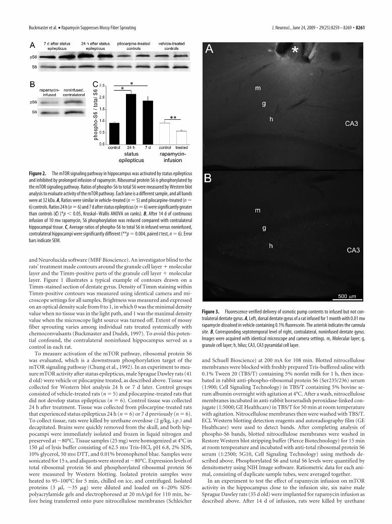
Figure 3. Fluorescence verified delivery of osmotic pump contents to infused but not con-tralateral dentate gyrus. A, Left, dorsal dentate gyrus of a rat infused for 1 month with 0.01 mM
rapamycin dissolved in vehicle containing 0.1% fluorescein. The asterisk indicates the cannulasite. B, Corresponding septotemporal level of right, contralateral, noninfused dentate gyrus.Images were acquired with identical microscope and camera settings. m, Molecular layer; g,granule cell layer; h, hilus; CA3, CA3 pyramidal cell layer.
Buckmaster et al. • Rapamycin Suppresses Mossy Fiber Sprouting J. Neurosci., June 24, 2009 • 29(25):8259 – 8269 • 8261

overdose (2 g/kg, i.p.) and decapitated. Brainswere quickly removed from the skull, placed ina chilled rat brain matrix (ASI Instruments),and blocked coronally to isolate a 3-mm-thickslice that contained the infusion site. On achilled platform, infused and contralateral hip-pocampi were isolated from slices, immediatelyfrozen in liquid nitrogen, and preserved at�80°C. Sample processing and Western analy-sis were performed as described above.
Values are reported as mean � SEM. Statis-tics were performed using SigmaStat (Systat). Avalue of p � 0.05 was considered significant.
ResultsStatus epilepticus activates themTOR pathwayTo test whether status epilepticus activatedthe mTOR signaling pathway in hip-pocampus, a downstream target of thepathway was evaluated. S6K-1, which isphosphorylated and activated by mTOR,in turn, phosphorylates ribosomal proteinS6 (Chung et al., 1992), which is a com-monly used readout of mTOR activity(Kwon et al., 2003; Huang et al., 2007;Meikle et al., 2008; Park et al., 2008; Zenget al., 2008). Hippocampal tissue was eval-uated in rats 24 h (n � 6) and 7 d (n � 6)after pilocarpine-induced status epilepti-cus. Control groups consisted of vehicle-treated rats (n � 5) and pilocarpine-treated rats that did not develop statusepilepticus (n � 6). Western blot analysiswas used to measure ratios of phospho-S6to total S6 (Fig. 2). Results from the twocontrol groups were similar (0.28 � 0.04vs 0.30 � 0.11 for vehicle- andpilocarpine-treated controls, respectively,p � 0.62, t test), so they were combined. At24 h and 7 d after status epilepticus, aver-age phospho-S6-to-total-S6 ratios weregreater than twice control values ( p �0.05, Kruskal–Wallis ANOVA on ranks).These findings suggest that status epilepti-cus activates the mTOR signaling pathway.
Rapamycin infusion inhibited themTOR signaling pathwayin hippocampusOsmotic pumps continuously deliveredrapamycin to a focal region of the left, dor-sal dentate gyrus, which was comparedwith the contralateral noninfused region.Vehicle contained fluorescein, and sec-tions were examined to verify fluorescencein infused hippocampi. Fluorescence wasclearly evident in sections near sites of in-fusion, but not in corresponding sectionsfrom contralateral hippocampi (Fig. 3). In three rats, fluores-cence was not observed, and they were excluded from analysis.These findings suggest that in all rats included in this study,pumps delivered their contents, which was concentrated in theinfused but not contralateral dentate gyrus.
Inhibition of the mTOR signaling pathway would be expectedto reduce levels of phospho-S6. Using the same rapamycin-infusion methods as used for experiments in which Timm stain-ing was measured, six rats were treated with 10 mM rapamycin for14 d. Subjects were naive, not pilocarpine treated, but we expectboth groups to respond similarly. Hippocampal tissue (3 mm
Figure 4. Prolonged, 1 month infusion of rapamycin reduced aberrant Timm staining. Timm stained dentate gyrus in vehicle-infused (A) and contralateral noninfused (B) hippocampus and in 10 mM rapamycin-infused (C) and contralateral noninfused (D)hippocampus. Asterisks indicate infusion sites (A, C). g, Granule cell layer; m, molecular layer; h, hilus; CA3, CA3 pyramidal celllayer. E, Higher-magnification views reveal less aberrant Timm staining in rapamycin-infused regions. E1, Vehicle-infused hip-pocampus (arrow in A). E2, Rapamycin-infused hippocampus at septotemporal level of cannula (arrow in C). E3, Rapamycin-infused hippocampus at 180 �m toward temporal pole of hippocampus from section shown in C and E2. E4, Contralateralnoninfused hippocampus (arrow in D). F, The average percentage of area of the granule cell layer (gcl)�molecular layer (ml) thatis Timm positive versus section position along the septotemporal axis relative to infusion site in rats infused with 10 mM rapamycin(n � 8). Infusion site � 0. The septal pole is to the left (negative values). The temporal pole is to the right (positive values). G,Difference in the percentage of the granule cell layer � molecular layer that is Timm positive in infused minus contralateralnoninfused hippocampi. Negative values indicate reduced aberrant Timm staining in infused hippocampi. Values calculated inindividual animals by averaging results of the three sections closest to the infusion site (between dashed vertical lines in F ) andcorresponding sections in the contralateral hippocampus. Hippocampi infused with 10 mM (n � 8), 1 mM (n � 4), and 0.1 mM
(n � 3) rapamycin displayed less aberrant Timm staining than those infused with 0.01 mM rapamycin (n � 4) or vehicle alone(n � 6) (*p � 0.05, ANOVA with Bonferroni t test). Error bars indicate SEM.
8262 • J. Neurosci., June 24, 2009 • 29(25):8259 – 8269 Buckmaster et al. • Rapamycin Suppresses Mossy Fiber Sprouting

thick, anterior–posterior) was isolated, which included dentategyrus, CA3, CA1, and subiculum and contained the infusion site.The same region was isolated from contralateral hippocampi.Western blot analysis was used to measure ratios of phospho-S6to total S6 in infused and contralateral noninfused hippocampi(Fig. 2). Ratios of phospho-S6 to total S6 were lower in infusedversus contralateral hippocampi in all individual animals. Groupaverages were significantly different (0.58 � 0.05 vs 0.92 � 0.09,p � 0.004, paired t test). These findings suggest that the rapamy-cin treatment protocol used in this study partially suppressed themTOR pathway in hippocampi near infusion sites.
Extent of inhibition is likely to be greatest near sites of infu-sion, where rapamycin concentration is likely to be highest withthis delivery method (Sendelbeck and Urquhart, 1985). In thepresent Western blot analysis, samples included all parts of hip-pocampus, not just dentate gyrus, and 3-mm-thick sections werecollected to provide sufficient material for analysis. This sam-pling method may have diluted focal rapamycin-infusion effects.Results, therefore, may underestimate inhibition of the mTORpathway in regions closest to infusion sites.
Rapamycin infusion for 1 month suppressed mossyfiber sproutingMossy fiber sprouting develops gradually over a period of weeksto months after pilocarpine-induced status epilepticus (Mello etal., 1993). By 1 month after status epilepticus, mossy fiber sprout-ing is developed to a sufficient baseline level for quantitative anal-ysis (Buckmaster, 2004b). In eight rats, 10 mM rapamycin wasinfused continuously and focally into the left, dorsal dentate gy-rus for 1 month after status epilepticus. Extent of aberrant Timmstaining was measured as the percentage of the area of the granulecell layer � molecular layer that was Timm positive. In sections�200 �m along the septotemporal axis from the infusion site,percentage of the granule cell layer � molecular layer that wasTimm positive was less than that of the corresponding region incontralateral noninfused hippocampi (Fig. 4). Inhibition of ab-errant Timm staining appeared maximal closest to infusion sites.In each animal, extent of aberrant Timm staining was calculatedby averaging values of the three sections closest to infusion sitesand the corresponding three sections in the contralateral nonin-fused hippocampus. In all rats, hippocampi infused with 10 mM
rapamycin for 1 month displayed less aberrant Timm stainingthan contralateral noninfused hippocampi. Group averages weresignificantly different (22.7 � 2.1 vs 27.2 � 1.9%, p � 0.001,paired t test).
To test whether suppression of aberrant Timm staining wasspecific to rapamycin, a range of concentrations was tested. Iden-tical infusion methods were used to test 1 mM rapamycin (n � 4),0.1 mM (n � 3), 0.01 mM (n � 4), and vehicle alone (n � 6). Ineach animal, average percentage of the granule cell layer � mo-lecular layer that was Timm positive was calculated for infusedand contralateral hippocampi, as described above. Then, the dif-ference was calculated by subtracting the average value of thecontralateral hippocampus from the average of the infused hip-pocampus. Accordingly, negative values indicate reduced aber-rant Timm staining in infused hippocampi. Group averages for10 mM, 1 mM, 0.1 mM, 0.01 mM, and vehicle alone were �6.5 �1.2, �7.5 � 1.2, �8.8 � 1.1, 2.5 � 0.8, and 0.3 � 1.9%, respec-tively. ANOVA revealed a significant difference between groups( p � 0.001). Pairwise multiple comparisons revealed signifi-cantly less aberrant Timm staining in hippocampi infused with10, 1, or 0.1 mM rapamycin (no significant differences betweenthese three groups) compared with 0.01 or 0 mM rapamycin (no
significant differences between these two groups) ( p � 0.05,Bonferroni t test). These findings suggest that infusion of rapa-mycin, but not vehicle alone, suppresses mossy fiber sprouting.
Rapamycin infusion for 2 months suppressed mossy fibersprouting moreDevelopment of mossy fiber sprouting is incomplete 1 monthafter pilocarpine-induced status epilepticus (Mello et al., 1993).To test whether longer treatment would further suppress mossyfiber sprouting, three rats were infused with 10 mM rapamycin for2 months (Fig. 5). Infusion methods were identical to those de-scribed above, except that osmotic pumps were replaced after 1month, which is the limit of their delivery duration. All rats dis-played less aberrant Timm staining in infused than in contralat-eral noninfused hippocampi. The average difference in percent-age of the granule cell layer � molecular layer that was Timmpositive (infused � contralateral) was �11.8 � 1.6%, which wasgreater than the average difference after only 1 month infusionwith 10 mM rapamycin (�6.5 � 1.2%, p � 0.04, unpaired t test).These findings suggest that longer treatment with rapamycinsuppressed mossy fiber sprouting more.
Suppression of mossy fiber sprouting diminished afterrapamycin infusion stoppedTo test whether rapamycin infusion permanently suppressedmossy fiber sprouting, four rats were infused with 10 mM rapa-mycin for 2 months and then survived 2 more months after pumpremoval. Infusion methods were identical to those describedabove, except that pumps were removed and tubing sealed after 2months of infusion, and rats were perfused 2 months later. In-tense, diffuse fluorescence was not evident as it was in rats per-fused immediately after infusion. Instead, moderate levels of par-ticulate fluorescence were observed in all rats (data not shown).These findings suggest that fluorescein had been delivered andlargely but incompletely cleared over the 2 months after pumpremoval. The average percentage of the granule cell layer � mo-lecular layer that was Timm positive in sections within �200 �mof infusion sites was similar to that in contralateral noninfusedhippocampi (30.5 � 2.5 vs 32.2 � 1.7%, respectively, p � 0.42,paired t test) (Fig. 5). The average difference in percentage of thegranule cell layer � molecular layer that was Timm positive (in-fused � contralateral) in rats that survived 2 months beyond a 2month period of 10 mM rapamycin infusion was significantly lessthan that of rats perfused immediately after 2 months infusionwith 10 mM rapamycin (�1.7 � 1.8 vs �11.8 � 1.6%, respec-tively, p � 0.01, unpaired t test). These findings suggest thatsuppression of mossy fiber sprouting by rapamycin is not perma-nent and instead may require continuous treatment.
Rapamycin infusion did not reverse established mossyfiber sproutingTo test whether rapamycin could reverse mossy fiber sproutingafter it had developed, infusion began 2 months after status epi-lepticus and lasted 1 month (n � 6). The average percentage ofthe granule cell layer � molecular layer that was Timm positive insections within �200 �m of infusion sites was similar to that incontralateral noninfused hippocampi (29.2 � 2.5 vs 29.7 � 2.5%,respectively, p � 0.72, paired t test) (Fig. 6). The average differ-ence in percentage of the granule cell layer � molecular layer thatwas Timm positive (infused � contralateral) in rats treated for 1month with 10 mM rapamycin after a latency of 2 months wassignificantly less than that of rats treated beginning immediatelyafter status epilepticus (�0.3 � 0.9 vs �6.5 � 1.2%, respectively,
Buckmaster et al. • Rapamycin Suppresses Mossy Fiber Sprouting J. Neurosci., June 24, 2009 • 29(25):8259 – 8269 • 8263

p � 0.002, unpaired t test). These findingssuggest that rapamycin infusion did notreverse mossy fiber sprouting after it haddeveloped.
Rapamycin infusion did not preventstatus epilepticus-induced hilarneuron lossExtent of aberrant Timm staining corre-lates with extent of hilar neuron loss (Babbet al., 1991; Masukawa et al., 1996; Buck-master and Dudek, 1997; Nissinen et al.,2001; Jiao and Nadler, 2007), and rapamy-cin has neuroprotective effects (Alirezaeiet al., 2008; Pan et al., 2008). These find-ings raise the possibility that rapamycinmight have protected hilar neurons fromstatus epilepticus-induced excitotoxicityand thereby indirectly reduced mossy fibersprouting. To avoid this potential con-founding factor, rapamycin infusion didnot begin until at least 1 h after suppres-sion of status epilepticus with diazepam.Nevertheless, to test whether hilar neuronswere spared in rapamycin-infused hip-pocampi, numbers of hilar neuron profileswere counted in the three sections closestto infusion sites and corresponding threesections in contralateral noninfused hip-pocampi. Average values from each hip-pocampus were calculated. Then, averagesfor experimental groups were calculated(Fig. 7). Average numbers of hilar neuronprofiles were slightly and consistentlylower in infused versus contralateral non-infused hippocampi (ANOVA, p � 0.006).The effect was apparent in all experimentalgroups, including those infused with only0.01 mM rapamycin or vehicle alone. Thesefindings suggest that hilar neuron loss wasslightly more severe in infused hip-pocampi, regardless of whether rapamycinwas present. Perhaps surgical implanta-tion of cannulae contributed to hilar neu-ron loss. These findings also suggest thatrapamycin infusion did not prevent statusepilepticus-induced hilar neuron loss.Therefore, suppression of mossy fibersprouting is not likely attributable to a sec-ondary effect on hilar neuron survival.
Another potential confound is densityof aberrant Timm staining. To testwhether intensities of Timm staining inthe granule cell layer � molecular layerwere different in infused versus nonin-fused contralateral hippocampi, opticaldensities within Timm-positive contourswere measured (Fig. 8). Densities of aber-rant Timm staining were similar withinoutlined areas of infused versus nonin-fused contralateral hippocampi (ANOVA,p � 0.4), including groups infused withmaximal doses of rapamycin and maximal
Figure 5. Longer rapamycin infusion suppressed mossy fiber sprouting more, but the effect reversed after infusion ceased.Timm stained dentate gyrus after 2 months of continuous infusion with 10 mM rapamycin (A) and contralateral noninfusedhippocampus (B). The asterisk indicates the infusion site. Arrows indicate reduced Timm staining in the granule cell layer (g) andmolecular layer (m) in the rapamycin-infused hippocampus. h, Hilus; CA3, CA3 pyramidal cell layer. Timm stained dentate gyrusafter 2 months of continuous infusion with 10 mM rapamycin followed by 2 more months without infusion (C) and contralateralnoninfused hippocampus (D). E, Experimental time line indicating duration of treatment (colored horizontal bars) and timing ofperfusion (red “x”). F, Difference in the percentage of the granule cell layer (gcl) � molecular layer (ml) that is Timm positive ininfused minus contralateral noninfused hippocampi. Negative values indicate reduced aberrant Timm staining in infused hip-pocampi. Values were calculated in individual animals by averaging results of the three sections closest to the infusion site andcorresponding sections in the contralateral hippocampus. The color of the vertical bars corresponds to the color of the horizontalbars in E, which indicate experimental group. Aberrant Timm staining was reduced more in rats infused with 10 mM rapamycin for2 months (n � 3) than in those infused only 1 month (n � 8, p � 0.04, unpaired t test) and those infused for 2 months but thenallowed to survive another 2 months after infusion ceased (n � 4, p � 0.01, unpaired t test). Results from vehicle-infused rats(n � 6) are displayed for comparison. Error bars indicate SEM.
8264 • J. Neurosci., June 24, 2009 • 29(25):8259 – 8269 Buckmaster et al. • Rapamycin Suppresses Mossy Fiber Sprouting

durations. These findings suggest that reduced areas of aberrantTimm staining in rapamycin-infused hippocampi were not com-pensated by more intense sprouting.
DiscussionThe principal findings of the present study are the following.Prolonged, continuous, focal infusion of rapamycin suppressed
development of aberrant mossy fibersprouting in a rat model of temporal lobeepilepsy. Longer infusions suppressedsprouting more. Sprouting that had beensuppressed initially with rapamycin devel-oped after infusion ceased, and rapamycinfailed to reverse established sprouting.These findings suggest that the mTOR sig-naling pathway may be a useful target forreducing granule cell axon reorganizationafter epileptogenic injuries.
Status epilepticus activates themTOR pathwayPilocarpine was used to cause status epi-lepticus, which resulted in development ofmossy fiber sprouting. Oxotremorine, an-other muscarinic acetylcholine receptoragonist, acutely increases phosphorylationof S6-kinase, and to a lesser extent mTOR,in hippocampus, but levels return to base-line by 16 h (Deguil et al., 2008). In thepresent study, there was no significant dif-ference in levels of phosphorylated S6 inpilocarpine- versus vehicle-treated con-trols 24 h after treatment. On the otherhand, pilocarpine-treated rats that experi-enced status epilepticus displayed signifi-cantly elevated levels of phosphorylated S624 h and 7 d after treatment. These find-ings suggest that the mTOR signalingpathway is activated by status epilepticus,but not by pilocarpine alone, during theperiod when mossy fibers sprout. Simi-larly, traumatic brain injury activates themTOR pathway (Chen et al., 2007) andcan cause mossy fiber sprouting (Golaraiet al., 2001; Kharatishvili et al., 2006).
Rapamycin suppressed mossyfiber sproutingIn the present study, granule cell axon ter-minals were identified by black Timmstaining. The Timm staining protocol gen-erates opaque silver particles at sites atwhich heavy metals are concentrated(Haug, 1967). The Timm stain is a specificmarker for granule cell axons becausemossy fibers concentrate zinc in synapticvesicles (Wenzel et al., 1997). Did rapamy-cin artifactually interfere with Timmstaining instead of suppressing mossy fibersprouting? This is unlikely, because Timmstaining was reduced specifically in regionsof sprouting, not in adjacent areas, includ-ing hilus and stratum lucidum of CA3.Furthermore, when infusion was delayed
until after mossy fiber sprouting developed, Timm staining in thegranule cell layer and molecular layer was intense, despite rapa-mycin treatment. In the present study, mossy fiber sprouting wasquantified by drawing contours around Timm-positive areaswithin the granule cell layer � molecular layer. Timm-positiveareas consist of many small, black spots, and defining borders of
Figure 6. Rapamycin infusion did not reverse mossy fiber sprouting after it had developed. Timm stained dentate gyrus after1 month infusion with 10 mM rapamycin, beginning 2 months after status epilepticus (A) and contralateral noninfused hippocam-pus (B). The asterisk indicates the infusion site. m, Molecular layer; g, granule cell layer; h, hilus; CA3, CA3 pyramidal cell layer. C,Experimental time line indicating duration of treatment (colored horizontal bars) and timing of perfusion (red “x”). D, Differencein the percentage of the granule cell layer (gcl) � molecular layer (ml) that is Timm positive in infused minus contralateralnoninfused hippocampi. Negative values indicate reduced aberrant Timm staining in infused hippocampi. Values were calculatedin individual animals by averaging results of the three sections closest to the infusion site and corresponding sections in thecontralateral hippocampus. The color of the vertical bars corresponds to the color of the horizontal bars in C, which indicateexperimental group. Aberrant Timm staining is reduced more in rats when 10 mM rapamycin is infused beginning immediatelyafter status epilepticus (n � 8) than in those in which onset of infusion was delayed 2 months (n � 6, p � 0.002, unpaired t test).Results from vehicle-infused rats (n � 6) are displayed for comparison. Error bars indicate SEM.
Buckmaster et al. • Rapamycin Suppresses Mossy Fiber Sprouting J. Neurosci., June 24, 2009 • 29(25):8259 – 8269 • 8265

those areas can be subjective. To avoid bias, investigators wereblind to experimental groups. Similar optical densities withinTimm-positive regions of infused versus contralateral hip-pocampi in each experimental group support the validity of com-paring relative areas of Timm-positive regions within the granulecell layer and molecular layer.
Suppression of mossy fiber sprouting appeared to be specificto rapamycin treatment. Sprouting occurred at untreated levelswhen vehicle or low-dose rapamycin was infused and was sup-pressed by higher doses. Rapamycin is a specific inhibitor of
mTOR complex 1 (Loewith et al., 2002), which is part of a signal-ing pathway that phosphorylates and activates ribosomal proteinS6 (Chung et al., 1992). Reduced phospho-S6 levels after infusionof 10 mM rapamycin are consistent with inhibition of mTORcomplex 1. Prolonged rapamycin treatment can sequester mTORand indirectly inhibit mTOR complex 2 (Sarbassov et al., 2006),which is part of another signaling pathway that controls cell pro-liferation and survival through the actin cytoskeleton and Akt/PKB (Jacinto et al., 2004). The present study cannot distinguishbetween rapamycin’s effects on mTOR complex 1 and its effectson mTOR complex 2. Despite mTOR’s role in cell survival, rapa-mycin infusion did not spare hilar neurons from excitotoxicity inthe present study, probably because infusion began after rats hadexperienced status epilepticus.
Hilar neuron loss was slightly more severe in infused than innoninfused hippocampi. However, the effect was not specific torapamycin. All groups displayed the difference, even those in-fused with little or no rapamycin. Slightly more severe hilar neu-ron loss in infused hippocampi might be attributable to surgicalplacement of cannulae. Nevertheless, rapamycin’s suppression ofmossy fiber sprouting was not attributable to hilar neuronsurvival.
Rapamycin binds FK506 binding protein 12 (Heitman et al.,1991), which is part of yet another signaling pathway that affectsactivity of calcineurin, a calcium-activated phosphatase that hasbeen proposed to play a role in mossy fiber sprouting (Moriwakiet al., 1996). However, this mechanism is unlikely to account forrapamycin’s effect, because infusion of FK506, an inhibitor ofcalcineurin, fails to suppress mossy fiber sprouting (Ingram et al.,2009). Rapamycin impairs potentiation of excitatory synapses(Casadio et al., 1999; Tang et al., 2002) and can reduce networkexcitability (Ruegg et al., 2007), suggesting that it might havesuppressed mossy fiber sprouting indirectly by affecting excit-ability or synapse strength. This possibility seems unlikely, how-ever, because prolonged infusion of the sodium channel blocker
Figure 7. Rapamycin infusion did not protect hilar neurons from status epilepticus-inducedloss. Nissl stained dentate gyrus after 1 month infusion with 10 mM rapamycin (A) and contralat-eral noninfused hippocampus (B). The asterisk indicates the infusion site. m, Molecular layer; g,granule cell layer; h, hilus; CA3, CA3 pyramidal cell layer. C, The average number of hilar neuronprofiles per section in the three sections closest to the infusion site and corresponding sectionsof contralateral hippocampus for all experimental groups. The number of hilar neuron profileswas slightly and consistently lower in infused than in contralateral hippocampi ( p � 0.006,ANOVA), regardless of experimental group. Error bars indicate SEM.
Figure 8. Density of staining within Timm-positive contours was not more intense inrapamycin-infused hippocampi. The average optical density (0, no tissue in light path; 1, mi-croscope light source turned off) within Timm-positive contours in three sections within �200�m of infusion site and corresponding sections of contralateral noninfused hippocampus for allexperimental groups is shown. Within each experimental group, densities of aberrant Timmstaining were similar in infused and contralateral, noninfused hippocampi (ANOVA, p � 0.4).Error bars indicate SEM.
8266 • J. Neurosci., June 24, 2009 • 29(25):8259 – 8269 Buckmaster et al. • Rapamycin Suppresses Mossy Fiber Sprouting

tetrodotoxin, which reduces neuronal activity substantially (Gal-van et al., 2000), fails to suppress mossy fiber sprouting (Buck-master, 2004b).
Findings of the present study suggest that rapamycin sup-pressed development of mossy fiber sprouting in a rodent modelof temporal lobe epilepsy. These findings also suggest that target-ing the mTOR signaling pathway may be useful for testing therole of mossy fiber sprouting on epileptogenesis. However, rapa-mycin also impairs long-term potentiation (Casadio et al., 1999;Tang et al., 2002) and learning (Dash et al., 2006; Parsons et al.,2006), alters neuronal expression of transporters (Li et al., 2006),voltage-gated channels (Raab-Graham et al., 2006), and ligand-gated channels (Sabatini et al., 1999; Wang et al., 2006), inhibitsdendritic growth (Jaworski et al., 2005; Kumar et al., 2005; Huanget al., 2007), and changes dendritic spine morphology (Tavazoieet al., 2005). Therefore, the mTOR signaling pathway’s effects onepileptogenesis and seizure threshold may be complex.
Mossy fiber sprouting may be permanent once establishedThe hypothesis that rapamycin might reverse already establishedmossy fiber sprouting was motivated by a report that mTORinhibition reverses dentate granule cell hypertrophy in adultPTEN-deficient mice (Kwon et al., 2003). However, in thepresent study, 1 month treatment with rapamycin was insuffi-cient to significantly reduce already established aberrant Timmstaining. Although these findings do not exclude the possibilitythat longer treatments may have been more effective, they suggestthat mossy fiber sprouting may be irreversible. Similarly, in ro-dent models of tuberous sclerosis, rapamycin administered aftercompletion of neuronal differentiation failed to reverse abnor-mally oriented neocortical dendrites (Meikle et al., 2008), andbeneficial effects of rapamycin, including reduced seizure fre-quency and increased survival, require continued long-termtreatment and reverse after treatment stops (Zeng et al., 2008). InPTEN-deficient mice, dentate granule cell hypertrophy resumesafter mTOR inhibition stops (Kwon et al., 2003). These results arenot surprising, because genetic disease models involve mutationsthat persist after rapamycin treatment ends. In the present study,continuous rapamycin treatment was required, which suggeststhat signals that promote mossy fiber sprouting may be persistentand, unfortunately, not transiently expressed only during a crit-ical window during which time targeted, temporary treatmentcould permanently block axon reorganization.
Mossy fiber sprouting is resilientMossy fiber sprouting is a common abnormality in patients, oc-curs in many laboratory animal models of epilepsy, and has re-sisted previous attempts to suppress it (see Introduction). In thepresent study, mossy fiber sprouting was reduced but not blockedentirely by rapamycin, even at high concentrations. These find-ings suggest redundancy in signaling pathways, such that inhibi-tion of a single important node may partially suppress but notabrogate axon sprouting (Bromberg et al., 2008). More completeblockade of mossy fiber sprouting may require targeting addi-tional steps in the mTOR cascade and in other signalingpathways.
ReferencesAlirezaei M, Kiosses WB, Flynn CT, Brady NR, Fox HS (2008) Disruption of
neuronal autophagy by infected microglia results in neurodegeneration.PLoS ONE 3:e2906.
Babb TL, Kupfer WR, Pretorius JK, Crandall PH, Levesque MF (1991) Syn-aptic reorganization by mossy fibers in human epileptic fascia dentata.Neuroscience 42:351–363.
Bromberg KD, Ma’ayan A, Neves SR, Iyengar R (2008) Design logic of acannabinoid receptor signaling network that triggers neurite outgrowth.Science 320:903–909.
Buckmaster PS (2004a) Laboratory animal models of temporal lobe epi-lepsy. Comp Med 54:473– 485.
Buckmaster PS (2004b) Prolonged infusion of tetrodotoxin does not blockmossy fiber sprouting in pilocarpine-treated rats. Epilepsia 45:452– 458.
Buckmaster PS, Dudek FE (1997) Neuron loss, granule cell axon reorgani-zation, and functional changes in the dentate gyrus of epileptic kainate-treated rats. J Comp Neurol 385:385– 404.
Buckmaster PS, Zhang GF, Yamawaki R (2002) Axon sprouting in a modelof temporal lobe epilepsy creates a predominantly excitatory feedbackcircuit. J Neurosci 22:6650 – 6658.
Cammalleri M, Lutjens R, Berton F, King AR, Simpson C, Francesconi W,Sanna PP (2003) Time-restricted role for dendritic activation of themTOR-p70 S6K pathway in the induction of late-phase long-term poten-tiation in the CA1. Proc Natl Acad Sci U S A 100:14368 –14373.
Campbell DS, Holt CE (2001) Chemotropic responses of retinal growthcones mediated by rapid local protein synthesis and degradation. Neuron32:1013–1026.
Casadio A, Martin KC, Giustetto M, Zhu H, Chen M, Bartsch D, Bailey CH,Kandel ER (1999) A transient, neuron-wide form of CREB-mediatedlong-term facilitation can be stabilized at specific synapses by local pro-tein synthesis. Cell 99:221–237.
Chen S, Atkins CM, Liu CL, Alonso OF, Dietrich WD, Hu BR (2007) Alter-ations in mammalian target of rapamycin signaling pathways after trau-matic brain injury. J Cereb Blood Flow Metab 27:939 –949.
Chung J, Kuo CJ, Crabtree GR, Blenis J (1992) Rapamycin-FKBP specifi-cally blocks growth-dependent activation of and signaling by the 70 kd S6protein kinases. Cell 69:1227–1236.
Dash PK, Orsi SA, Moore AN (2006) Spatial memory formation andmemory-enhancing effect of glucose involves activation of the tuberoussclerosis complex-mammalian target of rapamycin pathway. J Neurosci26:8048 – 8056.
Deguil J, Perault-Pochat MC, Chavant F, Lafay-Chebassier C, FaucconneauB, Pain S (2008) Activation of the protein p70S6K via ERK phosphory-lation by cholinergic muscarinic receptors stimulation in human neuro-blastoma cells and in mice brain. Toxicol Lett 182:91–96.
de Lanerolle NC, Kim JH, Robbins RJ, Spencer DD (1989) Hippocampalinterneuron loss and plasticity in human temporal lobe epilepsy. BrainRes 495:387–395.
Engel J Jr, Williamson PD, Wieser HG (1997) Mesial temporal lobe epilepsy.In: Epilepsy: a comprehensive textbook (Engel J Jr, Pedley TA, eds), pp2417–2426. Philadelphia: Lippincott-Raven.
Galvan CD, Hrachovy RA, Smith KL, Swann JW (2000) Blockade of neuro-nal activity during hippocampal development produces a chronic focalepilepsy in the rat. J Neurosci 20:2904 –2916.
Golarai G, Greenwood AC, Feeney DM, Connor JA (2001) Physiologicaland structural evidence for hippocampal involvement in persistent sei-zure susceptibility after traumatic brain injury. J Neurosci 21:8523– 8537.
Gong R, Park CS, Abbassi NR, Tang SJ (2006) Roles of glutamate receptorsand the mammalian target of rapamycin (mTOR) signaling pathway inactivity-dependent dendritic protein synthesis in hippocampal neurons.J Biol Chem 281:18802–18815.
Harris TE, Lawrence JC Jr (2003) TOR signaling. Sci STKE 2003:re15.Haug FM (1967) Electron microscopical localization of the zinc in hip-
pocampal mossy fibre synapses by a modified sulfide silver procedure.Histochemie 8:355–368.
Heitman J, Movva NR, Hall MN (1991) Targets for cell cycle arrest by theimmunosuppressant rapamycin in yeast. Science 253:905–909.
Holtzman DM, Lowenstein DH (1995) Selective inhibition of axon out-growth by antibodies to NGF in a model of temporal lobe epilepsy. J Neu-rosci 15:7062–7070.
Hou L, Klann E (2004) Activation of the phosphoinositide 3-kinase-Akt-mammalian target of rapamycin signaling pathway is required formetabotropic glutamate receptor-dependent long-term depression.J Neurosci 24:6352– 6361.
Houser CR, Miyashiro JE, Swartz BE, Walsh GO, Rich JR, Delgado-EscuetaAV (1990) Altered patterns of dynorphin immunoreactivity suggestmossy fiber reorganization in human hippocampal epilepsy. J Neurosci10:267–282.
Huang Y, Kang BN, Tian J, Liu Y, Luo HR, Hester L, Snyder SH (2007) The
Buckmaster et al. • Rapamycin Suppresses Mossy Fiber Sprouting J. Neurosci., June 24, 2009 • 29(25):8259 – 8269 • 8267
cationic amino acid transporters CAT1 and CAT3 mediate NMDA recep-tor activation-dependent changes in elaboration of neuronal processes viathe mammalian target of rapamycin mTOR pathway. J Neurosci27:449 – 458.
Ikegaya Y (1999) Abnormal targeting of developing hippocampal mossy fi-bers after epileptiform activities via L-type Ca 2� channel activation invitro. J Neurosci 19:802– 812.
Ikegaya Y, Nishiyama N, Matsuki N (2000) L-type Ca 2� channel blockerinhibits mossy fiber sprouting and cognitive deficits following pilocarpineseizures in immature mice. Neuroscience 98:647– 659.
Ingram EA, Toyoda I, Wen X, Buckmaster PS (2009) Prolonged infusionof inhibitors of calcineurin or L-type calcium channels does not blockmossy fiber sprouting in a model of temporal lobe epilepsy. Epilepsia50:56 – 64.
Jacinto E, Loewith R, Schmidt A, Lin S, Ruegg MA, Hall A, Hall MN (2004)Mammalian TOR complex 2 controls the actin cytoskeleton and is rapa-mycin insensitive. Nat Cell Biol 6:1122–1128.
Jaworski J, Spangler S, Seeburg DP, Hoogenraad CC, Sheng M (2005)Control of dendritic arborization by the phosphoinositide-3�-kinase-Akt-mammalian target of rapamycin pathway. J Neurosci25:11300 –11312.
Jiao Y, Nadler JV (2007) Stereological analysis of GluR2-immunoreactivehilar neurons in the pilocarpine model of temporal lobe epilepsy: corre-lation of cell loss with mossy fiber sprouting. Exp Neurol 205:569 –582.
Kadam SD, Dudek FE (2007) Neuropathological features of a rat model ofperinatal hypoxic-ischemic encephalopathy with associated epilepsy.J Comp Neurol 505:716 –737.
Kharatishvili I, Nissinen JP, McIntosh TK, Pitkanen A (2006) A model ofposttraumatic epilepsy induced by lateral fluid-percussion brain injury inrats. Neuroscience 140:685– 697.
Kim DH, Sarbassov DD, Ali SM, King JE, Latek RR, Erdjument-Bromage H,Tempst P, Sabatini DM (2002) mTOR interacts with raptor to form anutrient-sensitive complex that signals to the cell growth machinery. Cell110:163–175.
Kumar V, Zhang MX, Swank MW, Kunz J, Wu GY (2005) Regulation ofdendritic morphogenesis by Ras-PI3K-Akt-mTOR and Ras-MAPK sig-naling pathways. J Neurosci 25:11288 –11299.
Kwon CH, Zhu X, Zhang J, Baker SJ (2003) mTor is required for hypertro-phy of Pten-deficient neuronal soma in vivo. Proc Natl Acad Sci U S A100:12923–12928.
Li LB, Toan SV, Zelenaia O, Watson DJ, Wolfe JH, Rothstein JD, RobinsonMB (2006) Regulation of astrocytic glutamate transporter expression byAkt: evidence for a selective transcriptional effect on the GLT-1/EAAT2subtype. J Neurochem 97:759 –771.
Loewith R, Jacinto E, Wullschleger S, Lorberg A, Crespo JL, Bonenfant D,Oppliger W, Jenoe P, Hall MN (2002) Two TOR complexes, only one ofwhich is rapamycin sensitive, have distinct roles in cell growth control.Mol Cell 10:457– 468.
Longo BM, Mello LEAM (1997) Blockade of pilocarpine- or kainate-induced mossy fiber sprouting by cycloheximide does not prevent subse-quent epileptogenesis in rats. Neurosci Lett 226:163–166.
Longo BM, Mello LEAM (1998) Supragranular mossy fiber sprouting is notnecessary for spontaneous epileptogenesis in rats. Epilepsy Res32:172–182.
Lynch M, Sutula T (2000) Recurrent excitatory connectivity in the den-tate gyrus of kindled and kainic acid-treated rats. J Neurophysiol83:693–704.
Masukawa LM, Wang H, O’Connor MJ, Uruno K (1996) Prolonged fieldpotentials evoked by 1 Hz stimulation in the dentate gyrus of temporallobe epileptic human brain slices. Brain Res 721:132–139.
Meikle L, Pollizzi K, Egnor A, Kramvis I, Lane H, Sahin M, Kwiatkowski DJ(2008) Response of a neuronal model of tuberous sclerosis to mamma-lian target of rapamycin (mTOR) inhibitors: effects on mTORC1 and Aktsignaling lead to improved survival and function. J Neurosci28:5422–5432.
Mello LEAM, Cavalheiro EA, Tan AM, Kupfer WR, Pretorius JK, Babb TL,Finch DM (1993) Circuit mechanisms of seizures in the pilocarpinemodel of chronic epilepsy: cell loss and mossy fiber sprouting. Epilepsia34:985–995.
Moriwaki A, Lu YF, Hayashi Y, Tomizawa K, Tokuda M, Itano T, HataseO, Matsui H (1996) Immunosuppressant FK506 prevents mossy fi-
ber sprouting induced by kindling stimulation. Neurosci Res25:191–194.
Nadler JV, Perry BW, Cotman CW (1980) Selective reinnervation of hip-pocampal area CA1 and the fascia dentata after destruction of CA3-CA4afferents with kainic acid. Brain Res 182:1–9.
Nissinen J, Halonen T, Koivisto E, Pitkanen A (2000) A new model ofchronic temporal lobe epilepsy induced by electrical stimulation of theamygdala in rats. Epilepsy Res 38:177–205.
Nissinen J, Lukasiuk K, Pitkanen A (2001) Is mossy fiber sprouting presentat the time of the first spontaneous seizures in rat experimental temporallobe epilepsy? Hippocampus 11:299 –310.
Pan T, Kondo S, Zhu W, Xie W, Jankovic J, Le W (2008) Neuroprotection ofrapamycin in lactacystin-induced neurodegeneration via autophagy en-hancement. Neurobiol Dis 32:16 –25.
Park KK, Liu K, Hu Y, Smith PD, Wang C, Cai B, Xu B, Connolly L, KramvisI, Sahin M, He Z (2008) Promoting axon regeneration in the adult CNSby modulation of the PTEN/mTOR pathway. Science 322:963–966.
Parsons RG, Gafford GM, Helmstetter FJ (2006) Translational control viathe mammalian target of rapamycin pathway is critical for the formationand stability of long-term fear memory in amygdala neurons. J Neurosci26:12977–12983.
Qiao X, Noebels JL (1993) Developmental analysis of hippocampal mossyfiber outgrowth in mutant mouse with inherited spike-wave seizures.J Neurosci 13:4622– 4635.
Raab-Graham KF, Haddick PCG, Jan YN, Jan LY (2006) Activity- andmTOR-dependent suppression of Kv1.1 channel mRNA translation indendrites. Science 314:144 –148.
Ribak CE, Seress L, Weber P, Epstein CM, Henry TR, Bakay RAE (1998)Alumina gel injections into the temporal lobe of rhesus monkeys causecomplex partial seizures and morphological changes found in humantemporal lobe epilepsy. J Comp Neurol 401:266 –290.
Ruegg S, Baybis M, Juul H, Dichter M, Crino PB (2007) Effects of rapamycinon gene expression, morphology, and electrophysiological properties ofrat hippocampal neurons. Epilepsy Res 77:85–92.
Sabatini DM, Erdjument-Bromage H, Lui M, Tempst P, Snyder SH(1994) RAFT1: a mammalian protein that binds to FKBP12 in arapamycin-dependent fashion and is homologous to yeast TORs. Cell78:35– 43.
Sabatini DM, Barrow RK, Blackshaw S, Burnett PE, Lai MM, Field ME, Bahr BA,Kirsch J, Betz H, Snyder SH (1999) Interaction of RAFT1 with gephyrinrequired for rapamycin-sensitive signaling. Science 284:1161–1164.
Sarbassov DD, Ali SM, Sengupta S, Sheen JH, Hsu PP, Bagley AF, MarkhardAL, Sabatini DM (2006) Prolonged rapamycin treatment inhibitsmTORC2 assembly and Akt/PKB. Mol Cell 22:159 –168.
Scharfman HE, Sollas AL, Berger RE, Goodman JH (2003) Electrophysio-logical evidence of monosynaptic excitatory transmission between gran-ule cells after seizure-induced mossy fiber sprouting. J Neurophysiol90:2536 –2547.
Schmelzle T, Hall MN (2000) TOR, a central controller of cell growth. Cell103:253–262.
Sendelbeck SL, Urquhart J (1985) Spatial distribution of dopamine, meth-otrexate and antipyrine during continuous intracerebral microperfusion.Brain Res 328:251–258.
Sloviter RS (1992) Possible functional consequences of synaptic reorga-nization in the dentate gyrus of kainate-treated rats. Neurosci Lett137:91–96.
Sloviter RS, Zappone CA, Harvey BD, Frotscher M (2006) Kainic acid-induced recurrent mossy fiber innervation of dentate gyrus inhibitoryinterneurons: possible anatomical substrate of granule cell hyperinhibi-tion in chronically epileptic rats. J Comp Neurol 494:944 –960.
Sutula T, He XX, Cavazos J, Scott G (1988) Synaptic reorganization in thehippocampus induced by abnormal functional activity. Science239:1147–1150.
Sutula T, Cascino G, Cavazos J, Parada I, Ramirez L (1989) Mossy fibersynaptic reorganization in the epileptic human temporal lobe. Ann Neu-rol 26:321–330.
Swiech L, Perycz M, Malik A, Jaworski J (2008) Role of mTOR in physiologyand pathology of the nervous system. Biochem Biophys Acta1784:116 –132.
Takei N, Inamura N, Kawamura M, Namba H, Hara K, Yonezawa K, Nawa H(2004) Brain-derived neurotrophic factor induces mammalian target of
8268 • J. Neurosci., June 24, 2009 • 29(25):8259 – 8269 Buckmaster et al. • Rapamycin Suppresses Mossy Fiber Sprouting
rapamycin-dependent local activation of translation machinery and pro-tein synthesis in neuronal dendrites. J Neurosci 24:9760 –9769.
Tang SJ, Reis G, Kang H, Gingras AC, Sonenberg N, Schuman EM (2002) Arapamycin-sensitive signaling pathway contributes to long-term synapticplasticity in the hippocampus. Proc Natl Acad Sci U S A 99:467– 472.
Tauck DL, Nadler JV (1985) Evidence of functional mossy fiber sprouting inhippocampal formation of kainic acid-treated rats. J Neurosci 5:1016–1022.
Tavazoie SF, Alvarez VA, Ridenour DA, Kwiatkowski DJ, Sabatini BL (2005)Regulation of neuronal morphology and function by the tumor suppres-sors Tsc1 and Tsc2. Nat Neurosci 8:1727–1734.
Toyoda I, Buckmaster PS (2005) Prolonged infusion of cycloheximide doesnot block mossy fiber sprouting in a model of temporal lobe epilepsy.Epilepsia 46:1017–1020.
Verma P, Chierzi S, Codd AM, Campbell DS, Meyer RL, Holt CE, Fawcett JW(2005) Axonal protein synthesis and degradation are necessary for effi-cient growth cone regeneration. J Neurosci 25:331–342.
Wang Y, Barbaro MF, Baraban SC (2006) A role for the mTOR pathway insurface expression of AMPA receptors. Neurosci Lett 401:35–39.
Wenzel HJ, Cole TB, Born DE, Schwartzkroin PA, Palmiter RD (1997) Ul-trastructural localization of zinc transporter-3 (ZnT-3) to synaptic vesiclemembranes within mossy fiber boutons in the hippocampus of mouseand monkey. Proc Natl Acad Sci U S A 94:12676 –12681.
Williams PA, Wuarin JP, Dou P, Ferraro DJ, Dudek FE (2002) Reassessmentof the effects of cycloheximide on mossy fiber sprouting and epileptogen-esis in the pilocarpine model of temporal lobe epilepsy. J Neurophysiol88:2075–2087.
Wuarin JP, Dudek FE (1996) Electrographic seizures and new recurrent ex-citatory circuits in the dentate gyrus of hippocampal slices from kainate-treated epileptic rats. J Neurosci 16:4438 – 4448.
Zeng LH, Xu L, Gutmann DH, Wong M (2008) Rapamycin prevents epi-lepsy in a mouse model of tuberous sclerosis complex. Ann Neurol 63:444 – 453.
Buckmaster et al. • Rapamycin Suppresses Mossy Fiber Sprouting J. Neurosci., June 24, 2009 • 29(25):8259 – 8269 • 8269