Inhibition of Human Papillomavirus Type 16 E7 ...jvi.asm.org/content/79/2/1099.full.pdf · the S100...
14
JOURNAL OF VIROLOGY, Jan. 2005, p. 1099–1112 Vol. 79, No. 2 0022-538X/05/$08.000 doi:10.1128/JVI.79.2.1099–1112.2005 Copyright © 2005, American Society for Microbiology. All Rights Reserved. Inhibition of Human Papillomavirus Type 16 E7 Phosphorylation by the S100 MRP-8/14 Protein Complex Sharof Tugizov, 1,2 * Jennifer Berline, 1 Rossana Herrera, 1 Maria Elena Penaranda, 2 † Mayumi Nakagawa, 3 and Joel Palefsky 1,2 Department of Medicine 1 and Department of Stomatology, 2 University of California, San Francisco, San Francisco, California, and Department of Dermatology, University of Arkansas, Little Rock, Arkansas 3 Received 5 July 2004/Accepted 30 August 2004 The human papillomavirus type 16 (HPV16) E7 is a major viral oncoprotein that is phosphorylated by casein kinase II (CKII). Two S100 family calcium-binding proteins, macrophage inhibitory-related factor protein 8 (MRP-8) and MRP-14, form a protein complex, MRP-8/14, that inactivates CKII. The MRP-8/14 protein com- plex may inhibit CKII-mediated E7 phosphorylation and therefore may alter its interaction with cellular ligands and reduce E7 oncogenic activity. We examined the inhibitory effect of the MRP-8/14 complex on CKII activity and HPV16 E7 phosphorylation. We have shown that CKII activity and HPV16 E7 phosphorylation were inhibited by uptake of exogenous MRP-8/14 and activation of endogenous MRP-8/14. MRP-8/14-mediated inhibition of E7 phosphorylation occurred at the G 1 phase of the cell cycle. Analysis of MRP expression in primary keratinocytes and in HPV16- and 18-transformed cervical and foreskin epithelial cell lines showed that expression of MRP-8, MRP-14, and the MRP-8/14 complex was detected only in primary untransformed keratinocytes and not in the HPV-infected immortalized epithelial cells. CKII activity in HPV-immortalized keratinocytes was approximately fourfold higher than in HPV-negative primary keratinocytes. Treatment of HPV-positive immortalized epithelial cells with exogenous MRP-8/14 resulted in E7 hypophosphorylation and complete inhibition of cell growth within 2 weeks, compared with HPV-negative primary and immortalized HPV-negative cervical epithelial cells, which showed 25 and 40% growth inhibition, respectively. Together these results suggests that the MRP-8/14 protein complex in HPV-infected epithelial cells may play an important role in regulation of CKII-mediated E7 phosphorylation and inhibition of its oncogenic activity. Human papillomaviruses (HPV) are a large family of non- enveloped DNA viruses that infect epithelial cells and may cause condyloma, high-grade dysplasia, and invasive cancer. Certain high-risk genotypes of HPV, such as HPV type 16 (HPV16) and HPV18, are strongly involved in the pathogen- esis of anogenital cancer in men and women (65, 66). HPV- induced malignant transformation is mediated by cooperative expression of the HPV E6 and E7 oncoproteins and their interaction with their host cellular target proteins (36, 37, 46). HPV E6 binds to tumor suppressor protein p53, and this in- teraction leads to inactivation of p53 and chromosomal insta- bility (59). HPV E7 binds to pRb and to several other cellular proteins that are involved in cell cycle and transcriptional con- trol mechanisms, and these interactions stimulate cell cycle progression (14). The HPV16 E7 protein is a 98-amino-acid nuclear phospho- protein (49) that induces DNA synthesis in quiescent cells, cooperates with the ras oncogene to transform primary rodent cells, and interacts with pRb to promote cell cycle progression (41, 57). The HPV16 E7 CR2 domain plays a critical role in its oncogenic function by interaction with the Rb family of pro- teins via its LxCxE motif (8). This domain contains two phos- phorylation sites on its Ser 31 and Ser 32 positions that are phosphorylated by cellular casein kinase II (CKII) (3, 17). E7 is also phosphorylated at its C terminus at the serine 71 posi- tion by an unknown kinase (32). Furthermore, it has been shown that HPV E7 is differentially phosphorylated during the cell cycle at G 1 phase by CKII and at S phase by unidentified enzymes (32). HPV18 E7 phosphorylation has been shown to be critical to promote S-phase entry (9). Consistent with these data, muta- tions of the serine 31/32 residues and a consensus sequence for HPV16 E7 phosphorylation by CKII (amino acids 30 to 37) resulted in defective E7 phosphorylation and impaired its transforming ability (3, 18). Replacement of the serine 31/32 residues with two negatively charged aspartic acid residues restored E7 transforming activity (3, 18). The level of E7 phos- phorylation and transformation activity is lower in low-risk HPV types than in high-risk HPV types (3). These data suggest that E7 phosphorylation by CKII may play a critical role in enhancement of oncogenic functions of E7 and development of HPV-associated neoplasia. However, the molecular mech- anisms of regulation of E7 phosphorylation and its role in E7 interaction with its intracellular ligands are not fully under- stood. CKII is a pleiotropic, ubiquitous serine/threonine protein kinase composed of two catalytic subunits ( and ) and two regulatory subunits which phosphorylate many key cellular and viral proteins (30, 42). CKII has been detected within the cytoplasm and nucleus and is associated with plasma mem- brane and intracellular organelles, including the Golgi and the endoplasmic reticulum (15, 44). CKII is a key component of the regulatory protein kinase network and is involved in reg- * Corresponding author. Mailing address: Department of Medicine, University of California, San Francisco, 513 Parnassus Ave., San Fran- cisco, CA 94143. Phone: (415) 514-3177. Fax: (415) 476-9364. E-mail: [email protected]. † Present address: Sustainable Sciences Institute, San Francisco, CA 94102. 1099 on October 14, 2018 by guest http://jvi.asm.org/ Downloaded from
Transcript of Inhibition of Human Papillomavirus Type 16 E7 ...jvi.asm.org/content/79/2/1099.full.pdf · the S100...

JOURNAL OF VIROLOGY, Jan. 2005, p. 1099–1112 Vol. 79, No. 20022-538X/05/$08.00�0 doi:10.1128/JVI.79.2.1099–1112.2005Copyright © 2005, American Society for Microbiology. All Rights Reserved.
Inhibition of Human Papillomavirus Type 16 E7 Phosphorylation bythe S100 MRP-8/14 Protein Complex
Sharof Tugizov,1,2* Jennifer Berline,1 Rossana Herrera,1 Maria Elena Penaranda,2†Mayumi Nakagawa,3 and Joel Palefsky1,2
Department of Medicine1 and Department of Stomatology,2 University of California, San Francisco, San Francisco,California, and Department of Dermatology, University of Arkansas, Little Rock, Arkansas3
Received 5 July 2004/Accepted 30 August 2004
The human papillomavirus type 16 (HPV16) E7 is a major viral oncoprotein that is phosphorylated by caseinkinase II (CKII). Two S100 family calcium-binding proteins, macrophage inhibitory-related factor protein 8(MRP-8) and MRP-14, form a protein complex, MRP-8/14, that inactivates CKII. The MRP-8/14 protein com-plex may inhibit CKII-mediated E7 phosphorylation and therefore may alter its interaction with cellularligands and reduce E7 oncogenic activity. We examined the inhibitory effect of the MRP-8/14 complex on CKIIactivity and HPV16 E7 phosphorylation. We have shown that CKII activity and HPV16 E7 phosphorylationwere inhibited by uptake of exogenous MRP-8/14 and activation of endogenous MRP-8/14. MRP-8/14-mediatedinhibition of E7 phosphorylation occurred at the G1 phase of the cell cycle. Analysis of MRP expression inprimary keratinocytes and in HPV16- and 18-transformed cervical and foreskin epithelial cell lines showedthat expression of MRP-8, MRP-14, and the MRP-8/14 complex was detected only in primary untransformedkeratinocytes and not in the HPV-infected immortalized epithelial cells. CKII activity in HPV-immortalizedkeratinocytes was approximately fourfold higher than in HPV-negative primary keratinocytes. Treatment ofHPV-positive immortalized epithelial cells with exogenous MRP-8/14 resulted in E7 hypophosphorylation andcomplete inhibition of cell growth within 2 weeks, compared with HPV-negative primary and immortalizedHPV-negative cervical epithelial cells, which showed 25 and 40% growth inhibition, respectively. Together theseresults suggests that the MRP-8/14 protein complex in HPV-infected epithelial cells may play an important rolein regulation of CKII-mediated E7 phosphorylation and inhibition of its oncogenic activity.
Human papillomaviruses (HPV) are a large family of non-enveloped DNA viruses that infect epithelial cells and maycause condyloma, high-grade dysplasia, and invasive cancer.Certain high-risk genotypes of HPV, such as HPV type 16(HPV16) and HPV18, are strongly involved in the pathogen-esis of anogenital cancer in men and women (65, 66). HPV-induced malignant transformation is mediated by cooperativeexpression of the HPV E6 and E7 oncoproteins and theirinteraction with their host cellular target proteins (36, 37, 46).HPV E6 binds to tumor suppressor protein p53, and this in-teraction leads to inactivation of p53 and chromosomal insta-bility (59). HPV E7 binds to pRb and to several other cellularproteins that are involved in cell cycle and transcriptional con-trol mechanisms, and these interactions stimulate cell cycleprogression (14).
The HPV16 E7 protein is a 98-amino-acid nuclear phospho-protein (49) that induces DNA synthesis in quiescent cells,cooperates with the ras oncogene to transform primary rodentcells, and interacts with pRb to promote cell cycle progression(41, 57). The HPV16 E7 CR2 domain plays a critical role in itsoncogenic function by interaction with the Rb family of pro-teins via its LxCxE motif (8). This domain contains two phos-phorylation sites on its Ser 31 and Ser 32 positions that are
phosphorylated by cellular casein kinase II (CKII) (3, 17). E7is also phosphorylated at its C terminus at the serine 71 posi-tion by an unknown kinase (32). Furthermore, it has beenshown that HPV E7 is differentially phosphorylated during thecell cycle at G1 phase by CKII and at S phase by unidentifiedenzymes (32).
HPV18 E7 phosphorylation has been shown to be critical topromote S-phase entry (9). Consistent with these data, muta-tions of the serine 31/32 residues and a consensus sequence forHPV16 E7 phosphorylation by CKII (amino acids 30 to 37)resulted in defective E7 phosphorylation and impaired itstransforming ability (3, 18). Replacement of the serine 31/32residues with two negatively charged aspartic acid residuesrestored E7 transforming activity (3, 18). The level of E7 phos-phorylation and transformation activity is lower in low-riskHPV types than in high-risk HPV types (3). These data suggestthat E7 phosphorylation by CKII may play a critical role inenhancement of oncogenic functions of E7 and developmentof HPV-associated neoplasia. However, the molecular mech-anisms of regulation of E7 phosphorylation and its role in E7interaction with its intracellular ligands are not fully under-stood.
CKII is a pleiotropic, ubiquitous serine/threonine proteinkinase composed of two � catalytic subunits (� and ��) and two� regulatory subunits which phosphorylate many key cellularand viral proteins (30, 42). CKII has been detected within thecytoplasm and nucleus and is associated with plasma mem-brane and intracellular organelles, including the Golgi and theendoplasmic reticulum (15, 44). CKII is a key component ofthe regulatory protein kinase network and is involved in reg-
* Corresponding author. Mailing address: Department of Medicine,University of California, San Francisco, 513 Parnassus Ave., San Fran-cisco, CA 94143. Phone: (415) 514-3177. Fax: (415) 476-9364. E-mail:[email protected].
† Present address: Sustainable Sciences Institute, San Francisco, CA94102.
1099
on October 14, 2018 by guest
http://jvi.asm.org/
Dow
nloaded from

ulation of function of a broad spectrum of cellular proteins(30). Abnormally high levels of CKII expression have beendetected in solid tumors and leukemia (11, 16, 29, 51, 58, 61),and its dysregulated expression in transgenic mice induces lym-phoma (28, 48).
CKII activity is inhibited by mammalian cell proteins suchas calprotectin (38). Calprotectin or MRP-8/14 is a proteincomplex of S100 family calcium-binding proteins macrophageinhibitory-related factor protein 8 (MRP-8; S100A8) and MRP-14 (S100A9). MRP-8 was previously known as L1 light chain,calgranulin A, and cystic fibrosis antigen, and MRP-14 wasknown as calgranulin B, p14, and L1 heavy chain (2, 4, 10, 13,22, 60). MRP-8 and MRP-14, as well as their complex MRP-8/14, are expressed at high levels in neutrophils and monocytes(52, 54, 67). The MRP-8/14 complex has been shown to inhibitphosphorylation of casein by CKI and -II (38).
MRPs are expressed and secreted by monocyte/neutrophilsin chronic and acute inflammatory diseases, such as rheuma-toid arthritis and sarcoidosis (5, 20, 27). They may also beexpressed at high levels in epithelium in inflammatory condi-tions such as psoriasis and eczema (12, 21, 39). MRP-8 and -14are also found at high levels in the circulation (1).
Little is known about MRP expression in normal epitheliumor in viral infections of the epithelium. We have shown thatMRP-8 and MRP-14 expression is highly upregulated in HPV-associated dysplastic tissues compared with normal and can-cerous tissues (unpublished data). Given the role of the MRP-8/14 complex in inhibiting CKII and given the role of CKII inphosphorylation of HPV E7, we hypothesized that the MRP-8/14 protein complex may regulate CKII kinase activity inHPV-infected cells and may play an important role in modu-lation of the oncogenic functions of E7. To test this hypothesis,we investigated MRP-8/14-mediated CKII inactivation, its ef-fect on HPV16 E7 phosphorylation, and the antiproliferativeactivity of MRP-8/14 in HPV-infected and uninfected epithe-lial cells. Our data show that the MRP-8/14 complex inhibitsCKII catalytic activity and leads to hypophosphorylation of theHPV16 E7 protein. Treatment of HPV-infected epithelial celllines with exogenous MRP-8/14 protein complex inhibited E7phosphorylation and cell growth, suggesting that MRP-8/14-mediated inhibition of E7 phosphorylation may play an impor-tant role in regulating oncogenic functions of E7.
MATERIALS AND METHODS
Cells and media. Primary normal human foreskin keratinocytes (NFK) werepropagated from a neonatal foreskin as described previously (56). Primary nor-mal human tongue keratinocytes (OCO) were propagated from a lateral tonguebiopsy of a 35-year-old healthy volunteer. The protocol for use of primary OCOcells was approved by the Committee on Human Research Review Board of theUniversity of California, San Francisco (UCSF CHR approval no. RS00908).Primary normal cervical keratinocytes (NCK) were purchased from Clontech(Palo Alto, Calif.). Primary NFK and OCO cells were grown in keratinocytegrowth medium (Clontech). The HPV-negative human cervical carcinoma C33Aand human oral squamous carcinoma HSC-3 cell lines were obtained from theAmerican Type Culture Collection (Manassas, Va.) and Randall Kramer (Uni-versity of California, San Francisco), respectively. We have shown that the NFK,OCO, and NCK cells were HPV negative using PCR (data not shown). Thefollowing HPV-positive cell lines were used: 16MT (NFK described above andimmortalized with HPV16) (56), Caski (human cervical carcinoma cell linecontaining HPV16) (47), HOK-16 (oral carcinoma cell line immortalized withHPV16) (40), and HeLa (human cervical adenocarcinoma containing HPV18)and SiHa (human cervical squamous cell carcinoma containing HPV16) (47). Allimmortalized cell lines were grown in Dulbecco’s modified Eagle’s medium
(Gibco-BRL, Grand Island, N.Y.) with 10% fetal bovine serum (HyClone, Lo-gan, Utah), 2.5 �g of streptomycin/ml, and 5 �g of penicillin (Gibco-BRL)/ml.
Cloning, expression, and purification of MRPs. To clone MRP-8 and MRP-14genes, we purified mRNAs from white blood cells of a healthy volunteer and themRNAs were reverse transcribed into cDNAs, which were used as templates forPCR to amplify these genes. The sequences of the primers used were as follows:for MRP-8, 5�-CGCGGATCCGTGGGCATCATGTTG-3� and 5�-CCCAGTAACTCAGCTACTCTTTG-3�; for MRP-14, 5�-CGCGGATCCAAGACGATGACTTGCAAA-3� and 5�-TCTTGGCCACTGTGGTCTTAGG-3�. The ampliconswere TA cloned into the PCR II vector (Invitrogen, Carlsbad, Calif.). The clonesobtained were sequenced, and their identity and orientation were confirmed. Togenerate MRP-8 and MRP-14 proteins, the genes were subcloned in frame intothe pGEX-2T vector (Amersham Biosciences, Piscataway, N.J.). The pGEX-2Tvector expresses glutathione S-transferase (GST) and the thrombin cleavage sitein frame with the MRPs. To purify the fusion proteins, they were linked to aGST-Sepharose 4B column and eluted after cleavage with thrombin, and thepurified MRPs were collected in the eluate.
Cloning HPV16 E6 and E7 genes into a mammalian vector. A PCR fragment(0.8 kb) containing the HPV16 E6 and E7 genes was amplified from a plasmidcontaining the entire HPV16 genome (ATCC 45113) using oligonucleotide prim-ers (5�-CGGAAGCTTCCGAAATCGGTTGAA-3� and 5�-CGGGAATTCGGTACCTGCAGGATC-3�). The E6 and E7 genes are located tandemly and aretranslated using bicistronic mRNA. The PCR fragment was cloned into thepCR2.1-TOPO vector (Invitrogen). After confirming the orientation of the in-sert, the BamHI/XhoI fragment was cloned into a mammalian vector, pIRES-EGFP (Clontech). Expression of HPV16 E7 was confirmed in transiently trans-fected HSC-3 cells by green fluorescent protein (GFP) staining and Western blotassay using mouse monoclonal antibody (MAb) to HPV16 E7 (Zymed, SanFrancisco, Calif.).
Immunofluorescence assay. Expression of MRPs in HPV-positive and -nega-tive epithelial cell lines and untransformed primary cells was examined in im-munofluorescence assays. Cells were grown on chamber slides and fixed inmethanol-acetone (1:1) for 20 min at �20°C. The cells were then incubated withthe appropriate mouse MAbs to MRPs for 1 h at room temperature. MRP-8,MRP-14, and the MRP-8/14 complex were detected using mouse MAbs 8-5 C2,S 36.48, and 27 E 10, respectively (BMA Biomedicals AG, Augst, Switzerland).The cells were washed with phosphate-buffered saline and incubated with goatanti-mouse secondary antibodies conjugated with fluorescein isothiocyanate(Jackson Immunochemicals, West Grove, Pa.). Cell nuclei were stained withpropidium iodide (Sigma, St. Louis, Mo.). The simultaneous expression ofHPV16 E7, MRP-8/14, and CKII in HSC-3 cells transfected with HPV16 E7 wasdetected in a double immunofluorescence assay with antibodies to MRP-8/14and CKII and by GFP staining for E7. For CKII detection, goat polyclonalantibody to the ��-subunit of CKII (Santa Cruz Biotechnology, Santa Cruz,Calif.) was used. To visualize MRP-8/14 and CKII signals, anti-mouse and anti-goat secondary antibodies conjugated with Cy5 or Texas Red were used, respec-tively (Jackson Immunochemicals). Cells were analyzed by laser-scanning con-focal microscopy (MRC1024; Bio-Rad, Hercules, Calif.).
Northern blot assay. Total RNA was isolated from cell lines by using TRIzolreagent (Gibco BRL, Rockville, Md.). Equal quantities of total RNA fromtransformed and primary cell lines were loaded onto a formaldehyde-morpholi-nopropane sulfonic acid (MOPS) agarose gel and separated by electrophoresis.The RNA was blotted onto a Zetaprobe nylon membrane by using a semidrytransfer cell, following the manufacturer’s instructions (Bio-Rad). cDNA con-structs of MRP-8 and MRP-14 in pBKCMV were used as MRP-8- and MRP-14-specific probes. A probe for the housekeeping gene, glyceraldehyde-3-phos-phate dehydrogenase (GAPDH), was generated using gene-specific primers(kindly provided by Mark Scott, University of California, San Francisco) in aSuperScript one-step reverse transcription-PCR system (Gibco BRL) with totalRNA extracted from normal human keratinocytes. Northern blots were treatedwith prehybridization solution: 50% formamide, 0.12 M Na2HPO4 (pH 7.2), 0.25M NaCl, and 7% sodium dodecyl sulfate (SDS) for 30 min at 43°C. The �-32P-labeled probe was added (2 � 106 cpm/ml of hybridization solution) and incu-bated overnight at 43°C, blotted, and washed twice for 5 min each with 2� SSC(1� SSC is 0.15 M NaCl plus 0.015 M sodium citrate) containing 0.1% SDS.Blots were exposed to X-ray film.
Western blot assay. For Western blot analysis of endogenous MRP-8, MRP-14, and the MRP-8/14 complex, HSC-3 cells were extracted with radioimmuno-precipitation assay (RIPA) lysis buffer containing nonionic detergents, 1% NP-40, 1% sodium deoxycholate, 0.1% SDS, Tris (pH 8.0), and a cocktail of proteaseinhibitors: phenylmethylsulfonyl fluoride (1 mM), aprotinin (10 �g/ml), leupep-tin (10 �g/ml), and pepstatin A (10 �g/ml). Purified exogenous MRP-8 andMRP-14 proteins were denatured in sample buffer containing 2% SDS and 5%
1100 TUGIZOV ET AL. J. VIROL.
on October 14, 2018 by guest
http://jvi.asm.org/
Dow
nloaded from

�2-mercaptoethanol. To obtain the MRP-8/14 protein complex, MRP-8 andMRP-14 proteins in equal amounts were mixed in 0.5 mM CaCl2 solution andincubated at room temperature for 30 min. MRP-8 and -14 proteins were sep-arated using 16% denaturing reducing SDS-polyacrylamide gel electrophoresis(SDS-PAGE), and the MRP-8/14 protein complex was subjected to nonreducingSDS-PAGE. The proteins were transferred to an Immobilon-P membrane, andnonspecific binding was blocked using phosphate-buffered saline (pH 7.2) con-taining 5% skim milk and 0.01% Tween 20. Specific bands representing MRPswere detected using MAbs to MRP-8, MRP-14, and the MRP-8/14 complex(BMA). The protein bands were visualized using the enhanced chemilumines-cence (ECL) system (Amersham Life Sciences).
CKII kinase assay. CKII kinase activity in HPV-positive and -negative epi-thelial cell lines and untransformed primary cells was examined using purifiedCKII obtained by immunoprecipitation. For immunoprecipitation of CKII, cellswere extracted in RIPA buffer containing 1% NP-40, 1% sodium deoxycholate,0.1% SDS, 1 mM phenylmethylsulfonyl fluoride, 1 mg of aprotinin/ml, 1 mg ofpepstatin/ml, 1 mg of leupeptin/ml, 1 mM Na2V04, and 10 nM sodium pyrophos-phate. CKII was then immunoprecipitated using rabbit polyclonal antibody�-subunit (70 to 89 amino acids) to CKII (EMD Bioscience Inc., San Diego,Calif.) and washed six times with RIPA buffer. The concentration of immuno-precipitated CKII was measured using a bovine serum albumin protein concen-tration kit (Pierce, Rockford, Ill.). Kinase activity of immunoprecipitated CKIIwas measured with a CKII assay kit (Upstate Biotechnology, Lake Placid, N.Y.).Ten micrograms of immunoprecipitated CKII was added to 50 �l of reactionmixture containing assay dilution buffer (20 mM MOPS [pH 7.2], 25 mM �-glyc-erol phosphate, 5 mM EGTA, 1 mM sodium orthovanadate, 1 mM dithiothre-itol), substrate peptide (RRRDDDSDDD), inhibitor cocktail (2 �M proteinkinase A inhibitor peptide), and 100 �M [�32-P]ATP (6,000 Ci/mmol; ICNBiomedicals, Irvine, Calif.). The reaction mixture was incubated for 10 min at30°C, and the reaction was stopped by adding 20 �l of 40% trichloroacetic acidto each sample. Twenty-five microliters of the reaction mixture was transferredonto numbered P81 paper squares, and the papers were washed three times for15 min with 0.75% phosphoric acid and once with acetone. The papers weredried and transferred into scintillation vials, and radioactivity was counted inliquid scintillation counter (LS1701; Beckman, Fullerton, Calif.). As a control forCKII inactivation, we used a specific inhibitor for CKII, apigenin (Sigma). Cellswere treated with apigenin that was dissolved in dimethyl sulfoxide (DMSO;Fisher, Pittsburgh, Pa.).
HPV16 E7 phosphorylation assay. For in vitro HPV16 E7 phosphorylationassays, we used an E7-GST fusion protein. Phosphorylation of E7 was examinedby measuring incorporation of 10 �Ci of [�-32P]ATP (6,000 Ci/mmol; ICNBiomedicals). A 5-�g aliquot of E7-GST fusion protein was added to a phos-phorylation reaction buffer (75 mM NaCl, 3 mM MgCl2, 10 mM Tris-HCl; pH7.4), containing 500 U of purified CKII (New England Biolabs, Beverly, Mass.)reaction mixture and 0.1 mM ATP. As positive control for CKII kinase activity,we used 5 �g of partially dephosphorylated bovine casein (Sigma). A 100-ngaliquot each of MRP-8, MRP-14, and MRP-8/14 proteins in phosphorylationbuffer was added independently into the phosphorylation reaction mixture. Thereaction mixtures (50 �l) were then incubated for 30 min at 30°C, and thereaction was stopped by adding 2� loading sample buffer. E7 phosphorylationwas examined by SDS-PAGE and autoradiography. HPV16 E7 phosphorylationin vitro also was examined using rabbit antiphosphoserine polyclonal antibody(Zymed), which detects serine-phosphorylated proteins. In this assay, in vitrophosphorylation of HPV16 E7 was performed as described above except usingnonradioactive ATP as the source of phosphate molecules. The protein bandswere visualized using the ECL Western blot assay. The film was imaged using aMolecular Dynamics personal densitometer (Amersham Biosciences, LittleChalfont, England), and the level of phosphorylation was quantitated by mea-suring the intensity of pixels (mean density) of protein bands using NIH imagesoftware.
HPV16 E7 phosphorylation in vivo was examined in HSC-3 cells transientlytransfected with plasmid DNA containing HPV E6/E7 encoding sequences.Transfection of plasmid DNA was performed using the calcium-phosphate pre-cipitation method (23). At 48 h posttransfection, 3 � 106 cells were extractedwith RIPA buffer and E7 protein was immunoprecipitated with mouse MAb toHPV16 E7 (Zymed). Immunoprecipitated E7 was separated on a 16% gel bySDS-PAGE. Total and phosphorylated E7 were examined using mouse anti-E7MAb and rabbit antiphosphoserine polyclonal antibodies, respectively (Zymed).The phosphorylated protein bands were detected using an ECL Western blotassay, and the level of phosphorylation was quantitated using NIH image soft-ware as described above.
To examine E7 phosphorylation during the cell cycle, HSC-3 cells were trans-fected with HPV16 E7 and 12 h later were growth arrested in medium containing
0.2% fetal bovine serum for the next 38 h. HSC-3 cells do not express MRP-8/14without induction, and 10 ng of dexamethasone/ml was added at 24 h posttrans-fection to induce MRP-8/14 expression. At 38 h posttransfection, medium with0.2% serum was replaced with fresh medium containing 15% serum, 10 ng ofdexamethasone/ml, and 1 �Ci of [3H]thymidine/ml (1 �Ci 37 kBq) (Amer-sham Biosciences). At 2-h intervals, 106 cells were dissociated from the flask byusing a cell scraper and extracted with RIPA buffer. E7 was immunoprecipitated,and its phosphorylation was analyzed using rabbit antiphosphoserine antibodiesas described above. In the same experiment the cell growth, MRP-8/14 expres-sion, CKII activity, and total E7 and phosphorylated E7 were analyzed. CKIIactivity and E7 phosphorylation were analyzed as described above. Cell growthwas examined by measuring [3H]thymidine incorporation into DNA-synthesizingcells. Incorporated [3H]thymidine was extracted in 0.2 N NaOH and measured ina Beckman LS1701 liquid scintillation counter. For analysis of MRP-8/14 expres-sion, a small drop of cells before extraction was placed on a glass slide (dropslide), fixed, and subjected to an immunofluorescence assay with mouse MAb toMRP-8/14.
E7 phosphorylation during the cell cycle in the presence of exogenous MRP-8/14 was examined as described above. Experiments were performed as aboveexcept that 3 �g of exogenous MRP-8/14/ml was added to the culture medium at24 h posttransfection instead of dexamethasone.
To analyze HPV16 E7 phosphorylation in MRP-8/14-treated and untreatedSiHa and Caski cells, E7 was immunoprecipitated using MAb to HPV16 E7 andE7 phosphorylation was examined using rabbit antiphosphoserine antibodies asdescribed above. To examine dephosphorylation of E7, 100 �l of immunopre-cipitated E7 from MRP-8/14-untreated cells was incubated with 400 U of protein phosphatase (New England Biolabs) for 30 min at 30°C.
Cell viability assay in the presence of exogenous MRP-8/14. Primary andimmortalized cells were seeded in 24-well plates (Nunc; Fisher) at a density of 2� 104 cells per well in 1 ml of appropriate medium for each cell line. After 24 hpostseeding cells were treated with various concentrations of exogenous MRP-8/14 protein complex ranging from 0.1 to 10 �g/ml for 14 days. The culturemedium was changed every other day with fresh medium containing MRP-8/14.To quantify the live and dead cells, cells treated with MRP-8/14 were trypsinizedand stained every 2 days for 2 weeks with 2% trypan blue (1:1, vol/vol) for 5 min.Viable (unstained) and dead (stained) cells were counted from each well by usinga hemocytometer (Neubauer chamber). Experiments were performed in tripli-cate and repeated three times.
RESULTS
Characterization of MRP-GST proteins. MRP-8 and MRP-14 proteins were produced in endotoxin-free host bacterialcells and subsequently purified and cleaved from GST protein.Western blot analysis of MRP-8 and MRP-14 proteins showedthat their sizes were 10 and 14 kDa, respectively. These sizes ofproteins corresponded well to their predicted authentic size.To examine the ability of MRP-8 and MRP-14 to form MRP-8/14 protein complexes, we examined a mixture of MRP-8 andMRP-14 in the presence of 0.5 M CaCl2 in a Western blotassay with 27 E 10 MAbs that recognize only MRP-8/14 com-plexes. These experiments showed that MRP-8 and MRP-14proteins formed a complex with each other in a 1:1 ratio (Fig.1). MRP-8/14 complex formation was dependent on the pres-ence of a high concentration of calcium, i.e., 0.25 mM andhigher. Lower calcium concentrations of 0.05 mM and belowwere insufficient to form the MRP-8/14 protein complex.
Inhibition of CKII kinase activity and E7 phosphorylationby purified exogenous MRP-8/14. To determine whether exog-enous MRP proteins inhibit CKII kinase activity in HPV-trans-formed cell lines, we examined CKII activity in MRP-8/14-treated HeLa and SiHa cells, which do not express eitherMRP-8 or MRP-14 proteins. The MRP-8/14 complex at vari-ous concentrations was added to the cells for 24 h at 37°C. Inparallel experiments, the cells were treated with various con-centrations of apigenin, a specific inhibitor for CKII (7, 55).CKII kinase activity was measured in an in vitro phosphoryla-
VOL. 79, 2005 MRP-8/14 IN INHIBITION OF HPV16 E7 PHOSPHORYLATION 1101
on October 14, 2018 by guest
http://jvi.asm.org/
Dow
nloaded from

tion assay. These experiments showed that CKII activity wasinhibited by MRP-8/14 in a concentration-dependent manner(Fig. 2A). The 50% inhibitory concentration of MRP-8/14 wasabout 2.5 �g/ml. HeLa cells were then incubated with 2.5 �geach of MRP-8, MRP-14, and MRP-8/14 per ml for 24 h, andCK activity was examined. Approximately 50% inhibition ofCKII activity was observed only with the MRP-8/14 complex(Fig. 2B). MRP-8 alone did not inhibit CKII activity, butMRP-14 showed a weak inhibitory effect, i.e., approximate-ly10% inhibition of CKII. HeLa cells treated with MRP-8,MRP-14, and MRP-8/14 were also examined for the kinaseactivity of protein kinase C (PKC) and calcium/calmodulin-dependent protein kinase II (CaM kinase II). These experi-ments showed that neither MRP-8, MRP-14, nor the MRP-/14complex inhibited PKC or CaM kinase II activity (data notshown), indicating that MRP-8/14 specifically inhibited CKIIactivity. Similar data were obtained in SiHa cells (data notshown). These data confirmed the earlier observations that theMRP-8/14 complex purified from neutrophils can inhibit CKIIkinase activity.
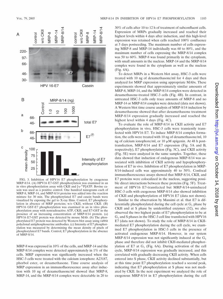
To determine the role of MRPs in HPV16 E7 phosphoryla-tion, we used the E7-GST fusion protein for an in vitro phos-phorylation assay. MRP-8, MRP-14, and MRP-8/14 proteinswere added to a phosphorylation reaction mixture containingpurified CKII and [�-32P]ATP. Casein was used as a controlsubstrate for CKII. E7-GST fusion protein and casein wereextensively phosphorylated within 30 min by CKII (Fig. 3A).The MRP-8/14 complex reduced the CKII-mediated phos-phorylation of E7 and casein by approximately 95 and 80%,respectively. MRP-8 alone did not inhibit E7 or casein phos-phorylation. MRP-14 alone resulted in approximately 40% in-hibition of E7 phosphorylation and casein phosphorylation byCKII (Fig. 3A). However, CKII inactivation in vivo in HeLacells by MRP-14 alone was not significant (about 10%). It ispossible that weaker inhibition of CKII catalytic activity byMRP-14 in the HeLa cells than was seen in the in vitro phos-phorylation assay was the result of slow internalization ofMRP-14. To address this possibility, we analyzed MRP inter-nalization in HeLa cells. The results indicated that efficiency of
internalization of MRP-8 and MRP-14 individually was signif-icantly lower than that of the MRP-8/14 protein complex (datanot shown).
CKII-mediated HPV16 E7 phosphorylation in the presenceof various concentrations of MRP-8/14 proteins was examinedin a Western blot assay using antiphosphoserine antibodies(Fig. 3B). These experiments showed that MRP-8/14-mediatedinhibition of E7 phosphorylation was concentration dependentand that significant inhibition of E7 phosphorylation was de-tected when MRP-8/14 was added to the phosphorylation re-action mixture at 2 ng/�l or higher.
Inhibition of CKII activity and E7 phosphorylation by en-dogenous MRP-8/14 protein complex. To study the inhibitoryrole of endogenous MRP-8/14 protein complex on CKII activ-ity and E7 phosphorylation, we established an epithelial cellmodel system using the HPV-negative human oral squamouscarcinoma HSC-3 cell line. Immunofluorescence analysis ofMRP protein expression in HSC-3 cells showed that theseproteins were expressed in only a small proportion of the cells.
FIG. 1. Complex formation of MRP-8 and MRP-14. Equal concen-trations, i.e., 1 �g each of MRP-8 (a) and MRP-14 (b), were mixed inthe presence of 0.5 mM CaCl2 (c) and incubated at room temperaturefor 30 min. To determine the dependence of MRP-8/14 complex for-mation on calcium ions, the MRP-8 and MRP-14 mixture was incu-bated with various concentrations of calcium (d). Proteins were re-solved by SDS-PAGE, and MRP-8, MRP-14, and their complexformation were analyzed by Western blot assay using mouse MAbs 8-5C2, S36.48, and 27 E10, which recognize MRP-8, MRP-14, and MRP-8/14, respectively. (a and b) Reducing; (c and d) nonreducing.
FIG. 2. Inhibition of CKII activity in HeLa cells by exogenousMRP-8/14. (A) HeLa cells were incubated with various concentrationsof MRP-8/14 proteins for 24 h at 37°C and extracted in RIPA buffer.In parallel experiments cells were treated with an increasing concen-tration of apigenin, a CKII inhibitor, as a positive control. (B) HeLacells were incubated with 2.5 �g of MRP-8, MRP-14, and MRP-8/14proteins per ml and 10 �M apigenin for 24 h at 37°C and then ex-tracted in RIPA buffer. CKII was immunoprecipitated, and kinaseactivity was measured in triplicate samples. Controls were untreatedcells and DMSO-treated cells, since DMSO was used as an organicsolvent for apigenin. Similar results were obtained in three indepen-dent experiments. The error bars indicate standard errors (n 3).
1102 TUGIZOV ET AL. J. VIROL.
on October 14, 2018 by guest
http://jvi.asm.org/
Dow
nloaded from
MRP-8 was expressed in 10% of the cells, and MRP-14 and theMRP-8/14 complex were detected approximately in 1% of thecells. MRP expression was significantly increased when theHSC-3 cells were treated with the calcium ionophore A23187,phorbol ester, or dexamethasone. Time course analysis ofMRP expression in an immunofluorescence assay after induc-tion with 10 ng of dexamethasone/ml showed that MRP-8,MRP-14, and the MRP-8/14 complex were detectable in 20 to
30% of cells after 10 to 12 h of treatment of subconfluent cells.Expression of MRPs gradually increased and reached theirhighest levels within 4 days after induction, and this high-levelexpression was retained when cells reached 100% confluenceat 5 days postseeding. The maximum number of cells express-ing MRP-8 and MRP-14 individually was 60 to 80%, and themaximum number of cells expressing the MRP-8/14 complexwas 50 to 60%. MRP-8 was found primarily in the cytoplasm,with small amounts in the nucleus. MRP-14 and the MRP-8/14complex were found in the cytoplasm as well as the nucleus(Fig. 4A).
To detect MRPs in a Western blot assay, HSC-3 cells weretreated with 10 ng of dexamethasone/ml for 4 days and thenanalyzed for MRP expression using appropriate MAbs. Theseexperiments showed that approximately similar amounts ofMRP-8, MRP-14, and the MRP-8/14 complex were detected indexamethasone-treated HSC-3 cells (Fig. 4B). In contrast, inuntreated HSC-3 cells only trace amounts of MRP-8 and noMRP-14 or MRP-8/14 complex were detected (data not shown).A Western blot time course analysis of MRP-8/14 induction bydexamethasone showed that after dexamethasone treatmentMRP-8/14 expression gradually increased and reached thehighest level within 4 days (Fig. 4C).
To evaluate the role of MRP-8/14 in CKII activity and E7phosphorylation in vivo, HSC-3 cells were transiently trans-fected with HPV16 E7. To induce MRP-8/14 complex forma-tion, the cells were treated with 10 ng of dexamethasone/ml, 10ng of calcium ionophore/ml, or 10 �M apigenin. At 48 h post-transfection, MRP-8/14 and E7 expression (Fig. 5A and B,respectively), E7 phosphorylation (Fig. 5C), and CKII activity(Fig. 5E) were analyzed in the same samples. Together, thesedata showed that induction of endogenous MRP-8/14 was as-sociated with inhibition of CKII activity and hypophosphory-lation of E7 in vivo. Inhibition of E7 phosphorylation in MRP-8/14-induced cells was approximately 40 to 50%. Confocalimmunofluorescence assays showed that MRP-8/14, CKII, andE7 proteins were colocalized in the nucleus, suggesting possi-ble direct interactions between these proteins (Fig. 5F). Treat-ment of HPV16 E7-transfected but MRP-8/14-uninducedHSC-3 cells with exogenous MRP-8/14 also showed inhibitionof CKII and phosphorylation of HPV16 E7 (data not shown).
Similar to the observation by Massimi et al. that E7 is dif-ferentially phosphorylated during the cell cycle at G1 phase byCKII and at S phase by unidentified enzymes (32), we alsoobserved the two highest peaks of E7 phosphorylation to be atG1 and S phases in the HSC-3 cell line transfected with HPV16E7 (data not shown). To study the role of MRP-8/14 in CKII-mediated E7 phosphorylation during the cell cycle, we exam-ined E7 phosphorylation in HSC-3 cells in the presence ofactivated endogenous MRP-8/14. However, in our systemMRP-8/14 expression was not significantly induced at the G1
phase and therefore did not inhibit CKII-mediated phosphor-ylation of E7 at G1 (Fig. 6A). During activation of the cellcycle, MRP-8/14 expression was gradually increased, and thiscorrelated with gradually decreasing CKII activity. When cellsentered into S phase, CKII activity declined substantially, butat this time point E7 phosphorylation reached a second peak,indicating that E7 phosphorylation at S phase was not medi-ated by CKII. In the next experiment we analyzed the role ofexogenous MRP-8/14 in E7 phosphorylation during the cell
FIG. 3. Inhibition of HPV16 E7 phosphorylation by exogenousMRP-8/14. (A) HPV16 E7-GST phosphorylation was examined in anin vitro phosphorylation assay with CKII and [�-32P]ATP. Bovine ca-sein was used as a positive control. One hundred nanograms each ofMRP-8, MRP-14, and MRP-8/14 proteins was added into the reactionmixture for 30 min. The phosphorylated E7 and casein bands werevisualized by exposing the gel to X-ray films. Control, E7 phosphory-lation in absence of MRP proteins; w/o CKII, without CKII. (B)HPV16 GST-E7 phosphorylation was examined in an in vitro phos-phorylation assay with nonradioactive ATP, CKII, and E7-GST in thepresence of an increasing concentration of MRP-8/14 protein. (a)HPV16 E7-GST protein was detected by mouse MAb. (b) The phos-phorylated E7 protein was detected by Western blot assay using rabbitpolyclonal antiphosphoserine antibodies. (c) Intensity of E7 phosphor-ylation was measured by determining the mean density of pixels ofphosphorylated E7 bands. Control, E7 phosphorylation in the absenceof MRP-8/14.
VOL. 79, 2005 MRP-8/14 IN INHIBITION OF HPV16 E7 PHOSPHORYLATION 1103
on October 14, 2018 by guest
http://jvi.asm.org/
Dow
nloaded from

cycle (Fig. 6B). This experiment showed that CKII activity wasdecreased by 45% in the G1 phase, and this correlated wellwith a 90% inhibition of E7 phosphorylation. In contrast, in-activation of CKII at S phase did not affect the second peak ofE7 phosphorylation. These data clearly demonstrate that ex-ogenous MRP 8/14 protein complex specifically inhibits CKII-mediated E7 phosphorylation at the G1 phase of the cell cycleand not at the S phase.
MRP expression in HPV-infected epithelial cell lines. Ourdata described above demonstrated that the MRP-8/14 proteincomplex inactivates CKII and inhibits phosphorylation of E7.Expression of MRP-8 and MRP-14 proteins in HPV-infectedepithelial cells may therefore play an important role in E7hypophosphorylation, with consequent reduction of its onco-genic potential. To examine the status of MRP-8 and MRP-14proteins in HPV-infected cells, we analyzed MRP expression
in HeLa, SiHa, and HOK-16 cell lines. Immunofluorescence(data not shown), Northern blotting (Fig. 7A), and Westernblotting (Fig. 7B) assays showed that these cell lines did notexpress either MRP-8 or MRP-14. Treatment of these cellswith dexamethasone or calcium ionophore did not induceMRP expression (data not shown). In contrast, HPV-negativeprimary oral (OCO), foreskin (NFK), and cervical (NCK) ep-ithelial cells expressed both MRP-8 and -14 (Fig. 7A, B, D, andE). In parallel experiments, these cell lines were examined forCKII kinase activity. These data showed that MRP-8/14 ex-pression in normal epithelial cells was associated with a lowlevel of CKII activity, and their absence in HPV-infected im-mortalized epithelial cells was associated with a three- to five-fold-higher level of CKII activity than in primary cells (Fig. 7Cand F).
We also compared MRP expression in HPV-negative (NFK)
FIG. 4. Expression of MRPs in HSC-3 cells by confocal microscopy (A) and Western blot analysis (B and C). (A) Cells were grown on chamberslides and treated with dexamethasone for 4 days. For confocal microscopy analysis, cells were fixed and immunostained for MRPs with appropriatemouse MAbs (in green). The cell nuclei were stained with propidium iodide (PI; red). Yellow in the merged panels shows nuclear localization ofMRPs. (B) For Western blot assays, approximately 106 HSC-3 cells were treated with 10 ng of dexamethasone/ml for 4 days and MRPs weredetected using mouse MAbs. (a and b) Reducing; (c) nonreducing. (C) To analyze induction of MRP-8/14 expression in HSC-3 cells, approximately3 � 106 cells were treated with 10 ng of dexamethasone/ml for 4 days. MRP-8/14 protein expression was examined in a Western blot assay undernonreducing conditions at 1, 2, 3, and 4 days postinduction.
1104 TUGIZOV ET AL. J. VIROL.
on October 14, 2018 by guest
http://jvi.asm.org/
Dow
nloaded from

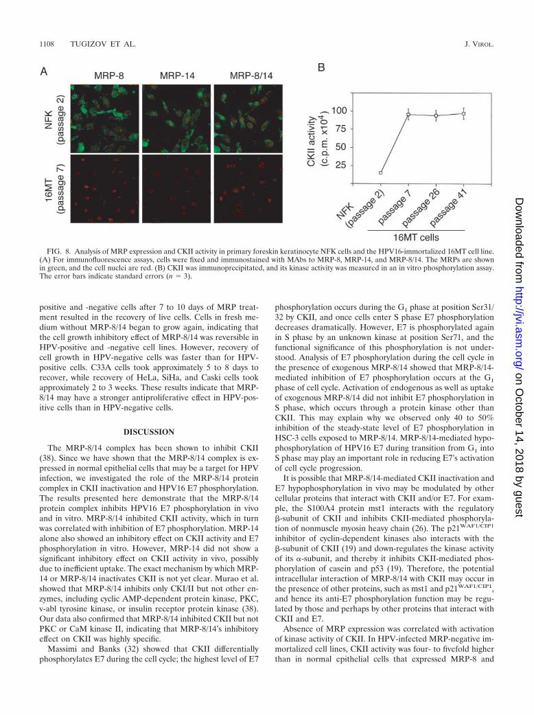
cells with the same cell line immortalized by HPV16 (16MT)cells of different passages to passage 100. Northern blotting(Fig. 7A), Western blotting (Fig. 7B), and immunofluores-cence (Fig. 8A) analysis of the NFK cells showed expression of
MRP-8 and MRP-14 and their MRP-8/14 complex, in contrastto the 16MT cells, which showed no expression of these pro-teins even at relatively early passages (passage 7 and up) (Fig.8A). Analysis of CKII activity in parental NFK and immortal-
FIG. 5. Inhibition of HPV16 E7 phosphorylation in HSC-3 cells by endogenous MRP-8/14 protein complex. A total of 3 � 106 HSC-3 cells weretransfected with HPV16 E7 and 8 h later were treated with 10 ng of dexamethasone/ml, 10 ng of calcium ionophore/ml, and 10 �M apigenin. (Aand B) MRP-8/14 (A) and HPV16 E7 (B) were detected using a Western blot assay with appropriate mouse mAbs. (C) At 48 h posttransfection,HPV16 E7 was immunoprecipitated and E7 phosphorylation was examined in a Western blot assay with rabbit antiphosphoserine antibodies. (D)Intensity of E7 phosphorylation was measured by determining the mean density of pixels of phosphorylated E7 bands. (E) CKII activity wasexamined in an in vitro phosphorylation assay. The error bars indicate standard errors (n 3). MRP-8/14 and E7 expression, E7 phosphorylation,and CKII activity were analyzed in the same samples. (F) Colocalization of MRP-8/14, E7, and CKII in HSC-3 cells. Cells were fixed andimmunostained for CKII (red) and MRP-8/14 (blue). HPV16 E7 protein was detected by GFP (green). In the merge panel, white shows co-localization of MRP-8/14, CKII, and E7.
VOL. 79, 2005 MRP-8/14 IN INHIBITION OF HPV16 E7 PHOSPHORYLATION 1105
on October 14, 2018 by guest
http://jvi.asm.org/
Dow
nloaded from

ized 16MT cells showed that CKII activity in 16MT cells wasfour- to fivefold higher than in the primary parental NFK cells(Fig. 8B).
To determine whether the loss of MRP-8/14 expression inHPV-infected cervical cancer cells was associated with HPVinfection, we examined MRP-8/14 expression in the HPV-neg-ative C33A cervical carcinoma cell line and normal primarycervical epithelial (NCK) cells. MRPs and their complex wereexpressed in NCK normal cervical epithelial cells, but not inthe C33A cells (Fig. 7D and E). CKII kinase activity in C33Acells was higher than in the NCK cells, i.e., activation of CKIIwas correlated with loss of MRP-8/14 (Fig. 7F). Thus, absenceof MRP expression in HPV-negative C33A cells indicated thatloss of MRP expression was not specifically related to HPVinfection.
Antiproliferative effect of MRPs on cancer cell lines. Inhi-bition of HPV16 E7 phosphorylation by the MRP-8/14 proteincomplex and its absence in HPV-associated tumor cell linessuggest that MRP-8/14 may play an important role in reductionof the oncogenic activity of E7. To study the antiproliferativeeffect of the MRP-8/14 complex, we examined the cell growth
rate of the HPV-positive HeLa, SiHa, and Caski cell lines, aswell as the HPV-negative C33A and NCK cervical epithelialcell lines, in the presence of exogenous MRP-8/14. As shownabove, HeLa, SiHa, Caski, and C33A cell lines did not expressMRP-8 and MRP-14 proteins and therefore did not form theMRP-8/14 complex. In contrast, NCK primary cervical epithe-lial cells did express MRP-8 and MRP-14 proteins and formedthe MRP-8/14 complex. Cells were grown in the presence ofpurified MRP-8/14 at various concentrations ranging from 0.1to 10 �g/ml for14 days. Concentrations higher than 3 �g ofMRP-8/14 per ml were toxic for all cell lines, and cells began todie at 3 to 5 days after addition of MRP-8/14. At MRP-8/14concentrations lower than 3 �g/ml, the HPV-negative and -posi-tive cells remained morphologically healthy during the first 7days. At the beginning of the second week the morphology ofthe HPV-positive cells at MRP-8/14 concentrations of 0.5 �g/ml and higher began to change. The cells detached from thesubstratum and began to die. At the end of the second week,almost 95% of these cells were dead (Fig. 9A). In contrast,HPV-negative NCK primary and C33A immortalized cervicalepithelial cells were more resistant to the growth inhibitory
FIG. 6. MRP-8/14-mediated inhibition of HPV16 E7 during the cell cycle. (A) Inhibition of E7 phosphorylation by endogenous MRP-8/14protein. HSC-3 cells were transfected with HPV16 E7, and 12 h later cells were growth arrested in 0.2% serum for the next 38 h. To induceMRP-8/14 expression at 24 h poststarvation, cells were treated with 10 ng of dexamethasone/ml. At 38 h posttransfection, cell growth was activatedby adding 15% serum in the presence of 10 ng of dexamethasone/ml and 1 �Ci of [3H]thymidine/ml. At 2-h intervals after growth activation, thecell cycle (a), MRP-8/14 (b), CKII activity (c), total E7 expression (d), and E7 phosphorylation (e) were analyzed. *, MRP-8/14 expression inE7-transfected HSC-3 cells before MRP-8/14 induction; **, CKII activity in E7-transfected HSC-3 cells before MRP-8/14 induction. (B) Inhibitionof E7 phosphorylation by exogenous MRP-8/14 protein. Experiments were performed as described above, but instead of induction of endogenousMRP-8/14 with dexamethasone, 3 �g of exogenous MRP-8/14/ml was added at 24 h posttransfection. At 2-h intervals after growth activation, thecell cycle (a), CKII activity (b), E7 expression (c), and E7 phosphorylation (d) were analyzed in the same samples. *, CKII activity in E7-transfectedHSC-3 cells before adding MRP-8/14.
1106 TUGIZOV ET AL. J. VIROL.
on October 14, 2018 by guest
http://jvi.asm.org/
Dow
nloaded from

effect of MRP-8/14 than the HPV-positive cells. At a concen-tration of 1.5 to 2 �g of MRP-8/14 per ml, detachment anddeath of the HPV-negative cells was moderate, i.e., about 15 to20% cells were dead. At a similar concentration of MRP-8/14,almost 90% of HPV-positive cells were dead. Substantial celldeath, i.e., 25 to 40% of HPV-negative NCK and C33A cells,respectively, was seen only at concentrations of MRP-8/14 of2.5 and 3 �g/ml (Fig. 9A). Continuous treatment of HPV-negative primary cervical NCK and tumor C33A cells withexogenous MRP-8/14 at concentrations of 1.5 �g/ml and abovefor 3 weeks completely inhibited their growth, indicating thathigh concentrations of exogenous MRP-8/14 were cytostatic,even for the HPV-negative cells.
To determine whether the antiproliferative effect of MRP-8/14 in HPV-infected cells was linked to E7 hypophosphoryla-tion, we examined E7 phosphorylation in MRP-8/14-treatedSiHa and Caski cell lines infected with HPV16 (Fig. 9B). Cellswere treated with 1, 2, and 3 �g of MRP-8/14 per ml for 7 days,and E7 phosphorylation was examined. These experimentsshowed that MRP-8/14 inhibited the steady-state level of phos-phorylation of HPV16 E7 in SiHa and Caski cells in a dose-dependent manner, similar to the dose-dependent antipro-liferative effect (Fig. 9B). Treatment of E7 with proteinphosphatase almost completely dephosphorylated it (Fig. 9C),showing high efficiency of phosphorylation of E7 in these cells.
Removal of MRP-8/14s from the culture medium of HPV-
FIG. 7. Expression of MRPs and assessment of CKII activity in normal and tumor cell lines. MRP expression was examined in the HPV-negative primary NFK, OCO, and NCK and immortalized C33A cell lines, and in the following HPV-positive immortalized cell lines: 16MT(derived by HPV16-mediated transformation of NFK cells), HOK-16, HeLa, and SiHa cells. (A) For Northern blot assays, cellular mRNA washybridized with probes to MRP-8 and MRP-14. A probe for GAPDH was used as a housekeeping gene control. (B and E) For Western blot assays,MRPs were separated under reducing (for MRP-8 and MRP-14) and nonreducing (for MRP-8/14 complex) conditions and immunoblotted withappropriate mouse MAbs to MRP-8, MRP-14, and MRP-8/14. (C and F) To measure CKII activity, CKII was immunoprecipitated and its kinaseactivity was measured in an in vitro phosphorylation assay. The error bars indicate standard errors (n 3). (D) For the immunofluorescence assay,NCK and C33A cells grown on chamber slides were fixed and immunostained with MAbs to MRP-8, MRP-14, and MRP-8/14. The MRPs areshown in green, and the cell nuclei are red.
VOL. 79, 2005 MRP-8/14 IN INHIBITION OF HPV16 E7 PHOSPHORYLATION 1107
on October 14, 2018 by guest
http://jvi.asm.org/
Dow
nloaded from
positive and -negative cells after 7 to 10 days of MRP treat-ment resulted in the recovery of live cells. Cells in fresh me-dium without MRP-8/14 began to grow again, indicating thatthe cell growth inhibitory effect of MRP-8/14 was reversible inHPV-positive and -negative cell lines. However, recovery ofcell growth in HPV-negative cells was faster than for HPV-positive cells. C33A cells took approximately 5 to 8 days torecover, while recovery of HeLa, SiHa, and Caski cells tookapproximately 2 to 3 weeks. These results indicate that MRP-8/14 may have a stronger antiproliferative effect in HPV-pos-itive cells than in HPV-negative cells.
DISCUSSION
The MRP-8/14 complex has been shown to inhibit CKII(38). Since we have shown that the MRP-8/14 complex is ex-pressed in normal epithelial cells that may be a target for HPVinfection, we investigated the role of the MRP-8/14 proteincomplex in CKII inactivation and HPV16 E7 phosphorylation.The results presented here demonstrate that the MRP-8/14protein complex inhibits HPV16 E7 phosphorylation in vivoand in vitro. MRP-8/14 inhibited CKII activity, which in turnwas correlated with inhibition of E7 phosphorylation. MRP-14alone also showed an inhibitory effect on CKII activity and E7phosphorylation in vitro. However, MRP-14 did not show asignificant inhibitory effect on CKII activity in vivo, possiblydue to inefficient uptake. The exact mechanism by which MRP-14 or MRP-8/14 inactivates CKII is not yet clear. Murao et al.showed that MRP-8/14 inhibits only CKI/II but not other en-zymes, including cyclic AMP-dependent protein kinase, PKC,v-abl tyrosine kinase, or insulin receptor protein kinase (38).Our data also confirmed that MRP-8/14 inhibited CKII but notPKC or CaM kinase II, indicating that MRP-8/14’s inhibitoryeffect on CKII was highly specific.
Massimi and Banks (32) showed that CKII differentiallyphosphorylates E7 during the cell cycle; the highest level of E7
phosphorylation occurs during the G1 phase at position Ser31/32 by CKII, and once cells enter S phase E7 phosphorylationdecreases dramatically. However, E7 is phosphorylated againin S phase by an unknown kinase at position Ser71, and thefunctional significance of this phosphorylation is not under-stood. Analysis of E7 phosphorylation during the cell cycle inthe presence of exogenous MRP-8/14 showed that MRP-8/14-mediated inhibition of E7 phosphorylation occurs at the G1
phase of cell cycle. Activation of endogenous as well as uptakeof exogenous MRP-8/14 did not inhibit E7 phosphorylation inS phase, which occurs through a protein kinase other thanCKII. This may explain why we observed only 40 to 50%inhibition of the steady-state level of E7 phosphorylation inHSC-3 cells exposed to MRP-8/14. MRP-8/14-mediated hypo-phosphorylation of HPV16 E7 during transition from G1 intoS phase may play an important role in reducing E7’s activationof cell cycle progression.
It is possible that MRP-8/14-mediated CKII inactivation andE7 hypophosphorylation in vivo may be modulated by othercellular proteins that interact with CKII and/or E7. For exam-ple, the S100A4 protein mst1 interacts with the regulatory�-subunit of CKII and inhibits CKII-mediated phosphoryla-tion of nonmuscle myosin heavy chain (26). The p21WAF1/CIP1
inhibitor of cyclin-dependent kinases also interacts with the�-subunit of CKII (19) and down-regulates the kinase activityof its �-subunit, and thereby it inhibits CKII-mediated phos-phorylation of casein and p53 (19). Therefore, the potentialintracellular interaction of MRP-8/14 with CKII may occur inthe presence of other proteins, such as mst1 and p21WAF1/CIP1,and hence its anti-E7 phosphorylation function may be regu-lated by those and perhaps by other proteins that interact withCKII and E7.
Absence of MRP expression was correlated with activationof kinase activity of CKII. In HPV-infected MRP-negative im-mortalized cell lines, CKII activity was four- to fivefold higherthan in normal epithelial cells that expressed MRP-8 and
FIG. 8. Analysis of MRP expression and CKII activity in primary foreskin keratinocyte NFK cells and the HPV16-immortalized 16MT cell line.(A) For immunofluorescence assays, cells were fixed and immunostained with MAbs to MRP-8, MRP-14, and MRP-8/14. The MRPs are shownin green, and the cell nuclei are red. (B) CKII was immunoprecipitated, and its kinase activity was measured in an in vitro phosphorylation assay.The error bars indicate standard errors (n 3).
1108 TUGIZOV ET AL. J. VIROL.
on October 14, 2018 by guest
http://jvi.asm.org/
Dow
nloaded from

FIG. 9. Antiproliferative and anti-E7 phosphorylation activity of the MRP-8/14 protein complex. (A) Antigrowth activity of MRP-8/14 inHPV-positive and HPV-negative cervical cell lines. Cells were grown in the presence of the MRP-8/14 protein complex at various concentrationsfor 14 days. The number of live cells was quantified with trypan blue staining at day 14. Similar results were obtained in three independentexperiments. The error bars indicate standard errors; n 3. Control, live cells without treatment. (B) Inhibition of HPV-16 E7 phosphorylationby MRP-8/14 in HPV16-infected SiHa and Caski cell lines. Approximately 5 � 106 cells were grown in the presence of 1, 2, and 3 �g of exogenousMRP-8/14 per ml. (a) At 5 days posttreatment, cells were extracted and E7 was immunoprecipitated with mouse MAb and E7 was detected byWestern blotting. (b) E7 phosphorylation in the same samples was examined using rabbit antiphosphoserine antibodies. (c) Intensity of E7phosphorylation was measured by the mean density of pixels in the E7 phosphorylated bands. (C) Dephosphorylation of immunoprecipitated E7by phosphatase treatment. Control (all panels), untreated cells.
VOL. 79, 2005 MRP-8/14 IN INHIBITION OF HPV16 E7 PHOSPHORYLATION 1109
on October 14, 2018 by guest
http://jvi.asm.org/
Dow
nloaded from

MRP-14 proteins. Treatment of HPV-infected MRP-negativecells with exogenous MRP-8/14 complex led to inhibition ofCKII activity in HPV16- and HPV18-infected cervical epithe-lial cells. Activation of endogenous MRP-8/14 also inhibitedCKII activity and HPV16 E7 phosphorylation in HPV-negativeHSC-3 oral squamous epithelial tumor cells. These data indi-cate that MRP-8/14 may play a role as a strong intracellularfactor that may negatively regulate the activity of CKII.
It has been shown that treatment of various normal andtransformed cell lines with 50 to 200 �g of MRP-8/14 per mlwithin 18 to 48 h leads to inhibition of DNA synthesis and cellgrowth (62–64). The minimum effective concentration to in-hibit cell growth was approximately 50 �g/ml. We have shownhere that prolonged uptake of a much lower concentration (1to 10 �g/ml) of MRP-8/14 for 7 to 14 days by HPV-positive andHPV-negative tumor cells, as well as normal epithelial cells,caused detachment of cells from the substratum and inhibitedtheir growth. CKII is a highly pleiotropic enzyme that phos-phorylates more than 160 cellular proteins with a wide varietyof functions, with consequent effects on gene expression, pro-tein synthesis, cell cycling, and differentiation (15, 30). There-fore, the antiproliferative effect of MRP-8/14 during prolongedtreatment may be due to its inactivation of CKII, which isrequired for cell proliferation and cell viability in both normaland cancer cells. However, MRP-8/14 more strongly inhibitedgrowth of HPV-positive cells than HPV-negative cells, a dif-ference that may have been due to their effect on E7. Our datatherefore suggest that MRP-8/14-mediated CKII inactivationmay lead to two groups of downstream antiproliferative effects.The first group of effects is more general, reflecting inhibitionof phosphorylation of CKII substrates, i.e., housekeeping pro-teins that are required for cell proliferation and viability. Thiskind of antiproliferative effect occurred in both HPV-positiveand HPV-negative cells. The second group of antiproliferativeeffects is more specific to HPV-positive cells, and it is possiblethis could be due to inhibition of HPV16 E7 phosphorylation.
Our data leave several questions open to future investiga-tion. The mechanisms by which CKII inhibition leads to inhi-bition of cell growth are not fully understood, nor is the extentto which inhibition of E7 phosphorylation mediates the HPV-specific effects. While our data are consistent with a key rolefor E7 phosphorylation, the downstream effects of MRP-8/14-mediated inhibition of E7 phosphorylation are not clear. Sto-rey et al. showed that substitution of one of the two serineresidues of the E7 CKII site, Ser31 or Ser32, only slightlydecreased its ability to cooperate with the EJ-ras oncogene totransform primary baby rat kidney cells (53). Barbosa et al.also showed that mutation of one of the two serines did notshow any significant biological activity (3). However, simulta-neous mutation of both serines impaired or significantly re-duced its transforming activity (3, 18), suggesting that phos-phorylation of both serines, Ser31 and Ser32, may be necessaryfor E7’s full transforming activity.
The best-characterized E7 ligand is pRb, but the role ofCKII-mediated E7 phosphorylation in its interaction with pRband pRb-associated proteins is not understood. Using in vitrobinding assays, it has previously been shown that mutation inthe CKII phosphorylation site of E7 Ser 31/32 did not affect itsbinding to pRb (3, 18). However, it is not clear how well thesein vitro binding assays reflect the more complex intracellular
environment. Moreover, since inhibition of transformation oc-curred with mutation of both serines despite the lack of changein pRb binding (3, 18), it is also likely that the E7-pRb inter-action is not the only mechanism for E7-mediated transforma-tion or stimulation of cell cycling (6, 24, 45). Consistent withthis, MRP-8/14-mediated CKII inactivation and E7 hypophos-phorylation in vivo may affect other pRb indirect and/or pRb-independent cell cycle regulatory pathways.
E7 phosphorylation of high-risk HPV types may be involvedin E7-mediated degradation of pRB (25). Phosphorylation ofHPV16 E7 has been shown to increase the binding affinity ofE7 to the basic subunit of the TFIID complex, the TATAbox-binding protein (TBP), and TBP-associated factor 110(TAF-110) protein (31, 34, 35, 36). E7 binding to F-actin re-quires HPV16 E7 phosphorylation (43), and HPV16 E7 phos-phorylation may play a role in E7 interaction with p53 (33). ACKII phosphorylation-defective HPV16 E7 mutant severelyimpaired its ability to induce tetrasomy in primary foreskinkeratinocytes, suggesting that E7 phosphorylation may be in-volved in induction of genetic instability (50). Taken together,these data indicate that CKII-mediated E7 phosphorylationmay be critical for its oncogenic functions and suggest thatMRP-8/14-mediated inhibition of CKII may affect HPV-in-duced cell cycling through multiple mechanisms.
In summary, we have found that the S100 calcium-bindingprotein complex MRP-8/14 inhibits CKII-mediated HPV16 E7phosphorylation in vivo and in vitro. We showed MRP expres-sion in normal primary epithelial cells in culture, with loss oftheir expression in several HPV-positive cervical cancer celllines. The mechanisms of loss of MRP-8 and -14 protein ex-pression in tumor cells are not well understood but may occurduring the multistep cell transformation process. Consistentwith this, loss of MRP expression was observed in 16MT cellsduring the process of HPV-induced immortalization. MRP-8/14 protein expression was detected only in parental cells,and once the primary keratinocytes were immortalized withHPV16 MRP-8/14 expression was lost and CKII activity waselevated. However, absence of MRP expression in HPV-neg-ative C33A cervical carcinoma cells indicated that inactivationof MRP expression was not specifically due to HPV infection.Activation of CKII has been reported in tumor cells of differ-ent origins (11, 16, 29, 51, 58, 61), and mechanisms of itsactivation, particularly the role of MRP-8/14 in its regulation,are not well understood. It is possible that loss of MRP ex-pression and elevation of CKII activity in high-risk HPV-in-fected precancerous lesions may lead to higher levels of phos-phorylation of E7, increasing E7 oncogenic activity andpossibly progression of HPV-associated neoplasia.
ACKNOWLEDGMENTS
This work was supported by National Institute of Dental and Cra-niofacial Research grants P01 DE13904 and P01 DE07946 and theHellman family award.
The HOK-16 cells were a kind gift from No-Hee Park (University ofCalifornia, Los Angeles).
REFERENCES
1. Ahmad, A., D. L. Bayley, S. He, and R. A. Stockley. 2003. Myeloid relatedprotein-8/14 stimulates interleukin-8 production in airway epithelial cells.Am. J. Respir. Cell. Mol. Biol. 29:523–530.
2. Andersson, K. B., K. Sletten, H. B. Berntzen, I. Dale, P. Brandtzaeg, E.Jellum, and M. K. Fagerhol. 1988. The leucocyte L1 protein: identity with
1110 TUGIZOV ET AL. J. VIROL.
on October 14, 2018 by guest
http://jvi.asm.org/
Dow
nloaded from

the cystic fibrosis antigen and the calcium-binding MRP-8 and MRP-14macrophage components. Scand. J. Immunol. 28:241–245.
3. Barbosa, M. S., C. Edmonds, C. Fisher, J. T. Schiller, D. R. Lowy, and K. H.Vousden. 1990. The region of the HPV E7 oncoprotein homologous toadenovirus E1a and Sv40 large T antigen contains separate domains for Rbbinding and casein kinase II phosphorylation. EMBO J. 9:153–160.
4. Bruggen, J., L. Tarcsay, N. Cerletti, K. Odink, M. Rutishauser, G. Hol-lander, and C. Sorg. 1988. The molecular nature of the cystic fibrosis antigen.Nature 331:570.
5. Brun, J. G., R. Jonsson, and H. J. Haga. 1994. Measurement of plasmacalprotectin as an indicator of arthritis and disease activity in patients withinflammatory rheumatic diseases. J. Rheumatol. 21:733–738.
6. Caldeira, S., E. M. de Villiers, and M. Tommasino. 2000. Human papillo-mavirus E7 proteins stimulate proliferation independently of their ability toassociate with retinoblastoma protein. Oncogene 19:821–826.
7. Channavajhala, P., and D. C. Seldin. 2002. Functional interaction of proteinkinase CK2 and c-Myc in lymphomagenesis. Oncogene 21:5280–5288.
8. Chellappan, S., V. B. Kraus, B. Kroger, K. Munger, P. M. Howley, W. C.Phelps, and J. R. Nevins. 1992. Adenovirus E1A, simian virus 40 tumorantigen, and human papillomavirus E7 protein share the capacity to disruptthe interaction between transcription factor E2F and the retinoblastomagene product. Proc. Natl. Acad. Sci. USA 89:4549–4553.
9. Chien, W. M., J. N. Parker, D. C. Schmidt-Grimminger, T. R. Broker, andL. T. Chow. 2000. Casein kinase II phosphorylation of the human papillo-mavirus-18 E7 protein is critical for promoting S-phase entry. Cell GrowthDiffer. 11:425–435.
10. Dale, I., P. Brandtzaeg, M. K. Fagerhol, and H. Scott. 1985. Distribution ofa new myelomonocytic antigen (L1) in human peripheral blood leukocytes.Immunofluorescence and immunoperoxidase staining features in compari-son with lysozyme and lactoferrin. Am. J. Clin. Pathol. 84:24–34.
11. Daya-Makin, M., J. S. Sanghera, T. L. Mogentale, M. Lipp, J. Parchomchuk,J. C. Hogg, and S. L. Pelech. 1994. Activation of a tumor-associated proteinkinase (p40TAK) and casein kinase 2 in human squamous cell carcinomasand adenocarcinomas of the lung. Cancer Res. 54:2262–2268.
12. Delabie, J., C. de Wolf-Peeters, J. J. van den Oord, and V. J. Desmet. 1990.Differential expression of the calcium-binding proteins MRP8 and MRP14 ingranulomatous conditions: an immunohistochemical study. Clin. Exp. Im-munol. 81:123–126.
13. Dorin, J. R., M. Novak, R. E. Hill, D. J. Brock, D. S. Secher, and V. vanHeyningen. 1987. A clue to the basic defect in cystic fibrosis from cloning theCF antigen gene. Nature 326:614–617.
14. Dyson, N., P. M. Howley, K. Munger, and E. Harlow. 1989. The humanpapilloma virus-16 E7 oncoprotein is able to bind to the retinoblastoma geneproduct. Science 243:934–937.
15. Faust, M., and M. Montenarh. 2000. Subcellular localization of proteinkinase CK2. A key to its function? Cell Tissue Res. 301:329–340.
16. Faust, R. A., M. Gapany, P. Tristani, A. Davis, G. L. Adams, and K. Ahmed.1996. Elevated protein kinase CK2 activity in chromatin of head and necktumors: association with malignant transformation. Cancer Lett. 101:31–35.
17. Firzlaff, J. M., D. A. Galloway, R. N. Eisenman, and B. Luscher. 1989. TheE7 protein of human papillomavirus type 16 is phosphorylated by caseinkinase II. New Biol. 1:44–53.
18. Firzlaff, J. M., B. Luscher, and R. N. Eisenman. 1991. Negative charge at thecasein kinase II phosphorylation site is important for transformation but notfor Rb protein binding by the E7 protein of human papillomavirus type 16.Proc. Natl. Acad. Sci. USA 88:5187–5191.
19. Gotz, C., P. Wagner, O. G. Issinger, and M. Montenarh. 1996. p21WAF1/CIP1 interacts with protein kinase CK2. Oncogene 13:391–398.
20. Hammer, H. B., T. K. Kvien, A. Glennås, and K. Melby. 1995. A longitudinalstudy of calprotectin as an inflammatory marker in patients with reactivearthritis. Clin. Exp. Rheumatol. 13:59–64.
21. Hessian, P. A., J. Edgeworth, and N. Hogg. 1993. MRP-8 and MRP-14, twoabundant Ca2�-binding proteins of neutrophils and monocytes. J. Leukoc.Biol. 53:197–204.
22. Hogg, N., C. Allen, and J. Edgeworth. 1989. Monoclonal antibody 5.5 reactswith p8,14, a myeloid molecule associated with some vascular endothelium.Eur. J. Immunol. 19:1053–1061.
23. Hsiung, N., R. S. Roginski, P. Henthorn, O. Smithies, R. Kucherlapati, andA. I. Skoultchi. 1982. Introduction and expression of a fetal human globingene in mouse fibroblasts. Mol. Cell. Biol. 2:401–411.
24. Jewers, R. J., P. Hildebrandt, J. W. Ludlow, B. Kell, and D. J. McCance.1992. Regions of human papillomavirus type 16 E7 oncoprotein required forimmortalization of human keratinocytes. J. Virol. 66:1329–1335.
25. Jones, D. L., D. A. Thompson, and K. Munger. 1997. Destabilization of theRB tumor suppressor protein and stabilization of p53 contribute to HPVtype 16 E7-induced apoptosis. Virology 239:97–107.
26. Kriajevska, M., I. B. Bronstein, D. J. Scott, S. Tarabykina, M. Fischer-Larsen, O. Issinger, and E. Lukanidin. 2000. Metastasis-associated proteinMts1 (S100A4) inhibits CK2-mediated phosphorylation and self-assembly ofthe heavy chain of nonmuscle myosin. Biochim. Biophys. Acta 1498:252–263.
27. Kuruto, R., R. Nozawa, K. Takeishi, K. Arai, T. Yokota, and Y. Takasaki.1990. Myeloid calcium binding proteins: expression in the differentiated
HL-60 cells and detection in sera of patients with connective tissue diseases.J. Biochem. 108:650–653.
28. Landesman-Bollag, E., P. L. Channavajhala, R. D. Cardiff, and D. C. Seldin.1998. p53 deficiency and misexpression of protein kinase CK2� collaboratein the development of thymic lymphomas in mice. Oncogene 16:2965–2974.
29. Landesman-Bollag, E., D. H. Song, R. Romieu-Mourez, D. J. Sussman, R. D.Cardiff, G. E. Sonenshein, and D. C. Seldin. 2001. Protein kinase CK2:signaling and tumorigenesis in the mammary gland. Mol. Cell. Biochem. 227:153–165.
30. Litchfield, D. W. 2003. Protein kinase CK2: structure, regulation and role incellular decisions of life and death. Biochem. J. 369:1–15.
31. Maldonado, E., M. E. Cabrejos, L. Banks, and J. E. Allende. 2002. Humanpapillomavirus-16 E7 protein inhibits the DNA interaction of the TATAbinding transcription factor. J. Cell Biochem. 85:663–669.
32. Massimi, P., and L. Banks. 2000. Differential phosphorylation of theHPV-16 E7 oncoprotein during the cell cycle. Virology 276:388–394.
33. Massimi, P., and L. Banks. 1997. Repression of p53 transcriptional activityby the HPV E7 proteins. Virology 227:255–259.
34. Massimi, P., D. Pim, A. Storey, and L. Banks. 1996. HPV-16 E7 and ade-novirus E1a complex formation with TATA box binding protein is enhancedby casein kinase II phosphorylation. Oncogene 12:2325–2330.
35. Mazzarelli, J. M., G. B. Atkins, J. V. Geisberg, and R. P. Ricciardi. 1995. Theviral oncoproteins Ad5 E1A, HPV16 E7 and SV40 TAg bind a commonregion of the TBP-associated factor-110. Oncogene 11:1859–1864.
36. McDougall, J. K. 1994. Immortalization and transformation of human cellsby human papillomavirus. Curr. Top. Microbiol. Immunol. 186:101–119.
37. Munger, K., W. C. Phelps, V. Bubb, P. M. Howley, and R. Schlegel. 1989. TheE6 and E7 genes of the human papillomavirus type 16 together are necessaryand sufficient for transformation of primary human keratinocytes. J. Virol.63:4417–4421.
38. Murao, S., F. R. Collart, and E. Huberman. 1989. A protein containing thecystic fibrosis antigen is an inhibitor of protein kinases. J. Biol. Chem. 264:8356–8360.
39. Odink, K., N. Cerletti, J. Bruggen, R. G. Clerc, L. Tarcsay, G. Zwadlo, G.Gerhards, R. Schlegel, and C. Sorg. 1987. Two calcium-binding proteins ininfiltrate macrophages of rheumatoid arthritis. Nature 330:80–82.
40. Park, N. H., B. M. Min, S. L. Li, M. Z. Huang, H. M. Cherick, and J.Doniger. 1991. Immortalization of normal human oral keratinocytes withtype 16 human papillomavirus. Carcinogenesis 12:1627–1631.
41. Phelps, W. C., C. L. Yee, K. Munger, and P. M. Howley. 1989. Functional andsequence similarities between HPV16 E7 and adenovirus E1A. Curr. Top.Microbiol. Immunol. 144:153–166.
42. Pinna, L. A., and F. Meggio. 1997. Protein kinase CK2 (“casein kinase-2”) andits implication in cell division and proliferation. Prog. Cell Cycle Res. 3:77–97.
43. Rey, O., S. Lee, M. A. Baluda, J. Swee, B. Ackerson, R. Chiu, and N. H. Park.2000. The E7 oncoprotein of human papillomavirus type 16 interacts withF-actin in vitro and in vivo. Virology 268:372–381.
44. Sarrouilhe, D., O. Filhol, D. Leroy, G. Bonello, M. Baudry, E. M. Chambaz,and C. Cochet. 1998. The tight association of protein kinase CK2 with plasmamembranes is mediated by a specific domain of its regulatory beta-subunit.Biochim. Biophys. Acta 1403:199–210.
45. Schmitt, A., J. B. Harry, B. Rapp, F. O. Wettstein, and T. Iftner. 1994.Comparison of the properties of the E6 and E7 genes of low- and high-riskcutaneous papillomaviruses reveals strongly transforming and high Rb-bind-ing activity for the E7 protein of the low-risk human papillomavirus type 1.J. Virol. 68:7051–7059.
46. Schwarz, E., U. K. Freese, L. Gissmann, W. Mayer, B. Roggenbuck, A.Stremlau, and H. zur Hausen. 1985. Structure and transcription of humanpapillomavirus sequences in cervical carcinoma cells. Nature 314:111–114.
47. Seedorf, K., T. Oltersdorf, G. Krammer, and W. Rowekamp. 1987. Identifi-cation of early proteins of the human papilloma viruses type 16 (HPV 16)and type 18 (HPV 18) in cervical carcinoma cells. EMBO J. 6:139–144.
48. Seldin, D. C., and P. Leder. 1995. Casein kinase II alpha transgene-inducedmurine lymphoma: relation to theileriosis in cattle. Science 267:894–897.
49. Smotkin, D., and F. O. Wettstein. 1987. The major human papillomavirusprotein in cervical cancers is a cytoplasmic phosphoprotein. J. Virol. 61:1686–1689.
50. Southern, S. A., M. H. Lewis, and C. S. Herrington. 2004. Induction oftetrasomy by human papillomavirus type 16 E7 protein is independent ofpRb binding and disruption of differentiation. Br. J. Cancer 90:1949–1954.
51. Stalter, G., S. Siemer, E. Becht, M. Ziegler, K. Remberger, and O. G.Issinger. 1994. Asymmetric expression of protein kinase CK2 subunits inhuman kidney tumors. Biochem. Biophys. Res. Commun. 202:141–147.
52. Steinbakk, M., C. F. Naess-Andresen, E. Lingaas, I. Dale, P. Brandtzaeg,and M. K. Fagerhol. 1990. Antimicrobial actions of calcium binding leuco-cyte L1 protein, calprotectin. Lancet 336:763–765.
53. Storey, A., N. Almond, K. Osborn, and L. Crawford. 1990. Mutations of thehuman papillomavirus type 16 E7 gene that affect transformation, transac-tivation and phosphorylation by the E7 protein. J. Gen. Virol. 71:965–970.
54. Teigelkamp, S., R. S. Bhardwaj, J. Roth, G. Meinardus-Hager, M. Karas, andC. Sorg. 1991. Calcium-dependent complex assembly of the myeloic differenti-ation proteins MRP-8 and MRP-14. J. Biol. Chem. 266:13462–13467.
VOL. 79, 2005 MRP-8/14 IN INHIBITION OF HPV16 E7 PHOSPHORYLATION 1111
on October 14, 2018 by guest
http://jvi.asm.org/
Dow
nloaded from

55. Tsai, S. C., and E. Seto. 2002. Regulation of histone deacetylase 2 by proteinkinase CK2. J. Biol. Chem. 277:31826–31833.
56. Turner, M. A., T. Darragh, and J. M. Palefsky. 1997. Epithelial-stromalinteractions modulating penetration of matrigel membranes by HPV 16-immortalized keratinocytes. J. Investig. Dermatol. 109:619–625.
57. Vousden, K. H., and P. S. Jat. 1989. Functional similarity betweenHPV16E7, SV40 large T and adenovirus E1a proteins. Oncogene 4:153–158.
58. Wang, L. G., X. M. Liu, H. Wikiel, and A. Bloch. 1995. Activation of caseinkinase II in ML-1 human myeloblastic leukemia cells requires IGF-1 andtransferrin. J. Leukoc. Biol. 57:332–334.
59. Werness, B. A., A. J. Levine, and P. M. Howley. 1990. Association of humanpapillomavirus types 16 and 18 E6 proteins with p53. Science 248:76–79.
60. Wilkinson, M. M., A. Busuttil, C. Hayward, D. J. Brock, J. R. Dorin, and V.Van Heyningen. 1988. Expression pattern of two related cystic fibrosis-associated calcium-binding proteins in normal and abnormal tissues. J. CellSci. 91:221–230.
61. Yenice, S., A. T. Davis, S. A. Goueli, A. Akdas, C. Limas, and K. Ahmed.1994. Nuclear casein kinase 2 (CK-2) activity in human normal, benignhyperplastic, and cancerous prostate. Prostate 24:11–16.
62. Yui, S., M. Mikami, K. Tsurumaki, and M. Yamazaki. 1997. Growth-inhib-itory and apoptosis-inducing activities of calprotectin derived from inflam-matory exudate cells on normal fibroblasts: regulation by metal ions. J. Leu-koc. Biol. 61:50–57.
63. Yui, S., M. Mikami, and M. Yamazaki. 1995. Induction of apoptotic celldeath in mouse lymphoma and human leukemia cell lines by a calcium-binding protein complex, calprotectin, derived from inflammatory peritonealexudate cells. J. Leukoc. Biol. 58:650–658.
64. Yui, S., Y. Nakatani, and M. Mikami. 2003. Calprotectin (S100A8/S100A9),an inflammatory protein complex from neutrophils with a broad apoptosis-inducing activity. Biol. Pharm. Bull. 26:753–760.
65. zur Hausen, H. 2002. Papillomaviruses and cancer: from basic studies toclinical application. Nat. Rev. Cancer 2:342–350.
66. zur Hausen, H. 1987. Papillomaviruses in human cancer. Cancer 59:1692–1696.
67. Zwadlo, G., J. Bruggen, G. Gerhards, R. Schlegel, and C. Sorg. 1988. Twocalcium-binding proteins associated with specific stages of myeloid cell dif-ferentiation are expressed by subsets of macrophages in inflammatory tis-sues. Clin. Exp. Immunol. 72:510–515.
1112 TUGIZOV ET AL. J. VIROL.
on October 14, 2018 by guest
http://jvi.asm.org/
Dow
nloaded from