Inhibition of Fibroblast Growth Factor-2-induced Vascular...
9
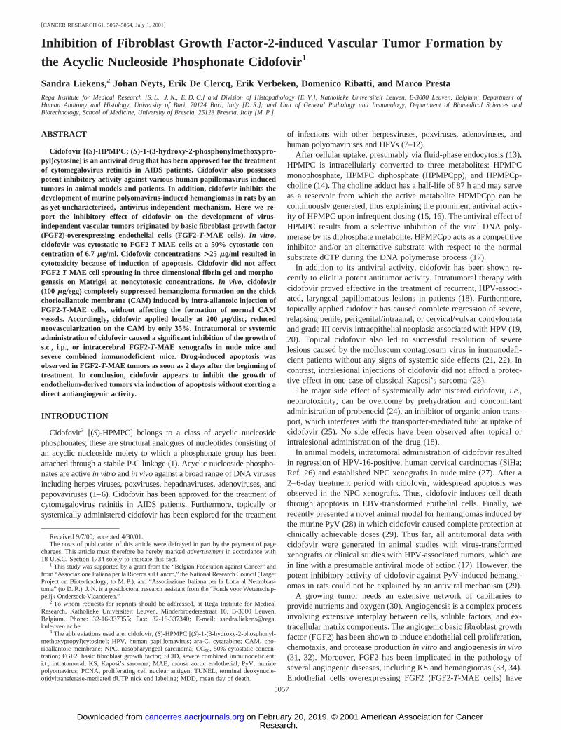
[CANCER RESEARCH 61, 5057–5064, July 1, 2001] Inhibition of Fibroblast Growth Factor-2-induced Vascular Tumor Formation by the Acyclic Nucleoside Phosphonate Cidofovir 1 Sandra Liekens, 2 Johan Neyts, Erik De Clercq, Erik Verbeken, Domenico Ribatti, and Marco Presta Rega Institute for Medical Research [S. L., J. N., E. D. C.] and Division of Histopathology [E. V.], Katholieke Universiteit Leuven, B-3000 Leuven, Belgium; Department of Human Anatomy and Histology, University of Bari, 70124 Bari, Italy [D. R.]; and Unit of General Pathology and Immunology, Department of Biomedical Sciences and Biotechnology, School of Medicine, University of Brescia, 25123 Brescia, Italy [M. P.] ABSTRACT Cidofovir [(S)-HPMPC; (S)-1-(3-hydroxy-2-phosphonylmethoxypro- pyl)cytosine] is an antiviral drug that has been approved for the treatment of cytomegalovirus retinitis in AIDS patients. Cidofovir also possesses potent inhibitory activity against various human papillomavirus-induced tumors in animal models and patients. In addition, cidofovir inhibits the development of murine polyomavirus-induced hemangiomas in rats by an as-yet-uncharacterized, antivirus-independent mechanism. Here we re- port the inhibitory effect of cidofovir on the development of virus- independent vascular tumors originated by basic fibroblast growth factor (FGF2)-overexpressing endothelial cells (FGF2-T-MAE cells). In vitro, cidofovir was cytostatic to FGF2-T-MAE cells at a 50% cytostatic con- centration of 6.7 mg/ml. Cidofovir concentrations >25 mg/ml resulted in cytotoxicity because of induction of apoptosis. Cidofovir did not affect FGF2-T-MAE cell sprouting in three-dimensional fibrin gel and morpho- genesis on Matrigel at noncytotoxic concentrations. In vivo, cidofovir (100 mg/egg) completely suppressed hemangioma formation on the chick chorioallantoic membrane (CAM) induced by intra-allantoic injection of FGF2-T-MAE cells, without affecting the formation of normal CAM vessels. Accordingly, cidofovir applied locally at 200 mg/disc, reduced neovascularization on the CAM by only 35%. Intratumoral or systemic administration of cidofovir caused a significant inhibition of the growth of s.c., i.p., or intracerebral FGF2-T-MAE xenografts in nude mice and severe combined immunodeficient mice. Drug-induced apoptosis was observed in FGF2-T-MAE tumors as soon as 2 days after the beginning of treatment. In conclusion, cidofovir appears to inhibit the growth of endothelium-derived tumors via induction of apoptosis without exerting a direct antiangiogenic activity. INTRODUCTION Cidofovir 3 [(S)-HPMPC] belongs to a class of acyclic nucleoside phosphonates; these are structural analogues of nucleotides consisting of an acyclic nucleoside moiety to which a phosphonate group has been attached through a stabile P-C linkage (1). Acyclic nucleoside phospho- nates are active in vitro and in vivo against a broad range of DNA viruses including herpes viruses, poxviruses, hepadnaviruses, adenoviruses, and papovaviruses (1– 6). Cidofovir has been approved for the treatment of cytomegalovirus retinitis in AIDS patients. Furthermore, topically or systemically administered cidofovir has been explored for the treatment of infections with other herpesviruses, poxviruses, adenoviruses, and human polyomaviruses and HPVs (7–12). After cellular uptake, presumably via fluid-phase endocytosis (13), HPMPC is intracellularly converted to three metabolites: HPMPC monophosphate, HPMPC diphosphate (HPMPCpp), and HPMPCp- choline (14). The choline adduct has a half-life of 87 h and may serve as a reservoir from which the active metabolite HPMPCpp can be continuously generated, thus explaining the prominent antiviral activ- ity of HPMPC upon infrequent dosing (15, 16). The antiviral effect of HPMPC results from a selective inhibition of the viral DNA poly- merase by its diphosphate metabolite. HPMPCpp acts as a competitive inhibitor and/or an alternative substrate with respect to the normal substrate dCTP during the DNA polymerase process (17). In addition to its antiviral activity, cidofovir has been shown re- cently to elicit a potent antitumor activity. Intratumoral therapy with cidofovir proved effective in the treatment of recurrent, HPV-associ- ated, laryngeal papillomatous lesions in patients (18). Furthermore, topically applied cidofovir has caused complete regression of severe, relapsing penile, perigenital/intraanal, or cervical/vulvar condylomata and grade III cervix intraepithelial neoplasia associated with HPV (19, 20). Topical cidofovir also led to successful resolution of severe lesions caused by the molluscum contagiosum virus in immunodefi- cient patients without any signs of systemic side effects (21, 22). In contrast, intralesional injections of cidofovir did not afford a protec- tive effect in one case of classical Kaposi’s sarcoma (23). The major side effect of systemically administered cidofovir, i.e., nephrotoxicity, can be overcome by prehydration and concomitant administration of probenecid (24), an inhibitor of organic anion trans- port, which interferes with the transporter-mediated tubular uptake of cidofovir (25). No side effects have been observed after topical or intralesional administration of the drug (18). In animal models, intratumoral administration of cidofovir resulted in regression of HPV-16-positive, human cervical carcinomas (SiHa; Ref. 26) and established NPC xenografts in nude mice (27). After a 2– 6-day treatment period with cidofovir, widespread apoptosis was observed in the NPC xenografts. Thus, cidofovir induces cell death through apoptosis in EBV-transformed epithelial cells. Finally, we recently presented a novel animal model for hemangiomas induced by the murine PyV (28) in which cidofovir caused complete protection at clinically achievable doses (29). Thus far, all antitumoral data with cidofovir were generated in animal studies with virus-transformed xenografts or clinical studies with HPV-associated tumors, which are in line with a presumable antiviral mode of action (17). However, the potent inhibitory activity of cidofovir against PyV-induced hemangi- omas in rats could not be explained by an antiviral mechanism (29). A growing tumor needs an extensive network of capillaries to provide nutrients and oxygen (30). Angiogenesis is a complex process involving extensive interplay between cells, soluble factors, and ex- tracellular matrix components. The angiogenic basic fibroblast growth factor (FGF2) has been shown to induce endothelial cell proliferation, chemotaxis, and protease production in vitro and angiogenesis in vivo (31, 32). Moreover, FGF2 has been implicated in the pathology of several angiogenic diseases, including KS and hemangiomas (33, 34). Endothelial cells overexpressing FGF2 (FGF2-T-MAE cells) have Received 9/7/00; accepted 4/30/01. The costs of publication of this article were defrayed in part by the payment of page charges. This article must therefore be hereby marked advertisement in accordance with 18 U.S.C. Section 1734 solely to indicate this fact. 1 This study was supported by a grant from the “Belgian Federation against Cancer” and from “Associazione Italiana per la Ricerca sul Cancro,” the National Research Council (Target Project on Biotechnology; to M. P.), and “Associazione Italiana per la Lotta al Neuroblas- toma” (to D. R.). J. N. is a postdoctoral research assistant from the “Fonds voor Wetenschap- pelijk Onderzoek-Vlaanderen.” 2 To whom requests for reprints should be addressed, at Rega Institute for Medical Research, Katholieke Universiteit Leuven, Minderbroedersstraat 10, B-3000 Leuven, Belgium. Phone: 32-16-337355; Fax: 32-16-337340; E-mail: sandra.liekens@rega. kuleuven.ac.be. 3 The abbreviations used are: cidofovir, (S)-HPMPC [(S)-1-(3-hydroxy-2-phosphonyl- methoxypropyl)cytosine]; HPV, human papillomavirus; ara-C, cytarabine; CAM, cho- rioallantoic membrane; NPC, nasopharyngeal carcinoma; CC 50 , 50% cytostatic concen- tration; FGF2, basic fibroblast growth factor; SCID, severe combined immunodeficient; i.t., intratumoral; KS, Kaposi’s sarcoma; MAE, mouse aortic endothelial; PyV, murine polyomavirus; PCNA, proliferating cell nuclear antigen; TUNEL, terminal deoxynucle- otidyltransferase-mediated dUTP nick end labeling; MDD, mean day of death. 5057 Research. on February 20, 2019. © 2001 American Association for Cancer cancerres.aacrjournals.org Downloaded from
Transcript of Inhibition of Fibroblast Growth Factor-2-induced Vascular...
[CANCER RESEARCH 61, 5057–5064, July 1, 2001]
Inhibition of Fibroblast Growth Factor-2-induced Vascular Tumor Formation bythe Acyclic Nucleoside Phosphonate Cidofovir1
Sandra Liekens,2 Johan Neyts, Erik De Clercq, Erik Verbeken, Domenico Ribatti, and Marco PrestaRega Institute for Medical Research [S. L., J. N., E. D. C.] and Division of Histopathology [E. V.], Katholieke Universiteit Leuven, B-3000 Leuven, Belgium; Department ofHuman Anatomy and Histology, University of Bari, 70124 Bari, Italy [D. R.]; and Unit of General Pathology and Immunology, Department of Biomedical Sciences andBiotechnology, School of Medicine, University of Brescia, 25123 Brescia, Italy [M. P.]
ABSTRACT
Cidofovir [(S )-HPMPC; (S)-1-(3-hydroxy-2-phosphonylmethoxypro-pyl)cytosine] is an antiviral drug that has been approved for the treatmentof cytomegalovirus retinitis in AIDS patients. Cidofovir also possessespotent inhibitory activity against various human papillomavirus-inducedtumors in animal models and patients. In addition, cidofovir inhibits thedevelopment of murine polyomavirus-induced hemangiomas in rats by anas-yet-uncharacterized, antivirus-independent mechanism. Here we re-port the inhibitory effect of cidofovir on the development of virus-independent vascular tumors originated by basic fibroblast growth factor(FGF2)-overexpressing endothelial cells (FGF2-T-MAE cells).In vitro,cidofovir was cytostatic to FGF2-T-MAE cells at a 50% cytostatic con-centration of 6.7 mg/ml. Cidofovir concentrations >25 mg/ml resulted incytotoxicity because of induction of apoptosis. Cidofovir did not affectFGF2-T-MAE cell sprouting in three-dimensional fibrin gel and morpho-genesis on Matrigel at noncytotoxic concentrations.In vivo, cidofovir(100 mg/egg) completely suppressed hemangioma formation on the chickchorioallantoic membrane (CAM) induced by intra-allantoic injection ofFGF2-T-MAE cells, without affecting the formation of normal CAMvessels. Accordingly, cidofovir applied locally at 200mg/disc, reducedneovascularization on the CAM by only 35%. Intratumoral or systemicadministration of cidofovir caused a significant inhibition of the growth ofs.c., i.p., or intracerebral FGF2-T-MAE xenografts in nude mice andsevere combined immunodeficient mice. Drug-induced apoptosis wasobserved in FGF2-T-MAE tumors as soon as 2 days after the beginning oftreatment. In conclusion, cidofovir appears to inhibit the growth ofendothelium-derived tumors via induction of apoptosis without exerting adirect antiangiogenic activity.
INTRODUCTION
Cidofovir3 [(S)-HPMPC] belongs to a class of acyclic nucleosidephosphonates; these are structural analogues of nucleotides consisting ofan acyclic nucleoside moiety to which a phosphonate group has beenattached through a stabile P-C linkage (1). Acyclic nucleoside phospho-nates are activein vitro andin vivoagainst a broad range of DNA virusesincluding herpes viruses, poxviruses, hepadnaviruses, adenoviruses, andpapovaviruses (1–6). Cidofovir has been approved for the treatment ofcytomegalovirus retinitis in AIDS patients. Furthermore, topically orsystemically administered cidofovir has been explored for the treatment
of infections with other herpesviruses, poxviruses, adenoviruses, andhuman polyomaviruses and HPVs (7–12).
After cellular uptake, presumably via fluid-phase endocytosis (13),HPMPC is intracellularly converted to three metabolites: HPMPCmonophosphate, HPMPC diphosphate (HPMPCpp), and HPMPCp-choline (14). The choline adduct has a half-life of 87 h and may serveas a reservoir from which the active metabolite HPMPCpp can becontinuously generated, thus explaining the prominent antiviral activ-ity of HPMPC upon infrequent dosing (15, 16). The antiviral effect ofHPMPC results from a selective inhibition of the viral DNA poly-merase by its diphosphate metabolite. HPMPCpp acts as a competitiveinhibitor and/or an alternative substrate with respect to the normalsubstrate dCTP during the DNA polymerase process (17).
In addition to its antiviral activity, cidofovir has been shown re-cently to elicit a potent antitumor activity. Intratumoral therapy withcidofovir proved effective in the treatment of recurrent, HPV-associ-ated, laryngeal papillomatous lesions in patients (18). Furthermore,topically applied cidofovir has caused complete regression of severe,relapsing penile, perigenital/intraanal, or cervical/vulvar condylomataand grade III cervix intraepithelial neoplasia associated with HPV (19,20). Topical cidofovir also led to successful resolution of severelesions caused by the molluscum contagiosum virus in immunodefi-cient patients without any signs of systemic side effects (21, 22). Incontrast, intralesional injections of cidofovir did not afford a protec-tive effect in one case of classical Kaposi’s sarcoma (23).
The major side effect of systemically administered cidofovir,i.e.,nephrotoxicity, can be overcome by prehydration and concomitantadministration of probenecid (24), an inhibitor of organic anion trans-port, which interferes with the transporter-mediated tubular uptake ofcidofovir (25). No side effects have been observed after topical orintralesional administration of the drug (18).
In animal models, intratumoral administration of cidofovir resultedin regression of HPV-16-positive, human cervical carcinomas (SiHa;Ref. 26) and established NPC xenografts in nude mice (27). After a2–6-day treatment period with cidofovir, widespread apoptosis wasobserved in the NPC xenografts. Thus, cidofovir induces cell deaththrough apoptosis in EBV-transformed epithelial cells. Finally, werecently presented a novel animal model for hemangiomas induced bythe murine PyV (28) in which cidofovir caused complete protection atclinically achievable doses (29). Thus far, all antitumoral data withcidofovir were generated in animal studies with virus-transformedxenografts or clinical studies with HPV-associated tumors, which arein line with a presumable antiviral mode of action (17). However, thepotent inhibitory activity of cidofovir against PyV-induced hemangi-omas in rats could not be explained by an antiviral mechanism (29).
A growing tumor needs an extensive network of capillaries toprovide nutrients and oxygen (30). Angiogenesis is a complex processinvolving extensive interplay between cells, soluble factors, and ex-tracellular matrix components. The angiogenic basic fibroblast growthfactor (FGF2) has been shown to induce endothelial cell proliferation,chemotaxis, and protease productionin vitro and angiogenesisin vivo(31, 32). Moreover, FGF2 has been implicated in the pathology ofseveral angiogenic diseases, including KS and hemangiomas (33, 34).Endothelial cells overexpressing FGF2 (FGF2-T-MAE cells) have
Received 9/7/00; accepted 4/30/01.The costs of publication of this article were defrayed in part by the payment of page
charges. This article must therefore be hereby markedadvertisementin accordance with18 U.S.C. Section 1734 solely to indicate this fact.
1 This study was supported by a grant from the “Belgian Federation against Cancer” andfrom “Associazione Italiana per la Ricerca sul Cancro,” the National Research Council (TargetProject on Biotechnology; to M. P.), and “Associazione Italiana per la Lotta al Neuroblas-toma” (to D. R.). J. N. is a postdoctoral research assistant from the “Fonds voor Wetenschap-pelijk Onderzoek-Vlaanderen.”
2 To whom requests for reprints should be addressed, at Rega Institute for MedicalResearch, Katholieke Universiteit Leuven, Minderbroedersstraat 10, B-3000 Leuven,Belgium. Phone: 32-16-337355; Fax: 32-16-337340; E-mail: [email protected].
3 The abbreviations used are: cidofovir, (S)-HPMPC [(S)-1-(3-hydroxy-2-phosphonyl-methoxypropyl)cytosine]; HPV, human papillomavirus; ara-C, cytarabine; CAM, cho-rioallantoic membrane; NPC, nasopharyngeal carcinoma; CC50, 50% cytostatic concen-tration; FGF2, basic fibroblast growth factor; SCID, severe combined immunodeficient;i.t., intratumoral; KS, Kaposi’s sarcoma; MAE, mouse aortic endothelial; PyV, murinepolyomavirus; PCNA, proliferating cell nuclear antigen; TUNEL, terminal deoxynucle-otidyltransferase-mediated dUTP nick end labeling; MDD, mean day of death.
5057
Research. on February 20, 2019. © 2001 American Association for Cancercancerres.aacrjournals.org Downloaded from
been shown to possess tumorigenic activity in nude mice, giving riseto highly vascularized lesions that histologically resemble KS (35,36). When inoculated into the allantoic sac of a chick embryo,FGF2-T-MAE cells cause an increase in vascular density and theformation of hemangiomas on the CAM (36, 37).
The present study was designed to gain further insight into themechanism of antitumor activity of cidofovir by using the virus-independent vascular tumor model represented by FGF2-T-MAEcells. Therefore, we evaluated the effect of cidofovir on angiogenesisand the growth of FGF2-overexpressing endothelial cellsin vitro andin vivo. The results demonstrate for the first time the antitumoractivity of cidofovir against lesions that are not induced by viruses.
MATERIALS AND METHODS
Materials. Cidofovir (Vistide; Fig. 1A) was kindly provided by GileadSciences (Foster City, CA). Ganciclovir (Cymevene) was obtained from Roche(Brussels, Belgium), and cytarabine (ara-C; Cytosar) was from Upjohn (Puurs,Belgium).
Cell Cultures. FGF2-transfected MAE cells (pZipbFGF2-MAE) expresshigh levels of theMr 18,000,Mr 22,000, andMr 24,000 molecular weightisoforms of FGF2 (36). After injection in nude mice, these cells induced theformation of vascular lesions from which FGF2-T-MAE cells could be iso-lated. When reinjected in nude mice, FGF2-T-MAE cells induced the forma-tion of tumors more rapidly than the parent pZipbFGF2-MAE cells (35).FGF2-T-MAE cells were grown in DMEM (Life Technologies, Inc., Rock-ville, MD) supplemented with 4 mM glutamine (Life Technologies, Inc.) and10% FCS (Integro, Zaandam, the Netherlands).
Cell Proliferation Assays. For measurement of cell growth, FGF2-T-MAEcells were seeded at 20,000 cells/cm2 in DMEM with 10% FCS. After 24 h, themedium was replaced, and cidofovir was added. The cell cultures were
incubated for 6 days, trypsinized, and counted. To examine the reversibility ofthe effect of cidofovir on FGF2-T-MAE cell proliferation, cells were washed2 days after addition of cidofovir and further incubated in the absence of thecompound for 4 days.
Detection of Apoptosis.To detect apoptosis in individual cells, FGF2-T-MAE cells were seeded in four-well chamber slides (Nunc, Naperville, IL) at50,000 cells/cm2. Subconfluent cells were treated with cidofovir at 10, 25, 50,100, or 250mg/ml for 1, 2, 3, or 4 days. At the indicated time points, cells werefixed in 4% paraformaldehyde in PBS for 1 h at room temperature, rinsed withPBS, and permeabilized for 2 min on ice in 0.1% Triton X-100 in 0.1% sodiumcitrate. Apoptotic cells were visualized by means of theIn Situ Cell DeathDetection kit (Boehringer Mannheim, Mannheim, Germany). This kit relies onthe use of terminal deoxynucleotidyl transferase, which catalyzes the polym-erization of fluorescein-labeled nucleotides to free 39-hydroxyl residues ofDNA fragments, generated by endonucleases during apoptosis. Fluorescein-labeled DNA strand breaks in apoptotic cells could subsequently be detectedwith a fluorescence microscope.
CAM Assay. The in vivo CAM angiogenesis model was performed asdescribed by Maragoudakiset al. (38). Briefly, fresh fertilized eggs wereincubated for 3 days at 37°C when 3 ml of albumen were removed (to detachthe shell from the developing CAM), and a window was opened on theeggshell, exposing the CAM. The window was covered with cellophane tape,and the eggs were returned to the incubator until day 9, when the testcompounds were applied. Cidofovir was placed on sterile plastic discs (Ø 8mm), which were allowed to dry under sterile conditions. A solution ofcortisone acetate (100mg/disc; Sigma Chemical Co., St. Louis, MO) wasincorporated in all discs to prevent an inflammatory response. The loaded anddried control discs (containing PBS) were placed on the CAM 1 cm away fromthe disc containing cidofovir. Next, the windows were covered, and the eggswere incubated until day 11, when angiogenesis was assessed. At day 11, theeggs were flooded with 10% buffered formalin (Janssen Chimica, Geel,Belgium), the plastic discs were removed, and the eggs were kept at roomtemperature for at least 2 h. A large area around the discs was cutoff and placedon a glass slide, and the vascular density index was measured by the methodof Harris-Hookeret al. (39). Briefly, a grid containing three concentric circlesof 4-, 5-, and 6-mm diameter was positioned on the surface of the CAMpreviously covered by the disc. Next, all vessels intersecting the circles werecounted. The two-tailed paired Student’st test was used to assess the signif-icance of the obtained results.
Chick Embryo Allantoic Sac Assay. Fertilized eggs were incubated for 3days at 37°C, when 3 ml of albumen were removed and a window was openedon the eggshell exposing the CAM. The window was covered with cellophanetape, and the eggs were returned to the incubator. At day 8, 200ml of PBScontaining 200,000 FGF2-T-MAE cells were injected into the allantoic sac.Cidofovir was injected into the allantoic sac at the same time. At day 12, alarge chorioallantoic vein of the CAM was perfused with India ink. Next, theCAMs were fixed, sectioned, rehydrated, and examined microscopically.
Tumor Growth in Mice. Eight-week-old female SCID mice, weighingabout 20 g, were inoculated i.p. with 200ml of MEM containing 53 105
FGF2-T-MAE cells. SCID mice, weighing;15 g, were inoculated intracere-brally into the right cerebral hemisphere with 50ml of MEM containing 105
FGF2-T-MAE cells. Cidofovir therapy was started 1 day after inoculation ofthe cells, according to a variety of treatment and administration schedules, asindicated in “Results.” Mortality was recorded daily, and statistical signifi-cance of the data was assessed using the two-tailed unpaired Student’st test.
Eight-week-old female, athymic, nudenu/numice, weighing;25 g, wereinoculated s.c. with 100ml of MEM containing 106 FGF2-T-MAE cells. i.t.administration of cidofovir or MEM was started 11 days after tumor cellinoculation,i.e., when tumors reached a volume of;100 mm3. Tumor sizewas monitored three times a week by means of a caliper, and the tumorvolume was calculated with the following formula: Tumor volume(mm3) 5 0.53 a 3 b2, wherea is the longest diameter andb is the shortestdiameter.
Histological Analysis and Immunohistochemistry of Vascular Tumors.Nontreated and cidofovir-treated mice were dissected at different times aftertumor cell inoculation. Tumors were fixed in 10% buffered formaldehyde andembedded in paraffin, and deparaffinized sections were subsequently stainedwith H&E and examined microscopically.
For immunohistochemistry, deparaffinized sections of the tumors were
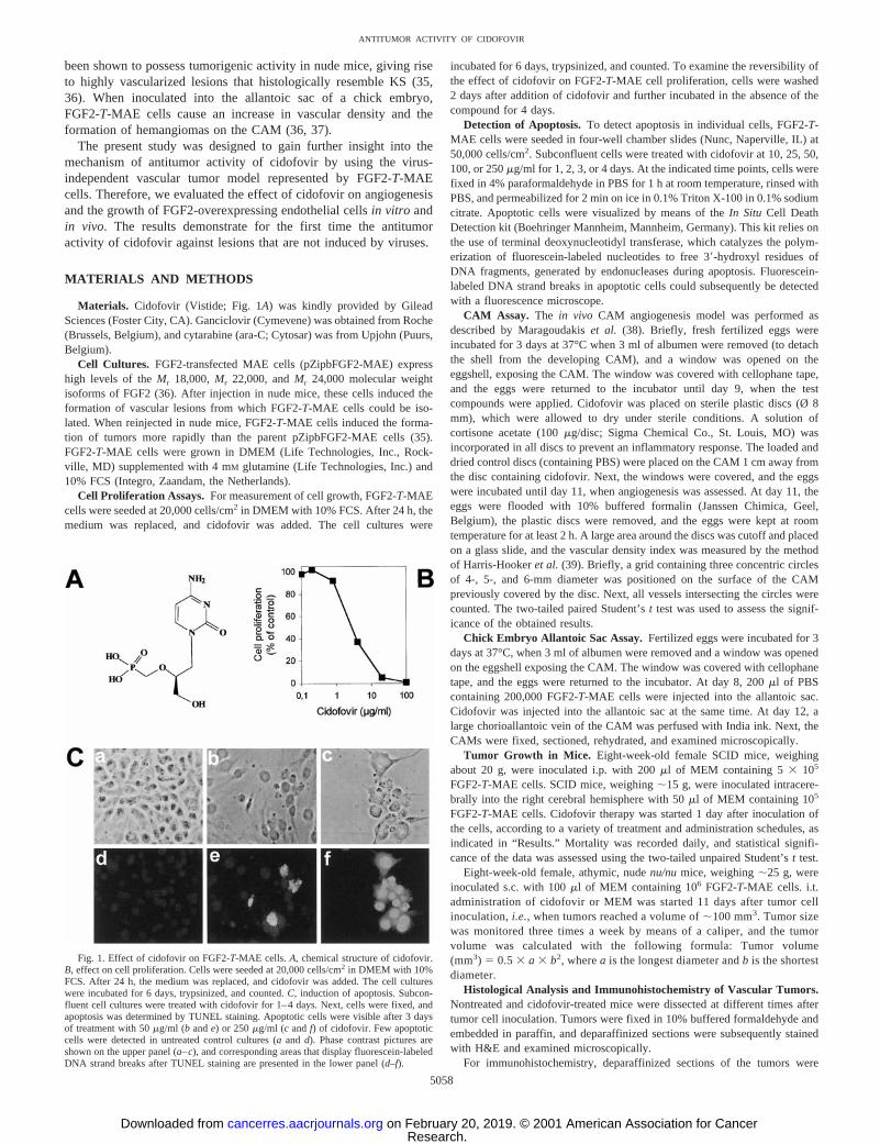
Fig. 1. Effect of cidofovir on FGF2-T-MAE cells.A, chemical structure of cidofovir.B, effect on cell proliferation. Cells were seeded at 20,000 cells/cm2 in DMEM with 10%FCS. After 24 h, the medium was replaced, and cidofovir was added. The cell cultureswere incubated for 6 days, trypsinized, and counted.C, induction of apoptosis. Subcon-fluent cell cultures were treated with cidofovir for 1–4 days. Next, cells were fixed, andapoptosis was determined by TUNEL staining. Apoptotic cells were visible after 3 daysof treatment with 50mg/ml (b ande) or 250mg/ml (c andf) of cidofovir. Few apoptoticcells were detected in untreated control cultures (aand d). Phase contrast pictures areshown on the upper panel (a–c), and corresponding areas that display fluorescein-labeledDNA strand breaks after TUNEL staining are presented in the lower panel (d–f).
5058
ANTITUMOR ACTIVITY OF CIDOFOVIR
Research. on February 20, 2019. © 2001 American Association for Cancercancerres.aacrjournals.org Downloaded from
incubated for 1 h with 1.5% blocking serum in PBS. After removal of theblocking serum, the slides were incubated with a primary mouse monoclonalantibody directed against human PCNA (clone PC10; Calbiochem-Novabio-chem, Nottingham, United Kingdom) for 30 min at room temperature. Theslides were subsequently washed in PBS for 5 min and then incubated withbiotinylated secondary antimouse antibody (Dako, Glostrup, Denmark) for 30min at room temperature. After a 5-min wash, all slides were incubated withhorseradish peroxidase-labeled streptavidine (Dako Corp.) for 30 min at roomtemperature. The immunoreactions were visualized as brown precipitates byincubation with the substrate diaminobenzidine (Dako) for 5 min. The reactionwas stopped by water, and the slides were counterstained with hematoxylin.Apoptosis detection in the tumors was performed by means of theIn SituCellDeath Detection kit (Boehringer Mannheim), as described above. AfterTUNEL staining, tissue sections were washed in PBS and exposed to theDNA-binding dye HOECHST 33342 (Sigma Chemical Co.) at a concentrationof 1.2 mg/ml in PBS for 15 min at room temperature. The slides wereextensively washed in PBS and examined with a Zeiss fluorescence micro-scope. Apoptotic cells were identified by positive TUNEL staining and chro-matin condensation.
RESULTS
Effect of Cidofovir on FGF2-T-MAE Cell Growth and Apo-ptosis in Vitro. We reported recently on the potent inhibition ofPyV-induced hemangioma development in rats by cidofovir. Thisactivity was not mediated by an antiviral effect (29). To elucidate themechanism by which cidofovir abrogates vascular tumor growth, weused FGF2-T-MAE cells. These endothelial cells are not transformedby an oncogenic virus (as is the case for PyV-induced hemangiomas)but overexpress FGF2 and induce vascular tumors on the CAM and innude mice (35, 36).
In vitro, cidofovir caused a dose-dependent inhibition of FGF2-T-MAE cell proliferation with a CC50 of 6.7 mg/ml after 6 days oftreatment (Fig. 1B). To study the reversibility of the observed growth-inhibitory effect on FGF2-T-MAE cells, cidofovir was removed after48 h, and cultures were kept for 4 days in the absence of thecompound. At 10mg/ml, removal of cidofovir resulted in acceleratedcell proliferation. In contrast, the cells did not recover if exposed tocidofovir at 30mg/ml, indicating that this concentration is toxic ratherthan cytostatic (data not shown).
Next, subconfluent FGF2-T-MAE cells, grown in four-well cham-ber slides, were incubated with various concentrations of cidofovir for1, 2, 3, or 4 days, after which induction of apoptosis was assessed. Noapoptotic cells were identified at cidofovir concentrations,25mg/ml.At 25 mg/ml, few apoptotic cells were visible after 3 days of cidofovirtreatment. At higher concentrations (50–100-250mg/ml), apoptosiswas evident after exposure of the cells to the compound for 48 h. Noviable cells were detected after 3 days at 250mg/ml (Fig. 1C). Themajority of these cells showed membrane blebbing and the presenceof apoptotic bodies, hallmarks of apoptotic cell death.
Effect of Cidofovir on Hemangioma Growth and Angiogenesison the CAM. Upon inoculation into the allantoic sac of chick em-bryos, FGF2-T-MAE cells induce the formation of hemangiomas on
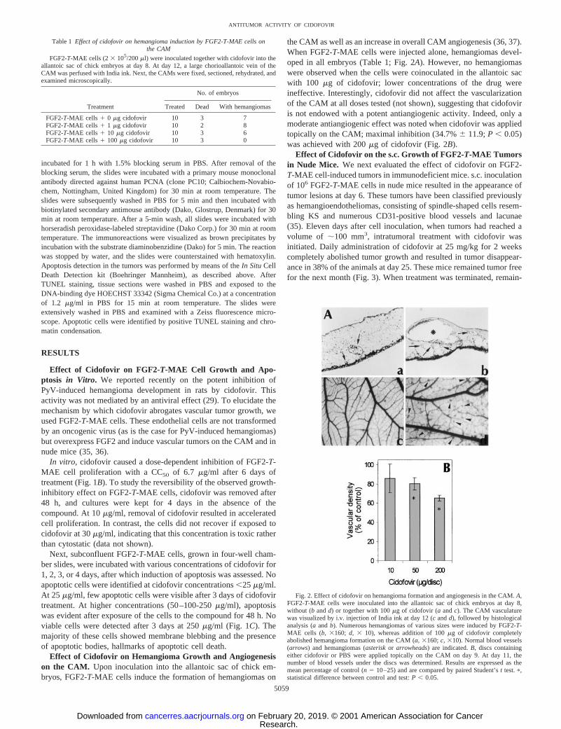
the CAM as well as an increase in overall CAM angiogenesis (36, 37).When FGF2-T-MAE cells were injected alone, hemangiomas devel-oped in all embryos (Table 1; Fig. 2A). However, no hemangiomaswere observed when the cells were coinoculated in the allantoic sacwith 100 mg of cidofovir; lower concentrations of the drug wereineffective. Interestingly, cidofovir did not affect the vascularizationof the CAM at all doses tested (not shown), suggesting that cidofoviris not endowed with a potent antiangiogenic activity. Indeed, only amoderate antiangiogenic effect was noted when cidofovir was appliedtopically on the CAM; maximal inhibition (34.7%6 11.9;P , 0.05)was achieved with 200mg of cidofovir (Fig. 2B).
Effect of Cidofovir on the s.c. Growth of FGF2-T-MAE Tumorsin Nude Mice. We next evaluated the effect of cidofovir on FGF2-T-MAE cell-induced tumors in immunodeficient mice. s.c. inoculationof 106 FGF2-T-MAE cells in nude mice resulted in the appearance oftumor lesions at day 6. These tumors have been classified previouslyas hemangioendotheliomas, consisting of spindle-shaped cells resem-bling KS and numerous CD31-positive blood vessels and lacunae(35). Eleven days after cell inoculation, when tumors had reached avolume of ;100 mm3, intratumoral treatment with cidofovir wasinitiated. Daily administration of cidofovir at 25 mg/kg for 2 weekscompletely abolished tumor growth and resulted in tumor disappear-ance in 38% of the animals at day 25. These mice remained tumor freefor the next month (Fig. 3). When treatment was terminated, remain-
Fig. 2. Effect of cidofovir on hemangioma formation and angiogenesis in the CAM.A,FGF2-T-MAE cells were inoculated into the allantoic sac of chick embryos at day 8,without (b andd) or together with 100mg of cidofovir (a andc). The CAM vasculaturewas visualized by i.v. injection of India ink at day 12 (candd), followed by histologicalanalysis (aandb). Numerous hemangiomas of various sizes were induced by FGF2-T-MAE cells (b, 3160; d, 3 10), whereas addition of 100mg of cidofovir completelyabolished hemangioma formation on the CAM (a,3160;c, 310). Normal blood vessels(arrows) and hemangiomas (asteriskor arrowheads) are indicated.B, discs containingeither cidofovir or PBS were applied topically on the CAM on day 9. At day 11, thenumber of blood vessels under the discs was determined. Results are expressed as themean percentage of control (n5 10–25) and are compared by paired Student’st test.p,statistical difference between control and test:P , 0.05.
Table 1 Effect of cidofovir on hemangioma induction by FGF2-T-MAE cells onthe CAM
FGF2-T-MAE cells (23 105/200ml) were inoculated together with cidofovir into theallantoic sac of chick embryos at day 8. At day 12, a large chorioallantoic vein of theCAM was perfused with India ink. Next, the CAMs were fixed, sectioned, rehydrated, andexamined microscopically.
Treatment
No. of embryos
Treated Dead With hemangiomas
FGF2-T-MAE cells1 0 mg cidofovir 10 3 7FGF2-T-MAE cells1 1 mg cidofovir 10 2 8FGF2-T-MAE cells1 10 mg cidofovir 10 3 6FGF2-T-MAE cells1 100 mg cidofovir 10 3 0
5059
ANTITUMOR ACTIVITY OF CIDOFOVIR
Research. on February 20, 2019. © 2001 American Association for Cancercancerres.aacrjournals.org Downloaded from
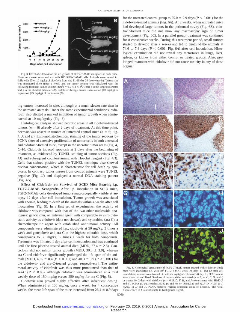
ing tumors increased in size, although at a much slower rate than inthe untreated animals. Under the same experimental conditions, cido-fovir also elicited a marked inhibition of tumor growth when admin-istered at 10 mg/kg/day (Fig. 3).
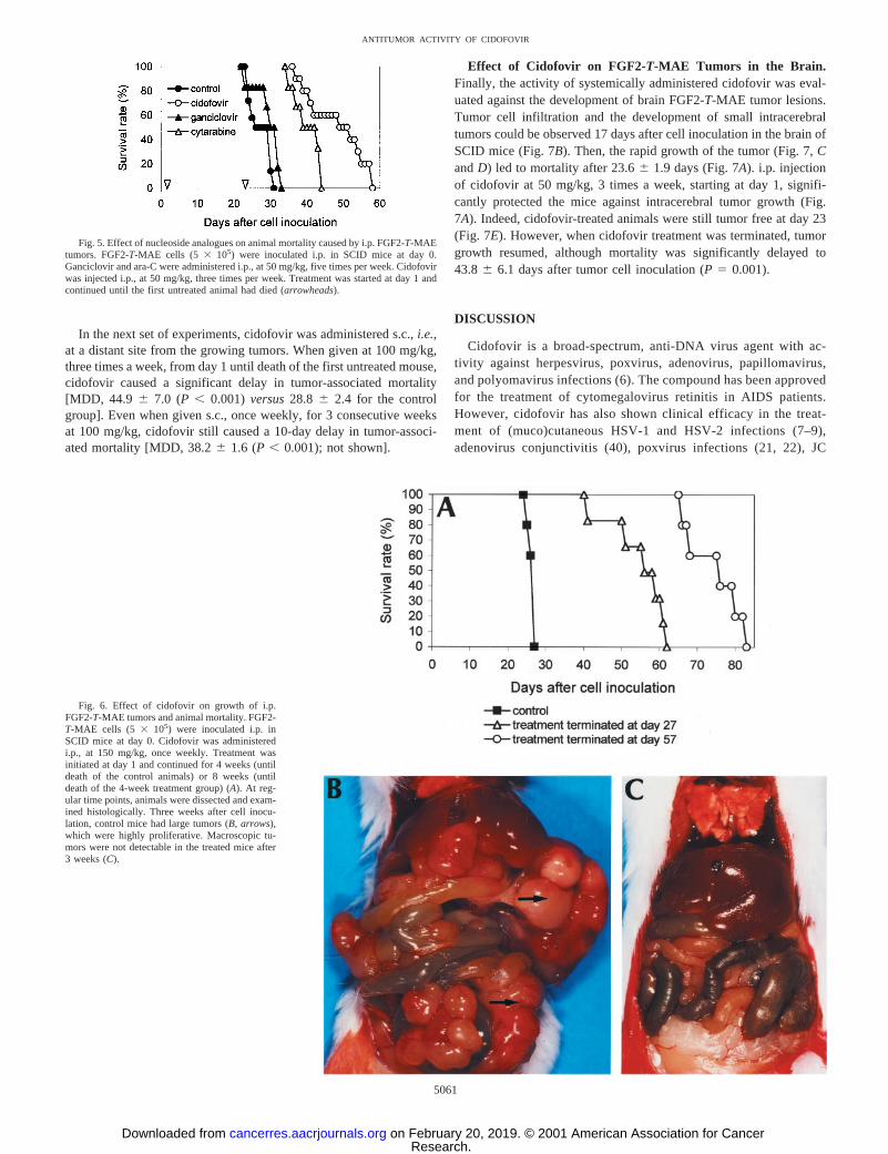
Histological analysis showed necrotic areas in all cidofovir-treatedtumors (n5 6) already after 2 days of treatment. At this time point,necrosis was absent in tumors of untreated control mice (n 5 6; Fig.4, A andB). Immunohistochemical staining of the tumor sections byPCNA showed extensive proliferation of tumor cells in both untreatedand cidofovir-treated mice, except in the necrotic tumor areas (Fig. 4,C–F). Cidofovir induced apoptosis at 2 days after the beginning oftreatment, as evidenced by TUNEL staining of tumor sections (Fig.4J) and subsequent counterstaining with Hoechst reagent (Fig. 4H).Cells that stained positive with the TUNEL technique also showednuclear condensation, which is characteristic for cell death by apo-ptosis. In contrast, tumor tissues from control animals were TUNELnegative (Fig. 4I) and displayed a normal DNA staining pattern(Fig. 4G).
Effect of Cidofovir on Survival of SCID Mice Bearing i.p.FGF2-T-MAE Xenografts. After i.p. inoculation in SCID mice,FGF2-T-MAE cells developed tumors macroscopically visible at au-topsy 12 days after cell inoculation. Tumor growth was associatedwith anemia, leading to death of the animals within 4 weeks after cellinoculation (Fig. 5). In a first set of experiments, the activity ofcidofovir was compared with that of the two other nucleoside ana-logues: ganciclovir, an antiviral agent with comparablein vitro cyto-static activity as cidofovir (data not shown); and cytarabine (ara-C), achemotherapeutic agent with established antitumoral activity. Allcompounds were administered i.p., cidofovir at 50 mg/kg, 3 times aweek and ganciclovir and ara-C at the highest tolerable dose, whichcorresponds to 50 mg/kg, 5 times a week for both compounds.Treatment was initiated 1 day after cell inoculation and was continueduntil the first placebo-treated animal died (MDD, 27.46 2.8). Gan-ciclovir did not inhibit tumor growth (MDD, 30.36 3.9), whereasara-C and cidofovir significantly prolonged the life span of the ani-mals [MDD, 49.56 8.4 (P, 0.001) and 40.36 3.9 (P, 0.001) forthe cidofovir- and ara-C-treated groups, respectively]. The antitu-moral activity of cidofovir was thus more pronounced than that ofara-C (P , 0.05), although cidofovir was administered at a totalweekly dose of 150 mg/kgversus250 mg/kg for ara-C (Fig. 5).
Cidofovir also proved highly effective after infrequent dosing.When administered at 150 mg/kg, once a week, for 4 consecutiveweeks, the mean life span of the mice increased from 26.46 0.9 days
for the untreated control group to 55.06 7.9 days (P, 0.001) for thecidofovir-treated animals (Fig. 6A). At 3 weeks, when untreated micehad developed large tumors in the peritoneal cavity (Fig. 6B), cido-fovir-treated mice did not show any macroscopic sign of tumordevelopment (Fig. 6C). In a parallel group, treatment was continuedfor 8 consecutive weeks. During this treatment period, small tumorsstarted to develop after 7 weeks and led to death of the animals at74.6 6 7.4 days (P, 0.001; Fig. 6A) after cell inoculation. Histo-logical examination did not reveal any metastases in lungs, liver,spleen, or kidney from either control or treated groups. Also, pro-longed treatment with cidofovir did not cause toxicity in any of theseorgans.
Fig. 4. Histological appearance of FGF2-T-MAE tumors treated with cidofovir. Nudemice were inoculated s.c. with 106 FGF2-T-MAE cells. At days 11 and 12 after cellinoculation, animals were treated i.t. with 25 mg/kg of cidofovir. At day 13, 3F2T tumorswere dissected and fixed. Sections of tumors, either untreated (n 5 6; A, C, E, G,andI)or treated for 2 days with cidofovir (n5 6; B, D, F, H,andJ) were stained with H&E (AandB), PCNA (C–F), Hoechst 33342 (GandH), or TUNEL (I andJ). A–D, 3125;E–J,3200. In D and F, PCNA-negative regions represent areas of necrosis. The weakfluorescence inI is attributable to background signal.
Fig. 3. Effect of cidofovir on the s.c. growth of FGF2-T-MAE xenografts in nude mice.Nude mice were inoculated s.c. with 106 FGF2-T-MAE cells. Animals were treated i.t.daily with 25 or 10 mg/kg of cidofovir from day 11 till day 24 (arrowheads). Tumor sizewas monitored three times a week, and the tumor volume was calculated with thefollowing formula: Tumor volume (mm3) 5 0.53 a 3 b2, wherea is the longest diameterandb is the shortest diameter (A). Cidofovir therapy caused stabilization (10 mg/kg) orregression (25 mg/kg) of the tumors (B).
5060
ANTITUMOR ACTIVITY OF CIDOFOVIR
Research. on February 20, 2019. © 2001 American Association for Cancercancerres.aacrjournals.org Downloaded from
In the next set of experiments, cidofovir was administered s.c.,i.e.,at a distant site from the growing tumors. When given at 100 mg/kg,three times a week, from day 1 until death of the first untreated mouse,cidofovir caused a significant delay in tumor-associated mortality[MDD, 44.9 6 7.0 (P , 0.001) versus28.8 6 2.4 for the controlgroup]. Even when given s.c., once weekly, for 3 consecutive weeksat 100 mg/kg, cidofovir still caused a 10-day delay in tumor-associ-ated mortality [MDD, 38.26 1.6 (P, 0.001); not shown].
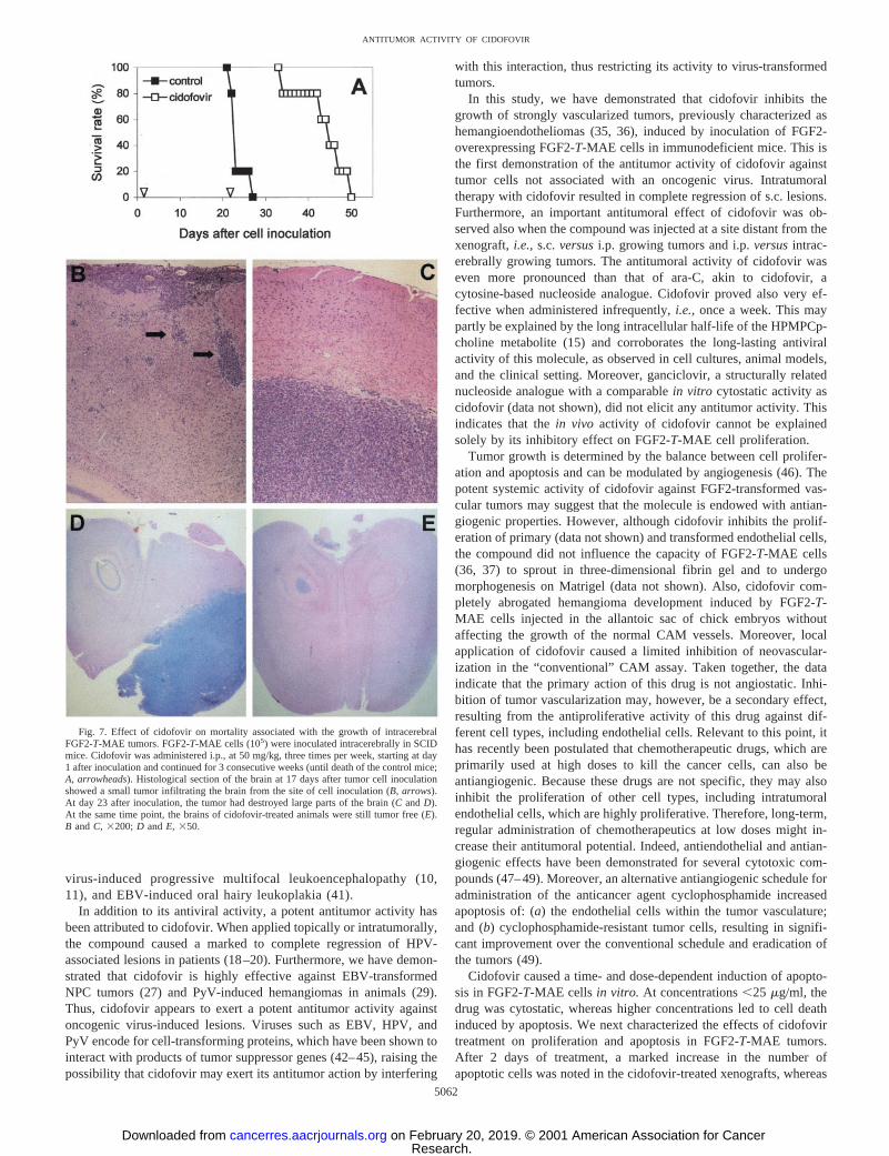
Effect of Cidofovir on FGF2-T-MAE Tumors in the Brain.Finally, the activity of systemically administered cidofovir was eval-uated against the development of brain FGF2-T-MAE tumor lesions.Tumor cell infiltration and the development of small intracerebraltumors could be observed 17 days after cell inoculation in the brain ofSCID mice (Fig. 7B). Then, the rapid growth of the tumor (Fig. 7,CandD) led to mortality after 23.66 1.9 days (Fig. 7A). i.p. injectionof cidofovir at 50 mg/kg, 3 times a week, starting at day 1, signifi-cantly protected the mice against intracerebral tumor growth (Fig.7A). Indeed, cidofovir-treated animals were still tumor free at day 23(Fig. 7E). However, when cidofovir treatment was terminated, tumorgrowth resumed, although mortality was significantly delayed to43.86 6.1 days after tumor cell inoculation (P 5 0.001).
DISCUSSION
Cidofovir is a broad-spectrum, anti-DNA virus agent with ac-tivity against herpesvirus, poxvirus, adenovirus, papillomavirus,and polyomavirus infections (6). The compound has been approvedfor the treatment of cytomegalovirus retinitis in AIDS patients.However, cidofovir has also shown clinical efficacy in the treat-ment of (muco)cutaneous HSV-1 and HSV-2 infections (7–9),adenovirus conjunctivitis (40), poxvirus infections (21, 22), JC
Fig. 5. Effect of nucleoside analogues on animal mortality caused by i.p. FGF2-T-MAEtumors. FGF2-T-MAE cells (53 105) were inoculated i.p. in SCID mice at day 0.Ganciclovir and ara-C were administered i.p., at 50 mg/kg, five times per week. Cidofovirwas injected i.p., at 50 mg/kg, three times per week. Treatment was started at day 1 andcontinued until the first untreated animal had died (arrowheads).
Fig. 6. Effect of cidofovir on growth of i.p.FGF2-T-MAE tumors and animal mortality. FGF2-T-MAE cells (5 3 105) were inoculated i.p. inSCID mice at day 0. Cidofovir was administeredi.p., at 150 mg/kg, once weekly. Treatment wasinitiated at day 1 and continued for 4 weeks (untildeath of the control animals) or 8 weeks (untildeath of the 4-week treatment group) (A). At reg-ular time points, animals were dissected and exam-ined histologically. Three weeks after cell inocu-lation, control mice had large tumors (B, arrows),which were highly proliferative. Macroscopic tu-mors were not detectable in the treated mice after3 weeks (C).
5061
ANTITUMOR ACTIVITY OF CIDOFOVIR
Research. on February 20, 2019. © 2001 American Association for Cancercancerres.aacrjournals.org Downloaded from
virus-induced progressive multifocal leukoencephalopathy (10,11), and EBV-induced oral hairy leukoplakia (41).
In addition to its antiviral activity, a potent antitumor activity hasbeen attributed to cidofovir. When applied topically or intratumorally,the compound caused a marked to complete regression of HPV-associated lesions in patients (18–20). Furthermore, we have demon-strated that cidofovir is highly effective against EBV-transformedNPC tumors (27) and PyV-induced hemangiomas in animals (29).Thus, cidofovir appears to exert a potent antitumor activity againstoncogenic virus-induced lesions. Viruses such as EBV, HPV, andPyV encode for cell-transforming proteins, which have been shown tointeract with products of tumor suppressor genes (42–45), raising thepossibility that cidofovir may exert its antitumor action by interfering
with this interaction, thus restricting its activity to virus-transformedtumors.
In this study, we have demonstrated that cidofovir inhibits thegrowth of strongly vascularized tumors, previously characterized ashemangioendotheliomas (35, 36), induced by inoculation of FGF2-overexpressing FGF2-T-MAE cells in immunodeficient mice. This isthe first demonstration of the antitumor activity of cidofovir againsttumor cells not associated with an oncogenic virus. Intratumoraltherapy with cidofovir resulted in complete regression of s.c. lesions.Furthermore, an important antitumoral effect of cidofovir was ob-served also when the compound was injected at a site distant from thexenograft,i.e., s.c.versusi.p. growing tumors and i.p.versusintrac-erebrally growing tumors. The antitumoral activity of cidofovir waseven more pronounced than that of ara-C, akin to cidofovir, acytosine-based nucleoside analogue. Cidofovir proved also very ef-fective when administered infrequently,i.e., once a week. This maypartly be explained by the long intracellular half-life of the HPMPCp-choline metabolite (15) and corroborates the long-lasting antiviralactivity of this molecule, as observed in cell cultures, animal models,and the clinical setting. Moreover, ganciclovir, a structurally relatednucleoside analogue with a comparablein vitro cytostatic activity ascidofovir (data not shown), did not elicit any antitumor activity. Thisindicates that thein vivo activity of cidofovir cannot be explainedsolely by its inhibitory effect on FGF2-T-MAE cell proliferation.
Tumor growth is determined by the balance between cell prolifer-ation and apoptosis and can be modulated by angiogenesis (46). Thepotent systemic activity of cidofovir against FGF2-transformed vas-cular tumors may suggest that the molecule is endowed with antian-giogenic properties. However, although cidofovir inhibits the prolif-eration of primary (data not shown) and transformed endothelial cells,the compound did not influence the capacity of FGF2-T-MAE cells(36, 37) to sprout in three-dimensional fibrin gel and to undergomorphogenesis on Matrigel (data not shown). Also, cidofovir com-pletely abrogated hemangioma development induced by FGF2-T-MAE cells injected in the allantoic sac of chick embryos withoutaffecting the growth of the normal CAM vessels. Moreover, localapplication of cidofovir caused a limited inhibition of neovascular-ization in the “conventional” CAM assay. Taken together, the dataindicate that the primary action of this drug is not angiostatic. Inhi-bition of tumor vascularization may, however, be a secondary effect,resulting from the antiproliferative activity of this drug against dif-ferent cell types, including endothelial cells. Relevant to this point, ithas recently been postulated that chemotherapeutic drugs, which areprimarily used at high doses to kill the cancer cells, can also beantiangiogenic. Because these drugs are not specific, they may alsoinhibit the proliferation of other cell types, including intratumoralendothelial cells, which are highly proliferative. Therefore, long-term,regular administration of chemotherapeutics at low doses might in-crease their antitumoral potential. Indeed, antiendothelial and antian-giogenic effects have been demonstrated for several cytotoxic com-pounds (47–49). Moreover, an alternative antiangiogenic schedule foradministration of the anticancer agent cyclophosphamide increasedapoptosis of: (a) the endothelial cells within the tumor vasculature;and (b) cyclophosphamide-resistant tumor cells, resulting in signifi-cant improvement over the conventional schedule and eradication ofthe tumors (49).
Cidofovir caused a time- and dose-dependent induction of apopto-sis in FGF2-T-MAE cellsin vitro. At concentrations,25 mg/ml, thedrug was cytostatic, whereas higher concentrations led to cell deathinduced by apoptosis. We next characterized the effects of cidofovirtreatment on proliferation and apoptosis in FGF2-T-MAE tumors.After 2 days of treatment, a marked increase in the number ofapoptotic cells was noted in the cidofovir-treated xenografts, whereas
Fig. 7. Effect of cidofovir on mortality associated with the growth of intracerebralFGF2-T-MAE tumors. FGF2-T-MAE cells (105) were inoculated intracerebrally in SCIDmice. Cidofovir was administered i.p., at 50 mg/kg, three times per week, starting at day1 after inoculation and continued for 3 consecutive weeks (until death of the control mice;A, arrowheads). Histological section of the brain at 17 days after tumor cell inoculationshowed a small tumor infiltrating the brain from the site of cell inoculation (B, arrows).At day 23 after inoculation, the tumor had destroyed large parts of the brain (CandD).At the same time point, the brains of cidofovir-treated animals were still tumor free (E).B andC, 3200; D andE, 350.
5062
ANTITUMOR ACTIVITY OF CIDOFOVIR
Research. on February 20, 2019. © 2001 American Association for Cancercancerres.aacrjournals.org Downloaded from
tumor cell proliferation was unaffected. These data are in agreementwith previous observations on NPC xenografts in nude mice in whichcidofovir caused extensive apoptosis leading to tumor regression (27).
A number of antitumor agents, including the nucleoside analoguescytarabine (ara-C), gemcitabine, and 5-fluorouracil, elicit their anti-tumor activity by induction of apoptosis (50, 51). Compared withthese drugs, cidofovir possesses lowin vitro cytostatic and cytotoxicaction. For instance, cidofovir is;500-fold less toxic than ara-C thatinduced apoptosis of FGF2-T-MAE cellsin vitro at 0.5mg/ml (datanot shown). The signaling pathway leading to drug-induced apoptosishas remained largely unknown and does not seem to be mediated byFAS receptor activation (52). The capacity of cidofovir to induceapoptosis in p53 deletion mutant NPC tumors rules out the involve-ment of p53 in mediating the antitumor activity of this drug (27).Gemcitabine monophosphate results in apoptosis because of incorpo-ration into cellular DNA (53). Interestingly, cidofovir also possessesthe potential to become integrated into DNA (17). Alternatively,cidofovir might disrupt the cell cycle, which has been shown to triggerapoptosis after 5-fluorouracil treatment (54). Recent experiments havedemonstrated a marked increase in CPP32 (caspase-3) protease activ-ity caused by cidofovir in HPV-containing cells (55).
FGF2 is an important mediator in the progression of KS (33). Inapparent contrast with its potent antitumoral activity against FGF2-overexpressing endothelial cells, intratumorally administered cidofo-vir did not afford a protective effect on KS lesion progression in mice4
as well as in the clinical setting (23). This indicates significantmolecular differences between the FGF2-T-MAE cell-induced he-mangioendotheliomas and KS lesions. Recent data indicate that che-motherapeutic resistance of KS cells could be attributed to the longdoubling times of these cells (56). Indeed, stimulation of KS cells withFGF2 rendered them more sensitive to the cytostatic activity ofcidofovir, as indicated by the markedly reduced CC50 of cidofovirafter FGF2 stimulation (56).
In conclusion, the data presented here indicate that cidofovir shouldalso be explored for the treatment of vascular tumors (that are notassociated with an oncogenic virus), such as juvenile hemangiomas.The ability to cross the blood-brain barrier and to suppress theprogression of angiogenic brain tumors after systemic treatment war-rants further investigation for the use of cidofovir in the therapy ofgliomas, highly proliferative vascular brain lesions for which at pres-ent no adequate treatment is available. Cidofovir appears to inhibittumor growth directly by induction of cell death (apoptosis) andindirectly by inhibiting the proliferation of the endothelial cells liningthe tumor vasculature. The trigger and subsequent signaling pathwayleading to apoptosis activation by cidofovir is currently under inves-tigation.
ACKNOWLEDGMENTS
We thank Willy Zeegers, Katrien Koninckx, Helga Van den Bosch, andJosiane Brebels for excellent technical assistance and Christiane Callebaut forfine editorial help.
REFERENCES
1. De Clercq, E., Holy, A., Rosenberg, I., Sakuma, T., Balzarini, J., and Maudgal, P. C.A novel selective broad-spectrum anti-DNA virus agent. Nature (Lond.),323: 464–467, 1986.
2. Andrei, G., Snoeck, R., Piette, J., Delvenne, P., and De Clercq, E. Antiproliferativeeffects of acyclic nucleoside phosphonates on human papillomavirus (HPV)-harbor-ing cell lines compared with HPV-negative cell lines. Oncol. Res.,10: 523–531,1998.
3. Jacobson, M. A. Treatment of cytomegalovirus retinitis in patients with the acquiredimmunodeficiency syndrome. N. Engl. J. Med.,337: 105–114, 1997.
4. Neyts, J., and De Clercq, E. Antiviral drug susceptibility of human herpesvirus 8.Antimicrob. Agents Chemother.,41: 2754–2756, 1997.
5. Kedes, D. H., and Ganem, D. Sensitivity of Kaposi’s sarcoma-associated herpesvirusreplication to antiviral drugs. Implications for potential therapy. J. Clin. Investig.,99:2082–2086, 1997.
6. De Clercq, E. In search of a selective antiviral chemotherapy. Clin. Microbiol. Rev.,10: 674–693, 1997.
7. Snoeck, R., Andrei, G., De Clercq, E., Gerard, M., Clumeck, N., Tricot, G., andSadzot-Delvaux, C. A new topical treatment for resistant herpes simplex infections.N. Engl. J. Med.,329: 968–969, 1993.
8. Lalezari, J., Schacker, T., Feinberg, J., Gathe, J., Lee, S., Cheung, T., Kramer, F.,Kessler, H., Corey, L., Drew, W. L., Boggs, J., McGuire, B., Jaffe, H. S., and Safrin,S. A randomized, double-blind, placebo-controlled trial of cidofovir gel for thetreatment of acyclovir-unresponsive mucocutaneous herpes simplex virus infection inpatients with AIDS. J. Infect. Dis.,176: 892–898, 1997.
9. Sacks, S. L., Shafran, S. D., Diaz-Mitoma, F., Trottier, S., Sibbald, R. G., Hughes, A.,Safrin, S., Rudy, J., McGuire, B., and Jaffe, H. S. A multicenter Phase I/II doseescalation study of single-dose cidofovir gel for treatment of recurrent genital herpes.Antimicrob. Agents Chemother.,42: 2996–2999, 1998.
10. Blick, G., Whiteside, M., Griegor, P., Hopkins, U., Garton, T., and LaGravinese, L.Successful resolution of progressive multifocal leukoencephalopathy after combina-tion therapy with cidofovir and 1-b-D-arabinofuranosylcytosine. Clin. Infect. Dis.,26:191–192, 1998.
11. De Luca, A., Fantoni, M., Tartaglione, T., and Antinori, A. Response to cidofovirafter failure of antiretroviral therapy alone in AIDS-associated progressive multifocalleukoencephalopathy. Neurology,52: 891–892, 1999.
12. Zabawski, E. J., Jr., and Cockerell, C. J. Topical and intralesional cidofovir: a reviewof pharmacology and therapeutic effects. J. Am. Acad. Dermatol.,39: 741–745, 1998.
13. Connelly, M. C., Robbins, B. L., and Fridland, A. Mechanism of uptake of the phospho-nate analog (S)-1-(3-hydroxy-2-phosphonylmethoxypropyl)cytosine (HPMPC) in Verocells. Biochem. Pharmacol.,46: 1053–1057, 1993.
14. Cihlar, T., and Chen, M. S. Identification of enzymes catalyzing two-step phospho-rylation of cidofovir and the effect of cytomegalovirus infection on their activities inhost cells. Mol. Pharmacol.,50: 1502–1510, 1996.
15. Aduma, P., Connelly, M. C., Srinivas, R. V., and Fridland, A. Metabolic diversity andantiviral activities of acyclic nucleoside phosphonates. Mol. Pharmacol.,47: 816–822, 1995.
16. Naesens, L., Snoeck, R., Andrei, G., Balzarini, J., Neyts, J., and De Clercq, E.HPMPC (cidofovir), PMEA (adefovir), and related acyclic nucleoside phosphonateanalogues: a review of their pharmacology and clinical potential in the treatment ofviral infections. Antiviral Chem. Chemother.,8: 1–23, 1997.
17. Xiong, X., Smith, J. L., and Chen, M. S. Effect of incorporation of cidofovir intoDNA by human cytomegalovirus DNA polymerase on DNA elongation. Antimicrob.Agents Chemother.,41: 594–599, 1997.
18. Snoeck, R., Wellens, W., Desloovere, C., Van Ranst, M., Naesens, L., De Clercq, E.,and Feenstra, L. Treatment of severe laryngeal papillomatosis with intralesionalinjections of cidofovir [(S)-1-(3-hydroxy-2-phosphonylmethoxypropyl)cytosine].J. Med. Virol.,54: 219–225, 1998.
19. Snoeck, R., Van Ranst, M., Andrei, G., De Clercq, E., De Wit, S., Poncin, M., andClumeck, N. Treatment of anogenital papillomavirus infections with an acyclicnucleoside phosphonate analogue. N. Engl. J. Med.,333: 943–944, 1995.
20. Snoeck, R., Noel, J. C., Muller, C., De Clercq, E., and Bossens, M. Cidofovir, a newapproach for the treatment of cervix intraepithelial neoplasia grade III (CIN III).J. Med. Virol.,60: 205–209, 2000.
21. Davies, E. G., Thrasher, A., Lacey, K., and Harper, J. Topical cidofovir for severemolluscum contagiosum. Lancet,353: 2042, 1999.
22. Meadows, K. P., Tyring, S. K., Pavia, A. T., and Rallis, T. M. Resolution ofrecalcitrant molluscum contagiosum virus lesions in human immunodeficiency virus-infected patients treated with cidofovir. Arch. Dermatol.,133: 987–990, 1997.
23. Simonart, T., Noel, J. C., De Dobbeleer, G., Parent, D., Van Vooren, J. P., De Clercq,E., and Snoeck, R. Treatment of classical Kaposi’s sarcoma with intralesionalinjections of cidofovir: report of a case. J. Med. Virol.,55: 215–218, 1998.
24. Cundy, K. C. Clinical pharmacokinetics of the antiviral nucleotide analogues cido-fovir and adefovir. Clin. Pharmacokinet.,36: 127–143, 1999.
25. Cihlar, T., Lin, D. C., Pritchard, J. B., Fuller, M. D., Mendel, D. B., and Sweet, D. H.The antiviral nucleotide analogs cidofovir and adefovir are novel substrates for humanand rat renal organic anion transporter 1. Mol. Pharmacol.,56: 570–580, 1999.
26. Andrei, G., Snoeck, R., Piette, J., Delvenne, P., and De Clercq, E. Inhibiting effectsof cidofovir (HPMPC) on the growth of the human cervical carcinoma (SiHa)xenografts in athymic nude mice. Oncol. Res.,10: 533–539, 1998.
27. Neyts, J., Sadler, R., De Clercq, E., Raab-Traub, N., and Pagano, J. S. The antiviralagent cidofovir [(S)-1-(3-hydroxy-2-phosphonyl-methoxypropyl)cytosine] has pro-nounced activity against nasopharyngeal carcinoma grown in nude mice. Cancer Res.,58: 384–388, 1998.
28. Liekens, S., Verbeken, E., Vandeputte, M., De Clercq, E., and Neyts, J. A novelanimal model for hemangiomas: inhibition of hemangioma development by theangiogenesis inhibitor TNP-470. Cancer Res.,59: 2376–2383, 1999.
29. Liekens, S., Andrei, G., Vandeputte, M., De Clercq, E., and Neyts, J. Potent inhibitionof hemangioma formation in rats by the acyclic nucleoside phosphonate analoguecidofovir. Cancer Res.,58: 2562–2567, 1998.
30. Hanahan, D. A. A flanking attack on cancer. Nat. Med.,4: 13–14, 1998.31. Presta, M., Moscatelli, D., Joseph-Silverstein, J., and Rifkin, D. B. Purification from
a human hepatoma cell line of a basic fibroblast growth factor-like molecule that4 Unpublished observations.
5063
ANTITUMOR ACTIVITY OF CIDOFOVIR
Research. on February 20, 2019. © 2001 American Association for Cancercancerres.aacrjournals.org Downloaded from
stimulates capillary endothelial cell plasminogen activator production. DNA synthesisand migration. Mol. Cell Biol.,6: 4060–4066, 1986.
32. Ribatti, D., Urbinati, C., Nico, B., Rusnati, M., Roncali, L., and Presta, M. Endog-enous basic fibroblast growth factor is implicated in the vascularization of the chickembryo chorioallantoic membrane. Dev. Biol.,170: 39–49, 1995.
33. Barillari, G., Sgadari, C., Palladino, C., Gendelman, R., Caputo, A., Morris, C. B.,Nair, B. C., Markham, P., Nel, A., Sturzl, M., and Ensoli, B. Inflammatory cytokinessynergize with the HIV-1 Tat protein to promote angiogenesis and Kaposi’s sarcomavia induction of basic fibroblast growth factor and theavb3 integrin. J. Immunol.,163: 1929–1935, 1999.
34. Takahashi, K., Mulliken, J. B., Kozakewich, H. P. W., Rogers, R. A., Folkman, J., andEzekowitz, R. A. B. Cellular markers that distinguish the phases of hemangiomaduring infancy and childhood. J. Clin. Investig.,93: 2357–2364, 1994.
35. Sola, F., Gualandris, A., Belleri, M., Giuliani, R., Coltrini, D., Bastaki, M., MolinariTosatti, M. P., Bonardi, F., Vecchi, A., Fioretti, F., Giavazzi, R., Ciomei, M., Grandi,M., Mantovani, A., and Presta, M. Endothelial cells overexpressing basic fibroblastgrowth factor (FGF-2) induce vascular tumors in immunodeficient mice. Angiogen-esis,1: 102–116, 1997.
36. Gualandris, A., Rusnati, M., Belleri, M., Nelli, E. E., Bastaki, M., Molinari-Tosatti,M. P., Bonardi, F., Parolini, S., Albini, A., Morbidelli, L., Ziche, M., Corallini, A.,Possati, L., Vacca, A., Ribatti, D., and Presta, M. Basic fibroblast growth factoroverexpression in endothelial cells: an autocrine mechanism for angiogenesis andangioproliferative diseases. Cell Growth Differ.,7: 147–160, 1996.
37. Ribatti, D., Gualandris, A., Belleri, M., Massardi, L., Nico, B., Rusnati, M., Dell’Era,P., Vacca, A., Roncali, L., and Presta, M. Alterations of blood vessel development byendothelial cells overexpressing fibroblast growth factor-2. J. Pathol.,189: 590–599,1999.
38. Maragoudakis, M. E., Panoutsakopoulou, M., and Sarmonika, M. Rate of basementmembrane biosynthesis as an index to angiogenesis. Tissue Cell,20: 531–539, 1988.
39. Harris-Hooker, S. A., Gajdusek, C. M., Wight, T. N., and Schwartz, S. M. Neovas-cular response induced by cultured aortic endothelial cells. J. Cell. Physiol.,114:302–310, 1983.
40. Gordon, Y. J., Naesens, L., De Clercq, E., Maudgal, P. C., and Veckeneer, M.Treatment of adenoviral conjunctivitis with topical cidofovir. Cornea,15: 546, 1996.
41. Lalezari, J. P., Drew, W. L., Glutzer, E., James, C., Miner, D., Flaherty, J., Fisher,P. E., Cundy, K., Hannigan, J., Martin, J. C., and Jaffe, H. S. (S)-1-[3-Hydroxy-2-(phosphonylmethoxy)propyl]cytosine (cidofovir). Results of a Phase I/II study of anovel antiviral nucleotide analogue. J. Infect. Dis.,171: 788–796, 1995.
42. Fries, K. L., Miller, W. E., and Raab-Traub, N. Epstein-Barr virus latent membraneprotein 1 blocks p53-mediated apoptosis through the induction of theA20 gene.J. Virol., 70: 8653–8659, 1996.
43. Scheffner, M., Werness, B. A., Huibregtse, J. M., Levine, A. J., and Howley, P. M.The E6 oncoprotein encoded by human papillomavirus types 16 and 18 promotes thedegradation of p53. Cell,63: 1129–1136, 1990.
44. Heck, D. V., Yee, C. L., Howley, P. M., and Munger, K. Efficiency of binding theretinoblastoma protein correlates with the transforming capacity of the E7 oncopro-teins of the human papillomaviruses. Proc. Natl. Acad. Sci. USA,89: 4442–4446,1992.
45. Sheng, Q., Love, T. M., and Schaffhausen, B. J domain-independent regulation of theRb family by polyomavirus large T antigen. J. Virol.,74: 5280–5290, 2000.
46. Macleod, K. F., and Jacks, T. Insights into cancer from transgenic mouse models.J. Pathol.,187: 43–60, 1999.
47. Presta, M., Rusnati, M., Belleri, M., Morbidelli, L., Ziche, M., and Ribatti, D. Purineanalogue 6-methylmercaptopurine riboside inhibits early and late phases of theangiogenesis process. Cancer Res.,59: 2417–2424, 1999.
48. Vacca, A., Iurlaro, M., Ribatti, D., Minischetti, M., Nico, B., Ria, R., Pellegrino, A.,and Dammacco, F. Antiangiogenesis is produced by nontoxic doses of vinblastine.Blood, 94: 4143–4155, 1999.
49. Browder, T., Butterfield, C. E., Kraling, B. M., Shi, B., Marshall, B., O’Reilly, M. S.,and Folkman, J. Antiangiogenic scheduling of chemotherapy improves efficacyagainst experimental drug-resistant cancer. Cancer Res.,60: 1878–1886, 2000.
50. Wang, C. Y., Cusack, J. C., Jr., Liu, R., and Baldwin, A. S., Jr. Control of induciblechemoresistance: enhanced anti-tumor therapy through increased apoptosis by inhi-bition of NF-kB. Nat. Med.,5: 412–417, 1999.
51. Grant, S. Ara-C: cellular and molecular pharmacology. Adv. Cancer Res.,72:197–233, 1998.
52. Ferreira, C. G., Tolis, C., Span, S. W., Peters, G. J., van Lopik, T., Kummer, A. J.,Pinedo, H. M., and Giaccone, G. Drug-induced apoptosis in lung cancer cells is notmediated by the Fas/FasL (CD95/APO1) signaling pathway. Clin. Cancer Res.,6:203–212, 2000.
53. Iwasaki, H., Huang, P., Keating, M. J., and Plunkett, W. Differential incorporation ofara-C, gemcitabine, and fludarabine into replicating and repairing DNA in prolifer-ating human leukemia cells. Blood,90: 270–278, 1997.
54. Yamane, N., Makino, M., and Kaibara, N. S-phase accumulation precedes apoptosisinduced by preoperative treatment with 5-fluorouracil in human colorectal carcinomacells. Cancer (Phila.),85: 309–317, 1999.
55. De Clercq, E., Andrei, G., Balzarini, J., Hatse, S., Liekens, S., Naesens, L., Neyts, J.,and Snoeck, R. Antitumor potential of acyclic nucleoside phosphonates. NucleosidesNucleotides,18: 759–771, 1999.
56. Simonart, T., Andrei, G., Parent, D., Van Vooren, J. P., De Clercq, E., and Snoeck,R. In vitro sensitivity of Kaposi’s sarcoma cells to various chemotherapeutic agentsincluding acyclic nucleoside phosphonates. Antiviral Chem. Chemother.,10: 129–134, 1999.
5064
ANTITUMOR ACTIVITY OF CIDOFOVIR
Research. on February 20, 2019. © 2001 American Association for Cancercancerres.aacrjournals.org Downloaded from
2001;61:5057-5064. Cancer Res Sandra Liekens, Johan Neyts, Erik De Clercq, et al. CidofovirTumor Formation by the Acyclic Nucleoside Phosphonate Inhibition of Fibroblast Growth Factor-2-induced Vascular
Updated version
http://cancerres.aacrjournals.org/content/61/13/5057
Access the most recent version of this article at:
Cited articles
http://cancerres.aacrjournals.org/content/61/13/5057.full#ref-list-1
This article cites 54 articles, 22 of which you can access for free at:
Citing articles
http://cancerres.aacrjournals.org/content/61/13/5057.full#related-urls
This article has been cited by 10 HighWire-hosted articles. Access the articles at:
E-mail alerts related to this article or journal.Sign up to receive free email-alerts
Subscriptions
Reprints and
To order reprints of this article or to subscribe to the journal, contact the AACR Publications
Permissions
Rightslink site. Click on "Request Permissions" which will take you to the Copyright Clearance Center's (CCC)
.http://cancerres.aacrjournals.org/content/61/13/5057To request permission to re-use all or part of this article, use this link
Research. on February 20, 2019. © 2001 American Association for Cancercancerres.aacrjournals.org Downloaded from