Inhibition by Iodine Release of Thyroxine Thyroid Glands...
9
Inhibition by Iodine of the Release of Thyroxine from the Thyroid Glands of Patients with Thyrotoxicosis LEONARD WARTOFSKY, BERNARD J. RANSIL, and SIDNEY H. INGBAR From the Thorndike Memorial Laboratory, Harvard (Second and Fourth) Medical Services, Boston City Hospital, and the Department of Medicine, Harvard Medical School, Boston, Massachusetts 02118 A B S T R A C T A method has been devised which is free of many of the shortcomings of serial epithyroid counting techniques as an index of the rate of thyroid hormone seeretion. By means of this method, the effect of treatment with Lugol's iodine on the rate of thyroidal secretion of thyroxine (T4) has been assessed in eight patients with thyrotoxicosis due to diffuse or multinodu- lar goiter. The technique involves administration of a tracer dose of inorganic 'I followed several days later by an intravenous tracer dose of "'I-labeled T4. Serial observations of serum protein-bound (PB) 'I and 'I are accompanied by frequent measurements of en- dogenous serum T4 (T4-'I) concentration. Regardless of whether or not its administration was anteceded and accompanied by the administration of large doses of methimazole, iodine induced a rapid decrease in serum T4-RI concentration which could not be explained by an increase in the peripheral turnover of T., as judged from the metabolism of the "I-labeled hormone. Hence, the decreased serum T4 concentration could only have re- sulted from decreased secretion of the hormone by the gland. Analyses of specific activity relationships between PB'I or T4-1'I and PB1I made possible estimations of the extent to which iodine had decreased the rate of secretion of T4. From such analysis, and in view of other considerations, it is concluded that the rapid de- crease in T4 secretion induced by iodine is not the re- sult of an acute, sustained inhibition of T4 synthesis, but rather results from an abrupt decrease in the fractional rate of thyroidal T4 release. INTRODUCTION The mechanism whereby iodine alleviates thyrotoxicosis in patients with Graves' disease has been a subject of This work was presented in part at the Annual Meeting of the American Federation for Clinical Research, Atlantic City, N. J., May 1968. Received for publication 19 June 1969. long-standing interest and intermittent debate (1-7). The beneficial effect of iodine is usually manifested far more rapidly than would be the case even with very large doses of antithyroid agents. This alone would suggest that iodine does not act by inhibiting hormonal synthe- sis, if it does so at all, but probably inhibits hormonal release. Attempts to resolve this question through ob- servation of the effect of iodine on the rate of release of glandular radioiodine, as judged by serial epithyroid counting, have led to conflicting results, depending on whether antithyroid agents were administered in associ- ation with the iodine (1-5). In view of these discrep- ancies, and because of both interpretive and technical shortcomings in the technique of serial epithyroid count- ing, we have devised a method which permits an assess- ment of the influence of agents, such as iodine, on the rate of release of thyroxine (T4) from the thyroid gland. By means of this technique, iodine has been shown to inhibit abruptly the thyroidal release of T4 in patients with hyperthyroidism, an effect which seems adequate to account for its rapid therapeutic action. METHODS Studies were performed in eight patients with untreated thyrotoxicosis due to either diffuse or multinodular goiter. Pertinent clinical and laboratory data are recorded in Table I. All studies were conducted in patients hospitalized on a metabolic ward. Each study consisted of at least two periods, usually three: a control period, a period of iodine adminis- tration, and a period after iodine was withdrawn. The total duration of the studies varied from 21 to 30 days (Figs. 1 and 2). The general experimental protocol was as follows. Each patient was given 150 Ac of inorganic 1"5I intravenously. Thereafter, bloods were drawn at 12-hr intervals and 12-hr urine collections were made for the duration of the study. Aliquots of serum were subjected to trichloracetic acid pre- cipitation and both the concentration of protein-bound "25I in serum (PB1'"I) and total urinary 'I were measured. When the concentration of PB1'I had reached an approximate plateau (usually at 5-7 days), 50 ,uc of 'I-labeled T4 was 78 The Journal of Clinical Investigation Volume 49 1970
Transcript of Inhibition by Iodine Release of Thyroxine Thyroid Glands...

Inhibition by Iodine of the Release of Thyroxine from
the Thyroid Glands of Patients with Thyrotoxicosis
LEONARDWARTOFSKY,BERNARDJ. RANSIL, and SIDNEY H. INGBAR
From the Thorndike Memorial Laboratory, Harvard (Second and Fourth)Medical Services, Boston City Hospital, and the Department of Medicine,Harvard Medical School, Boston, Massachusetts 02118
A B S T R A C T A method has been devised which isfree of many of the shortcomings of serial epithyroidcounting techniques as an index of the rate of thyroidhormone seeretion. By means of this method, the effectof treatment with Lugol's iodine on the rate of thyroidalsecretion of thyroxine (T4) has been assessed in eightpatients with thyrotoxicosis due to diffuse or multinodu-lar goiter. The technique involves administration of atracer dose of inorganic 'I followed several days laterby an intravenous tracer dose of "'I-labeled T4. Serialobservations of serum protein-bound (PB) 'I and 'Iare accompanied by frequent measurements of en-dogenous serum T4 (T4-'I) concentration. Regardlessof whether or not its administration was anteceded andaccompanied by the administration of large doses ofmethimazole, iodine induced a rapid decrease in serumT4-RI concentration which could not be explained by anincrease in the peripheral turnover of T., as judged fromthe metabolism of the "I-labeled hormone. Hence, thedecreased serum T4 concentration could only have re-sulted from decreased secretion of the hormone by thegland. Analyses of specific activity relationships betweenPB'I or T4-1'I and PB1I made possible estimations ofthe extent to which iodine had decreased the rate ofsecretion of T4. From such analysis, and in view ofother considerations, it is concluded that the rapid de-crease in T4 secretion induced by iodine is not the re-sult of an acute, sustained inhibition of T4 synthesis, butrather results from an abrupt decrease in the fractionalrate of thyroidal T4 release.
INTRODUCTIONThe mechanism whereby iodine alleviates thyrotoxicosisin patients with Graves' disease has been a subject of
This work was presented in part at the Annual Meetingof the American Federation for Clinical Research, AtlanticCity, N. J., May 1968.
Received for publication 19 June 1969.
long-standing interest and intermittent debate (1-7).The beneficial effect of iodine is usually manifested farmore rapidly than would be the case even with very largedoses of antithyroid agents. This alone would suggestthat iodine does not act by inhibiting hormonal synthe-sis, if it does so at all, but probably inhibits hormonalrelease. Attempts to resolve this question through ob-servation of the effect of iodine on the rate of release ofglandular radioiodine, as judged by serial epithyroidcounting, have led to conflicting results, depending onwhether antithyroid agents were administered in associ-ation with the iodine (1-5). In view of these discrep-ancies, and because of both interpretive and technicalshortcomings in the technique of serial epithyroid count-ing, we have devised a method which permits an assess-ment of the influence of agents, such as iodine, on therate of release of thyroxine (T4) from the thyroid gland.By means of this technique, iodine has been shown toinhibit abruptly the thyroidal release of T4 in patientswith hyperthyroidism, an effect which seems adequateto account for its rapid therapeutic action.
METHODS
Studies were performed in eight patients with untreatedthyrotoxicosis due to either diffuse or multinodular goiter.Pertinent clinical and laboratory data are recorded in TableI. All studies were conducted in patients hospitalized on ametabolic ward. Each study consisted of at least two periods,usually three: a control period, a period of iodine adminis-tration, and a period after iodine was withdrawn. The totalduration of the studies varied from 21 to 30 days (Figs. 1and 2).
The general experimental protocol was as follows. Eachpatient was given 150 Ac of inorganic 1"5I intravenously.Thereafter, bloods were drawn at 12-hr intervals and 12-hrurine collections were made for the duration of the study.Aliquots of serum were subjected to trichloracetic acid pre-cipitation and both the concentration of protein-bound "25I inserum (PB1'"I) and total urinary 'I were measured. Whenthe concentration of PB1'I had reached an approximateplateau (usually at 5-7 days), 50 ,uc of 'I-labeled T4 was
78 The Journal of Clinical Investigation Volume 49 1970

TABLE IClinical Data in Patients Studied
24 hrthyroid
1311 SerumPatient Age Sex Diagnosis uptake thyroxine
%dose pg/100 mlA. J. 33 F Graves' disease 80 15.8W. H. 60 M Toxic multinodular goiter 64 10.8M. M. 33 F Graves' disease 82 20.5K. K. 70 F Toxic multinodular goiter 62 21.0E. B. 44 F Graves' disease 75 20.0M. D.* 75 F Toxic multinodular goiter 70 15.1C. F.* 29 F Graves' disease 62 13.2M. D. M.* 49 F Toxic multinodular goiter 62 17.0
* Received methimazole (30 mg every 6 hr) during period of study.
administered intravenously in a single dose.' Thereafter, bothPB'I and PB`31I in serum, as well as urinary 'lI and
I, were measured in a dual-channel well-type scintillationcounter, corrections being made for crossover of counts fromone isotope into the counting range of the other.' Valuesfor each isotope were calculated as a per cent of the originaldose. Measurements of thyroidal 'I and "JI were madedaily by means of an external scintillation probe and spec-trometer. After a control period of 72-96 hr, Lugol'ssolution, five drops three times daily, was administered for6-7 days. In most patients, observations were continued forabout 5 days after withdrawal of iodine.
In five patients, studies were carried out precisely asdescribed above. In the remaining three, methimazole (30 mgevery 6 hr) was begun 1 or 2 days before administration ofthe l"lI-labeled T4 and was continued throughout the periodof study.
Estimations of serum stable T4 (T4AXI) concentrationwere made by the method of Murphy, Pattee, and Gold onmultiple samples obtained during each experimental period(8) .3
From the foregoing data a number of calculations weremade. The kinetics of the peripheral metabolism of 'I-labeled T4 were assessed by methods described in detailelsewhere (9). The fractional rate of peripheral turnoverof T4 was calculated from the semilogarithmic regressionslope of the serum PB'"I, as determined by the method ofleast squares. T4 distribution space was calculated from thezero time intercept of the least squares regression equation.Peripheral T4 clearance rate was calculated as the productof the T4 distribution space and the fractional turnover rate,and the T4 disposal rate as the product of the T4 clearancerate and the serum T4-I concentration (10).
The ratio PB'I: PB'11 was calculated for each specimenof serum obtained. In addition, the ratio T4-sI: PB'I wascalculated for each of the frequently obtained specimens in
"1NI-abeled T4 was obtained from Abbott Laboratories,Chicago, Ill.
2 Data obtained concerning the urinary excretion of 'Iand 131I are not employed in the methods of analysis used inthe present report, but will be discussed in a later publica-tion.
3Performed by the Boston Medical Laboratory, Boston,Mass.
which T4-11II had been determined. For each treatmentperiod, the slopes and standard errors of the curves describedby these ratios were calculated as a semilogarithmic functionof time by the method of least squares. For each patient, thesignificance of the differences between the slopes in thediffering treatment periods was calculated by the t test. Inaddition, the paired t test was employed to assess the effectof iodine administration on the several functions studied inthe group of patients as a whole. The foregoing statisticalanalyses were based on methods described by Snedecor andCochran (11).
A formulation was developed from which the maximumfractional rate of T4 release from the thyroid during theadministration of iodine, relative to that present during theantecedent control period, could be calculated. This methodand the underlying assumptions are presented in the Ap-pendix.
RESULTS
Turnover of exogenous '"2I-labeled To (Table II).Values for various aspects of the peripheral turnover ofexogenously labeled T. are shown in Table II, and arecharacteristic of those found previously in patients withthyrotoxicosis (9, 12, 13). The thyroxine distributionspace averaged 11.59 ±2.28 liters (mean ±SD), a valuewithin the normal range for adults. Fractional rate ofT4 turnover was greater than normal, averaging 16.0 +
3.0%/day. As a consequence, To clearance rate wasalso increased (1.83 ±0.41 liters). Values for TA-mIconcentration during control periods were generally in-creased (16.7 ±3.4 lsg/100 ml), as was the daily rateof disposal of T4 (298 ±68 sg).4
In all five patients studied in the absence of anti-thyroid blockade, the curve describing the disappearanceof "I-labeled T4 from the serum displayed an apparentslowing during the later portion of each study (Fig.
'Throughout this presentation, values for Trw7I are in-tended to represent total T4 concentrations in serum. Thesecan be converted to values for To iodine by multiplying by0.65.
Inhibition of T4 Secretion by Iodine 79

TABLE I IVarious Aspects of the Peripheral Metabolism of Thyroxine (T4) in Patients with
Thyrotoxicosis Given Lugol's Iodine
Fractional T4Body T4 To T4 Serum disposal
Patient wt space turnover (k) clearance T4 rate
kg liters %/day liters/day Asg/100 ml pg T4/dayA. J. 42 8.7 16.6 1.45 15.8 229W. H. 59 16.0 13.4 2.14 11.0 235M. M. 43 10.1 20.0 2.02 20.5 414K. K. 40 9.5 13.0 1.23 21.0 258E. B. 41 10.0 16.9 1.69 20.0 338M. D.* 43 12.4 20.8 2.58 15.1 390C. F.* 54 12.8 15.7 2.01 13.2 265M. D. M.* 53 13.2 11.6 1.53 17.0 260
Mean 1:SEM 11.6 i40.8 16.0 4:1.1 1.83 410.14 16.7 41:1.2 300 4:22
*%Received methimazole (30 mgevery 6 hr) during period of study.
1). This phenomenon, which has been described previ-ously (9) can be ascribed to thyroidal secretion as T4of radioiodine accumulated by the gland after liberationby the peripheral degradation of the exogenous labeledhormone. As would be expected, therefore, no such slow-ing was evident in the curve of disappearance of JI-labeled T4 from the serum of patients given methimazoleduring the period of study. In no case did administra-tion of iodine either accelerate the disappearance or
15
alter the volume of distribution of the exogenous '3MI-labeled T4.
Concentration of T4-127I. Before the administrationof Lugol's solution, values for the concentration of se-rum To were essentially constant, regardless of whetheror not patients were receiving methimazole during thisperiod. After institution of iodine therapy, serum T.concentrations decreased abruptly. This decrease wasusually not progressive, however, since values tended to
/V PB125i -'-(% Dose/Liter) I'\ \
IfSERUMT4-127" l .
(JLg/lOOml) I
! !
LLsIs| 2 s | l | I i | I I ie I|p5 10 15 20 25
DURATIONOF STUDY (DAYS)
FIGURE 1 The effect of Lugol's iodine on the thyroidal release and periph-eral metabolism of thyroxine (T4) in a patient (E. B.) with hyperthyroid-ism. Patient given inorganic 'I and several days later 'I-labeled T4.Serial measurements made of serum protein-bound 'SI and 'I and ofT4-MI concentrations.
80 L. Wartofsky, B. J. Ransil, and S. H. Ingbar
I
.

*-* PB'251 (Endoenous)asto_ or _ "^-1 %
10.0- (% 096/fitff.OQ5,IIT6YK' I
- me131PBI (Exogenous)
5.0 -
2.0
1.0
14F SERUMT4-1" I
12 A(g/IOOmI)
6 _ 1
L I I I I I LI5 10 15 20 25
DURATIONOF STUDY (DAYS)
FIGURE 2 The effect of Lugol's iodine in the presence of methimazoleblockade on the thyroidal release and peripheral metabolism of thyroxine(T4) in a patient with hyperthyroidism. Patient given inorganic 'I andseveral days later 'I-labeled T4. Serial measurements made of serumprotein-bound 'I and 'I and of T4-'I concentrations.
plateau within 31-6 days after institution of iodine ther- dine, little or no increase in serum T4 occurred in twoapy (Fig. 1). At that time, values had decreased to a patients who received methimazole (Fig. 2).mean of 10.2 +1.9 Ag/100 ml from a control mean of Ratio of T4-lnI: PB"11 (Table III). Before the ad-16.7 ±3.4 Ag/100 ml. Four patients who received no' ministration of iodine, the numerical value of the ratioantithyroid drug during the study were studied after of T4-'I: PB'I (l'g/% dose) in the serum increased ex-iodine was withdrawn, and in all the sharp rise in se- ponentially with time, since T4-7I remained constant andrum To concentration into the thyrotoxic range occurred PB'I declined exponentially. In all patients, the slopewithin 4 or 5 days. In contrast, after withdrawal of io- of the ratio with time decreased markedly when iodine
TABLE IIIEffect of Lugol's Iodine on the Release of Thyroxine (T4) from the Thyroids of Patients with Thyrotoxicosis
as Assessed from the Slope with Time of the Ratio T4-271I:PB131I after Administration of T4-311Ii
A. Control period B. Lugol's iodine C. Post-Lugol's period P values*
Patient Slope 4SEMI r§ Slope ISEM r Slope dSEM r A vs. B B vs. C
A. J. 0.230 40.014 0.99 0.079 ±0.011 0.77 0.199 40.015 0.92 <0.02 <0.05W. H. 0.180 ±0.013 0.96 0.092 40.015 0.77 <0.05M. M. 0.133 ±0.009 0.95 0.080 ±0.003 0.99 0.162 ±0.013 0.93 <0.05 <0.05K. K. 0.273 ±0.017 0.94 0.013 ±0.007 0.30 0.132 A0.003 0.98 <0.001 <0.001E. B. 0.242 ±0.009 0.98 0.069 ±0.005 0.93 0.197 ±0.009 0.97 <0.001 <0.001M. D.II 0.152 ±0.005 0.98 0.053 ±0.013 0.82 <0.01C. F.j1 0.258 ±0.029 0.95 0.056 ±0.010 0.82 0.192 ±0.010 0.98 <0.01 <0.01M. D. M.Jj 0.188 ±0.019 0.93 0.066 ±0.009 0.80 0.166 ±0.012 0.97 <0.02
* Calculated by the t test. Only significant differences are shown.Standard error of the slope with time (fraction/day) of the T4-127: PB1M1I ratio (Gsg/% dose); calculated by the method of
least squares.§ Correlation coefficient of the T4 127I PB131I ratio vs. time.11 Received methimazole (30 mgevery 6 hr) during period of study.
Inhibition of T. Secretion by Iodine 81
was administered. During the control period, slopesaveraged 20.7 +4.8%/day, whereas during treatmentwith iodine the mean slope decreased to 6.4 +2.2%/day.After withdrawal of iodine, the slope of the T4-mI: PB'1Iratio with time increased markedly, averaging 17.5 +
2.4%/day in the six patients studied, as compared to anaverage of 6.0 ±2.3%/day in the same patients duringthe administration of Lugol's solution.
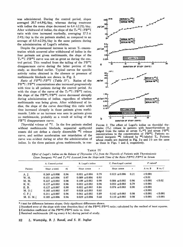
Despite the pronounced increase in serum T4 concen-tration which occurred after withdrawal of iodine in thefour patients not given methimazole, the slope of theT4-1"I: PB"'I curve was not as great as during the con-trol period. This resulted from the tailing of the PB"31Idisappearance curve during the latter portion of thestudy, as described earlier. Typical curves for specificactivity ratios obtained in the absence or presence ofmethimazole blockade are shown in Fig. 3.
Ratio of PB1I51: PB"I1 (Table IV). Ratios of thePB'I: PB1'I concentrations also increased progressivelywith time in all patients during the control period. Aswith the slope of the curve of the T4-'I: PB"..I ratios,the slope of the PB'I/PB'I curve decreased abruptlyduring administration of iodine, regardless of whethermethimazole was being given. After withdrawal of io-dine, the slope of the curve describing this ratio withtime increased abruptly in those patients given methi-mazole. Little or no increase was seen in patients givenno methimazole, probably as a result of tailing of thePB"31I disappearance curve.
Thyroidal release of "'I1. In the five patients studiedwithout methimazole blockade, sequential epithyroidcounts did not define a clearly discernible 'I releasecurve, and neither acceleration nor retardation of thecurve was evident during or after the administration ofiodine. In the three patients given methimazole, in con-
.-9) [SERUMT*-'7i][SERUM P1S1'I]
)c I/AC
. I * . .
a lo
(CF),6040 _25 _
#9/1ACv10 _
1.50
0.50 -
0.250.1 8 -
.I . ..
I5 20 25 30DURATIONOF STUDY (DAYS)
[SERUMT-""TIJ[SERUMP@"'3]
[ERUM PBl"I][SERUM PB131 ]
y7/X, LUV60t 'i'As
I . . 1. . . .
b 10 15 20 25DURATIONOF STUDY (DAYS)
FIGURE 3 The effect of Lugol's iodine on thyroidal thy-roxine (To) release in patients with hyperthyroidism asjudged from the ratios of serum T4-'I and serum PB'Iconcentrations to the concentration of PBmI. Patients re-ceived inorganic 'I followed by MIIlabeled T4. Patientswhose results are depicted in Fig. 3 a and 3 b are the sameas those in Figs. 1 and 2, respectively.
TABLE IVEffect of Lugol's Iodine on the Release of Thyroxine (T4) from the Thyroids of Patients with Thyrotoxicosis
Gizven Inorganic 125I and T4-1311 Assessed from the Slope with Time of the Ratio PB125I: PB1311I in Serum
A. Control period B. Lugol's iodine C. Post-Lugol's period P values*
Patient Slope ISEM: r§ Slope ISEM r Slope ±SEM r A vs. B B vs. C
A. J. 0.249 ±0.008 0.96 0.051 +0.004 0.79 0.023 ±0.006 0.21 <0.001W. H. 0.151 ±0.004 0.97 0.089 ±0.004 0.93 <0.01M. M. 0.227 ±0.003 0.98 0.109 ±0.002 0.99 0.084 ±0.002 0.96 <0.001 <0.02K. K. 0.127 ±0.003 0.96 0.052 ±0.002 0.96 0.053 ±0.003 0.79 <0.001E. B. 0.127 ±0.007 0.90 0.052 ±0.002 0.84 0.074 ±0.003 0.90 <0.001M. D.II 0.185 ±0.003 0.97 0.028 ±0.003 0.65 <0.001C. F.jj 0.211 ±0.007 0.96 0.041 ±0.002 0.89 0.142 ±0.003 0.98 <0.001 <0.001M. D. M.jj 0.185 ±0.006 0.94 0.029 ±0.004 0.48 0.110 ±0.002 0.98 <0.001 <0.001
* t test for difference between slopes. Only significant differences shown.t Standard error of the slope with time (fraction/day) of the PB125I: PB131I ratio; calculated by the method of least squares.
§ Correlation coefficient of the PB125I PB131I ratio vs. time.Received methimazole (30 mgevery 6 hr) during period of study.
82 L. Wartofsky, B. J. Ransil, and S. H. Ingbar

trast, a clearly exponential release curve for 125I in thethyroid was evident during the control period. As hasbeen previously described, this slowed abruptly duringiodine administration and accelerated after its with-drawal (1-3).
Estimate of fractional inhibition of T4-"'I release byiodine. The formulation described in the Appendix pro-
vides an estimate of the rate of fractional To release fromthe thyroid during the period of iodine administration,relative to that present during the control period. Theformulation rests on the assumption that thyroidal T,exists in a single pool and does not increase in amount
as a result of iodine administration. The former as-
sumption cannot be validated, but probably the latter as-
sumption is nearly the case in patients receiving anti-thyroid drugs. In patients not receiving antithyroiddrugs, thyroidal content of Ti-mI almost certainly didincrease during administration of iodine. Even so, theassumption of constancy of thyroidal T4&-9I would leadto an underestimate of the extent to which fractionalsecretion rate for T4-'I had been decreased.
In five patients studied in the absence of methimazoleblockade, the calculated percentage decrease in frac-tional T4-mI release rate averaged 74.1 ±+12.8. In thethree patients who were studied during methimazoleblockade, the estimated percentage decreases in frac-tional T4-7I release rates were 61.2, 63.4, and 87.3,respectively (Table V).
DISCUSSION
The striking discrepancy between the speed with whichiodine often ameliorates the manifestations of thyro-
toxicosis and the generally delayed response to antithy-roid drugs suggests that these agents differ in their basicmechanisms of action. It has been presumed that theearly action of iodine can be explained by an inhibitionof the release of hormone from the thyroid gland, a sug-
gestion which is supported by numerous demonstrationsof a rapid decline in serum protein-bound iodine (PBI)after iodine administration. This conclusion was
strengthened by the findings of Goldsmith and Eisele(1), who employed direct epithyroid counting to demon-strate that in patients with hyperthyroidism who were
receiving antithyroid drugs, iodine abruptly decreasedthe rate of loss of 'I from the thyroid gland. Thesefindings were subsequently confirmed in other studiessimilarly conducted (2, 3, 5). More recently, however,the observations of Mitchell, Bradford, and Gilboa (4)have raised some doubt as to whether iodine does indeedinhibit the release of thyroidal radioiodine and, by in-ference, of T4. Among 16 patients studied in the absenceof antithyroid blockade, eight displayed an apparent ac-
celeration of radioiodine release during iodine adminis-tration, while the remaining eight showed no effect.
This discrepancy highlights certain shortcomings ofthe serial epithyroid counting technique as an indexof the rate of thyroid hormone release. Accurate mea-
surement of glandular radioiodine release rates, es-
pecially in hyperthyroid patients, requires administra-tion of antithyroid agents to prevent secondary reac-
cumulation of radioiodine which has already traversedthe thyroid, been secreted as hormone, and been liberatedas iodide by peripheral deiodination. Such recyclingwould retard and obscure the primary glandular radio-
TABLE VInhibition by Lugol's Iodine of the Fractional Rate of Release of Thyroxine (T4) from
the Thyroids of Patients with Thyrotoxicosis
Per centFractional inhibition of
Control Serum T4 at Days to rate of T4 fractional T4Patient serum T4 time t time t* turnover; k release rate:
ig/100 ml Ag/100 ml %/day
A. J. 15.8 9.0 6.5 16.6 65.0W. H. 10.8 8.0 4.0 13.4 63.0M. M. 20.5 14.0 3.5 20.0 63.0K. K. 21.0 11.0 6.0 13.0 87.8E. B. 20.0 11.0 4.0 16.9 91.5M. D.§ 15.1 8.5 6.0 20.8 61.2C. F.§ 13.2 8.5 5.0 15.7 63.4M. D. M.§ 17.0 11.5 4.0 11.6 87.3
Mean ±SEM 16.7 ±1.2 10.2 40.7 4.9 ±0.4 16.0 ±1.1 72.8 ±4.4
* Approximate duration of Lugol's iodine therapy at which serum T4 concentration no longercontinued to decrease.T See Appendix for method of calculation from the primary data shown in this table.§ Received methimazole (30 mgevery 6 hr) during period of study.
Inhibition of T. Secretion by Iodine 83

iodine release curve. In studies of the effects of iodine inthe absence of antithyroid blockade, inhibition by stableiodine of radioiodine reaccumulation would tend to ac-celerate the net loss of "31I from the thyroid and couldthereby obscure any slowing in primary 'lI releasewhich iodine might produce. On the other hand, asMitchell and coworkers suggested, it is possible thatantithyroid agents alter intrathyroidal iodine metabolismin such a way as to change the response of over-all "Irelease rates to either pharmacological agents or disease,thus explaining the apparently different effects of iodinein the presence or absence of methimazole blockade(4). Other shortcomings of the serial epithyroid count-ing technique are also apparent. It provides no informa-tion concerning the nature of the iodinated materialsbeing released, an important consideration since io-dinated materials other than T4 are released from boththe normal and diseased thyroid gland (14 6). Finally, thetechnique makes no allowance for the specific activity ofthe radioiodinated materials released, a considerationwhich becomes particularly cogent in studies of the ef-fects of iodine administration.
The method which we have described is free of theshortcomings of serial epithyroid counting as a tech-nique for demonstrating changes in the rate of T4 re-lease, and involves few, if any, intrinsic assumptions.Basically, we have demonstrated that Lugol's iodine6abruptly decreases the serum concentration of T4-'"I inpatients with hyperthyroidism and that this cannot beexplained by a change in the distribution or turnoverof the hormone in the periphery, since the metabol sm ofexogenous "'I-labeled T4 was unaffected. This responsewas observed irrespective of whether complete blockingdoses of antithyroid drugs were given before the ad-ministration of iodine. Hence, the absolute rate of T4-"2I secretion must have been decreased by iodine if,as seems most likely, the exogenous T4-131I is a suitabletag for the metabolism of T4-17I secreted by the thyroid.
A more quantitative evaluation of the influence ofiodine on the rate of T4 secretion can be obtained by anexamination of the curves depicting the change with timeof the ratio of T4-17I: PB..1T. As would be expected fromthe constancy of T4-1'I and the exponential decline inPB 3..L the ratios increased exponentially during the con-trol phase. Had iodine produced a complete inhibitionof T4 release, the serum T4-17I concentration would havedecreased at the same exponential rate as did the concen-tration of exogenously labeled PB13`I. Hence, the slope ofthe curve depicting the change with time in the ratio ofT4-'I: PB13.I would have been zero. In the entire group
6Wartofsky, L., and S. H. Ingbar. To be published.'The effects herein demonstrated should not be construed
as being due to iodine per se, since they can be reproducedby pharmacological doses of inorganic iodine.
of eight patients in the present study, the slope of theratio averaged 20.7 ±4.8%/day during the control pe-riod and decreased to 6.4 +2.2%/day during iodine ad-ministration. The lowest individual value for the slopeobserved during iodine administration was 1.3%/day,indicating almost complete inhibition of T4 secretion.
A decrease in T4 secretion induced by iodine couldhave come about in several ways: an inhibition of T4synthesis, a decrease in the fractional rate of T4 release,or both. The ability of iodine acutely to inhibit T4 syn-thesis (Wolff-Chaikoff effect) is well known and hasbeen demonstrated to occur in patients with thyrotoxi-cosis (15, 16). This effect is usually transient (16), andsuch transiency might be considered as evidence againstits primary role in decreasing T4 secretion. On the otherhand, the transient nature of the Wolff-Chaikoff effectcould be taken to be responsible for the observation thatserum T4 concentration generally did not continue to fallduring the entire period of iodine administration. Nev-ertheless, several lines of evidence indicate that even ifa decrease in synthesis occurs during iodine administra-tion, the major effect of iodine is to inhibit the mecha-nism by which T4 is released. First, very large doses ofmethimazole do not produce the sharp decl;ne in serumT4 or PBI that is produced by iodine. This differenceis evident in the methimazole-treated patients in thepresent study, as well as in earlier studies of the periph-eral turnover of T4 in thyrotoxic patients in whommethimazole was given to prevent recycling of iodine(9).
A second line of evidence is provided by the responseof the serum T4 to withdrawal of iodine in methimazole-treated and untreated groups. In the former group, se-rum T4-'I concentration remained constant or increasedonly slightly in the 4-6 days after withdrawal of iodine.In the patients who had received no methimazole, incontrast, withdrawal of iodine was followed both by arapid rise in T4-1271I concentration to values characteristicof thyrotoxicosis and by reappearance of clinical mani-festations. If iodine had acted only to inhibit hormonalsynthesis, the response to its withdrawal should havebeen uninfluenced by concomitant methimazole adminis-tration. The rapid increase in serum T4 which occurredafter iodine was withdrawn from patients given nomethimazole is more consistent with restoration of arapid fractional T4 release from a pool which had re-mained unchanged, or even increased, during iodine ad-ministration.
The third, and most important line of evidence derivesfrom observations of the endogenously-labeled PBI(PB"1I). In the present technique, an intrathyroidalpool of "SI-labeled T was present before iodine adminis-tration. As noted earlier, administration of iodine wasnot accompanied by an abrupt decrease in thyroidal con-
84 L. Wartofsky, B. J. Ransil, and S. H. Ingbar

TABLE VIEstimated Values for the Thyroid Content of Thyroxine (T4) Based on the Assumption of
Complete Inhibition of T4 Synthesis by Lugol's Iodine*
Estimated EstimatedTi release Estimated T4 thyroid Predicted T4
Patient rate; r content, Vo weight content; Vpt Vp/Vo
%/day pg T4 g pg T4A. J. 32.3 706 100 30,800 43.6W. H. 53.7 431 35 10,780 25.0M. M. 60.3 687 65 20,020 29.1E. B. 224.7 150 50 15,400 103.0K. K. 103.7 250 25 7,700 30.8M. D.§ 29.9 1301 30 9,240 7.1-C. F.§ 45.0 589 40 12,320 20.9M. D. M.§ 164.1 159 35 10,780 67.8
* Estimations of thyroid content of T4 (Vo) and fractional rate of T4 release (r) based on for-mulation and assumptions presented in Appendix and upon values for serum T4 concen-tration and duration of Lugol's iodine therapy shown in Table V.I Based upon the estimated gland weight and upon analyses by Braasch, Albert, Keating,and Black (17), which revealed a mean T4 concentration of 20 ,ug T4 iodine/100 mgwet wtin untreated diffuse toxic goiter.J Received methimazole (30 mgevery 6 hr) during period of study.
tent of HI. Hence, the abrupt decrease in T4-'I secretionduring iodine administration, evident from an examina-tion of PB'I: PBmI ratios, is best explained by a de-crease in the fractional rate of T4-'I release, and not adecrease in its synthesis.
Finally, the Appendix presents a mathematical formu-lation relating the size of the intrathyroidal T4 pool andits fractional rate of release to the size and turnoverrate of the extrathyroidal T4 pool. As indicated above,the formulation has demonstrated a marked 'reductionin the fractional rate of T. release during iodine ad-ministration, assuming no change in the intrathyroidalT4 pool (Table V). The same formulation can be em-ployed to calculate the content of the glandular To poolwhich must have been present had iodine decreased theserum To to the observed extent, not by affecting itsfractional rate of release, but solely by inhibiting new Tlsynthesis (see Appendix). The values thereby derivedare far below available estimates of thyroidal T4 contentin Graves' disease (17), making the assumption of apredominant effect of iodine on synthesis rather thanrelease of To highly unlikely (Table VI).
For these reasons we would conclude that a decreasein the fractional rate of To release is at least the majorreason that iodine acutely decreases To secretion, therebydecreasing serum T. and ameliorating the clinical mani-festations in patients with thyrotoxicosis. Nevertheless,several aspects of the action of iodine in such patientsremain unclear. It is uncertain why, after a few days oftreatment, T. secretion rate is adjusted to maintain aeuthyroid state and a normal concentration of serum T4.
Also unexplained is the later reemergence of thyrotoxi-cosis in many patients, despite continued iodine adminis-tration (16). Additional and more prolonged observa-tions will be required to clarify the origin of these re-sponses.
APPENDIXLet
Vo = glandular content of thyroxine (T4) during the controlperiod;
V = glandular content of T4 at time t;P0 = T4 content of peripheral T4 pool during the control
period;P = T4 content of the peripheral T4 pool at time t;t = time after initiation of Lugol's iodine therapy;
ro = fractional rate of glandular T4 release during thecontrol period (day-1);
r = fractional rate of glandular T4 release during Lugol'siodine therapy; and
k = fractional rate of turnover of peripheral T4 pool (un-changed by Lugol's iodine therapy).
The equations governing changes in the content of theperipheral T4 pool (P) are
r kV -+P
dp/dt = rV - kPDuring the control period a steady state exists; hence
dp/dt = rV - kP = 0and
(1)(2)
(3)
roVo = kPo (4)
A. If Lugol's iodine therapy is introduced, and if Lugol'siodine does not change the glandular pool of TW(V = Vo), butchanges only the fractional release rate for T4, then
dp/dt = rVo - kP (5)
Inhibition of To Secretion by Iodine 85

The solution for this equation is
P = eko [NO (1 e-kt) + Po] (6)Rearranging:
k (P Poekt) (7)
Under these conditions, from equations 7 and 4, the ratio offractional release rates
ro =_ (1- kt)r (P/Po - ekt) (8)
B. On the other hand, it can be assumed that the only actionof Lugol's iodine is to inhibit the synthesis of T4. The equationdescribing the changes in the content of the peripheral T4 poolunder these conditions has been derived and presented in anearlier publication (18).
P = e-kt [(rVe ;r)))+ PO - k 1 (9)Rearranging:
P = edkt [kiO (e(k-r)t-1) +Pr (10)k --r)Since the assumption dictates that during Lugol's iodine
therapy r = ro, equations 4 and 10 can be solved simultane-ously to determine the two unknowns, ro and Vo.Equation 10 is rearranged to yield
from whence
Let
Pekt = krO (e(k-r)t - 1) + Po
rVo-P= k-PO r (e (k-r)t - 1)
f X = k-r. (13)By substitution into equation 12
pekl Po =rVo (ext 1) (14)
WhencePek - Po ext - 1 (15)
rVo XLet
Pekt - Po(16)rVo
Substituting into equation 15 and rearranging, we arrive at thetranscendental equation
ext-YX=1 (17)Equation 14 is solved for the root Y. Since k is known, r(= ro)can be determined from equation 13 and Vo from equation 4.
ACKNOWLEDGMENTSThis work was supported in part by Research Grant No.AM-09753 from the National Institute of Arthritis andMetabolic Diseases and by Grant No. FR-76 from theDivision of Health Resources and Facilities, National Insti-tutes of Health, Bethesda, Md.
REFERENCES1. Goldsmith, R. E., and M. L. Eisele. 1956. The effect of
iodide on the release of thyroid hormone in hyperthyroid-ism. J. Clin. Endocrinol. Metab. 16: 130.
2. Greer, M. A., and L. J. DeGroot. 1956. The effect ofstable iodide on thyroid secretion in man. Metabolism.5: 682.
3. Solomon, D. H. 1956. Factors affecting the fractionalrate of release of radioiodine from the thyroid glandin man. Metabolism. 5: 667.
4. Mitchell, M. L., A. H. Bradford, and Y. Gilboa. 1966.Paradoxical response of the unblocked hyperthyroidgland to iodide. J. Clin. Endocrinol. Metab. 26: 639.
5. Goldsmith, R., C. Herbert, and G. Lutsch. 1958. Theeffect of iodide on the release of thyroid hormone inhyperthyroidism: further observations. J. Clin. Endo-crinol. Metab. 18: 367.
6. Ansell, G., and H. Miller. 1952. Influence of iodine onthe release of thyroid hormone in thyrotoxicosis. Lancet.2: 5.
7. Volpe, R., and M. W. Johnston. 1962. The effect of smalldoses of stable iodine in patients with hyperthyroidism.Ann. Intern. Med. 56: 577.
8. Murphy, B. E., C. J. Pattee, and A. Gold. 1966. Clinicalevaluation of a new method for the determination ofserum thyroxine. J. Clin. Endocrinol. Metab. 26: 247.
9. Ingbar, S. H., and N. Freinkel. 1955. Simultaneous esti-mation of rates of thyroxine degradation and thyroidhormone synthesis. J. Clin. Invest. 34: 808.
10. Ingbar, S. H., and N. Freinkel. 1960. Regulation of theperipheral metabolism of the thyroid hormones. Re-cent Progr. Hormone Res. 16: 353.
11. Snedecor, G. W., and W. G. Cochran. 1967. StatisticalMethods. Iowa State Univ. Press, Ames. 6th edition.
12. Sterling, K., and R. B. Chodos. 1956. Radiothyroxineturnover studies in myxedema, thyrotoxicosis, and hy-permetabolism without endocrine disease. J. Clin. In-vest. 35: 806.
13. Ingbar, S. H., and N. Freinkel. 1958. Studies of thyroidfunction and the peripheral metabolism of II-labeledthyroxine in patients with treated Graves' disease. J.Clin. Invest. 37: 1603.
14. DeGroot, L. J. 1966. Kinetic analysis of iodine metabo-lism. J. Clin. Endocrinol. Metab. 26: 149.
15. Childs, D. S., Jr., F. R. Keating, Jr., J. E. Rall, M. M.D. Williams, and M. H. Power. 1950. The effect ofvarying quantities of inorganic iodide (carrier) on theurinary excretion and thyroidal accumulation of radio-iodine in exophthalmic goiter. J. Clin. Invest. 29: 726.
16. Harden, R. McG., D. A. Koutras, W. D. Alexander, andE. J. Wayne. 1964. Quantitative studies of iodine metab-olism in iodide-treated thyrotoxicosis. Clin. Sci. 27: 399.
17. Braasch, J. W., A. Albert, F. R. Keating, Jr., and B. M.Black. 1955. A note on the iodinated constituents of nor-mal thyroids and of exophthalmic goiters. J. Clin. En-docrinol. Metab. 15: 732.
18. Ingbar, S. H., and N. Freinkel. 1956. ACTH, cortisone,and the metabolism of iodine. Metabolism. 5: 652.
86 L. Wartofsky, B. 1. Ransil, and S. H. Ingbar


