Ingegneria delle tecnologie per la salute · system-controlled internal...
85
Ingegneria delle tecnologie per la salute Fondamenti di anatomia e istologia Il sistema genito-urinario
Transcript of Ingegneria delle tecnologie per la salute · system-controlled internal...

Ingegneria delle tecnologie per la salute
Fondamenti di anatomia e istologia
Il sistema genito-urinario

Ingegneria delle tecnologie per la salute
Fondamenti di anatomia e istologia
Il sistema urinario
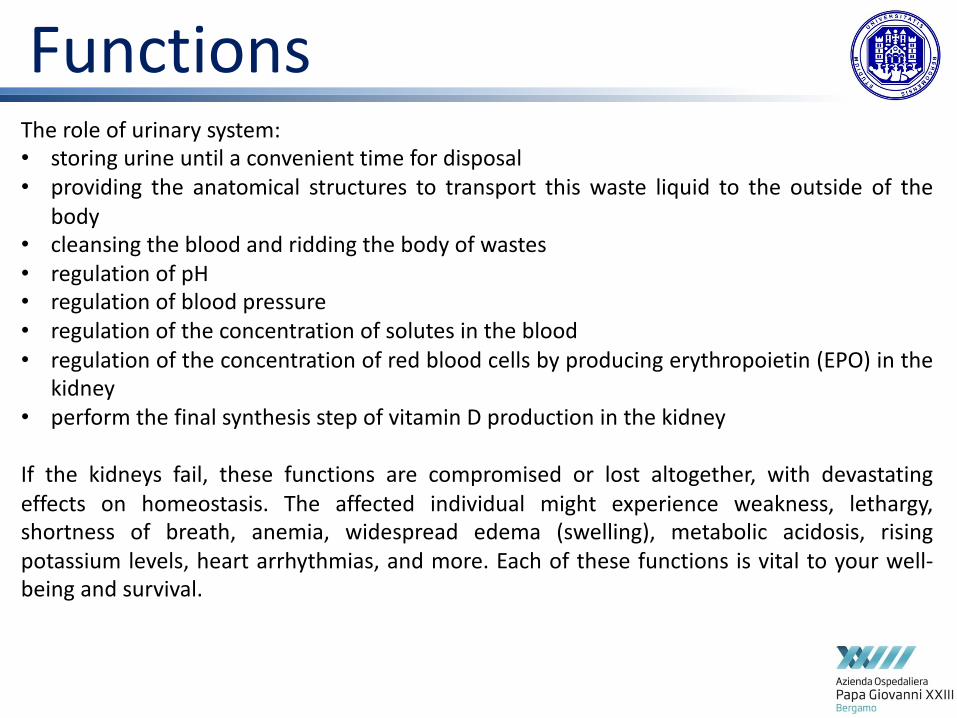
The role of urinary system:• storing urine until a convenient time for disposal• providing the anatomical structures to transport this waste liquid to the outside of the
body• cleansing the blood and ridding the body of wastes• regulation of pH• regulation of blood pressure• regulation of the concentration of solutes in the blood• regulation of the concentration of red blood cells by producing erythropoietin (EPO) in the
kidney• perform the final synthesis step of vitamin D production in the kidney
If the kidneys fail, these functions are compromised or lost altogether, with devastatingeffects on homeostasis. The affected individual might experience weakness, lethargy,shortness of breath, anemia, widespread edema (swelling), metabolic acidosis, risingpotassium levels, heart arrhythmias, and more. Each of these functions is vital to your well-being and survival.
Functions

The urinary system’s ability to filter the blood resides in about 2 to 3 million tufts ofspecialized capillaries—the glomeruli—distributed more or less equally between the twokidneys. Because the glomeruli filter the blood based mostly on particle size, large elementslike blood cells, platelets, antibodies, and albumen are excluded. The glomerulus is the firstpart of the nephron, which then continues as a highly specialized tubular structureresponsible for creating the final urine composition. The glomeruli create about 200 liters offiltrate every day, yet you excrete less than two liters of waste you call urine.
Characteristics of the urine change,depending on influences such aswater intake, exercise, environmentaltemperature, nutrient intake, andother factors. Some of thecharacteristics such as color and odorare rough descriptors of your state ofhydration.
Urine
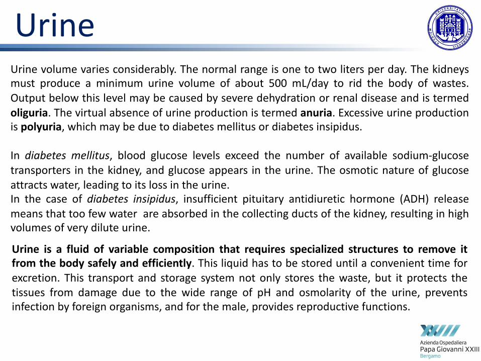
Urine volume varies considerably. The normal range is one to two liters per day. The kidneysmust produce a minimum urine volume of about 500 mL/day to rid the body of wastes.Output below this level may be caused by severe dehydration or renal disease and is termedoliguria. The virtual absence of urine production is termed anuria. Excessive urine productionis polyuria, which may be due to diabetes mellitus or diabetes insipidus.
In diabetes mellitus, blood glucose levels exceed the number of available sodium-glucosetransporters in the kidney, and glucose appears in the urine. The osmotic nature of glucoseattracts water, leading to its loss in the urine.In the case of diabetes insipidus, insufficient pituitary antidiuretic hormone (ADH) releasemeans that too few water are absorbed in the collecting ducts of the kidney, resulting in highvolumes of very dilute urine.
Urine
Urine is a fluid of variable composition that requires specialized structures to remove itfrom the body safely and efficiently. This liquid has to be stored until a convenient time forexcretion. This transport and storage system not only stores the waste, but it protects thetissues from damage due to the wide range of pH and osmolarity of the urine, preventsinfection by foreign organisms, and for the male, provides reproductive functions.

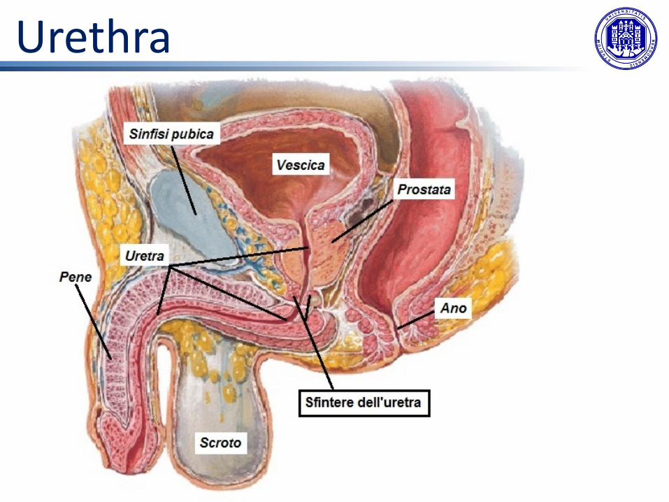
UrethraThe urethra transports urine from the bladder to the outside of the body for disposal. The urethra is the only urologic organ that shows any significant anatomic difference between males and females; all other urine transport structures are identical. The urethra in both males and females begins inferior and central to the two ureteral openings forming the three points of a triangular-shaped area at the base of the bladder called the trigone. The urethra tracks posterior and inferior to the pubic symphysis. In both males and females, the proximal urethra is lined by transitional epithelium, whereas the terminal portion is a nonkeratinized, stratified squamous epithelium. In the male, pseudostratified columnar epithelium lines the urethra between these two cell types.
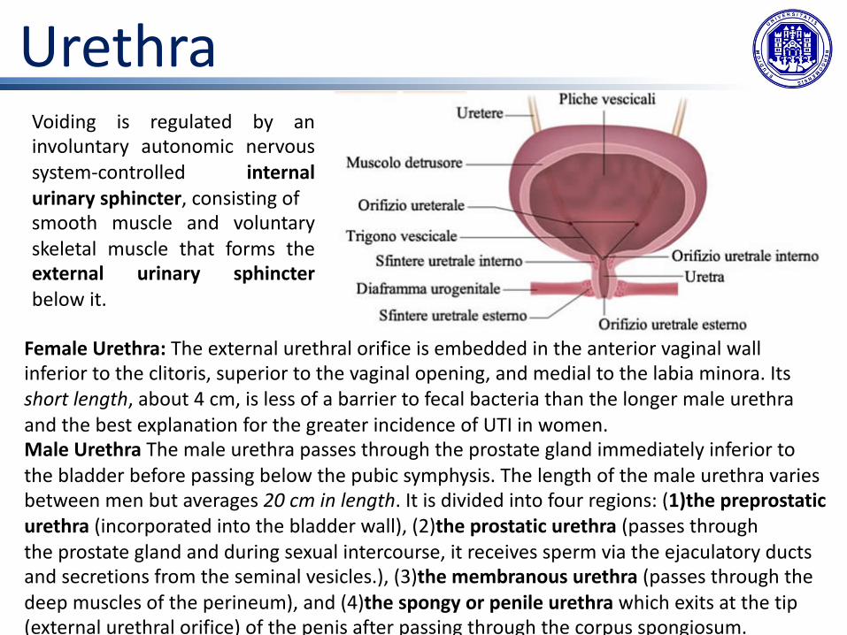
Voiding is regulated by aninvoluntary autonomic nervoussystem-controlled internalurinary sphincter, consisting ofsmooth muscle and voluntaryskeletal muscle that forms theexternal urinary sphincterbelow it.
Female Urethra: The external urethral orifice is embedded in the anterior vaginal wall inferior to the clitoris, superior to the vaginal opening, and medial to the labia minora. Its short length, about 4 cm, is less of a barrier to fecal bacteria than the longer male urethra and the best explanation for the greater incidence of UTI in women. Male Urethra The male urethra passes through the prostate gland immediately inferior to the bladder before passing below the pubic symphysis. The length of the male urethra varies between men but averages 20 cm in length. It is divided into four regions: (1)the preprostaticurethra (incorporated into the bladder wall), (2)the prostatic urethra (passes throughthe prostate gland and during sexual intercourse, it receives sperm via the ejaculatory ducts and secretions from the seminal vesicles.), (3)the membranous urethra (passes through the deep muscles of the perineum), and (4)the spongy or penile urethra which exits at the tip (external urethral orifice) of the penis after passing through the corpus spongiosum.
Urethra
Urethra

BladderThe urinary bladder collectsurine from both ureters. Thebladder lies anterior to theuterus in females, posterior tothe pubic bone and anterior tothe rectum. In males, theanatomy is similar, minus theuterus, and with the addition ofthe prostate inferior to thebladder. The bladder is partiallyretroperitoneal (outside theperitoneal cavity) with itsperitoneal-covered “dome”projecting into the abdomenwhen the bladder is distendedwith urine.
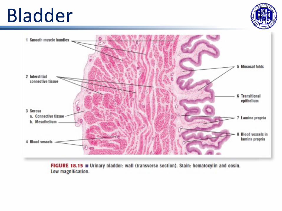
The bladder is a highly distensible organ comprised ofirregular crisscrossing bands of smooth muscle collectivelycalled the detrusor muscle. The interior surface is made oftransitional cellular epithelium that is structurally suited forthe large volume fluctuations of the bladder. When empty, itresembles columnar epithelia, but when stretched, it“transitions” (hence the name) to a squamous appearanceVolumes in adults can range from nearly zero to 500–600 mL.
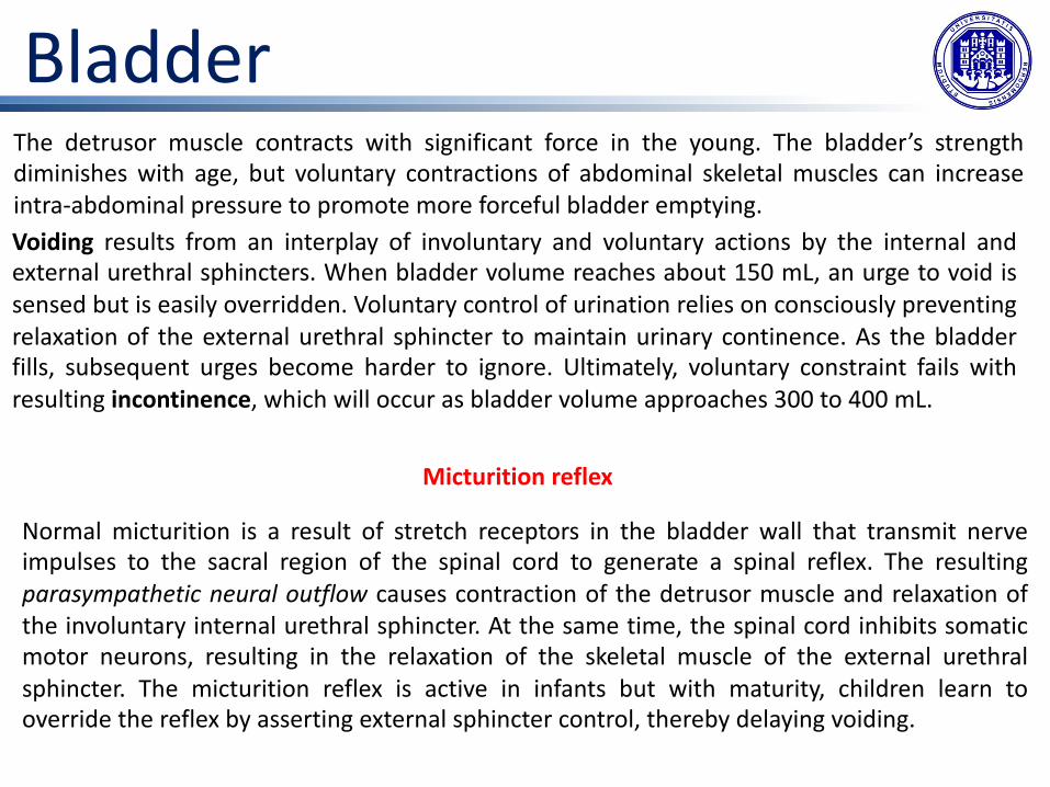
BladderThe detrusor muscle contracts with significant force in the young. The bladder’s strengthdiminishes with age, but voluntary contractions of abdominal skeletal muscles can increaseintra-abdominal pressure to promote more forceful bladder emptying.Voiding results from an interplay of involuntary and voluntary actions by the internal andexternal urethral sphincters. When bladder volume reaches about 150 mL, an urge to void issensed but is easily overridden. Voluntary control of urination relies on consciously preventingrelaxation of the external urethral sphincter to maintain urinary continence. As the bladderfills, subsequent urges become harder to ignore. Ultimately, voluntary constraint fails withresulting incontinence, which will occur as bladder volume approaches 300 to 400 mL.
Normal micturition is a result of stretch receptors in the bladder wall that transmit nerveimpulses to the sacral region of the spinal cord to generate a spinal reflex. The resultingparasympathetic neural outflow causes contraction of the detrusor muscle and relaxation ofthe involuntary internal urethral sphincter. At the same time, the spinal cord inhibits somaticmotor neurons, resulting in the relaxation of the skeletal muscle of the external urethralsphincter. The micturition reflex is active in infants but with maturity, children learn tooverride the reflex by asserting external sphincter control, thereby delaying voiding.
Micturition reflex
Bladder

Bladder
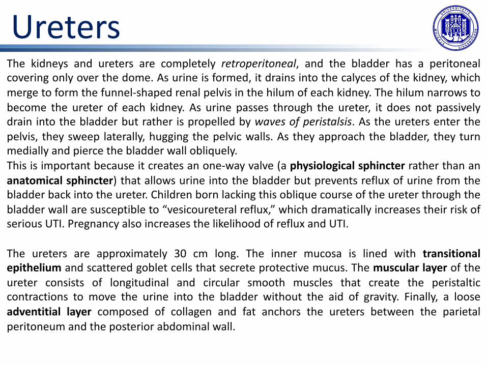
UretersThe kidneys and ureters are completely retroperitoneal, and the bladder has a peritonealcovering only over the dome. As urine is formed, it drains into the calyces of the kidney, whichmerge to form the funnel-shaped renal pelvis in the hilum of each kidney. The hilum narrows tobecome the ureter of each kidney. As urine passes through the ureter, it does not passivelydrain into the bladder but rather is propelled by waves of peristalsis. As the ureters enter thepelvis, they sweep laterally, hugging the pelvic walls. As they approach the bladder, they turnmedially and pierce the bladder wall obliquely.This is important because it creates an one-way valve (a physiological sphincter rather than ananatomical sphincter) that allows urine into the bladder but prevents reflux of urine from thebladder back into the ureter. Children born lacking this oblique course of the ureter through thebladder wall are susceptible to “vesicoureteral reflux,” which dramatically increases their risk ofserious UTI. Pregnancy also increases the likelihood of reflux and UTI.
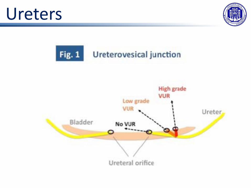
The ureters are approximately 30 cm long. The inner mucosa is lined with transitionalepithelium and scattered goblet cells that secrete protective mucus. The muscular layer of theureter consists of longitudinal and circular smooth muscles that create the peristalticcontractions to move the urine into the bladder without the aid of gravity. Finally, a looseadventitial layer composed of collagen and fat anchors the ureters between the parietalperitoneum and the posterior abdominal wall.
Ureters
Ureters

Ureters

Ureters

KidneyThe kidneys lie on either side of the spine in the retroperitoneal space between the parietal peritoneum and the posterior abdominal wall, well protected by muscle, fat, and ribs. They are roughly the size of your fist, and the male kidney is typically a bit larger than the female kidney. The kidneys are well vascularized, receiving about 25 percent of the cardiac output atrest.

KidneyThe left kidney is located at about the T12 to L3 vertebrae, whereas the right is lower due toslight displacement by the liver.Upper portions of the kidneys are somewhat protected by the eleventh and twelfth ribs.Each kidney weighs about 125–175 g in males and 115–155 g in females. They are about 11–14 cm in length, 6 cm wide, and 4 cm thick, and are directly covered by a fibrous capsulecomposed of dense, irregular connective tissue that helps to hold their shape and protectthem. This capsule is covered by a shock-absorbing layer of adipose tissue called the renal fatpad, which in turn is encompassed by a tough renal fascia. The fascia and, to a lesser extent,the overlying peritoneum serve to firmly anchor the kidneys to the posterior abdominal wallin a retroperitoneal position.

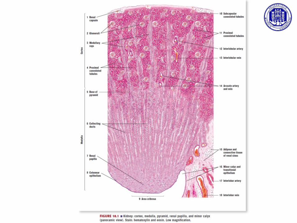
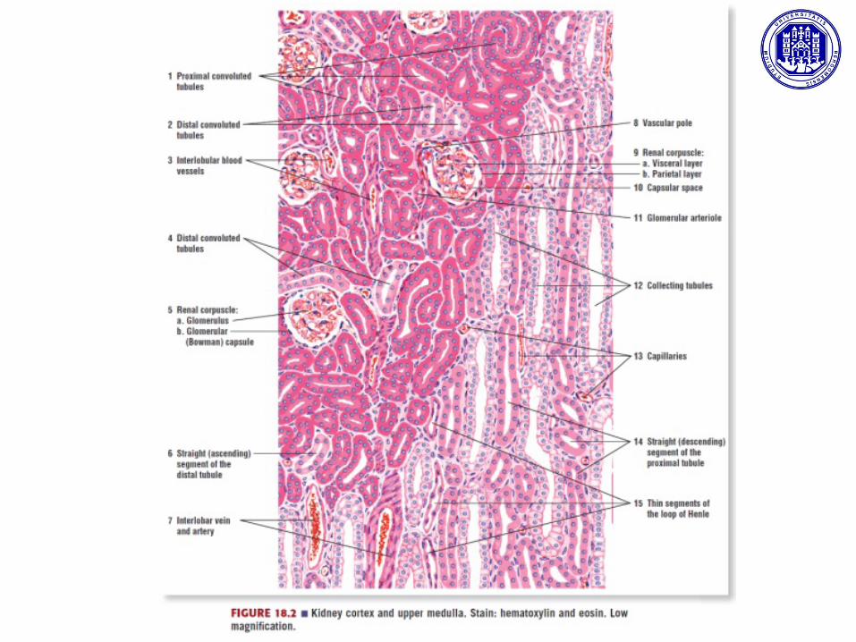
KidneyA frontal section through the kidney reveals an outer region called the renal cortex and an inner region called the medulla. The renal columns are connective tissue extensions that radiate downward from the cortex through the medulla to separate the most characteristic features of the medulla, the renal pyramids and renal papillae. The papillae are bundles of collecting ducts that transport urine made by nephrons to the calyces of the kidney for excretion. The renal columns also serve to divide the kidney into 6–8 lobes and provide a supportive framework for vessels that enter and exit the cortex. The pyramids and renal columns taken together constitute the kidney lobes.

KidneyThe renal hilum is the entry and exit site for structures servicing the kidneys:• the renal pelvis, which is formed from the major and minor calyxes in the kidney• the renal arteries form directly from the descending aorta,• the renal veins which return cleansed blood directly to the inferior vena cava.The artery, vein, and renal pelvis are arranged in an anterior-to-posterior order.
The renal artery first divides into segmental arteries, followed by further branching to forminterlobar arteries that pass through the renal columns to reach the cortex. The interlobararteries, in turn, branch into arcuate arteries, interlobular arteries, and then into afferentarterioles. The afferent arterioles service about 1.3 million nephrons in each kidney.
Nephrons are the “functional units” of the kidney; they cleanse the blood and balance theconstituents of the circulation. Nephrons are made of:• A renal corpuscle• A renal tubule
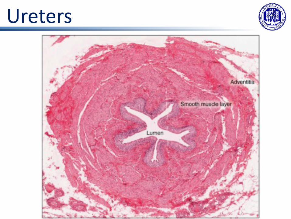
KidneyRenal corpuscle:The afferentarterioles form a tuftof high-pressurecapillaries about 200μm in diameter, theglomerulus. The restof the nephronconsists of acontinuoussophisticated tubulewhose proximal endsurrounds theglomerulus in anintimate embrace—this is Bowman’scapsule. Theglomerulus andBowman’s capsuletogether form therenal corpuscle.

KidneyRenal tubule:It is made of 3portion:• Proximal
convoluted tubule(PCT)
• Loop of Henle• Distal convolute
tubule (DCT)
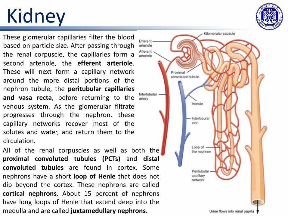
These glomerular capillaries filter the bloodbased on particle size. After passing throughthe renal corpuscle, the capillaries form asecond arteriole, the efferent arteriole.These will next form a capillary networkaround the more distal portions of thenephron tubule, the peritubular capillariesand vasa recta, before returning to thevenous system. As the glomerular filtrateprogresses through the nephron, thesecapillary networks recover most of thesolutes and water, and return them to thecirculation.
Kidney
All of the renal corpuscles as well as both theproximal convoluted tubules (PCTs) and distalconvoluted tubules are found in cortex. Somenephrons have a short loop of Henle that does notdip beyond the cortex. These nephrons are calledcortical nephrons. About 15 percent of nephronshave long loops of Henle that extend deep into themedulla and are called juxtamedullary nephrons.

Nephrons: The Functional UnitNephrons take a simple filtrate of the blood and modify it into urine.
They do this by accomplishing three principle functions—filtration, reabsorption, and secretion.
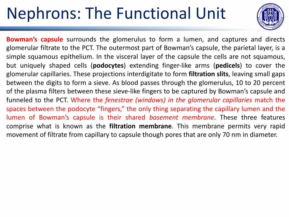
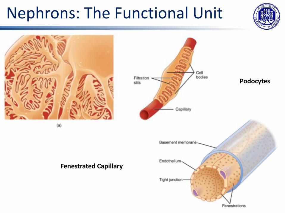
Nephrons: The Functional UnitBowman’s capsule surrounds the glomerulus to form a lumen, and captures and directsglomerular filtrate to the PCT. The outermost part of Bowman’s capsule, the parietal layer, is asimple squamous epithelium. In the visceral layer of the capsule the cells are not squamous,but uniquely shaped cells (podocytes) extending finger-like arms (pedicels) to cover theglomerular capillaries. These projections interdigitate to form filtration slits, leaving small gapsbetween the digits to form a sieve. As blood passes through the glomerulus, 10 to 20 percentof the plasma filters between these sieve-like fingers to be captured by Bowman’s capsule andfunneled to the PCT. Where the fenestrae (windows) in the glomerular capillaries match thespaces between the podocyte “fingers,” the only thing separating the capillary lumen and thelumen of Bowman’s capsule is their shared basement membrane. These three featurescomprise what is known as the filtration membrane. This membrane permits very rapidmovement of filtrate from capillary to capsule though pores that are only 70 nm in diameter.
Nephrons: The Functional Unit
Podocytes
Fenestrated Capillary
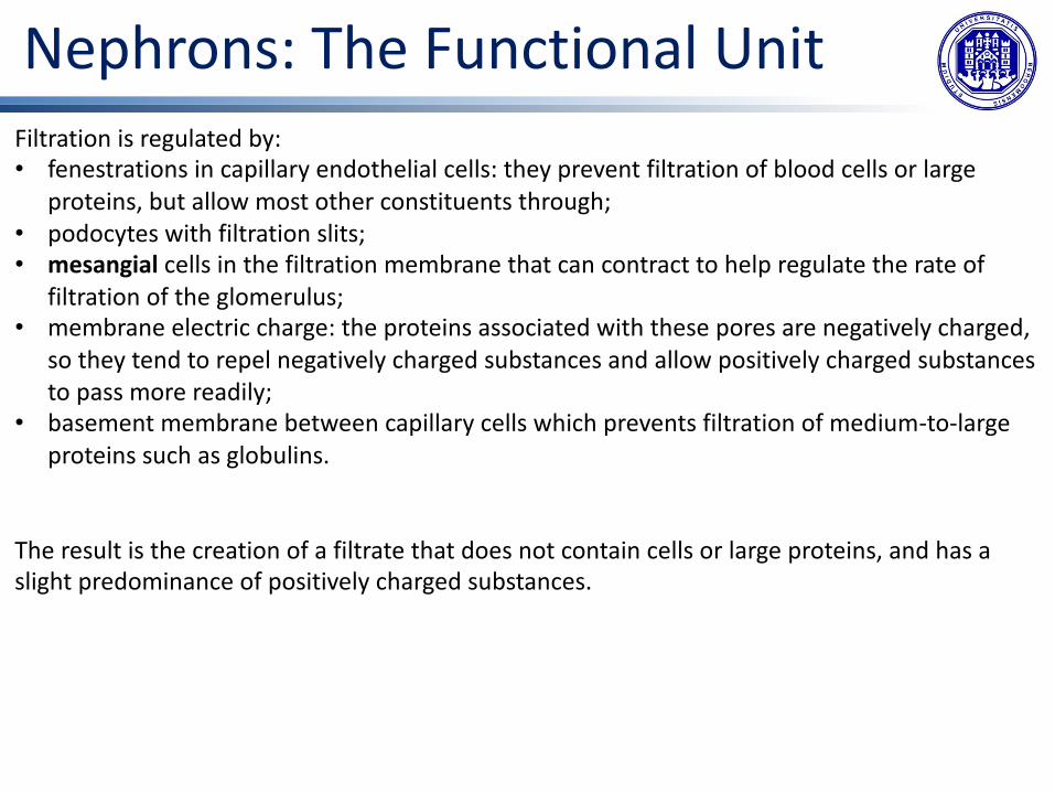
Nephrons: The Functional UnitFiltration is regulated by:• fenestrations in capillary endothelial cells: they prevent filtration of blood cells or large
proteins, but allow most other constituents through;• podocytes with filtration slits;• mesangial cells in the filtration membrane that can contract to help regulate the rate of
filtration of the glomerulus;• membrane electric charge: the proteins associated with these pores are negatively charged,
so they tend to repel negatively charged substances and allow positively charged substances to pass more readily;
• basement membrane between capillary cells which prevents filtration of medium-to-large proteins such as globulins.
The result is the creation of a filtrate that does not contain cells or large proteins, and has a slight predominance of positively charged substances.

Nephrons: The Functional UnitLying just outside Bowman’s capsule and the glomerulus is the juxtaglomerular apparatus (JGA). At the juncture where the afferent and efferent arterioles enter and leave Bowman’s capsule, the initial part of the distal convoluted tubule (DCT) comes into direct contact with the arterioles. The wall of the DCT at that point forms a part of the JGA known as the macula densa. This cluster of cuboidal epithelial cells monitors the fluid composition of fluid flowing through the DCT. In response to the concentration of Na+ in the fluid flowing past them, these cells release paracrine signals.
Nephrons: The Functional Unit
A second cell type in this apparatus is the juxtaglomerular cell. This is a modified, smoothmuscle cell lining the afferent arteriole that can contract or relax in response to paracrinesignals released by the macula densa. Such contraction and relaxation regulate blood flow tothe glomerulus.Ø If the osmolarity of the filtrate is too high (↑Na+, hyperosmotic), the juxtaglomerular cells
will contract, decreasing the glomerular filtration rate (GFR) so less plasma is filtered, leadingto less urine formation and greater retention of fluid. This will ultimately decrease bloodosmolarity toward the physiologic norm.
Ø If the osmolarity of the filtrate is too low (↓Na+), the juxtaglomerular cells will relax,increasing the GFR and enhancing the loss of water to the urine, causing blood osmolarity torise.
In other words, when osmolarity goes up, filtration and urine formation decrease and water isretained. When osmolarity goes down, filtration and urine formation increase and water is lostby way of the urine.

Active renin activate renin-angiotensin-aldosterone system which stimulates the release of the hormonealdosterone from the adrenal cortex. Aldosterone stimulates Na+ reabsorption by the kidney, which resultsin water retention and increased blood pressure.
A second function of themacula densa cells is toregulate renin release fromthe juxtaglomerular cells ofthe afferent arteriole.
Nephrons: The Functional UnitProximal Convoluted Tubule (PCT)Filtered fluid collected by Bowman’s capsule enters into the PCT. It is called convoluted due toits tortuous path. Simple cuboidal cells form this tubule with prominent microvilli on theluminal surface, forming a brush border. These microvilli create a large surface area tomaximize the absorption and secretion of solutes (Na+, Cl–, glucose, etc.), the most essentialfunction of this portion of the nephron. These cells actively transport ions across theirmembranes.
Loop of HenleThe descending and ascending portions of the loop of Henle (sometimes referred to as the nephron loop) are, of course, just continuations of the same tubule. They run adjacent and parallel to each other after having made a hairpin turn at the deepest point of their descent. The descending and ascending thick portion consists of simple cuboidal epithelium similar to that of the PCT. The descending and ascending thin portions consists of simple squamous epithelium. These are important differences, since different portions of the loop have different permeabilities for solutes and water.
Nephrons: The Functional UnitDistal Convoluted Tubule (DCT)The DCT, like the PCT, is very tortuous and formed by simple cuboidal epithelium, but it isshorter than the PCT. These cells are not as active as those in the PCT; thus, there are fewermicrovilli on the apical surface. However, these cells must also pump ions against theirconcentration gradient.
Collecting DuctsThe collecting ducts are continuous with the nephron but not technically part of it. In fact, eachduct collects filtrate from several nephrons for final modification. They are lined with simplesquamous epithelium with receptors for ADH.
When stimulated by ADH, these cells will insert aquaporinchannel proteins into their membranes, which as theirname suggests, allow water to pass from the duct lumenthrough the cells and into the interstitial spaces to berecovered by the vasa recta. This process allows for therecovery of large amounts of water from the filtrate backinto the blood. In the absence of ADH, these channels arenot inserted, resulting in the excretion of water in the formof dilute urine.


Ingegneria delle tecnologie per la salute
Fondamenti di anatomia e istologia
Il sistema riproduttivo maschile
Male Reproductive SystemA gamete is a specialized sex cell carrying 23 chromosomes—one half the number in bodycells. At fertilization, the chromosomes in one male gamete, called a sperm (orspermatozoon), combine with the chromosomes in one female gamete, called an oocyte.
The function of the male reproductive system is to produce sperm and transfer them to thefemale reproductive tract.
The paired testes are a crucial component in this process, as they produce both sperm andandrogens, the hormones that support male reproductive physiology. In humans, the mostimportant male androgen is testosterone. Several accessory organs and ducts aid the processof sperm maturation and transport the sperm and other seminal components to the penis,which delivers sperm to the female reproductive tract.
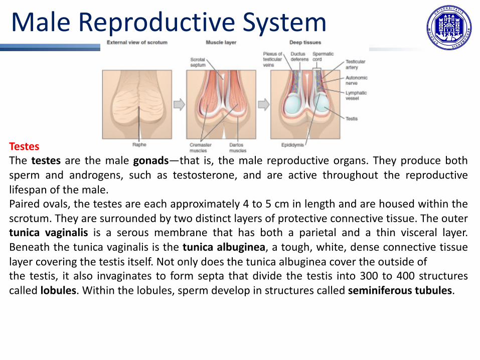
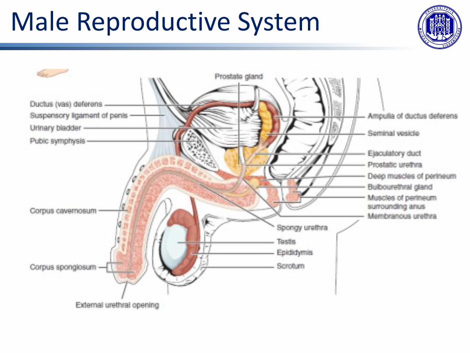
Male Reproductive SystemScrotumThe testes are located in a skin-covered, highly pigmented, muscular sack called the scrotum that extends from the body behind the penis. This location is important in sperm production, which occurs within the testes, and proceeds more efficiently when the testes are kept 2 to 4°C below core body temperature.The dartos muscle makes up the subcutaneous muscle layer of the scrotum. It continues internally to make up the scrotal septum, a wall that divides the scrotum into two compartments, each housing one testis. Descending from the internal oblique muscle of the abdominal wall are the two cremaster muscles, which cover each testis like a muscular net.By contracting simultaneously, the dartos and cremaster muscles can elevate the testes in cold weather (or water), moving the testes closer to the body and decreasing the surface area of the scrotum to retain heat. Alternatively, as the environmental temperature increases, the scrotum relaxes, moving the testes farther from the body core and increasing scrotal surface area, which promotes heat loss. Externally, the scrotum has a raised medial thickening on the surface called the raphae.
During the seventh month of the developmental period of a male fetus, each testis moves through the abdominal musculature to descend into the scrotal cavity. This is called the “descent of the testis.” Cryptorchidism is the clinical term used when one or both of the testes fail to descend into the scrotum prior to birth.
Male Reproductive System
TestesThe testes are the male gonads—that is, the male reproductive organs. They produce bothsperm and androgens, such as testosterone, and are active throughout the reproductivelifespan of the male.Paired ovals, the testes are each approximately 4 to 5 cm in length and are housed within thescrotum. They are surrounded by two distinct layers of protective connective tissue. The outertunica vaginalis is a serous membrane that has both a parietal and a thin visceral layer.Beneath the tunica vaginalis is the tunica albuginea, a tough, white, dense connective tissuelayer covering the testis itself. Not only does the tunica albuginea cover the outside ofthe testis, it also invaginates to form septa that divide the testis into 300 to 400 structurescalled lobules. Within the lobules, sperm develop in structures called seminiferous tubules.

Male Reproductive System
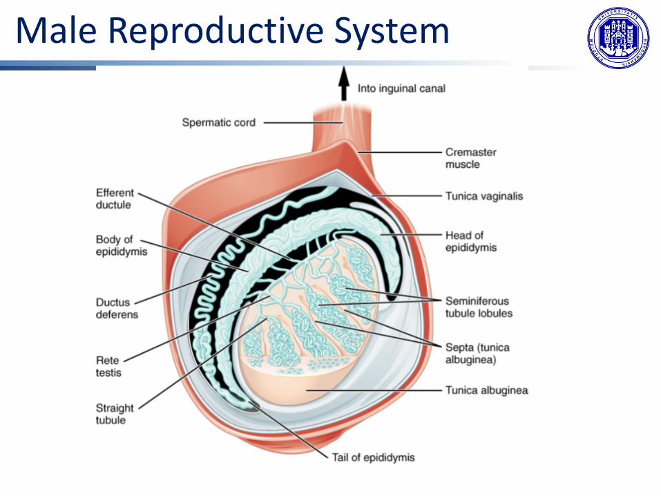
The tightly coiled seminiferous tubulesform the bulk of each testis. They arecomposed of developing sperm cellssurrounding a lumen, the hollow centerof the tubule, where formed sperm arereleased into the duct system of the testis.Specifically, from the lumens of theseminiferous tubules, sperm move intothe straight tubules (or tubuli recti), andfrom there into a fine meshwork oftubules called the rete testes. Sperm leavethe rete testes, and the testis itself,through the 15 to 20 efferent ductulesthat cross the tunica albuginea.
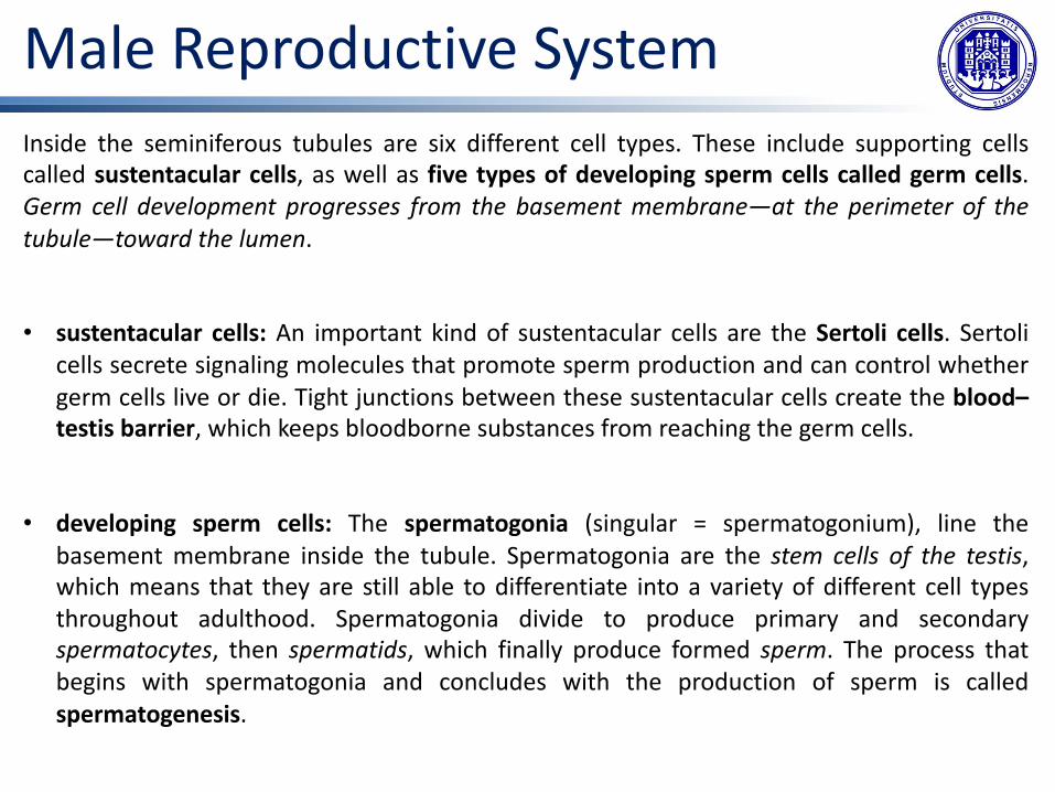
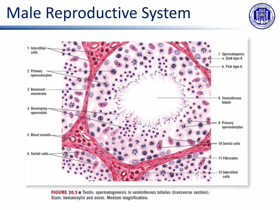
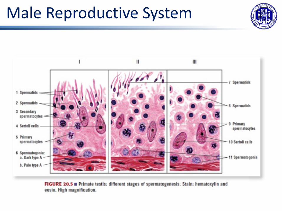
Male Reproductive SystemInside the seminiferous tubules are six different cell types. These include supporting cellscalled sustentacular cells, as well as five types of developing sperm cells called germ cells.Germ cell development progresses from the basement membrane—at the perimeter of thetubule—toward the lumen.
• sustentacular cells: An important kind of sustentacular cells are the Sertoli cells. Sertolicells secrete signaling molecules that promote sperm production and can control whethergerm cells live or die. Tight junctions between these sustentacular cells create the blood–testis barrier, which keeps bloodborne substances from reaching the germ cells.
• developing sperm cells: The spermatogonia (singular = spermatogonium), line thebasement membrane inside the tubule. Spermatogonia are the stem cells of the testis,which means that they are still able to differentiate into a variety of different cell typesthroughout adulthood. Spermatogonia divide to produce primary and secondaryspermatocytes, then spermatids, which finally produce formed sperm. The process thatbegins with spermatogonia and concludes with the production of sperm is calledspermatogenesis.

Male Reproductive System
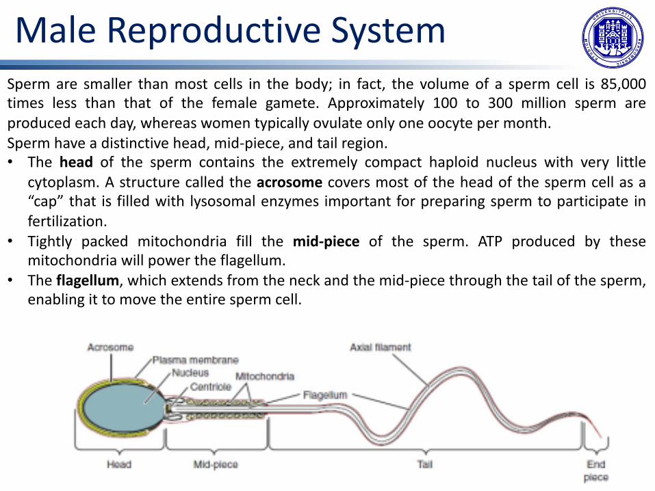
Male Reproductive SystemSperm are smaller than most cells in the body; in fact, the volume of a sperm cell is 85,000times less than that of the female gamete. Approximately 100 to 300 million sperm areproduced each day, whereas women typically ovulate only one oocyte per month.Sperm have a distinctive head, mid-piece, and tail region.• The head of the sperm contains the extremely compact haploid nucleus with very little
cytoplasm. A structure called the acrosome covers most of the head of the sperm cell as a“cap” that is filled with lysosomal enzymes important for preparing sperm to participate infertilization.
• Tightly packed mitochondria fill the mid-piece of the sperm. ATP produced by thesemitochondria will power the flagellum.
• The flagellum, which extends from the neck and the mid-piece through the tail of the sperm,enabling it to move the entire sperm cell.
Male Reproductive System

Male Reproductive System
Male Reproductive System
Male Reproductive System
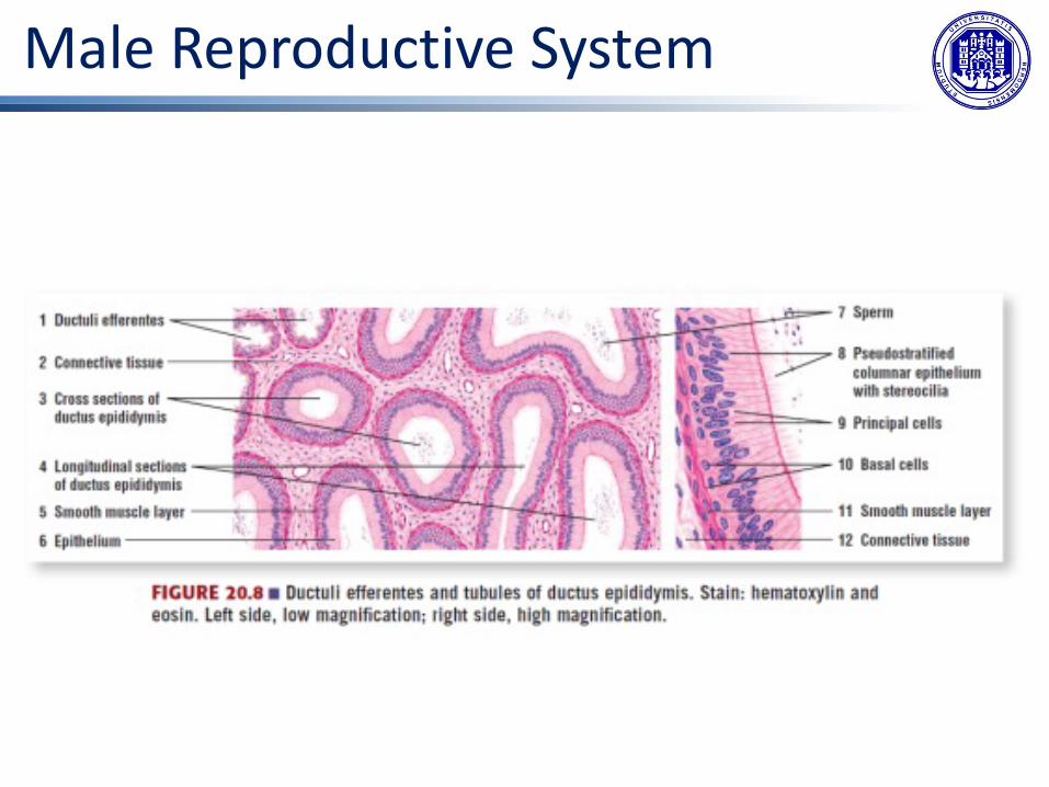
Male Reproductive SystemEpididymisFrom the lumen of the seminiferous tubules, the immotile sperm are surrounded by testicularfluid and moved to the epididymis, a coiled tube attached to the testis where newly formedsperm continue to mature. It would be approximately 6 m long if straightened. It takes anaverage of 12 days for sperm to move through the coils of the epididymis.Sperm enter the head of the epididymis and are moved along predominantly by the contractionof smooth muscles lining the epididymal tubes. As they are moved along the length of theepididymis, the sperm further mature and acquire the ability to move under their own power.Once inside the female reproductive tract, they will use this ability to move independentlytoward the unfertilized egg. The more mature sperm are then stored in the tail of theepididymis (the final section) until ejaculation occurs.Pseudstratified columnar epithelium with stereocilia.
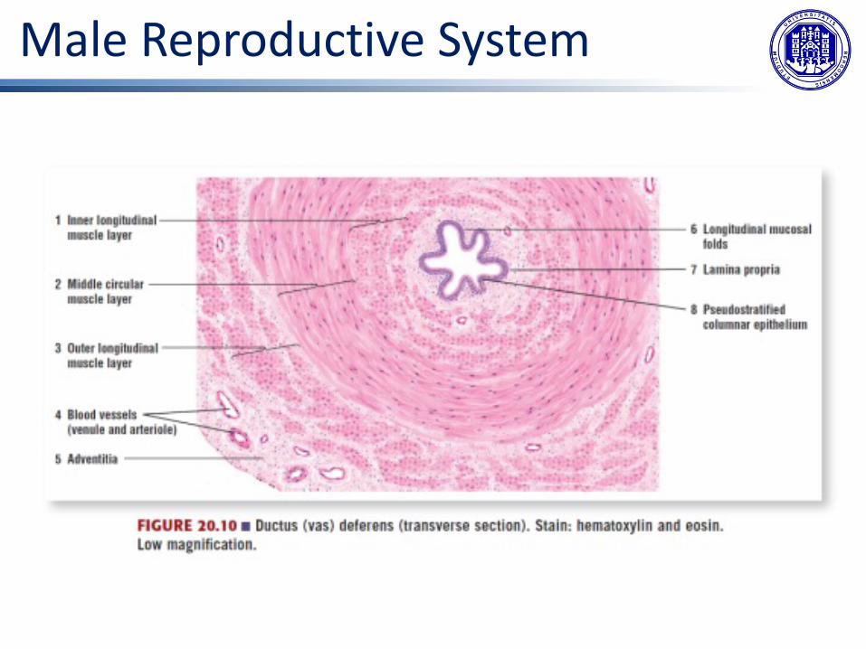
Duct SystemDuring ejaculation, sperm exit the tail of the epididymis and are pushed by smooth musclecontraction to the ductus deferens (also called the vas deferens). The ductus deferens is a thick,muscular tube that is in the spermatic cord. From each epididymis, each ductus deferensextends superiorly into the abdominal cavity through the inguinal canal in the abdominal wall.From here, the ductus deferens continues posteriorly to the pelvic cavity, ending posterior tothe bladder where it dilates in a region called the ampulla.
Male Reproductive System
Male Reproductive System
Male Reproductive System

Male Reproductive System
Male Reproductive System
Male Reproductive SystemSperm make up only 5 percent of the final volume of semen, the thick, milky fluid that the male ejaculates. The bulk of semen is produced by three critical accessory glands of the male reproductive system: the seminal vesicles, the prostate, and the bulbourethral glands.
Seminal VesiclesAs sperm pass through the ampulla of the ductus deferens at ejaculation, they mix with fluid from the associated seminal vesicle. The paired seminal vesicles are glands that contribute approximately 60 percent of the semen volume. Seminal vesicle fluid contains large amounts of fructose, which is used by the sperm mitochondria to generate ATP to allow movement through the female reproductive tract.The fluid, now containing both sperm and seminal vesicle secretions, next moves into the associated ejaculatory duct, a short structure formed from the ampulla of the ductus deferens and the duct of the seminal vesicle. The paired ejaculatory ducts transport the seminal fluid into the next structure, the prostate gland.

Male Reproductive System

Male Reproductive SystemProstate GlandThe centrally located prostate gland sits anterior to the rectum at the base of the bladdersurrounding the prostatic urethra (the portion of the urethra that runs within the prostate).The prostate is formed of both muscular and glandular tissues. It excretes an alkaline, milkyfluid to the passing seminal fluid that is critical to the consistence of semen followingejaculation. The prostate normally doubles in size during puberty. At approximately age 25, itgradually begins to enlarge again. This enlargement does not usually cause problems;however, abnormal growth of the prostate, or benign prostatic hyperplasia (BPH), can causeconstriction of the urethra as it passes through the middle of the prostate gland, leading to anumber of lower urinary tract symptoms, such as a frequent and intense urge to urinate, aweak stream, and a sensation that the bladder has not emptied completely. By age 60,approximately 40 percent of men have some degree of BPH. By age 80, the number ofaffected individuals has jumped to as many as 80 percent.Bulbourethral GlandsThe final addition to semen is made by two bulbourethral glands (or Cowper’s glands) thatrelease a thick, salty fluid that lubricates the end of the urethra and the vagina, and helps toclean urine residues from the penile urethra. The fluid from these accessory glands is releasedshortly before the release of the semen. It is therefore sometimes called pre-ejaculate. It isimportant to note that, in addition to the lubricating proteins, it is possible for bulbourethralfluid to pick up sperm already present in the urethra, and therefore it may be able to causepregnancy.
Male Reproductive System
The PenisThe penis is the male organ of copulation (sexual intercourse). The shaft of the penissurrounds the urethra. The shaft is composed of 3 column-like chambers of erectile tissue:• two larger lateral chambers called corpi cavernosi.• one corpus spongiosum, which is a smaller chamber that surrounds the spongy, or penile,
urethra.The end of the penis, called the glans penis, has a high concentration of nerve endings,resulting in very sensitive skin. The skin from the shaft extends down over the glans and formsa collar called the prepuce (or foreskin). The foreskin also contains a dense concentration ofnerve endings, and both lubricate and protect the sensitive skin of the glans penis. A surgicalprocedure called circumcision, often performed for religious or social reasons, removes theprepuce, typically within days of birth.

Male Reproductive System
Ingegneria delle tecnologie per la salute
Fondamenti di anatomia e istologia
Il sistema riproduttivo femminile

Female Reproductive System
The female reproductive system functions to produce gametes and reproductive hormones,just like the male reproductive system; however, it also has the additional task of supportingthe developing fetus and delivering it to the outside world. Unlike its male counterpart, thefemale reproductive system is located primarily inside the pelvic cavity.
The ovaries are the female gonads. The gamete they produce is called an oocyte.

Female Reproductive System
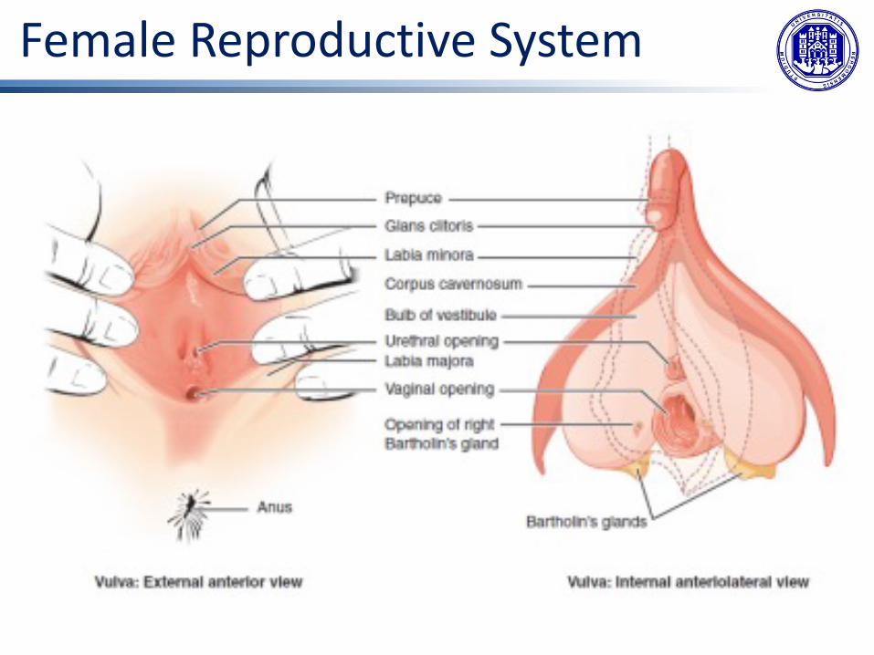
Female Reproductive SystemExternal Female GenitalsThe external female reproductive structures are referred to collectively as the vulva.• The mons pubis is a pad of fat that is located at the anterior, over the pubic bone. After
puberty, it becomes covered in pubic hair.• The labia majora are folds of hair-covered skin that begin just posterior to the mons pubis.• The thinner and more pigmented labia minora extend medial to the labia majora. They
labia minora serve to protect the female urethra and the entrance to the femalereproductive tract.
• The superior, anterior portions of the labia minora come together to encircle the clitoris,an organ that has abundant nerves that make it important in sexual sensation and orgasm.
• The hymen is a thin membrane that sometimes partially covers the entrance to the vagina.An intact hymen cannot be used as an indication of “virginity”; even at birth, this is only apartial membrane, as menstrual fluid and other secretions must be able to exit the body,regardless of penile–vaginal intercourse.
• The vaginal opening is located between the opening of the urethra and the anus.• It is flanked by outlets to the Bartholin’s glands (or greater vestibular glands).
Female Reproductive System
Female Reproductive SystemVaginaThe vagina, is a muscular canal (approximately 10 cm long) that serves as the entrance to thereproductive tract. It also serves as the exit from the uterus during menses and childbirth. Thesuperior portion of the vagina—called the fornix—meets the protruding uterine cervix. Thewalls of the vagina are lined with:• an outer, fibrous adventitia;• a middle layer of smooth muscle;• an inner mucous membrane with transverse folds called rugae.Together, the middle and inner layers allow the expansion of the vagina to accommodateintercourse and childbirth.The vagina is home to a normal population of microorganisms that help to protect againstinfection by pathogenic bacteria, yeast, or other organisms that can enter the vagina. In ahealthy woman, the most predominant type of vaginal bacteria is from the genusLactobacillus. This family of beneficial bacterial flora secretes lactic acid, and thus protects thevagina by maintaining an acidic pH (below 4.5). Potential pathogens are less likely to survive inthese acidic conditions.
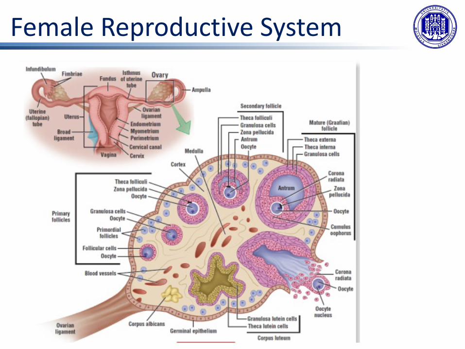
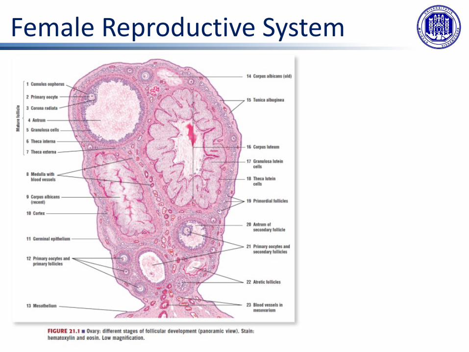
Female Reproductive SystemOvariesThe ovaries are the female gonads. Paired ovals, they are each about 2 to 3 cm in length. Theovaries are located within the pelvic cavity, and are supported by the mesovarium, an extensionof the peritoneum that connects the ovaries to the broad ligament. Extending from themesovarium itself is the suspensory ligament that contains the ovarian blood and lymphvessels. Finally, the ovary itself is attached to the uterus via the ovarian ligament.
The ovary comprises an outer covering of cuboidal epithelium called the ovarian surfaceepithelium that is superficial to a dense connective tissue covering called the tunica albuginea.
Beneath the tunica albuginea is the cortex, or outer portion, of the organ. Oocytes developwithin the outer layer of this stroma, each surrounded by supporting cells. This grouping of anoocyte and its supporting cells is called a follicle.
Beneath the cortex lies the inner ovarian medulla, the site of blood vessels, lymph vessels, andthe nerves of the ovary.
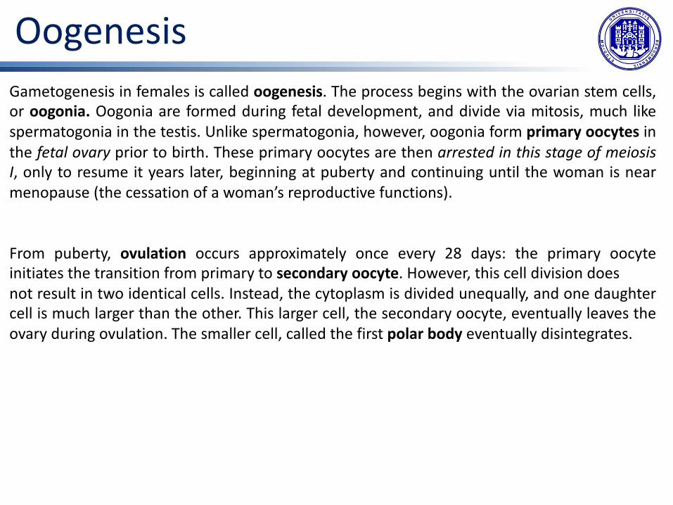
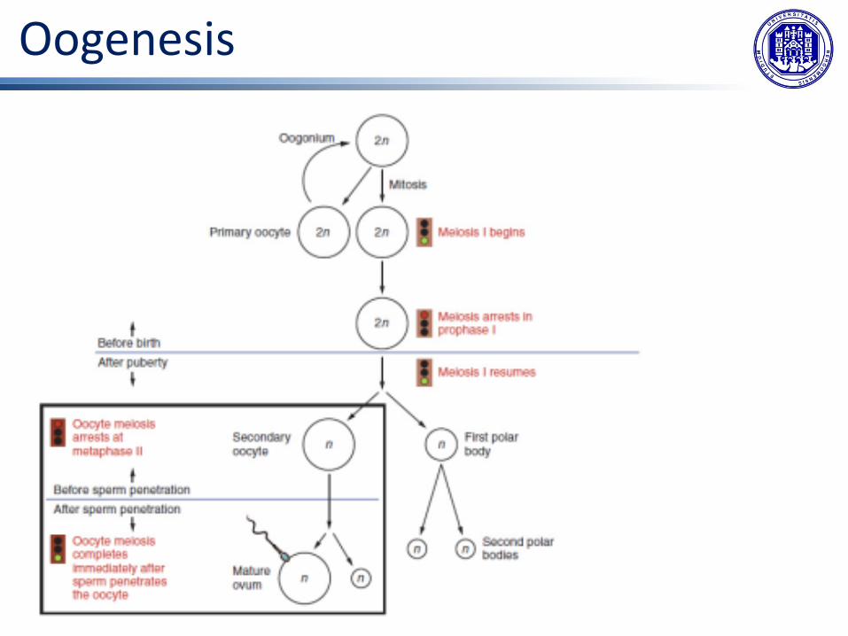
OogenesisGametogenesis in females is called oogenesis. The process begins with the ovarian stem cells,or oogonia. Oogonia are formed during fetal development, and divide via mitosis, much likespermatogonia in the testis. Unlike spermatogonia, however, oogonia form primary oocytes inthe fetal ovary prior to birth. These primary oocytes are then arrested in this stage of meiosisI, only to resume it years later, beginning at puberty and continuing until the woman is nearmenopause (the cessation of a woman’s reproductive functions).
From puberty, ovulation occurs approximately once every 28 days: the primary oocyteinitiates the transition from primary to secondary oocyte. However, this cell division doesnot result in two identical cells. Instead, the cytoplasm is divided unequally, and one daughtercell is much larger than the other. This larger cell, the secondary oocyte, eventually leaves theovary during ovulation. The smaller cell, called the first polar body eventually disintegrates.

OogenesisMeiosis of a secondary oocyte is completed only if a sperm succeeds in penetrating itsbarriers. Meiosis II then resumes, producing one haploid ovum (ovulo) that, at the instant offertilization by a (haploid) sperm, becomes the first diploid cell of the new offspring (azygote).The larger amount of cytoplasm contained in the female gamete is used to supply thedeveloping zygote with nutrients during the period between fertilization and implantationinto the uterus. Interestingly, sperm contribute only DNA at fertilization —not cytoplasm.Therefore, the cytoplasm and all of the cytoplasmic organelles in the developing embryo areof maternal origin. This includes mitochondria, which contain their own DNA. Scientificresearch in the 1980s determined that mitochondrial DNA was maternally inherited.
Oogenesis

FolliculogenesisOvarian follicles are oocytes and their supporting cells. They grow and develop in a processcalled folliculogenesis, which typically leads to ovulation of one follicle approximately every28 days, along with death to multiple other follicles (atresia).
Follicles progress from primordial, to primary, to secondary and tertiary stages prior toovulation—with the oocyte inside the follicle remaining as a primary oocyte until right beforeovulation.
Folliculogenesis begins with follicles in aresting state.primordial follicles à primary folliclesàsecondary folliclesà tertiary follicles
Several follicles reach the tertiary stage atthe same time, and most of these willundergo atresia. The one that does notdie will continue to grow and developuntil ovulation, when it will expel itssecondary oocyte surrounded by severallayers of granulosa cells from the ovary.
Female Reproductive System
Female Reproductive System

Female Reproductive System

Female Reproductive System
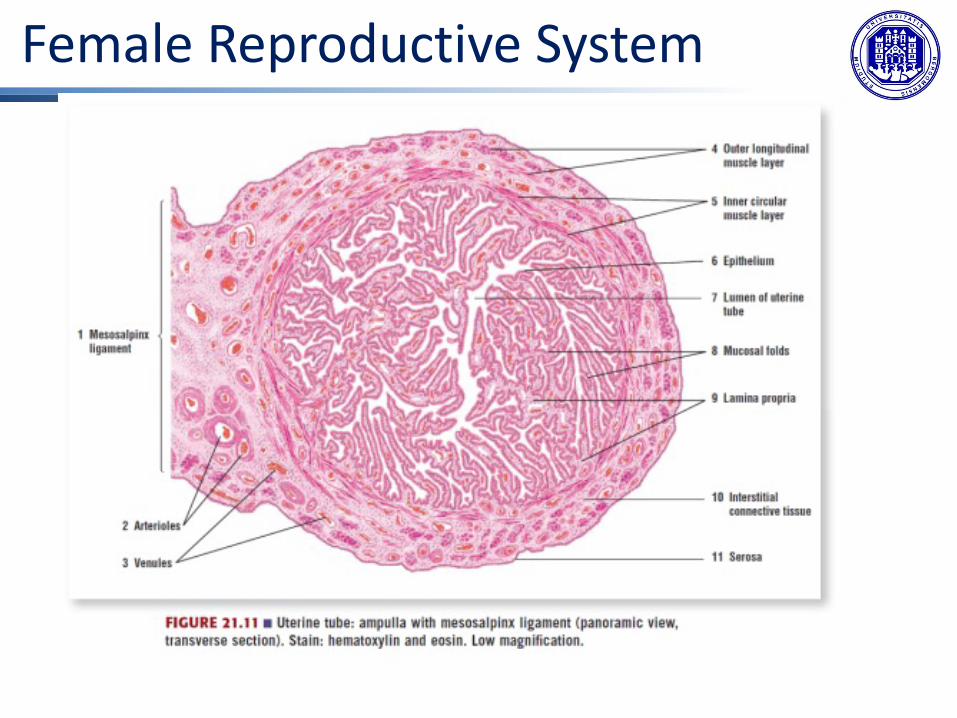
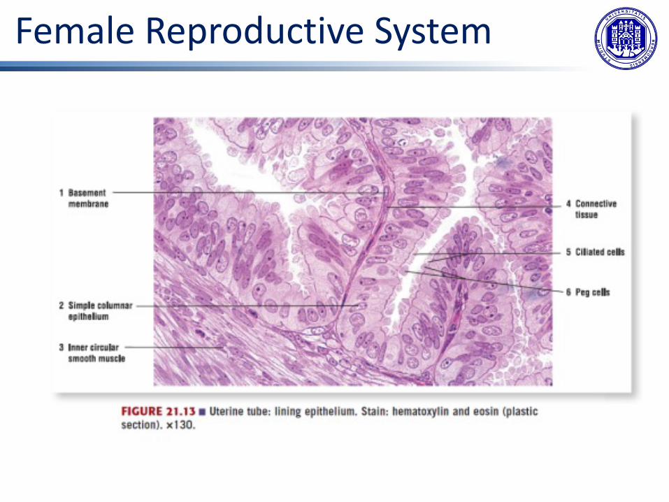
The Uterine TubesThe uterine tubes (also called fallopian tubes or oviducts) serve as the conduit of the oocytefrom the ovary to the uterus. Each of the two uterine tubes is close to, but not directlyconnected to, the ovary and divided into sections.The isthmus is the narrow medial end of each uterine tube that is connected to the uterus.The wide distal infundibulum flares out with slender, finger-like projections called fimbriae.The middle region of the tube, called the ampulla, is where fertilization often occurs. Theuterine tubes also have three layers:• an outer serosa,• a middle smooth muscle layer,• an inner mucosal layer.In addition to its mucus-secreting cells, the inner mucosa contains ciliated cells that beat in thedirection of the uterus, producing a current that will be critical to move the oocyte.
When fertilization does occur, sperm typically meet the egg while it is still moving through the ampulla. If the oocyte is successfully fertilized, the resulting zygote will begin to divide into two cells, then four, and so on, as it makes its way through the uterine tube and into the uterus. There, it will implant and continue to grow. If the egg is not fertilized, it will simply degrade.
Female Reproductive System

Female Reproductive System
Female Reproductive System
Female Reproductive System
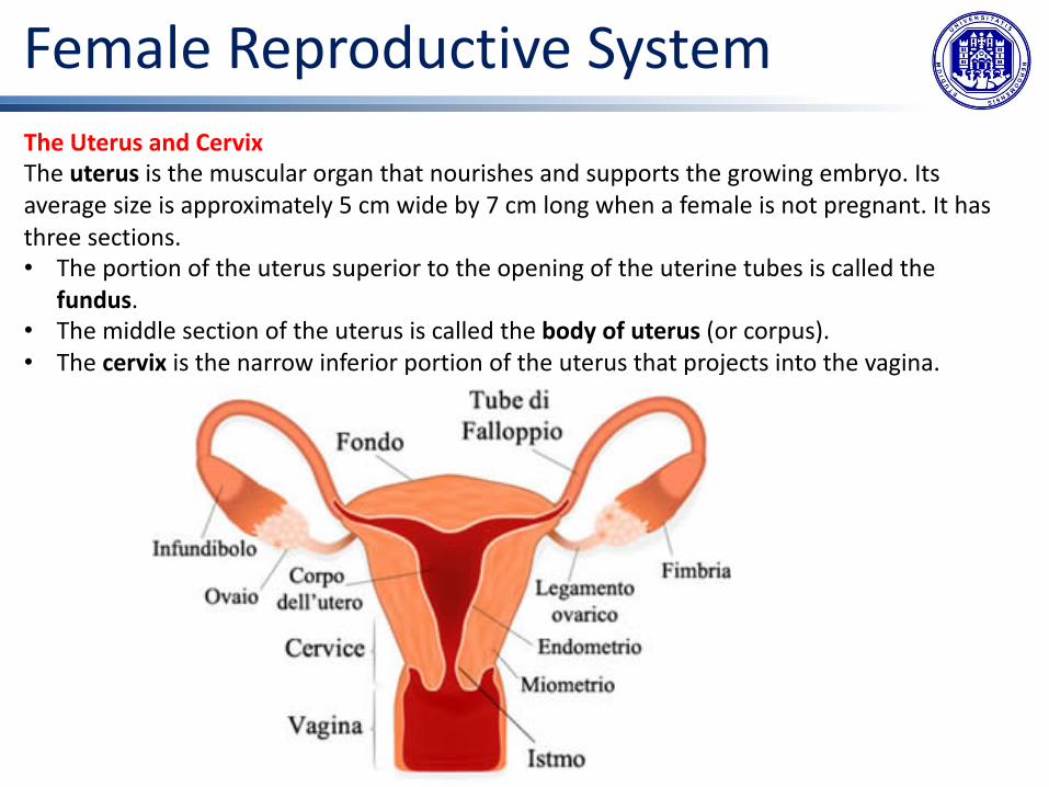
Female Reproductive SystemThe Uterus and CervixThe uterus is the muscular organ that nourishes and supports the growing embryo. Its average size is approximately 5 cm wide by 7 cm long when a female is not pregnant. It has three sections.• The portion of the uterus superior to the opening of the uterine tubes is called the
fundus. • The middle section of the uterus is called the body of uterus (or corpus). • The cervix is the narrow inferior portion of the uterus that projects into the vagina.

Female Reproductive SystemThe wall of the uterus is made up of three layers:• The most superficial layer is the serous membrane, or perimetrium, which consists of
epithelial tissue that covers the exterior portion of the uterus. • The middle layer, or myometrium, is a thick layer of smooth muscle responsible for uterine
contractions. Most of the uterus is myometrial tissue, and the muscle fibers run horizontally, vertically, and diagonally, allowing the powerful contractions that occur during labor and the less powerful contractions (or cramps) that help to expel menstrual blood during a woman’s period.
• The innermost layer of the uterus is called the endometrium. The endometrium contains a connective tissue lining, the lamina propria, which is covered by epithelial tissue that lines the lumen. Structurally, the endometrium consists of two layers: ü The stratum basalis layer is part of the lamina propria and is adjacent to the
myometrium; this layer does not shed during menses. ü The stratum functionalis layer contains the glandular portion of the lamina propria
and the endothelial tissue that lines the uterine lumen. It is the stratum functionalisthat grows and thickens in response to increased levels of estrogen and progesterone. This inner functional layer provides the proper site of implantation for the fertilized egg, and—should fertilization not occur—it is only the stratum functionalis layer of the endometrium that sheds during menstruation.