Feasibility of Low-Cost Infrared Thermal Imaging to Assess ...
NeuroImage 47 (2009) T154–T162
Contents lists available at ScienceDirect
NeuroImage
j ourna l homepage: www.e lsev ie r.com/ locate /yn img
Infrared thermal imaging: A review of the literature and case report
Babak Kateb a,b,⁎, Vicky Yamamoto d, Cheng Yu a, Warren Grundfest b,c, John Peter Gruen a
a Department of Neurological Surgery, University of Southern California, Keck School of Medicine, Los Angeles, CA, USAb Brain Mapping Foundation and International Brain Mapping and Intraoperative Surgical Planning Society, Santa Monica, CA, USAc UCLA School of Medicine, Department of Surgery, Los Angeles, CA, USAd Eli and Edythe Broad Center for Stem Cell and Regenerative Medicine, USC-Keck School of Medicine, Los Angeles, CA, USA
⁎ Corresponding author. Department of Neurological SCalifornia, Keck School of Medicine, Los Angeles, CA, US
E-mail address: [email protected] (B. Kateb).
1053-8119/$ – see front matter © 2009 Elsevier Inc. Aldoi:10.1016/j.neuroimage.2009.03.043
a b s t r a c t
a r t i c l e i n f oArticle history:Received 25 January 2009Revised 14 March 2009Accepted 18 March 2009Available online 28 March 2009
Keywords:Brain tumorBrain tumor imagingBrain tumor biophotonicThermal imagingInfrared imagingIntraoperative imagingImage-guided surgeryBrain MetastasisMelanomaMulti-Modality Imaging
Intraoperative Thermal Imaging (ITI) is a novel neuroimaging technique that can potentially locate themargins of primary and metastatic brain tumors. As a result, the additional real-time anatomical andpathophysiological information may significantly contribute to an improved extent of tumor resection.Our objectives in this article are i) to briefly discuss the current status of intraoperative imaging modalitiesincluding ITI and ii) to present a case report that evaluates the usefulness of ITI in detection of brain tumorand its margins. In this case report, ITI was used in a patient with a metastatic intracortical melanoma. Thethermal profile of the tumor and surrounding normal cerebral cortex were mapped with a ThermaCAMP60 (TCP60) infrared camera by FLIR Systems. The data obtained by TCP60, intra-operatively, revealed aclear demarcation of tumor with significant temperature differences, up to 3.3 °C, between the tumor core(36.4 °C) and the surrounding normal tissue (33.1 °C). Ultrasound and pre-resection MR and CT confirmedthe position and size of the metastasis. The volume of the tumor was preoperatively calculated using theCyberKnife™ software and postoperative volumetric measurement of the tumor residual was calculatedby the Gamma Knife™ software. Our result, along with previously published results of others, suggeststhat thermal imaging could be used to provide a rapid, non-invasive, and real-time intra-operativeimaging.
© 2009 Elsevier Inc. All rights reserved.
Introduction
Despite considerable advances in diagnosis and treatment, thesurvival rate of patients with malignant brain tumors has notsignificantly improved. The mortality from malignant brain tumorsremains high, as the median survival rate is 12 to 18 months inpatients with glioblastoma and 41 months in patients with anaplasticastrocytomas (Wen and Kesari, 2008; Stupp et al., 2006; Mitchellet al., 2005; DeAngelis, 2001). Surgical resection followed by radio-therapy and chemotherapy offers a survival benefit, particularly whenresection is complete (Sanai and Berger, 2008; Mitchell et al., 2005;Nazzaro and Neuwelt, 1990; Mineo et al., 2002; Laws et al., 2003).Despite all advances made in the field of brain imaging in the lastdecades, brain shift, also known as post-imaging brain distortion,often makes intraoperative delineation of the tumors difficult as itspreoperative imaging can no longer be fully relied on (Reinertsenet al., 2007; Nabavi et al., 2001; Hill et al., 1998; Knauth et al., 1999).Moreover, it is difficult to distinguish brain tumors from normal
urgery, University of SouthernA
l rights reserved.
surrounding tissue if they exhibit infiltrative nature, which makes itvirtually impossible to achieve near total resection. Intraoperativemulti-modality imaging can be helpful in resolving these issues, andthus in increasing the extent of resection. Therefore, there is a greatneed for development and integration of new intra-operative brainimaging/mapping technologies such as thermography. Many of thesetechnologies are currently available for research purposes and havebeen shown to hold a great promise in assisting surgeons to achievenear total resection through a more objective detection and up-to-date delineation of the tumor margins.
Background
Brief history and current state of thermal imaging
Human body temperature distribution is influenced by manycomplex factors; the heat exchange processes between skin tissues,metabolic activity, vasculature, circadian rhythm, and the regulating ofthe sympathetic and parasympathetic activity for maintaining home-ostasis (Merla and Romani, 2006; Saxena and Willital, 2008). Apresence of disease often alter the homeostasis, hence thermobiolo-gical diagnosis has been used as one of the important diagnostic
T155B. Kateb et al. / NeuroImage 47 (2009) T154–T162
measurements, as the first documented use of it can be found in thewritings of Hippocrates (circa 480 BC) (Jiang et al., 2005; Samaras andGreenblatt, 1983).
Thermal, or infrared, energy is the part of electromagneticradiation that an observer perceives as heat. Infrared thermographyallows us to visualize temperature distribution of the human body andhas been used inmedical practice since the 1950s. The earliest modernthermography was used to screen breast tumor, although due to thedrawback of less-sensitivity and low resolution, it is now mainly usedas an alternative method to detect breast tumor mass (Barrett et al.,1980; Head et al., 2000). Effort has been made by many researchers toimprove accuracy of tumor detection by increasing resolutions ofcameras, formulating better algorithm, and producing more sophis-ticated instruments (Mital and Scott, 2007; Gonzalez, 2007; Aroraet al., 2008).
Although thermoimaging has been used extensively and is wellestablished in aerospace, military and industry, its use in themedical setting is still under investigation, with the exception ofbreast tumor screening. Thermoimaging has potentially broadapplications in practical and experimental medicine, as manyclinicians and scientists have recently published exciting newreports covering a wide range of medical specialties. Among theexamples are the use of thermal imaging for the monitoring andassessment of renal ischemia (Gorbach et al., 2003, 2008), surfaceblood flow and local thermodynamics during surgery in patientswith moyamoya disease (Nakagawa et al., 2008) and viability ofgastric tube during esophagectomy (Nishikawa et al., 2006).Thermography is also used in determining the location of sensorymotor cortex during surgery by measuring cortical surface tempera-ture after median nerve stimulation. A slight temperature increaseafter the stimulation of thalamic cortical tract was elicited bymetabolic activity and heating the sensory cortex following neuronalactivity (Ueda et al., 1997).
Functional mapping using thermoencephaloscopy (TES) wasdocumented in 1990 by Shevelev and coworkers in which changeson IR radiation were measured through intact skull of rats. TES hasbeen utilized for many years to detect areas of hyperthermia infunctional imaging and brain mapping and to study associativelearning, movement, and sleep (Gorbach, 1993; Shevelev, 1992,1998). This method was non-invasive and had a reasonable spatialand temporal resolution. The pitfalls of this method were a failure toseparate cerebral blood flow (CBF) and metabolic events as well aslimited resolution of imaging instruments (Shevelev, 1992, 1998;Shevelev et al., 1993).
In 2008,Saxena andWillital (2008) reported on the use of infraredthermography in pediatric cases. 483 examinations on 285 patientsshowed that thermoimaging could recognize abnormal temperaturedistributions on skin surface caused by hemangiomas, vascularmalformations, varicoceles, extremity thrombosis, abscess, sternalwound infections, gangrene, and other symptoms/disorders. Theauthors concluded from the collection of 10 years of data thatthermoimaging is proven to be a very valuable, quick, cost-efficient,and non-invasive way to identify or to verify clinical states andtherefore used as an effective diagnostic parameter in the pediatricage group.
Current state of intraoperative diagnostics for brain tumor other than ITI
The loss of cerebrospinal fluid, tumor resection, edema, andplacing surgical apparatus during surgery can often contribute to the“brain shift” phenomenon, causing significant inconsistencies withpreoperative imaging. However, recent advances in intraoperativebiomedical imaging such as CT and MRI scans have greatly improvedour effort to identify tumors and delineate their margins. One suchtechnological advance is the use of intraoperativeMagnetic ResonanceImaging (iMRI) in the operating room, which was first introduced by
Dr. Peter Black and his colleagues (Schulder and Carmel, 2003; Blacket al., 1997). At present, this technology offers surgeons the bestintraoperative image guidance during brain tumor resection as itprovides various surgically relevant parameters such as location of thetumor and its borders, as well as functional, metabolic and vascularparameters at the highest resolution (Mittal and Black, 2006;Fahlbusch and Samii, 2007). The use of iMRI can detect brain shift,which greatly maximizes tumor resections and helps avoid harmfulresection of normal tissues and critical structures (Frangioni, 2008;Tempany and McNeil, 2001). However, its use creates challenges forsurgeons and operating room (OR) staff, such as the size of theequipment to be placed in the OR, maintaining sterility and MR-compatible surgical instruments. In an attempt to overcome some ofthese challenges, Black et al. (1997) constructed a specialized OR.Others integrated MRI suites in the OR that can be transformed intosterile surgical areas or designed a compact, portable MRI (Hall et al.,2000; Hadani et al., 2001; Schulder et al., 2001; Hall and Truwit,2008). Nevertheless, the cost of the iMRI remains high and using theprocedure can prolong the operating time as each scan takes about20 min (Fahlbusch and Samii, 2007). Therefore, it is imperative toexplore faster and more cost-efficient non-invasive alternatives toiMRI.
Aside from iMRI, there are reports of intraoperative imagingutilizing computer tomography and ultrasound, in an attempt toimprove maximum brain tumor resection. For instance, Nakao et al.(2003) used a mobile CT which is readily available in an operatingroom to acquire intraoperative images during tumor resection.Intra-operative 3D ultrasound is another emerging alternativeintraoperative modality that scientists and clinicians are exploring.Intraoperative 3D ultrasound (3D-iUS) is proven to be excellent incapturing images of metastasis, meningeoma and angioma overthose in malignant glioma and can detect brain shifting (range: 2–25 mm) (Lindner et al., 2006; Unsgaard et al., 2006). Recently,Rasmussen et al. (2007) reported neuronavigation utilizing intrao-perative 3D ultrasound that integrate preoperative structural andfunctional MR data as well as diffusion tensor imaging data tocorrect brain shift in twelve patients. They demonstrated thatautomatic brain shift correction using the method was feasible, andwere able to optimize surgical planning successfully. Both CT andultrasound imaging modalities allow surgeons to obtain severalupdates during surgery and thus are able to minimize the problemassociated with brain shift during an operation. However, theresolution of CT and ultrasound imaging is still inferior to iMRI andtheir ability to delineate tumor border remains unsatisfactory(Fahlbusch and Samii, 2007).
Current state of intraoperative thermal imaging in brain tumors
Although not as common as intraoperative MRI, there areseveral experimental models and case reports utilizing thermalimaging in brain tumor detection. In both animal and humanmodels, primary brain tumors of glial origins consistently havelower temperature than the surrounding normal tissue (Gorbachet al., 2004; Ecker et al., 2002; Papaioannou et al., 2002; Konerdinget al., 1998), which is consistent with the earlier study using humanmodel by Koga et al. (1987). Papaioannou et al. (2002) reported aC6 glioma mouse model with 0.3–0.7 °C temperature difference.The temperature was lower at the center of the tumor, with thedifference growing larger as the tumor growth progresses. Inpatients, Gorbach et al. (2004) showed that brain tumors of glialorigin generally have 0.5–2.0 °C difference (lower) compared tonormal surrounding tissue. Similar cases were reported by Ecker etal. (2002), where 11 out of 14 primary tumors were hypothermic.Both Papaiannou and Gorbach also reported delayed recovery oftemperature in tumors after irrigation of the tumor surface withcold saline (Papaioannou et al., 2002; Gorbach et al., 2004).
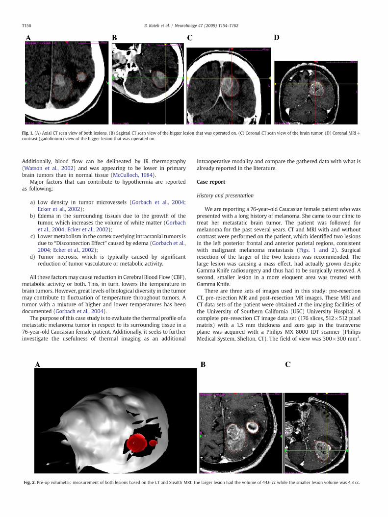
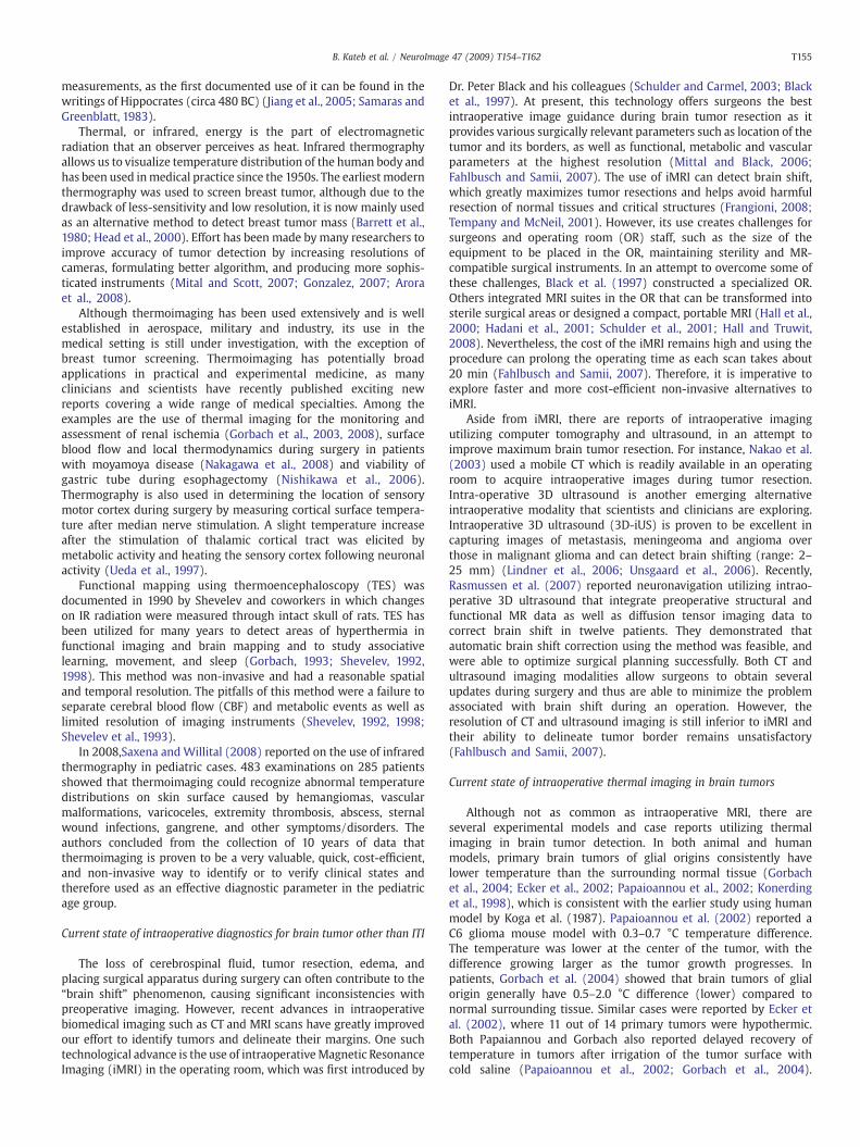
Fig. 1. (A) Axial CT scan view of both lesions. (B) Sagittal CT scan view of the bigger lesion that was operated on. (C) Coronal CT scan view of the brain tumor. (D) Coronal MRI+contrast (gadolinium) view of the bigger lesion that was operated on.
T156 B. Kateb et al. / NeuroImage 47 (2009) T154–T162
Additionally, blood flow can be delineated by IR thermography(Watson et al., 2002) and was appearing to be lower in primarybrain tumors than in normal tissue (McCulloch, 1984).
Major factors that can contribute to hypothermia are reportedas following:
a) Low density in tumor microvessels (Gorbach et al., 2004;Ecker et al., 2002);
b) Edema in the surrounding tissues due to the growth of thetumor, which increases the volume of white matter (Gorbachet al., 2004; Ecker et al., 2002);
c) Lowermetabolism in the cortex overlying intracranial tumors isdue to “Disconnection Effect” caused by edema (Gorbach et al.,2004; Ecker et al., 2002);
d) Tumor necrosis, which is typically caused by significantreduction of tumor vasculature or metabolic activity.
All these factors may cause reduction in Cerebral Blood Flow (CBF),metabolic activity or both. This, in turn, lowers the temperature inbrain tumors. However, great levels of biological diversity in the tumormay contribute to fluctuation of temperature throughout tumors. Atumor with a mixture of higher and lower temperatures has beendocumented (Gorbach et al., 2004).
The purpose of this case study is to evaluate the thermal profile of ametastatic melanoma tumor in respect to its surrounding tissue in a76-year-old Caucasian female patient. Additionally, it seeks to furtherinvestigate the usefulness of thermal imaging as an additional
Fig. 2. Pre-op volumetric measurement of both lesions based on the CT and Stealth MRI: t
intraoperative modality and compare the gathered data with what isalready reported in the literature.
Case report
History and presentation
We are reporting a 76-year-old Caucasian female patient who waspresented with a long history of melanoma. She came to our clinic totreat her metastatic brain tumor. The patient was followed formelanoma for the past several years. CT and MRI with and withoutcontrast were performed on the patient, which identified two lesionsin the left posterior frontal and anterior parietal regions, consistentwith malignant melanoma metastasis (Figs. 1 and 2). Surgicalresection of the larger of the two lesions was recommended. Thelarge lesion was causing a mass effect, had actually grown despiteGamma Knife radiosurgery and thus had to be surgically removed. Asecond, smaller lesion in a more eloquent area was treated withGamma Knife.
There are three sets of images used in this study: pre-resectionCT, pre-resection MR and post-resection MR images. These MRI andCT data sets of the patient were obtained at the imaging facilities ofthe University of Southern California (USC) University Hospital. Acomplete pre-resection CT image data set (176 slices, 512×512 pixelmatrix) with a 1.5 mm thickness and zero gap in the transverseplane was acquired with a Philips MX 8000 IDT scanner (PhilipsMedical System, Shelton, CT). The field of view was 300×300 mm2.
he larger lesion had the volume of 44.6 cc while the smaller lesion volume was 4.3 cc.
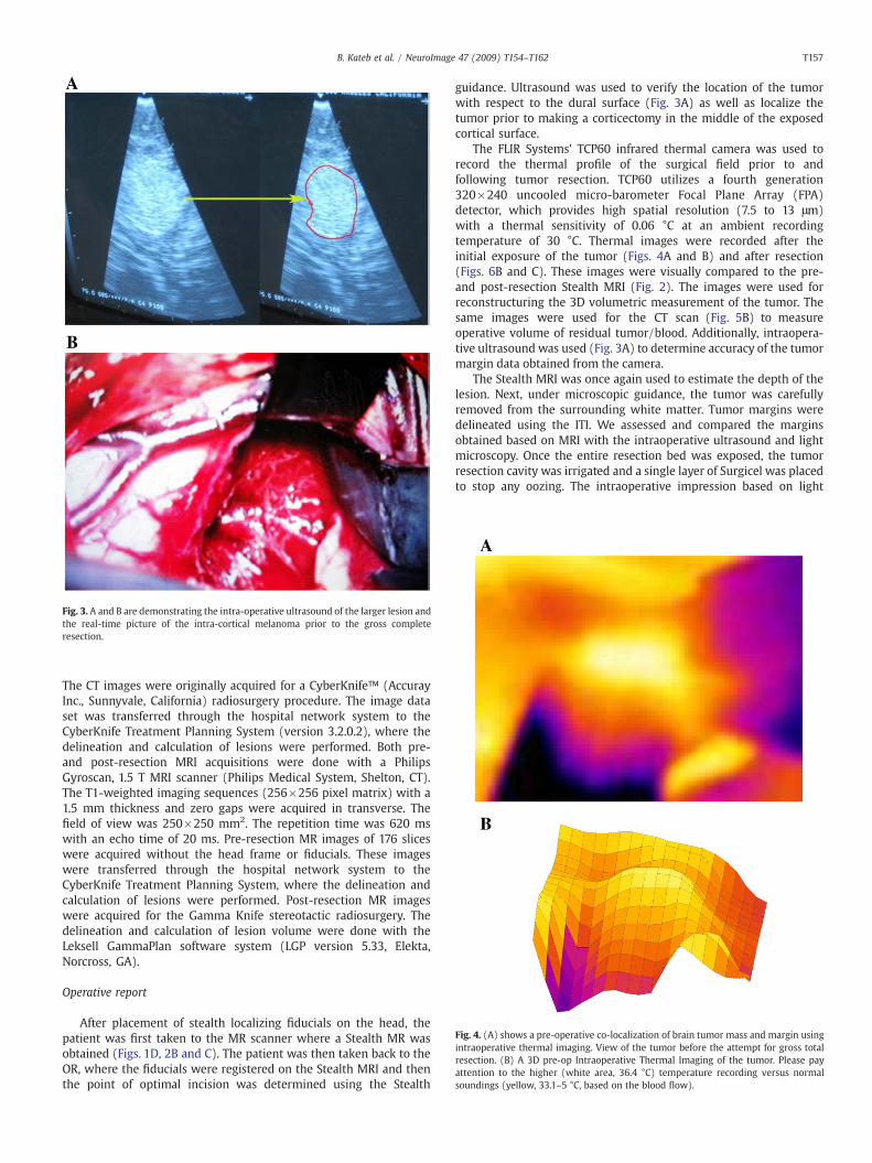
Fig. 3. A and B are demonstrating the intra-operative ultrasound of the larger lesion andthe real-time picture of the intra-cortical melanoma prior to the gross completeresection.
Fig. 4. (A) shows a pre-operative co-localization of brain tumor mass and margin usingintraoperative thermal imaging. View of the tumor before the attempt for gross totalresection. (B) A 3D pre-op Intraoperative Thermal Imaging of the tumor. Please payattention to the higher (white area, 36.4 °C) temperature recording versus normalsoundings (yellow, 33.1–5 °C, based on the blood flow).
T157B. Kateb et al. / NeuroImage 47 (2009) T154–T162
The CT images were originally acquired for a CyberKnife™ (AccurayInc., Sunnyvale, California) radiosurgery procedure. The image dataset was transferred through the hospital network system to theCyberKnife Treatment Planning System (version 3.2.0.2), where thedelineation and calculation of lesions were performed. Both pre-and post-resection MRI acquisitions were done with a PhilipsGyroscan, 1.5 T MRI scanner (Philips Medical System, Shelton, CT).The T1-weighted imaging sequences (256×256 pixel matrix) with a1.5 mm thickness and zero gaps were acquired in transverse. Thefield of view was 250×250 mm2. The repetition time was 620 mswith an echo time of 20 ms. Pre-resection MR images of 176 sliceswere acquired without the head frame or fiducials. These imageswere transferred through the hospital network system to theCyberKnife Treatment Planning System, where the delineation andcalculation of lesions were performed. Post-resection MR imageswere acquired for the Gamma Knife stereotactic radiosurgery. Thedelineation and calculation of lesion volume were done with theLeksell GammaPlan software system (LGP version 5.33, Elekta,Norcross, GA).
Operative report
After placement of stealth localizing fiducials on the head, thepatient was first taken to the MR scanner where a Stealth MR wasobtained (Figs. 1D, 2B and C). The patient was then taken back to theOR, where the fiducials were registered on the Stealth MRI and thenthe point of optimal incision was determined using the Stealth
guidance. Ultrasound was used to verify the location of the tumorwith respect to the dural surface (Fig. 3A) as well as localize thetumor prior to making a corticectomy in the middle of the exposedcortical surface.
The FLIR Systems' TCP60 infrared thermal camera was used torecord the thermal profile of the surgical field prior to andfollowing tumor resection. TCP60 utilizes a fourth generation320×240 uncooled micro-barometer Focal Plane Array (FPA)detector, which provides high spatial resolution (7.5 to 13 μm)with a thermal sensitivity of 0.06 °C at an ambient recordingtemperature of 30 °C. Thermal images were recorded after theinitial exposure of the tumor (Figs. 4A and B) and after resection(Figs. 6B and C). These images were visually compared to the pre-and post-resection Stealth MRI (Fig. 2). The images were used forreconstructuring the 3D volumetric measurement of the tumor. Thesame images were used for the CT scan (Fig. 5B) to measureoperative volume of residual tumor/blood. Additionally, intraopera-tive ultrasound was used (Fig. 3A) to determine accuracy of the tumormargin data obtained from the camera.
The Stealth MRI was once again used to estimate the depth of thelesion. Next, under microscopic guidance, the tumor was carefullyremoved from the surrounding white matter. Tumor margins weredelineated using the ITI. We assessed and compared the marginsobtained based on MRI with the intraoperative ultrasound and lightmicroscopy. Once the entire resection bed was exposed, the tumorresection cavity was irrigated and a single layer of Surgicel was placedto stop any oozing. The intraoperative impression based on light
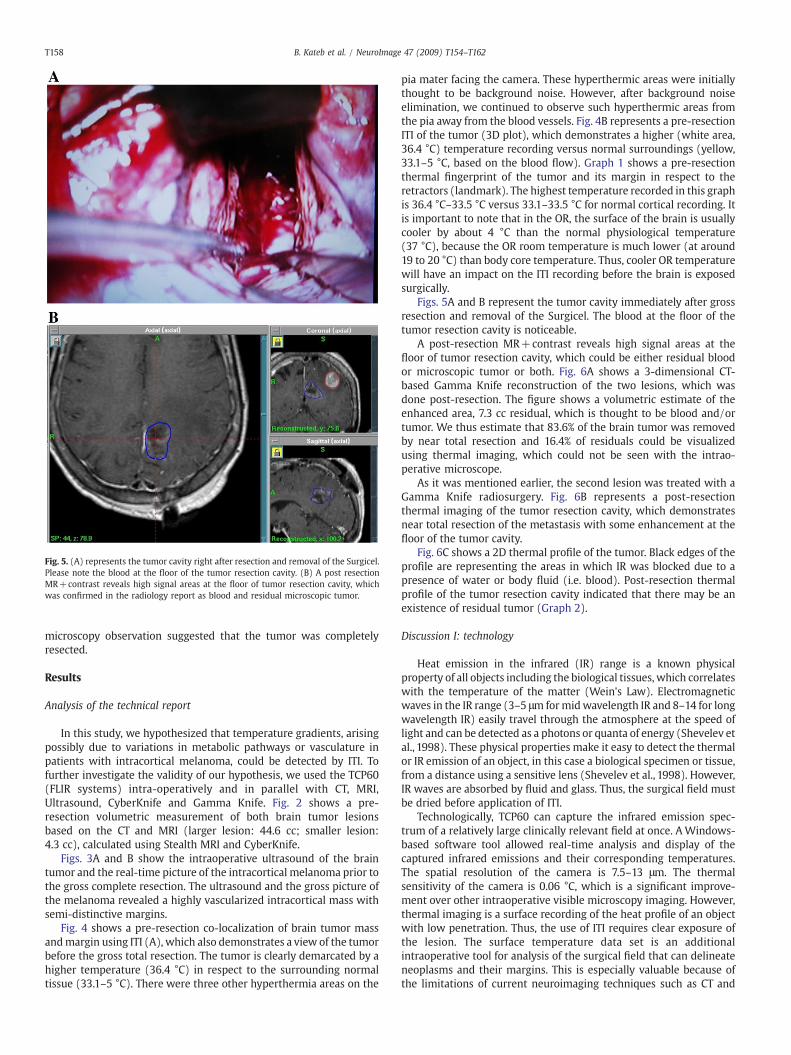
Fig. 5. (A) represents the tumor cavity right after resection and removal of the Surgicel.Please note the blood at the floor of the tumor resection cavity. (B) A post resectionMR+contrast reveals high signal areas at the floor of tumor resection cavity, whichwas confirmed in the radiology report as blood and residual microscopic tumor.
T158 B. Kateb et al. / NeuroImage 47 (2009) T154–T162
microscopy observation suggested that the tumor was completelyresected.
Results
Analysis of the technical report
In this study, we hypothesized that temperature gradients, arisingpossibly due to variations in metabolic pathways or vasculature inpatients with intracortical melanoma, could be detected by ITI. Tofurther investigate the validity of our hypothesis, we used the TCP60(FLIR systems) intra-operatively and in parallel with CT, MRI,Ultrasound, CyberKnife and Gamma Knife. Fig. 2 shows a pre-resection volumetric measurement of both brain tumor lesionsbased on the CT and MRI (larger lesion: 44.6 cc; smaller lesion:4.3 cc), calculated using Stealth MRI and CyberKnife.
Figs. 3A and B show the intraoperative ultrasound of the braintumor and the real-time picture of the intracortical melanoma prior tothe gross complete resection. The ultrasound and the gross picture ofthe melanoma revealed a highly vascularized intracortical mass withsemi-distinctive margins.
Fig. 4 shows a pre-resection co-localization of brain tumor massandmargin using ITI (A), which also demonstrates a view of the tumorbefore the gross total resection. The tumor is clearly demarcated by ahigher temperature (36.4 °C) in respect to the surrounding normaltissue (33.1–5 °C). There were three other hyperthermia areas on the
pia mater facing the camera. These hyperthermic areas were initiallythought to be background noise. However, after background noiseelimination, we continued to observe such hyperthermic areas fromthe pia away from the blood vessels. Fig. 4B represents a pre-resectionITI of the tumor (3D plot), which demonstrates a higher (white area,36.4 °C) temperature recording versus normal surroundings (yellow,33.1–5 °C, based on the blood flow). Graph 1 shows a pre-resectionthermal fingerprint of the tumor and its margin in respect to theretractors (landmark). The highest temperature recorded in this graphis 36.4 °C–33.5 °C versus 33.1–33.5 °C for normal cortical recording. Itis important to note that in the OR, the surface of the brain is usuallycooler by about 4 °C than the normal physiological temperature(37 °C), because the OR room temperature is much lower (at around19 to 20 °C) than body core temperature. Thus, cooler OR temperaturewill have an impact on the ITI recording before the brain is exposedsurgically.
Figs. 5A and B represent the tumor cavity immediately after grossresection and removal of the Surgicel. The blood at the floor of thetumor resection cavity is noticeable.
A post-resection MR+contrast reveals high signal areas at thefloor of tumor resection cavity, which could be either residual bloodor microscopic tumor or both. Fig. 6A shows a 3-dimensional CT-based Gamma Knife reconstruction of the two lesions, which wasdone post-resection. The figure shows a volumetric estimate of theenhanced area, 7.3 cc residual, which is thought to be blood and/ortumor. We thus estimate that 83.6% of the brain tumor was removedby near total resection and 16.4% of residuals could be visualizedusing thermal imaging, which could not be seen with the intrao-perative microscope.
As it was mentioned earlier, the second lesion was treated with aGamma Knife radiosurgery. Fig. 6B represents a post-resectionthermal imaging of the tumor resection cavity, which demonstratesnear total resection of the metastasis with some enhancement at thefloor of the tumor cavity.
Fig. 6C shows a 2D thermal profile of the tumor. Black edges of theprofile are representing the areas in which IR was blocked due to apresence of water or body fluid (i.e. blood). Post-resection thermalprofile of the tumor resection cavity indicated that there may be anexistence of residual tumor (Graph 2).
Discussion I: technology
Heat emission in the infrared (IR) range is a known physicalproperty of all objects including the biological tissues, which correlateswith the temperature of the matter (Wein's Law). Electromagneticwaves in the IR range (3–5 μm formidwavelength IR and 8–14 for longwavelength IR) easily travel through the atmosphere at the speed oflight and can be detected as a photons or quanta of energy (Shevelev etal., 1998). These physical properties make it easy to detect the thermalor IR emission of an object, in this case a biological specimen or tissue,from a distance using a sensitive lens (Shevelev et al., 1998). However,IR waves are absorbed by fluid and glass. Thus, the surgical field mustbe dried before application of ITI.
Technologically, TCP60 can capture the infrared emission spec-trum of a relatively large clinically relevant field at once. AWindows-based software tool allowed real-time analysis and display of thecaptured infrared emissions and their corresponding temperatures.The spatial resolution of the camera is 7.5–13 μm. The thermalsensitivity of the camera is 0.06 °C, which is a significant improve-ment over other intraoperative visible microscopy imaging. However,thermal imaging is a surface recording of the heat profile of an objectwith low penetration. Thus, the use of ITI requires clear exposure ofthe lesion. The surface temperature data set is an additionalintraoperative tool for analysis of the surgical field that can delineateneoplasms and their margins. This is especially valuable because ofthe limitations of current neuroimaging techniques such as CT and
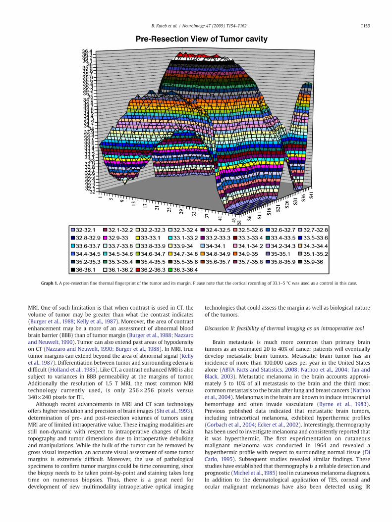
Graph 1. A pre-resection fine thermal fingerprint of the tumor and its margin. Please note that the cortical recording of 33.1–5 °C was used as a control in this case.
T159B. Kateb et al. / NeuroImage 47 (2009) T154–T162
MRI. One of such limitation is that when contrast is used in CT, thevolume of tumor may be greater than what the contrast indicates(Burger et al., 1988; Kelly et al., 1987). Moreover, the area of contrastenhancement may be a more of an assessment of abnormal bloodbrain barrier (BBB) than of tumor margin (Burger et al., 1988; Nazzaroand Neuwelt, 1990). Tumor can also extend past areas of hypodensityon CT (Nazzaro and Neuwelt, 1990; Burger et al., 1988). In MRI, truetumor margins can extend beyond the area of abnormal signal (Kellyet al., 1987). Differentiation between tumor and surrounding edema isdifficult (Holland et al., 1985). Like CT, a contrast enhanced MRI is alsosubject to variances in BBB permeability at the margins of tumor.Additionally the resolution of 1.5 T MRI, the most common MRItechnology currently used, is only 256×256 pixels versus340×240 pixels for ITI.
Although recent advancements in MRI and CT scan technologyoffers higher resolution and precision of brain images (Shi et al., 1993),determination of pre- and post-resection volumes of tumors usingMRI are of limited intraoperative value. These imaging modalities arestill non-dynamic with respect to intraoperative changes of braintopography and tumor dimensions due to intraoperative debulkingand manipulations. While the bulk of the tumor can be removed bygross visual inspection, an accurate visual assessment of some tumormargins is extremely difficult. Moreover, the use of pathologicalspecimens to confirm tumor margins could be time consuming, sincethe biopsy needs to be taken point-by-point and staining takes longtime on numerous biopsies. Thus, there is a great need fordevelopment of new multimodality intraoperative optical imaging
technologies that could assess the margin as well as biological natureof the tumors.
Discussion II: feasibility of thermal imaging as an intraoperative tool
Brain metastasis is much more common than primary braintumors as an estimated 20 to 40% of cancer patients will eventuallydevelop metastatic brain tumors. Metastatic brain tumor has anincidence of more than 100,000 cases per year in the United Statesalone (ABTA Facts and Statistics, 2008; Nathoo et al., 2004; Tan andBlack, 2003). Metastatic melanoma in the brain accounts approxi-mately 5 to 10% of all metastasis to the brain and the third mostcommonmetastasis to the brain after lung and breast cancers (Nathooet al., 2004). Melanomas in the brain are known to induce intracranialhemorrhage and often invade vasculature (Byrne et al., 1983).Previous published data indicated that metastatic brain tumors,including intracortical melanoma, exhibited hyperthermic profiles(Gorbach et al., 2004; Ecker et al., 2002). Interestingly, thermographyhas been used to investigate melanoma and consistently reported thatit was hyperthermic. The first experimentation on cutaneousmalignant melanoma was conducted in 1964 and revealed ahyperthermic profile with respect to surrounding normal tissue (DiCarlo, 1995). Subsequent studies revealed similar findings. Thesestudies have established that thermography is a reliable detection andprognostic (Michel et al., 1985) tool in cutaneousmelanoma diagnosis.In addition to the dermatological application of TES, corneal andocular malignant melanomas have also been detected using IR
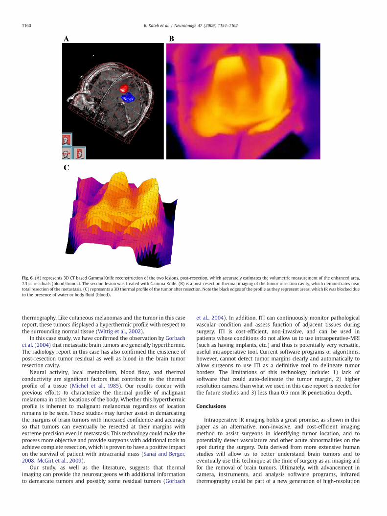
Fig. 6. (A) represents 3D CT based Gamma Knife reconstruction of the two lesions, post-resection, which accurately estimates the volumetric measurement of the enhanced area,7.3 cc residuals (blood/tumor). The second lesion was treated with Gamma Knife. (B) is a post-resection thermal imaging of the tumor resection cavity, which demonstrates neartotal resection of the metastasis. (C) represents a 3D thermal profile of the tumor after resection. Note the black edges of the profile as they represent areas, which IR was blocked dueto the presence of water or body fluid (blood).
T160 B. Kateb et al. / NeuroImage 47 (2009) T154–T162
thermography. Like cutaneous melanomas and the tumor in this casereport, these tumors displayed a hyperthermic profile with respect tothe surrounding normal tissue (Wittig et al., 2002).
In this case study, we have confirmed the observation by Gorbachet al. (2004) that metastatic brain tumors are generally hyperthermic.The radiology report in this case has also confirmed the existence ofpost-resection tumor residual as well as blood in the brain tumorresection cavity.
Neural activity, local metabolism, blood flow, and thermalconductivity are significant factors that contribute to the thermalprofile of a tissue (Michel et al., 1985). Our results concur withprevious efforts to characterize the thermal profile of malignantmelanoma in other locations of the body. Whether this hyperthermicprofile is inherent to malignant melanomas regardless of locationremains to be seen. These studies may further assist in demarcatingthe margins of brain tumors with increased confidence and accuracyso that tumors can eventually be resected at their margins withextreme precision even in metastasis. This technology could make theprocess more objective and provide surgeons with additional tools toachieve complete resection, which is proven to have a positive impacton the survival of patient with intracranial mass (Sanai and Berger,2008; McGirt et al., 2009).
Our study, as well as the literature, suggests that thermalimaging can provide the neurosurgeons with additional informationto demarcate tumors and possibly some residual tumors (Gorbach
et al., 2004). In addition, ITI can continuously monitor pathologicalvascular condition and assess function of adjacent tissues duringsurgery. ITI is cost-efficient, non-invasive, and can be used inpatients whose conditions do not allow us to use intraoperative-MRI(such as having implants, etc.) and thus is potentially very versatile,useful intraoperative tool. Current software programs or algorithms,however, cannot detect tumor margins clearly and automatically toallow surgeons to use ITI as a definitive tool to delineate tumorborders. The limitations of this technology include: 1) lack ofsoftware that could auto-delineate the tumor margin, 2) higherresolution camera than what we used in this case report is needed forthe future studies and 3) less than 0.5 mm IR penetration depth.
Conclusions
Intraoperative IR imaging holds a great promise, as shown in thispaper as an alternative, non-invasive, and cost-efficient imagingmethod to assist surgeons in identifying tumor location, and topotentially detect vasculature and other acute abnormalities on thespot during the surgery. Data derived from more extensive humanstudies will allow us to better understand brain tumors and toeventually use this technique at the time of surgery as an imaging aidfor the removal of brain tumors. Ultimately, with advancement incamera, instruments, and analysis software programs, infraredthermography could be part of a new generation of high-resolution

Graph 2. Post-resection thermal profile of the tumor resection cavity, which reveals a presence of a residual mass. This could be either blood or tumor. The normal corticaltemperature recorded in this patient was 33.1–5 °C. In this case the highest temperature recorded at the floor of the tumor cavity was 34.1 °C. There is no thermal profile of metallicretractors appearing on this graph. The radiology report in this case has also confirmed existence of post resection tumor residual as well as blood in the tumor resection cavity.
T161B. Kateb et al. / NeuroImage 47 (2009) T154–T162
neurosurgical microscopes that can distinguish between normal andneoplastic tissue with exceptional precision using other multimod-ality biophotonic technologies.
Conflict of interest statementThe authors declare there is no conflict of interest.
Acknowledgments
Wewould like to thank Mr. Brad Risser, FLIR system, who providedus with the TCP60. This project could not be possible without thesignificant administrative and technical assistance of professorsMartin Weiss, Steven Geonnatta, Dr. Darcy Spicer (chair of the IRBcommittee) and Dr. Crohen (the anesthesiologist). We thank Dr.Payman Moin for his logistical assistant and Dr. Sean MacNatt forhelping Dr. Gruen in the surgery. We also thank Dr. Ilya Eckstein for hiseditorial comments. This work is made possible by Brain MappingFoundation and International Brain Mapping and IntraoperativeSurgical Planning Society (www.IBMISPS.ORG).
Appendix A. Supplementary data
Supplementary data associated with this article can be found, inthe online version, at doi:10.1016/j.neuroimage.2009.03.043.
References
ABTA, 2008 ABTA (American Brain Tumor Association). “Facts & Statistics, 2008”www.abta.org.
Arora, N., Martins, D., Ruggerio, D., Tousimis, E., Swistel, A.J., Osborne, M.P., Simmons,R.M., 2008. Effectiveness of a noninvasive digital infrared thermal imaging systemin the detection of breast cancer. Am. J. Surg. 196 (4), 523–526.
Barrett, A.H., Myers, P.C., Sadowsky, N.L., 1980. Microwave thermography in thedetection of breast cancer. AJR, Am. J. Roengenol. 34 (2), 365–368.
Black, P.M., Moriarty, T., Alexander 3rd, E., Stieg, P., Woodard, E.J., Gleason, P.L., Martin,C.H., Kikinis, R., Schwartz, R.B., Jolesz, F.A., 1997. Development and implementationof intraoperative magnetic resonance imaging and its neurosurgical applications.Neurosurgery 41 (4), 831–842.
Burger, P.C., Heinz, E.R., Shibata, T., Kleihues, P., 1988. Topographic anatomy and CTcorrelations in the untreated glioblastoma multiforme. J. Neurosurg. 68, 698–704.
Byrne, T.N., Cascino, T.L., Posner, J.B., 1983. Brain metastasis from melanoma. J. Neuro-Oncol. 1, 313–317.
DeAngelis, L.M., 2001. Brain tumors. N. Engl. J. Med. 344, 114–123.Di Carlo, A., 1995. Thermography and the possibilities for its applications in clinical and
experimental dermatology. Clin. Dermatol. 13, 329–336.Ecker, R.D., Goerrss, S.J., Meyer, F.B., Cohen-Gadol, A.A., Britton, J.W., Levine, J.A., 2002.
Vision of the future: initial experience with real-time intraoperative high-resolution Dynamic Infrared Imaging (DIRI). J. Neurosurg. 97 (6), 1460–1471.
Fahlbusch, R., Samii, A., 2007. A review of cranial imaging techniques: potential andlimitations, clinical neurosurgery. Congress Neurol. Surg. 54 (17), 100–104.
Frangioni, J.V., 2008. New technologies for human cancer imaging. J. Clin. Oncol. 26(24), 4012–4021.
Gonzalez, F.J., (2007). Infrared Imager Requirements for Breast Cancer Detection.Proceedings of the 29th Annual Int'l Conference of the IEEE EMBS. 3312–3314.
Gorbach, A.M., 1993. Infrared imaging of brain function. Adv. Exp. Med. Biol. 333,95–123.
Gorbach, A., Simonton, D., Hale, D.A., Swanson, S.J., Kirk, A.D., 2003. Objective, real-time,intraoperative assessment of renal perfusion using infrared imaging. Am. J.Transplant. 3 (8), 988–993.
Gorbach, A.M., Heiss, J.D., Kopylev, L., Oldfield, E.H., 2004. Intraoperative infraredimaging of brain tumors. J. Neurosurg. 101, 960–969.
Gorbach, A.M., Wang, H., Dhanani, N.N., Gage, F.A., Pinto, P.A., Smith, P.D., Kirk, A.D.,Elster, E.A., 2008. Assessment of critical renal ischemia with real-time infraredimaging. J. Surg. Res. 149 (2), 310–318.
Hadani, M., Spiegelman, R., Feldman, Z., et al., 2001. Novel, compact, intraoperativemagnetic resonance imaging-guided system for conventional neurosurgicaloperating rooms. Neurosurgery 48, 799–809.
T162 B. Kateb et al. / NeuroImage 47 (2009) T154–T162
Hall, W.A., Truwit, C.L., 2008. Intraoperative MR-guided neurosurgery. J. Magn. Reson.Imaging 27 (2), 368–375 Feb.
Hall, W., Liu, H., Martin, A.J., et al., 2000. Safety, efficacy, and functionality of high-fieldstrength interventional magnetic resonance imaging for neurosurgery. Neurosur-gery 46, 632–642.
Head, J.F., Wang, F., Lipari, C.A., Elliot, R.L., 2000. The important role of infrared imagingin breast cancer. IEEE Eng Med. Biol. Mag. 19 (2), 52–57.
Hill, D., Maurer, C., Maciunas, R., Barwise, J., Fitzpatrick, J., Wang, M., 1998. Measurementof intraoperative brain surface deformation under a craniotomy. Neurosurgery 43(3), 514–526.
Holland, B.A., Brant-Zawadzki, M., Norman, D., Newton, T.H., 1985. Magnetic resonanceimaging of primary intracranial tumors: a review. Int. J. Radiat. Oncol. Biol. Phys. 11,315–321.
Jiang, L.J., Ng, E.Y., Yeo, A.C., Wu, S., Pan, F., Yau, W.Y., Chen, J.H., Yang, Y., 2005. Aperspective on medical infrared imaging. J. Med. Eng. Technol. 29 (6), 257–267.
Knauth, M., Wirtz, C., Tronnier, V., Aras, N., Kunze, S., Sartor, K., 1999. Intraoperative MRimaging increases the extent of tumor resection in patients with high-gradegliomas. Am. J. Neuroradiology 9, 1642–1646.
Kelly, P.J., Daumas-Duport, C., Scheithauer, B.W., Kall, B.A., Kispert, D.B., 1987.Stereotactic histologic correlations of computed tomography- and magneticresonance imaging-defined abnormalities in patients with glial neoplasms. MayoClinic Proceeding 62, 450–459.
Konerding, M.A., Konerding, M.A., Fait, E., Dimitropoulou, C., Malkusch, W., Ferri, C.,Giavazzi, R., Coltrini, D., Presta, M., 1998. Impact of fibroblast growth factor-2 ontumor microvascular architecture. A tridimensional morphometric study. Am. J.Pathol. 152 (6), 1607–1616.
Laws, E.R., Shaffrey, M.E., Morris, A., Anderson Jr, F.A., 2003. Surgical management ofintracranial gliomas—does radical resection improve outcome? Acta Neurochir.Suppl. 85, 47–53.
Lindner, D., Trantakis, C., Renner, C., Arnold, S., Schmitgen, A., Schneider, J.,Meixensberger, J., 2006. Application of intraoperative 3D ultrasound duringnavigated tumor resection. Minim. Invasive Neurosurg. 49 (4), 197–202.
McCulloch, J., 1984. Perivascular nerve fibres and the cerebral circulation. TrendsNeurosci. 7, 135–138.
McGirt, M.J., Chaichana, K.L., Gathinji, M., Attenello, F.J., Than, K., Olivi, A., Weingart, J.D.,Brem, H., Quiñones-Hinojosa, A.R., 2009. Independent association of extent ofresection with survival in patients with malignant brain astrocytoma. J. Neurosurg.110 (1), 156–162.
Merla, A., Romani, G.L., 2006. Functional infrared imaging in medicine: a quantitativediagnostic approach. Conf. Proc. IEEE Eng. Med. Biol. Soc. 1, 224–227.
Michel, U., Hornstein, O.P., Schonberger, A., 1985. Infrared thermography in malignantmelanoma. Diagnostic potential and limits [in German]. Hautarzt 36, 83–89.
Mineo, J.F., Quintin-Roue, I., Lucas, B., Buburusan, V., Besson, G., 2002. Glioblastomas: clinicalstudy and search for prognostic factors [in French]. Neurochirurgie 48, 500–509.
Mital, M., Scott, E.P., 2007. Thermal detection of embedded tumors using infraredimaging. J. Biomech. Eng. 129, 33–39.
Mitchell, P., Ellison, D.W., Mendelow, A.D., 2005. Surgery for malignant gliomas:mechanistic reasoning and slippery statistics. Lancet Neurol. 4, 413–422.
Mittal, S., Black, P.M., 2006. Intraoperative magnetic resonance imaging in neurosur-gery: the Brigham concept. Acta Neurochir. Suppl. 98, 77–86.
Nabavi, A., Black, P.M., Gering, D.T., Westin, C.F., Mehta, V., Pergolizzi Jr, R.S., Ferrant, M.,Warfield, S.K., Hata, N., Schwartz, R.B., Wells 3rd, W.M., Kikinis, R., Jolesz, F.A., 2001.Serial intraoperative magnetic resonance imaging of brain shift. Neurosurgery 48(4), 787–797.
Nakagawa, A., Fujimura, M., Arafune, T., Sakuma, I., Tominaga, T., 2008. Intraoperativeinfrared brain surface blood flow monitoring during superficial temporal artery–middle cerebral artery anastomosis in patients with childhood moyamoya disease.Childs Nerv. Syst. 24 (11), 1299–1305.
Nakao, N., Nakai, K., Itakura, T., 2003. Updating of neuronavigation based on imagesintraoperatively acquired with a mobile computerized tomographic scanner:technical note. Minim. Invasive Neurosurg. 46 (2), 117–120.
Nathoo, N., Toms, S.A., Barnett, G.H., 2004. Metastases to the brain: current manage-ment perspectives. Expert Rev. Neurotherapeutics 4 (4), 633–640.
Nazzaro, J.M., Neuwelt, E.A., 1990. The role of surgery in the management ofsupratentorial intermediate and high-grade astrocytomas in adults. J. Neurosurg.73, 331–344.
Nishikawa, K., Matsudaira, H., Suzuki, H., Mizuno, R., Hanyuu, N., Iwabuchi, S., Yanaga,K., 2006. Intraoperative thermal imaging in esophageal replacement: its use in theassessment of gastric tube viability. Surg. Today 36 (9), 802–806.
Papaioannou, T., Thompson, R.C., Kateb, B., Sorokoumov, O., Grundfest, W.S., Black, K.L.,2002. Thermal imaging of brain tumors in a rat glioma model. Proc. SPIE 4615, 32DOI:10.1117/12.466653.
Rasmussen, I.A., Lindseth, F., Rygh, O.M., Berntsen, E.M., Selbekk, T., Xu, J., NagelhusHernes, T.A., Harg, E., Haberg, A., Unsgaard, G., 2007. Functional neuronavigationcombined with intra-operative 3D ultrasound: initial experiences during surgicalresections close to eloquent brain areas and future directions in automatic brainshift compensation of preoperative data. Acta Neurochir. 149, 365–378.
Reinertsen, I., Descoteaux, M., Siddiqi, K., Collins, D.L., 2007. Validation of vessel-basedregistration for correction of brain shift. Med. Image Anal. 11 (4), 374–388.
Samaras, C.A., Greenblatt, R.B., 1983. The role of thermography in breast cancer.Contemp. Surg. 22, 31–38.
Sanai, N., Berger, M.S., 2008. Glioma extent of resection and its impact on patientoutcome. Neurosurgery 62 (4), 753–766.
Saxena, A.K., Willital, G.H., 2008. Infrared thermography: experience from a decade ofpediatric imaging. Eur. J. Pediatr. 167, 757–764.
Schulder, M., Carmel, P.W., 2003. Intraoperative magnetic resonance imaging: impacton brain tumor surgery. Cancer Control 10 (2), 115–124.
Schulder, M., Liang, D., Carmel, P.W., 2001. Cranial surgery navigation aided by acompact intraoperative magnetic resonance imager. J. Neurosurg. 94, 936–945.
Shevelev, I.A., 1992. Temperature topography of the brain cortex: thermoencephalo-scopy. Brain Topogr. 5, 77–85.
Shevelev, I.A., 1998. Functional imaging of the brain by infrared radiation (thermo-encephaloscopy). Prog. Neurobiol. 56, 269–305.
Shevelev, I.A., Tsicalov, E.N., Gorbach, A.M., Budko, K.P., Sharaev, G.A., 1993. Thermo-imaging of the brain. J. Neurosci. Methods 46 (1993), 49–57.
Shi, W.M., Wildrick, D.M., Sawaya, R., 1993. Volumetric measurement of brain tumorsfrom MR imaging. J. Neuro-Oncol. 37, 87–93.
Stupp, R., Mason, W.P., et al., 2006. Radiotherapy plus concomitant and adjuvanttemozolomide for glioblastoma. N. Engl. J. Med. (10), 987–996.
Tan, T.C., Black, P.M., 2003. Image-guided craniotomy for cerebral metastases:techniques and outcomes. Neurosurgery 53 (1), 82–90.
Tempany, C., McNeil, B., 2001. Advances in biomedical imaging. JAMA 285 (5),562–567.
Unsgaard, G., Rygh, O.M., Selbekk, T., Müller, T.B., Kolstad, F., Lindseth, F., Hernes, T.A.,2006. Acta Neurochir. 148 (3), 235–253.
Ueda, M., Sakurai, T., Kasai, K., Ushikubo, Y., Samejima, H., 1997. Localisation of sensorymotor cortex during surgery by changes of cortical surface temperature aftermedian nerve stimulation. Lancet 350 (9077), 561.
Watson, J.C., Gorbach, A.M., Pluta, R.M., Rak, R., Heiss, J.D., Oldfield, E.H., 2002. Real-timedetection of vascular occlusion and reperfusion of the brain during surgery by usinginfrared imaging. J. Neurosurg. 96, 918–923.
Wen, P.Y., Kesari, S., 2008. Malignant gliomas in adults. N. Engl. J. Med. 5, 492–507.Wittig, I., Kohlmann, H., Lommatzsch, P.K., Kruger, L., Herold, H., 2002. Static and
dynamic infrared thermometry and thermography in malignant melanoma of theuvea and conjunctiva [in German]. Klinische Monatsblatter fur Augenheilkunde201, 317–321.