Influence of goals on observation of actions: functional ... of goals on observation of actions:...
249
Influence of goals on observation of actions: functional neuroimaging studies Johannes Wolfram Robert Schultz Wellcome Department of Imaging Neuroscience University College London Submitted for PhD in Neurological Studies 1
Transcript of Influence of goals on observation of actions: functional ... of goals on observation of actions:...
Influence of goals on observation of
actions: functional neuroimaging studies
Johannes Wolfram Robert Schultz
Wellcome Department of Imaging Neuroscience
University College London
Submitted for PhD in Neurological Studies
1

Abstract
Mentalising or Theory-of-Mind (ToM) is defined as the attribution of mental
states to other agents. While this capacity develops progressively in children, an
important step is reached by passing the false-belief task, normally at about four years
of age. Measures of brain activity during performance of a wide range of tasks
requiring ToM have repeatedly demonstrated involvement of a particular set of brain
regions. But how each of these regions contributes to this process is not yet clear.
Based on previous data and a model of the cognitive components necessary for ToM,
I performed three experiments using event-related functional magnetic resonance
imaging in healthy volunteers to clarify the involvement of brain regions in important
components of the ToM capacity. Two different cognitive processes were studied: 1)
the identification of potential living entities in the environment and 2) the observation
of human actions. In both types of processes, one variable appears to play an
important role according to the literature: the presence of goals in the observed
actions. This variable was therefore manipulated in all experiments. When healthy
subjects watched two disks, moving in a seemingly animate way, interact with each
other, activity in a region known to respond to biological motion (the posterior part of
the superior temporal sulcus area, or pSTS) increased parametrically with the
presence of a goal in the behaviour of the disks, as did attribution of animacy. In a
second experiment using moving disks, the pSTS showed greater activation when a
chasing disk appeared to attribute goals to the target rather than simply following it.
The third experiment showed a role of the pSTS in the analysis of human movement
kinematics during categorisation of actions depending on goal-directedness. The role
of goals in the neural basis of mentalising is discussed.
2

Table of contents
Abstract 2
Table of contents 3
Acknowledgements 7
List of figures 8
List of tables 10
List of appendices 10
List of abbreviations 11
Part 1: Literature review 12
Chapter 1. General introduction 12 1.1 Definitions of commonly used terms 13
Mental states 13 Intentions and intentionality 13 Goals and actions 15 Agents and animacy 17
1.1. A brief history of Theory-of-Mind 18 Does the chimpanzee have a Theory-of-Mind? 18 The False-belief task 19 A good control 21
1.2 Possible neural substrates for ToM 23 The medial prefrontal cortex 24 The temporal poles 26 The posterior part of the superior temporal sulcus 28 Other regions 28
Chapter 2. Cognitive processes in ToM 30 2.1. Representing mental states 30
Intentional relations 30 Simulation Theory and Theory Theory 31 But maybe we use both? 32
2.2. Recent cognitive models for ToM 34 Understanding others' actions with one’s own action system 34 Internal models for prediction and control 35 An internal model-based system for understanding mental states 38
3

2.3. An extended model 41 Components 41 What it can do 42 The A-M module 43 Neural bases 44
Chapter 3. Processes for mentalising studied in this thesis 45 3.1.1 Animacy and intentionality in moving abstract shapes 45
Parameters for animacy and intentionality attribution 46 Animacy in single moving dots 47 Contingency 48 Identification of actions in terms of goals 49 Self-propelled movements and goal-directedness? 51
3.1.2 Biological bases for the perception of animate moving objects 52 Biological motion and the superior temporal sulcus 53 Attention effects 58
3.2.1 Action observation and imitation: the role of goals 58 Imitation and action observation in adults: the ideomotor principle 58 Goal-directed imitation in children 61
3.2.2 Biological bases for action observation 62 a) Studies in monkeys 63 b) Studies in humans 67 c) Anatomical regions of interest in this thesis 70
Chapter 5. Conclusions 72
Part 2: Rationale for experiments 73
Chapter 1. General rationale 73
Chapter 2. Specific rationale for experiments involving observation of intentional objects. 73 2.1. Experiment 1 74 2.2. Experiment 2 75 2.3. The role of attention 75
Chapter 3. Specific rationale for action observation 77
Part 3. Materials and methods 79
Chapter 1. MRI and fMRI 79 1.1. Basic MRI and fMRI physics 80
Spin 81 Equilibrium magnetisation 82 Radiofrequency magnetic fields 83 Relaxation 85
4

Image formation: frequency and phase encoding 88 Voxels 90 Image contrast 90 Ultrafast MRI sequences: Echo-Planar Imaging 91
1.2. FMRI and neural activity 93 BOLD contrast in fMRI 93 Physiology of BOLD 94 Correspondence between neural activity and the BOLD signal 95
Chapter 2. FMRI analysis 97 2.1. Introduction 97 2.2. Spatial preprocessing 99
Realignment 99 Slice-time correction 99 Normalisation 100 Spatial smoothing 101
2.3. Model-based analysis of fMRI data 102 Basic description of the SPM approach 102 Construction of the model 102 Eliminating temporal confounds 104 Fitting the model 104 t and F-statistics 106
2.4. Inference and the Theory of Gaussian Random Fields 107 i) Whole-brain inferences 107 ii) Anatomically constrained inferences 109 Inferences at the population level: Random Effects 110 Threshold used in this thesis 112
Part 4. Experiments 113
Overview 113
Chapter 1. Experiment 1: Parametric study of animate motion 114 Movement equation 114 Methods 121 Results 126 Discussion 140
Chapter 2. Experiment 2: Study of goal-attribution in a chasing paradigm 145 Methods 145 Results 149 Discussion 158
Chapter 3. Experiment 3: action observation 164
5

3.1. Introduction and overview 164 3.2. The fMRI experiment 166
Methods 166 Results 175 Discussion 189
Part 5. General discussion 199
1. So what is this thesis all about? 199
2. Activation in pSTS during observation of abstract and human movements 201
3. Other functions of the superior temporal sulcus and gyrus 204
4. Potential confounding factors 208
5. How the 3 mentalising regions could interact 210
6. What comes next? Future directions 212
7. Conclusion 214
Part 6. Appendices 215
Appendix 1: Behavioural reports of animacy testing in Experiment 1. 215
Appendix 2: kinematic recordings of object manipulations used in Experiment 3 217
Part 7. References 220
6

Acknowledgements
My greatest thanks go to my principal supervisor Chris Frith, for his ideas, his
calm, his humour and his constant support, and to my second supervisor Daniel
Wolpert for his enthusiasm and technical prowesses. Without these two persons, I
would probably never have come to the Wellcome Department nor written a single
line of this thesis. Further thanks are due to collaborators John O’Doherty, Hiroshi
Imamizu, Karl Friston and Mitsuo Kawato. These people, and many others as well,
have shown me how fascinating full-time research at a very high level can be, and
have helped me in my attempts to enter this world. Additional thanks go to Katja
Wiech and Frédérique de Vignemont for critical comments on this thesis. I would like
to thank the Wellcome Trust for the Wellcome Prize Studentship that financed me
during 3 years, the James McDonnell Foundation whose grant to my supervisors
supplied additional funding for 9 months and enabled the collaboration with the ATR
Lab in Japan, and the British Council whose Overseas Research Student Award paid
my overseas graduate student fees. I would also like to thank the radiographers at the
Functional Imaging Laboratory in London and at the ATR Brain Activity Imaging
Center, and all the volunteers (from outside and inside the FIL!) who participated in
my studies. Additional personal thanks go to my parents for their unconditional
support, my friends at the FIL for help on various topics ranging from statistics to
relationships and beer appreciation, and to the Queen's Larder for an unending supply
of Guinness. Finally, thanks to my sister's cat Guinness (not the beer), whose sarcastic
glances and relaxed attitude helped things to stay in perspective and my head to stay
on my shoulders.
7
List of figures
Figure Intro.1. The Sally-Ann task 20
Figure Intro.2. The Picture task. 22
Figure Intro.3. Three areas generally associated with mentalising. 24
Figure Intro.4. Gallese and Goldman’s “retrodictive simulation” routine. 36
Figure Intro.5. MOSAIC (A) and HMOSAIC (B) models. 38
Figure Intro.6. Application of the MOSAIC model to action observation. 40
Figure Intro.7. A new cognitive model of mentalising. 43
Figure Intro.8. Action - Mental state (A-M) matching system. 43
Figure Intro.9. Johnson and colleagues’ furry object. 49
Figure Intro.10. The classic point-light display of biological motion 54
Figure Intro.11. Activation in the superior temporal sulcus and gyrus during
observation of biological motion. 57
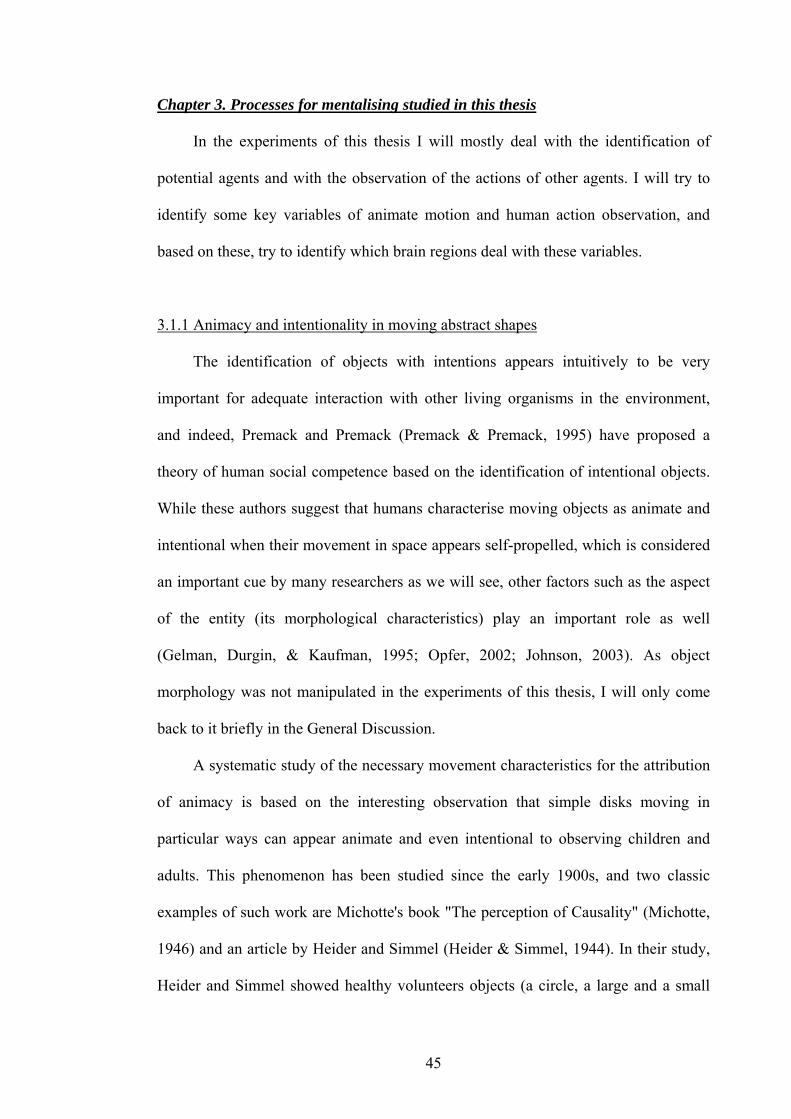
Figure Intro.12. Imitation in young infants 62
Figure Intro.13. Visual and motor responses of a mirror neuron in area F5. 65
Figure Intro.14. Activity of a mirror neuron in F5 in response to action observation in
full vision and in hidden conditions. 66
Figure Physics.1. Magnetic properties of hydrogen protons. 81
Figure Physics.2. Example of precession movement. 83
Figure Physics.3. RF excitation and the effect on proton orientation. 84
Figure Physics.4. T1 relaxation. 86
Figure Physics.5. T2 and T2* decay. 88
Figure Exp 1.1. Example of display. 113
Figure Exp 1.2. Results from the behavioural pilot study. 115
Figure Exp 1.3. Speed. 116
8
Figure Exp 1.4. Interactive and Control animations. 117
Figure Exp 1.5: Animacy ratings. 120
Figure Exp 1.6. Example of the 8 conditions. 122
Figure Exp 1.7. Interactivity and Speed ratings. 127
Figure Exp 1.8. Response times. 130
Figure Exp 1.9. Eye movements. 131
Figure Exp 1.10. Brain regions whose activation increased linearly with the amount of
interactive motion. 135
Figure Exp 1.11. VOI anatomy and data. 137
Figure Exp 1.12. Parameter estimates and time-course data pSTS. 138
Figure Exp 1.13. Time-course of activation in other regions identified in the
Interaction contrast (see Table Exp 1.4). 140
Figure Exp 2.1. Design and stimuli. 146
Figure Exp 2.2. Ratings in the Strategy and the Outcome task. 150
Figure Exp 2.3. Response times of the 14 participants. 152
Figure Exp 2.4. Brain areas responding more to Predict than Follow trials, irrespective
of task. 154
Figure Exp 2.5. Brain regions: task effects. 156
Figure Exp 2.6. Activation time-courses in the superior temporal gyrus in both
Strategy and Outcome tasks. 157
Figure Exp 2.7. Comparison between STS/STG activation in all 3 experiments. 163
Figure Exp 3.1. Experimental design and stimuli. 168
Figure Exp 3.2. Movement kinematics and animation. 170
Figure Exp 3.3. Behavioural results. 176
Figure Exp 3.4. Correlation between kinematic information and behaviour. 178
9
Figure Exp 3.5. Brain activation during all conditions with a moving actor vs. the
resting actor image. 179
Figure Exp 3.6. Effects of object presence. 183
Figure Exp 3.7. Congruence effects. 186
Figure Exp 3.8. Correlation between stimuli, and pSTS activation. 189
Figure Discussion.1. Possible neural correlates of ToM cognitive components. 212
List of tables
Exp 1.1 – Response times 129
Exp 1.2 – Eye movements 131
Exp 1.3 – fMRI 1 – Main effects 132
Exp 1.4 – fMRI 2 – Interaction 133
Exp 1.5 – fMRI 3 – Regions of interest 136
Exp 2.1 – fMRI data 153
Exp 3.1 – Number of joint measures with significant differences between Mimed and
Actual movement kinematics 172
Exp 3.2 – fMRI 1 – commonly activated regions for all manipulation types 180
Exp 3.3 – fMRI 2 – differences between manipulation types 181
Exp 3.4 – fMRI 3 – object presence effects 184
Exp 3.5 – fMRI 4 – artificial vs. realistic conditions 186
List of appendices
Appendix 1 – Behavioural reports from animacy rating task in Experiment 1 215
Appendix 2 – Joint variables time-series from Experiment 3 217
10

List of abbreviations
ACC: Anterior Cingulate Cortex
ATR: Advanced Telecommunications Research laboratories
BOLD: Blood Oxygenation-Level Dependent signal
EEG: Electro-Encephalogram
FIL: Functional Imaging Laboratory
fMRI: functional Magnetic Resonance Imaging
HRF: Hemodynamic Response Function
MEG: Magneto-Encephalography
MIP: Maximum Intensity Projection
MPFC: medial Prefrontal Cortex
PET: Positron Emission Tomography
SPM: Statistical Parametric Mapping
STS, pSTS, STG and pSTG: (posterior) Superior Temporal Sulcus and Gyrus
TMPp: Temporal pole
TPJ: Temporo-Parietal Junction, an area including the pSTS, the inferior parietal
lobule (angular and supramarginal gyri).
RT: Response Time
WDIN: Wellcome Department of Imaging Neuroscience
11

Part 1: Literature review
Chapter 1. General introduction
This thesis will deal with a basic process that enables human beings to interact
socially with each other. The first part of the introduction sets the background of the
thesis, defines keywords and processes that will be used and referred to frequently,
shows connections between them and mentions previous neuroscientific work that led
to the association of specific brain structures to some of the processes.
Human beings and other animals live in environments that change frequently,
and these changes can be good or bad for them. Also, to survive, humans as well as
animals need to interact with each other. To understand the changes that happen in the
world, predict future events and influence them, people need to understand underlying
principles that cause these events. A classic philosophical view is that humans explain
events in the world using either mentalistic or physicalist explanations. Mentalistic
explanations are used for animate things, physicalist explanations for inanimate
things. Therefore, an essential step for this process is the discrimination between
animate and inanimate things; this will be discussed further and become an important
part of this thesis. Mentalising, the process of applying mentalistic explanations by
attributing mental states to entities in the world, is also known as "Theory-of-Mind"
(ToM) (Premack & Woodruff, 1978). This is a useful heuristic for dealing with
animate agents (i.e. entities that "do things", meaning that they behave in a goal-
directed manner, see below), for predicting and influencing their behaviour if it is not
compatible with one's own desires, beliefs, wishes, thoughts or intentions. Mentalising
is the key background concept that will be used in this thesis, and experiments will
address basic processes thought to be necessary for it.
12

1.1 Definitions of commonly used terms
In this thesis, mental states, goals, actions, intentions and intentionality, agency
and animacy will be central concepts, so I would like to define these terms at the
beginning in order not to confuse readers.
Mental states
Mental states in the sense used in this thesis, such as remembering, believing,
desiring, hoping, knowing, intending, feeling, experiencing are mental representations
of a state of the world; this state could be present, past or future, real or hypothetical.
For example, the sentence “Carolina wants to eat chocolate” describes the mental state
of Carolina, which is the desire to eat chocolate. Mental states are useful for
understanding and predicting the behaviour of others: to continue the previous
example, if I knew that Carolina wants to eat chocolate, I will not be very surprised if
I see her reaching for a chocolate bar. It is therefore worthwhile to be able to identify
mental states of other organisms. But being mental states, they are solely present in
the mind of the organism that has the mental state. However, as the organism’s
behaviour can be related to its mental states, an observer can use the behaviour to
identify potential mental states explaining that behaviour. This is done by mentalising.
Intentions and intentionality
Intentions and intentionality are classic concepts in philosophy, and as I will use
them repeatedly, I will briefly review the way they are used usually and then define
how I will use them. They are classically defined in three ways:
Intentionality. This refers to the “aboutness” of mental states: mental states have
an intrinsic relationship to things in the world, and they would not exist if not
13
completed by something other than themselves (this was defined by Brentano; see
also Searle, 1983). For example, Carolina’s desire to eat chocolate relates to Carolina
and chocolate, which are an objective organism and item in the world, eating
chocolate is an action which also exists in the world, and Carolina eating chocolate is
(possibly) a future state of the world. Mental states such as believing, desiring, and
others listed above all relate to states of the world; in that, they are said to be
intentional. But this is not the way in which I will use the word “intentional”, I will
use it as relating to intentions. See below.
Intentions in actions. These are specific intentional states of mind that, unlike
beliefs, judgments, hopes, desires or fears, play a clear role in the etiology of actions.
While all intentions are intentional, not all intentional states are intentions. For
example, “Carolina intends to eat chocolate” is quite likely to play a role in making
her reach for a chocolate bar, whereas “Carolina believes that there is chocolate in the
cupboard” does not, by itself, explain her act of taking the chocolate bar. When
referring to intentions in this thesis, I will be referring to intentions in actions (Note: I
will not make the difference between 'intention in action' and 'intention to act', as
defined in Searle, 1983).
Communicative intention. A speech act has two types of intention: the
informative intention (the desire to inform the listener of something), and the
communicative intention, which is the intention to “make mutually manifest to
audience and speaker the informative intention of the speaker” (Sperber & Wilson,
1986). The communicative intention only exists when dealing with other humans.
Although this type of intention is certainly relevant for Theory-of-Mind, this will be
only interesting at higher levels of ToM than those examined in this thesis. I will
therefore not use this definition.
14

So I will use intention to mean intention in an action, and intentional as the
adjective for intention, meaning something that has intentions. Intentional actions are
therefore opposed to accidental actions, which are defined by not being directed by an
intention.
Goals and actions
Goals in actions are defined differently by different authors, as discussed in a
recent study by Koski and colleagues (Koski et al., 2002). The goal-directed theory of
imitation (Bekkering, Wohlschlaeger, & Gattis, 2002) defines goals as physical
objects that can be targets for reaching and grasping movements, but also as a
representation of the goal in neural codes, in a “functional mechanism necessary to
initiate an imitative action” (Koski et al., 2002). Tomasello separates action goals
from the means to achieve them (Tomasello, 1999). Travis defines a goal as a “mental
state representing a desired state of affairs in the world” (Travis, 1997). In Meltzoff
and Moore’s active intermodal mapping theory, an infant’s goal is to match the
relations between their own body parts with those of the observed model, again a
functional definition (Meltzoff & Moore, 1997). Dickinson and Balleine consider as
goal-directed an action mediated by 1) instrumental knowledge of the causal
relationship between the action and the outcome or goal, and 2) the current goal or
incentive value of the outcome (Dickinson & Balleine, 2000). In summary, a goal can
be defined as an object, the outcome of an action or a representation (mental or
neuronal) of either the object or the desired end-state of an action. In this thesis, I will
use the word “goal” only as the physical object toward which an action is directed, not
a mental representation, by contrast to an intention. Therefore a goal-directed action is
akin to an object-directed action, and I will sometimes use the second terminology to
15

make sure that what I mean is clear. However, some authors I cite will use a different
definition, which I hope will appear clearly enough.
Depending on the definition of goal used, some actions might also be considered
non-goal-directed. When someone mimes or pretends to perform an action, her or his
movements are not directed towards a present object, and are thought to be based on a
stored representation of the target object rather than perceptual input (Goodale,
Jakobson, & Keillor, 1994). The following data suggest that mimed movements are
controlled by different processes from object-directed actions. Mimed movements can
have different kinematics from object-directed or actual movements (Goodale,
Jakobson, & Keillor, 1994) and are much less affected by visual illusions (Westwood,
Chapman, & Roy, 2000) than are actual movements (Aglioti, DeSouza, & Goodale,
1995; Haffenden & Goodale, 1998; Ellis, Flanagan, & Lederman, 1999; Flanagan &
Beltzner, 2000). Together with the case of a patient with ventral visual stream lesions
(James, Culham, Humphrey, Milner, & Goodale, 2003) who is unable to perform
mimed movements based on perceptual cues (Goodale, Jakobson, & Keillor, 1994),
this suggests that neural structures underlying control of mimed actions could be
located in the ventral rather than the dorsal visual stream (Milner & Goodale, 1995;
Westwood, Chapman, & Roy, 2000). Thus, the fact that actual and mimed movements
differ based on their object-directedness, and maybe even goal-directedness, might
determine their control by different visual streams. As will be mentioned below,
observation of non-object-directed actions does not activate neurons responding both
during execution and observation of human actions (the so-called “Mirror Neurons”,
see below). These data will serve as the basis for an imaging experiment of action
observation, Experiment 3 in this thesis. Also, I will use the terminology “non-object-
16

directed” as being similar to “non-goal-directed”, as for goal-directed and object-
directed actions.
While most goal-directed actions imply that the agent has an intention, this does
not need to be the case: an accidental action can have a goal, but does not have an
intention. The opposite is true as well: an intentional action can be non-goal-directed:
if one considers that a mimed movement is not a goal-directed action, it can
nevertheless be executed on the basis of an intention.
Agents and animacy
I will use the word agent as referring to an entity or an organism that “does
something”, i.e. performs an action. Agents can perform goal-directed or (more
rarely) non-goal-directed actions, which might be directed by intentions and other
mental states, or not. Sometimes, we would explain actions of agents by mentalistic
terms including intentions, even if we know that the agent has no mental states (ex: a
computer “wants to connect to the web”).
Animacy I will use to mean “being alive”. A process I will study in this thesis is
the identification of animate entities in the world. Animate things often perform
actions, and are therefore often agents, and as will be discussed in more detail,
performing a goal-directed action is a good cue for attributing animacy to an entity.
17

1.1. A brief history of Theory-of-Mind
Does the chimpanzee have a Theory-of-Mind?
Premack and Woodruff published an article in 1978 discussing the existence of
Theory-of-Mind in chimpanzees, which sparked off discussions about Theory-of-
Mind in other fields as well (Premack & Woodruff, 1978). The authors describe an
interaction with a chimpanzee called Sarah, in which Sarah watched a person trying to
solve practical problems, such as trying to reach bananas hanging from the ceiling.
Sarah had to choose the most plausible continuation of the person’s action among
various options, and chose a continuation showing the person stacking boxes to reach
for the bananas. The authors argued that Sarah understood what the person was trying
to do and used this information to predict the next actions of the person. They
suggested that Sarah was able to understand a person’s actions based on their desires
and goals, an essential component for Theory-of-Mind.
But another explanation for Sarah’s behaviour can be put forward. She might
just have shown what she would do in the person’s situation (stack boxes to reach the
banana). She might just have projected her behaviour onto the situation of the person
on the screen, and might therefore just have shown what she would do and not what
she thought the person wanted to do. Her behaviour therefore does not need to involve
understanding or manipulating mental states, but just understanding of the physical
situation a person is in. In a comment on Premack and Woodruff’s article, Bennett,
Dennett and Harman suggested that a convincing demonstration for mentalising in
animals or humans1 would be to show that one can understand that someone has a
1 NOTE: As the question of whether or not only humans are capable of mentalising is not directly relevant to the present thesis, I will only discuss behavioural data from studies with humans from now on, and only refer to animal work regarding single-cell neurophysiological recording data.
18
false belief about the world, and deduct his actions from it (Bennett, 1978; Dennett,
1978; Harman, 1978).
The False-belief task
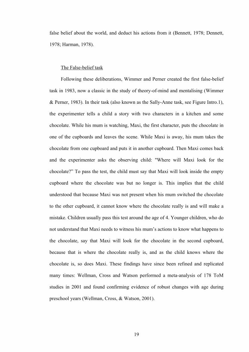
Following these deliberations, Wimmer and Perner created the first false-belief
task in 1983, now a classic in the study of theory-of-mind and mentalising (Wimmer
& Perner, 1983). In their task (also known as the Sally-Anne task, see Figure Intro.1),
the experimenter tells a child a story with two characters in a kitchen and some
chocolate. While his mum is watching, Maxi, the first character, puts the chocolate in
one of the cupboards and leaves the scene. While Maxi is away, his mum takes the
chocolate from one cupboard and puts it in another cupboard. Then Maxi comes back
and the experimenter asks the observing child: "Where will Maxi look for the
chocolate?” To pass the test, the child must say that Maxi will look inside the empty
cupboard where the chocolate was but no longer is. This implies that the child
understood that because Maxi was not present when his mum switched the chocolate
to the other cupboard, it cannot know where the chocolate really is and will make a
mistake. Children usually pass this test around the age of 4. Younger children, who do
not understand that Maxi needs to witness his mum’s actions to know what happens to
the chocolate, say that Maxi will look for the chocolate in the second cupboard,
because that is where the chocolate really is, and as the child knows where the
chocolate is, so does Maxi. These findings have since been refined and replicated
many times: Wellman, Cross and Watson performed a meta-analysis of 178 ToM
studies in 2001 and found confirming evidence of robust changes with age during
preschool years (Wellman, Cross, & Watson, 2001).
19

Figure Intro.1. The Sally-Ann task.
Originally by Wimmer and Perner (1983). Drawing by Axel Scheffler, in Frith, 2001.
This task has since been tested in children with various developmental disorders,
and a
n interesting finding has emerged: children with autism with a verbal mental age
of 4 fail the test, but children with Down syndrome with a verbal mental age of 4
perform as well as healthy 4-year olds (Baron-Cohen, Leslie, & Frith, 1985; Frith,
2001). This and other findings led to the hypothesis that children with autism suffer
20

from "mind-blindness", a specific deficit in "reading other people's minds" (Baron-
Cohen, 1995; for a recent review, see: Frith, 2001).
Gopnik and Astington developed a variant of the false-belief task to test whether
the age of false belief understanding is similar when attributed to the self as to others:
the Smarties test (Gopnik & Astington, 1988). In their test, a child is shown a tube of
candies with “Smarties” written on it. The experimenter asks the child what he thinks
is in it. The child, of course, answers: “Smarties!” The experimenter then opens the
tube and shows the child that there are no Smarties in the tube, but pencils instead.
Now the child is asked the decisive question: “When you saw the box first, what did
you think was in it?” To pass the test, the child has to remember that at the time, it
didn’t know that there were pencils in the tube, but thought it was Smarties.
Confirming previous findings, this usually happens at age 4-5, whereas 3-year-olds
fail the test and say “Pencils”.
A good control
To refine findings of a deficit in the Sally-Anne task and show that it is really
due to a deficit in attribution of mental states and not of understanding of complex
situations, Leslie and Thaiss compared the False Photograph Task with the Sally-
Anne task in autistic and healthy children (Leslie & Thaiss, 1992). In this test (Figure
Intro.2), the child is shown a teddy bear sitting on a chair. The experimenter takes a
Polaroid photograph of the teddy on the chair in front of the child, and puts the
photograph in his pocket. He then moves the teddy bear onto a bed beside the chair.
Then he asks the child: "On the photograph, is the teddy on the chair or the bed?" To
pass the test, the child has to answer "On the chair", as the photograph will not change
even if the reality it used to represent changes. Autistic children that fail the Sally-
21

Anne Task pass the False Photograph Task, whereas normally developing children
succeed in both tasks, even though the False Photograph Task appears more difficult
than the Sally-Anne Task. This suggests that children with autism have a specific
problem with an aspect of the Sally-Anne Task, most probably with the attribution of
mental states (Frith, 2001).
Figure Intro.2. The Picture task.
By Leslie and Thaiss (1992), also known as the (False) Photograph task. From Frith, 2001.
22

1.2 Possible neural substrates for ToM
The first two studies attempting to identify neural structures activated during
attribution of mental states were performed by Fletcher and colleagues (Fletcher et al.,
1995) and Goel and colleagues (Goel, Grafman, & Hallett, 1995) almost 10 years ago.
Fletcher and colleagues asked volunteers to explain the behaviour of people using
pretence and deception in one series of short stories, and presented them with stories
in which the mental states of the characters did not play a role as a control. Activity in
medial prefrontal cortex, posterior cingulate cortex and right temporo-parietal
junction was increased during mentalising. Goel and colleagues asked healthy
volunteers to judge whether someone like Christopher Columbus, living in the 15th
century, would have known the use of a series of objects. Medial prefrontal cortex and
left temporo-parietal junction were more activated during this task than during
memory retrieval and simple inferencing.
Since these original studies, a number of fMRI and PET studies have followed,
using verbal and non-verbal, on- and off-line tasks with a variety of media (pictures,
stories, cartoons, animations, games…). In reviews of such studies, Chris and Uta
Frith and colleagues (Frith & Frith, 1999; Gallagher & Frith, 2003; Frith & Frith,
2003) showed that three regions appear to show increased activation during
mentalising (Figure Intro.3): the posterior superior temporal gyrus or temporo-parietal
junction, the temporal poles and the medial prefrontal cortex.
23

Medial prefrontal cortex Superior temporal sulcus Temporal pole
Figure Intro.3. Three areas generally associated with mentalising.
FMRI and PET studies using diverse mentalising tasks, from the meta-analysis by Frith and Frith,
2003, activate the medial prefrontal cortex, the superior temporal sulcus and the temporal pole.
Displayed are data from 10 studies, with tasks including inferred knowledge (Goel, Grafman, &
Hallett, 1995), social transgressions (Berthoz, Armony, Blair, & Dolan, 2002), cartoons (Brunet,
Sarfati, Hardy-Bayle, & Decety, 2000), interactive games (Gallagher, Jack, Roepstorff, & Frith, 2002),
animations (Castelli, Happe, Frith, & Frith, 2000; Schultz et al., 2003) and stories (Fletcher et al., 1995;
Gallagher et al., 2000; Vogeley et al., 2001; Ferstl & von Cramon, 2002).
The medial prefrontal cortex
It is possible that the medial prefrontal cortex (mPFC, also referred to as anterior
paracingulate cortex) is the region most specifically associated with mentalising, and
that the pSTS and temporal poles reflect activities that aid mentalising and from
which mentalising possibly developed (Gallagher & Frith, 2003). In their latest review
(Frith & Frith, 2003), Uta and Chris Frith discuss the currently available information
on the mPFC. This region has direct connections to TMPp and pSTS (Bachevalier,
Meunier, Lu, & Ungerleider, 1997). It is the most anterior part of the paracingulate
(BA 32), partly overlapping but mostly anterior to the anterior rostral cingulate area
(RCZa) as defined by Picard and Strick in their review of premotor areas (Picard &
Strick, 2001), and overlapping with the (functionally defined) “emotional” part of the
ACC (Bush, Luu, & Posner, 2000). The paracingulate cortex is often considered part
of ACC (BA 24, 25 and 33), but the mPFC has been described cytoarchitectonically
as a cingulo-frontal transition area, different from the ACC proper (Devinsky,
24

Morrell, & Vogt, 1995). Frith and Frith also mention a specific particularity of the
ACC which might correlate with the mentalising aptitude: the presence of spindle
cells, a type of neurons found only in apes and hominids (Nimchinsky et al., 1999),
appearing at the age of 4 months in humans (Allman, Hakeem, Erwin, Nimchinsky, &
Hof, 2001). If the ACC has undergone recent evolutionary changes and is involved in
mentalising, it could explain why only humans can perform mentalising fully, while
apes are at best limited to representing very simple psychological states of others,
such as seeing (Tomasello, Call, & Hare, 2003; Povinelli & Vonk, 2003).
Functionally also, cognitive processes associated with the anterior paracingulate
cortex hint at a connection to mentalising. This area is activated by attention to
diverse sorts of events and sensations, such as emotion, pain, tickling and irrelevant
thoughts, and could therefore store 2nd-order representations (i.e. decoupled from the
"physical world") of these sensations and events, used for attention and report.
Representing mental states of the self or others appears to be a similar type of process
and could therefore be supported by the same neural structure. Studies of
autobiographical memory also yielded activation in the mPFC, and they might
implicate a common component to mentalising by calling upon representations of the
self. A simpler explanation would be that mentalising tasks might be forms of
complex problem-solving. While there are connections between mentalising and
executive functions in the development of cognitive functions, they are still not fully
understood (Perner & Lang, 1999), and imaging studies of executive functions
suggest that different regions are involved in these processes (Frith & Frith, 2003).
Neuropsychological studies in patients with frontal lesions showed that some of
these patients have deficits in Theory-of-Mind tasks. One study of patients with
frontal variant frontotemporal dementia showed that these patients have deficits in
25

first-order and second-order false belief tasks, faux pas detection and a mentalising
task based on pictures of the eye region of the face (Baron-Cohen, Jolliffe,
Mortimore, & Robertson, 1997), while having no problem in control tasks testing
memory and general comprehension. Alzheimer patients only had deficits in the
second-order false belief task (Gregory et al., 2002). Interestingly, damage to
ventromedial frontal cortex was associated with deficit in the ToM tasks, while
deficits in executive function did not correlate with deficits in ToM. In another study
comparing patients with unilateral left and right frontal lesions to matched controls,
patients of both groups were found to have deficits in first- and second-order ToM
tasks (Rowe, Bullock, Polkey, & Morris, 2001). No effect of laterality or lesion size
was found. Comparing patients with orbitofrontal and dorsolateral prefrontal lesions
showed that only the former performed like individuals with Asperger’s syndrome,
with deficits in recognizing social faux pas; the latter had problems only when task
demands on working memory were high (Stone, Baron-Cohen, & Knight, 1998). In
another study, patients with medial frontal lesions showed impaired detection of
deception; the authors suggest that this deficit may depend on connections between
the medial frontal lobe and the amygdala (Stuss, Gallup, & Alexander, 2001). But in a
very recent study, a patient with symmetric medial prefrontal lesions showed no
impairment on a range of ToM tasks (Bird, Castelli, Malik, Frith, & Husain, 2004).
The temporal poles
In their review mentioned above, Frith and Frith (Frith & Frith, 2003) also
discuss explanations for the association between mentalising and the temporal poles,
particularly in the left hemisphere. The authors mention that the anterior temporal
lobe has been considered a potential convergence point for all sensory modalities
26

(Moran, Mufson, & Mesulam, 1985). Reviewing neuroimaging data available on the
area, they mention that activation increases in the temporal poles, particularly in the
left hemisphere, are found in language paradigms, such as the comparison of
sentences to word strings or unrelated sentence strings, or the comparison between
more coherent vs. less coherent narratives, or in semantic decision tasks. Another
process associated with this brain area is retrieval of autobiographic memory, retrieval
of emotional context in single-word recognition, and the recognition of familiar faces,
scenes and voices. Episodic memory in itself might be useful for mentalising:
remembering past interactions with a person might help us to recall what we said to
them at the last encounter, what their likely attitude towards us could be, or which
mental states were associated with a particular behaviour they exhibited (Gallagher &
Frith, 2003).
Frith and Frith (Frith & Frith, 2003) suggest that an overall function of the
temporal poles might be involved in generating a wider semantic and emotional
context for material being processed. A part of this wider semantic context that could
be useful for mentalising are the so-called scripts: the habitual sequences of events
and activities that take place in a given setting and time. One popular example is the
restaurant script: we choose a restaurant, then expect what we will find on the menu,
then that we will order, then taste the wine, then receive and enjoy the food, then pay
the bill. Such scripts could be useful for understanding mental states of other people
by matching their behaviour with possible scripts for their situation, and noticing the
deviations. These scripts are gradually lost in patients with semantic dementia, who
show atrophy in the anterior temporal lobes, particularly in the left hemisphere.
27

The posterior part of the superior temporal sulcus
A recent study suggests that the pSTS is not only involved in mentalising tasks
but is also the region that shows the most consistent activation during ToM stories,
and no activation during control stories about a false photograph, mechanical
inference, human actions and other controls (Saxe & Kanwisher, 2003). The authors
of this study therefore conclude that it is the pSTS that is the most important region
for mentalising. Arguing that pSTS is necessary for mentalising, a recent
neuropsychological study showed that three patients with lesions of the left temporo-
parietal junction including the posterior STS all had deficits in a false-belief task,
while not all showed deficits in story-based and video-based control tasks involving
memory, counterfactual reasoning, reality checking, or response inhibition (Samson,
Apperly, Chiavarino, & Humphreys, 2004). Other processes associated with the
posterior part of the STS will be discussed in greater detail later on.
Other regions
Other researchers suggest that there might not be a specific neural circuit for
mentalising, but that this ability could instead rely on neural structures associated with
other cognitive processes (Siegal & Varley, 2002). Candidate processes and brain
structures are executive functioning in the frontal lobes, visuo-spatial processing
(particularly identification of animate entities in higher-order visual cortex, see Frith
& Frith, 1999; Frith, 2001), language abilities (particularly grammatical abilities) in
the left hemisphere or emotional processing in the amygdala. Although language is
helpful in solving ToM tasks, disorders of language or grammar only (either acquired
from a brain lesion or present during development) do not eliminate mentalising
abilities (Siegal & Varley, 2002). Patients with frontal lesions (specially right) have
28

difficulties with deception tasks, but deficits in tests of executive functioning do not
correlate with deficits in ToM in patients with prefrontal brain lesions (Rowe,
Bullock, Polkey, & Morris, 2001). Brain regions associated with social cognition,
often termed the "social brain" (orbitofrontal cortex, the amygdala and the superior
temporal gyrus, see Brothers, 1990; Adolphs, 1999; Adolphs, 2003) are certainly
important and probably necessary for the emergence of ToM, but these regions are
probably not sufficient for this cognitive process once it is developed. Some
researchers argue that the "social brain" is the core component for ToM, but others
argue that it is rather the above-mentioned triad, identified in functional imaging
studies, that is the network underlying ToM. Also important for the development of
ToM are conversational skills and access to interactions with other human beings,
with which a child gets exposed to mental states of other people.
29
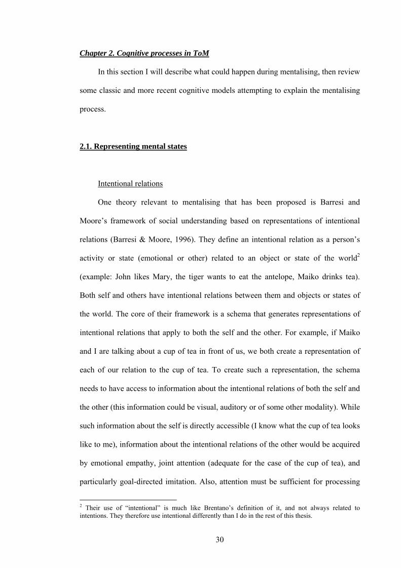
Chapter 2. Cognitive processes in ToM
In this section I will describe what could happen during mentalising, then review
some classic and more recent cognitive models attempting to explain the mentalising
process.
2.1. Representing mental states
Intentional relations
One theory relevant to mentalising that has been proposed is Barresi and
Moore’s framework of social understanding based on representations of intentional
relations (Barresi & Moore, 1996). They define an intentional relation as a person’s
activity or state (emotional or other) related to an object or state of the world2
(example: John likes Mary, the tiger wants to eat the antelope, Maiko drinks tea).
Both self and others have intentional relations between them and objects or states of
the world. The core of their framework is a schema that generates representations of
intentional relations that apply to both the self and the other. For example, if Maiko
and I are talking about a cup of tea in front of us, we both create a representation of
each of our relation to the cup of tea. To create such a representation, the schema
needs to have access to information about the intentional relations of both the self and
the other (this information could be visual, auditory or of some other modality). While
such information about the self is directly accessible (I know what the cup of tea looks
like to me), information about the intentional relations of the other would be acquired
by emotional empathy, joint attention (adequate for the case of the cup of tea), and
particularly goal-directed imitation. Also, attention must be sufficient for processing
2 Their use of “intentional” is much like Brentano’s definition of it, and not always related to intentions. They therefore use intentional differently than I do in the rest of this thesis.
30
both types of information at the same time. Once this common representation is
created, it can be applied to both self and other and enables social understanding.
This framework emphasizes the link between perception of goals and
understanding of intentional relations (culminating in mentalising), and in this, is
relatively close to the relationship between actions, goals and intentions which will
also be defended in this thesis.
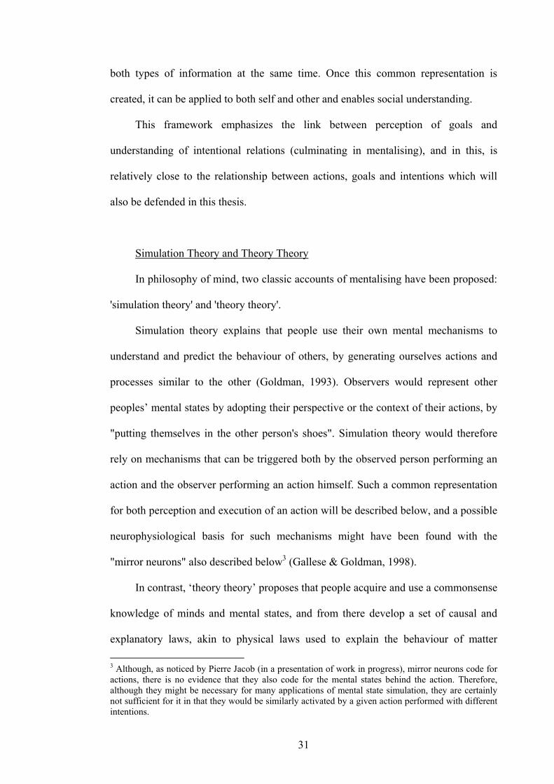
Simulation Theory and Theory Theory
In philosophy of mind, two classic accounts of mentalising have been proposed:
'simulation theory' and 'theory theory'.
Simulation theory explains that people use their own mental mechanisms to
understand and predict the behaviour of others, by generating ourselves actions and
processes similar to the other (Goldman, 1993). Observers would represent other
peoples’ mental states by adopting their perspective or the context of their actions, by
"putting themselves in the other person's shoes". Simulation theory would therefore
rely on mechanisms that can be triggered both by the observed person performing an
action and the observer performing an action himself. Such a common representation
for both perception and execution of an action will be described below, and a possible
neurophysiological basis for such mechanisms might have been found with the
"mirror neurons" also described below3 (Gallese & Goldman, 1998).
In contrast, ‘theory theory’ proposes that people acquire and use a commonsense
knowledge of minds and mental states, and from there develop a set of causal and
explanatory laws, akin to physical laws used to explain the behaviour of matter 3 Although, as noticed by Pierre Jacob (in a presentation of work in progress), mirror neurons code for actions, there is no evidence that they also code for the mental states behind the action. Therefore, although they might be necessary for many applications of mental state simulation, they are certainly not sufficient for it in that they would be similarly activated by a given action performed with different intentions.
31

(Gopnik, 1993). Therefore, to understand the mental states of another, I would enter
into my database the observed behaviour and on its basis infer which mental state
could explain the behaviour. To develop this theory, one would act like a child
scientist, performing experiments with oneself and others to understand the mind.
But maybe we use both?
The explanation of what happens during mentalising that I will try to defend is
as follows. To attribute mental states to another agent, the observer relies mainly on
the behaviour of this agent. Using his own knowledge of mental states associated with
the observed behaviour and the information he has about the other agent (the context
of the person’s action), he will then try to select the mental state that is most likely to
be present in the mind of the other agent. To test and refine his selection, the observer
can make predictions about the behaviour of the other agent based on the mental state
he has chosen and compare them to the real behaviour, then modify his choice of a
mental state. In this, he will act like Gopnik’s child scientist (see above), and compile
a database of mental states associated with different actions and contexts. I would not
like to exclude however the possibility that the observer’s action system is directly
activated by the actions of another, which would call up mental states associated with
these actions when performed himself, which would be useful to compare the action-
mental state associations of the other and the self (For example: I cry when I am sad,
but she cries when she is happy. Same action, but different mental state. Good to
know when I observe her the next time!). Also, in different situations simulation of
the other’s action might help (when one can put oneself in the other’s shoes) but in
others it will be necessary to theorise (how can I understand a mother’s actions when
32

she has lost a child, when I have never experienced this?). I believe that I am therefore
following completely neither Theory Theory nor Simulation Theory.
In my view, the basic processes for the accomplishment of mentalising are: 1)
the identification of potential agents or detection of agency, 2) observation and
recognition / understanding of their actions, 3) knowledge of potential mental states
that the other agent could have, 4) the correct association between the mental state and
the behaviour and 5) the evaluation and testing of the mental state by comparison
between expected and actual behaviour of the other agent. Different models of how
these processes interact will be discussed below.
33

2.2. Recent cognitive models for ToM
Recently, more detailed models based on control systems used by engineers and
on neuroscientific data have been proposed.
Understanding others' actions with one’s own action system
Based on previous psychophysical and neuroimaging work, Blakemore and
Decety (Blakemore & Decety, 2001) have suggested that humans automatically infer
intentions from observed actions of other people and other types of biological motion.
Inferring intentions of others by observing their actions could be a basic form of
theory of mind and the basis of higher levels of understanding of others’ minds. They
propose that such a mechanism for intention inference might be based on the system
labelling the consequences of one’s own actions from one’s own intentions. This
system could be implemented as a forward model predicting the sensory
consequences from a given intention, on the basis of a store of sensory predictions
associated with actions of the self. Understanding others’ intentions could be based on
simulating (covertly imitating) the observed action and estimating the intentions of the
actor on the basis of one’s own intentions. The authors propose the following
sequence of events: “the observed sensory consequences (of another person’s actions)
would be mapped onto stored sensory predictions (of the sensory consequences of
one’s own actions). These stored representations could then be used to estimate the
motor commands and intentions that would normally precede such an action. This
could be achieved by automatically and unconsciously simulating the observed action
and estimating what our intentions would be if we produced the same action within
the same context” (Blakemore & Decety, 2001). In their article, Blakemore and
Decety suggest that the forward model could use efference copy signals created in
34

parallel with the motor commands of an action to predict the sensory consequences
from that action (psychological evidence is provided by the example of tickling:
Weiskrantz, Elliott, & Darlington, 1971; Blakemore, Frith, & Wolpert, 1999), but
they do not explain how this mechanism could be reversed to retrieve the intentions
associated with the mapped sensory consequences of the observed actor’s actions.
Internal models for prediction and control
In a recent article, Gallese and Goldman (Gallese & Goldman, 1998) detail the
"simulation routine" that represents the mechanism of Simulation Theory (Figure
Intro.4). Based on Goldmans's model (Goldman, 1989), they propose that while an
observer is watching another person, a "pretend belief and desire generator" would
generate potential mental states that the observed person might have, on the basis of
all information available about the observed person, such as previous actions or
contextual information. This mental state would be fed into a "decision-making
system", which would predict the appropriate behaviour of the observed person
corresponding to the mental state. The decision-making system used for this purpose
would be the same as used by the observer himself to perform everyday actions. This
predicted behaviour can then be used to anticipate the next actions of the other and
interact appropriately with him.
35
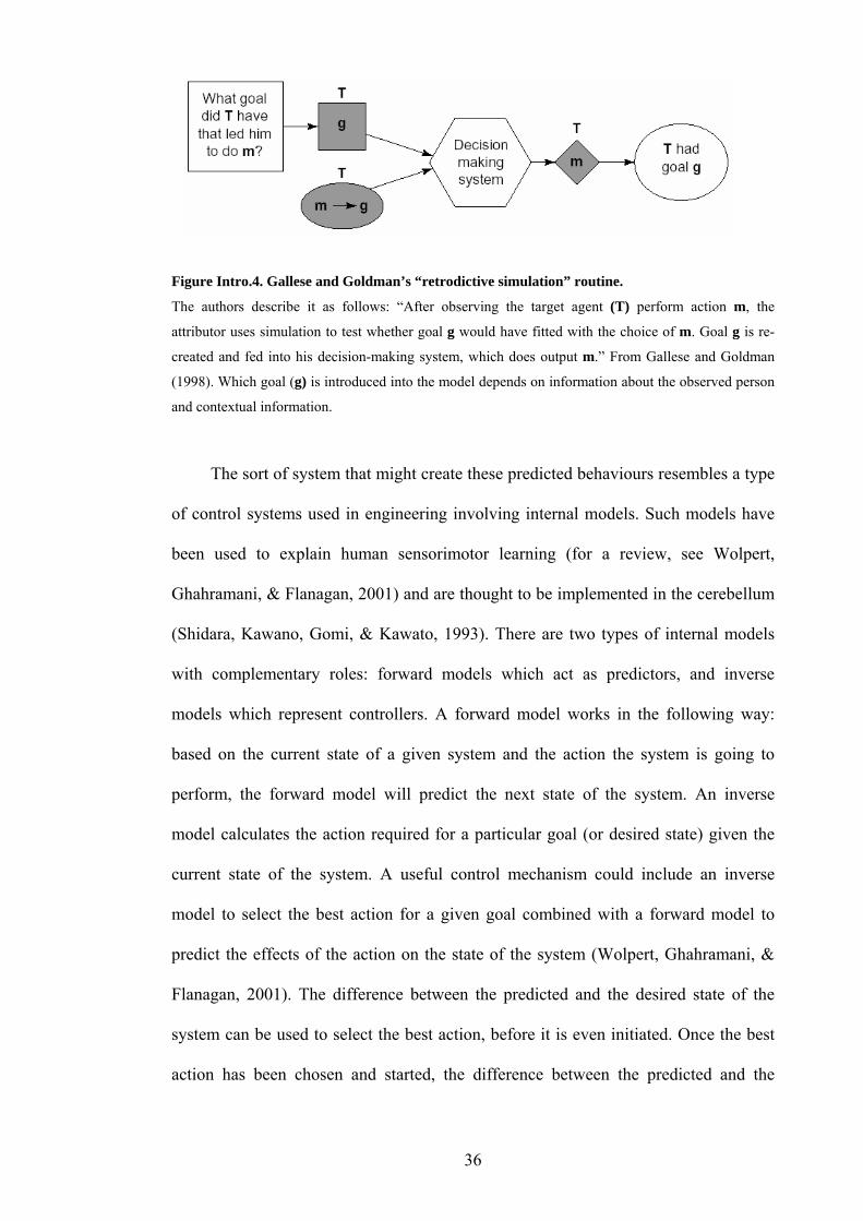
Figure Intro.4. Gallese and Goldman’s “retrodictive simulation” routine.
The authors describe it as follows: “After observing the target agent (T) perform action m, the
attributor uses simulation to test whether goal g would have fitted with the choice of m. Goal g is re-
created and fed into his decision-making system, which does output m.” From Gallese and Goldman
(1998). Which goal (g) is introduced into the model depends on information about the observed person
and contextual information.
The sort of system that might create these predicted behaviours resembles a type
of control systems used in engineering involving internal models. Such models have
been used to explain human sensorimotor learning (for a review, see Wolpert,
Ghahramani, & Flanagan, 2001) and are thought to be implemented in the cerebellum
(Shidara, Kawano, Gomi, & Kawato, 1993). There are two types of internal models
with complementary roles: forward models which act as predictors, and inverse
models which represent controllers. A forward model works in the following way:
based on the current state of a given system and the action the system is going to
perform, the forward model will predict the next state of the system. An inverse
model calculates the action required for a particular goal (or desired state) given the
current state of the system. A useful control mechanism could include an inverse
model to select the best action for a given goal combined with a forward model to
predict the effects of the action on the state of the system (Wolpert, Ghahramani, &
Flanagan, 2001). The difference between the predicted and the desired state of the
system can be used to select the best action, before it is even initiated. Once the best
action has been chosen and started, the difference between the predicted and the
36

actual state of the system would be fed back into the inverse model, modifying or
fine-tuning the chosen action to minimise the difference and finally achieve the goal.
To make such a control system more flexible for various sensorimotor contexts,
Wolpert and Kawato have proposed to use multiple parallel systems: the modular
selection and identification for control model, or MOSAIC model (Wolpert &
Kawato, 1998; Haruno, Wolpert, & Kawato, 2001, see Figure Intro.11 A). In this
system, prior information about the context is given by sensory information such as
visual input. Given the state of the system and the goal to be achieved, the parallel
inverse models corresponding best to the context then calculate actions corresponding
to the specified goal. The predicted effects of each action on the state of the system
are compared with the desired state of the system. The inverse model that proposed
the action which results in the closest state to the desired state receives the greatest
confidence rating, and the action is executed. The actual state of the system is then
compared to the desired state. When the inverse model is performing well and has
selected the right context and action, there is no difference between the desired and
the actual state of the system, the goal is reached.
A recent development of this architecture was to propose a hierarchical version
of MOSAIC: the HMOSAIC (Wolpert, Doya, & Kawato, 2003, see Figure Intro.11
B). This structure is composed of higher-level MOSAICs that control lower-level
ones. The higher level systems deal with action goals and intentions, intermediary
ones with action sequences, and the lowest with the actions themselves. Different
pathways between the higher and the lower models allow a flexible use of actions to
achieve a given goal. Such a system might allow control of multiple objects, and a
very flexible adaptation to a number of situations.
37

AA
BB
Figure Intro.5. MOSAIC (A) and HMOSAIC (B) models.
From Wolpert, Doya and Kawato (2003).
An internal model-based system for understanding mental states
Instead of controlling the effects of actions on physical states of the world, such
a system could also be used to control interactions between people. Blakemore and
Decety propose that such a control system composed of a forward model coupled to
an inverse model could be used to understand other people’s mental states (Blakemore
38

& Decety, 2001). These authors build on the idea of a common representation for
perceived and executed actions as postulated in the Ideomotor theory and embodied in
the mirror-neuron system. They propose that such a representation would link an
action with its sensory consequences such as lifting a glass and the proprioceptive
feedback and visual aspect of the action. This representation is itself associated with
mental states, such as the desire to quench a thirst. When observing a person lifting a
glass, the representation of the sensory consequences of this action would be
activated, and used to estimate what the motor commands for this action might have
been, and the mental states associated with the action would be retrieved (probably by
using an inverse model, although this is not specified by Blakemore and Decety). The
system would then attribute this mental state to the observed person: it would suppose
that the person is trying to quench his thirst. To test whether this is correct, the mental
state would be fed to a forward model, which would calculate the expected next
actions of the observed person. The actual actions of the person and the predicted
actions would then be compared, if there is no difference, the system would assume to
have successfully attributed a mental state to the observed person.
Wolpert, Doya and Kawato (Wolpert, Doya, & Kawato, 2003) have proposed
that such a system could be based on a Hierarchical MOSAIC model (Figure Intro.6).
As described above, hierarchic MOSAIC models are made of multiple MOSAICs
coding different levels of the action, from the actions themselves to the goals and
intentions behind them. As it is assumed that two humans have a roughly similar
HMOSAIC, observation of another person’s actions could activate the observer’s own
model. An observed movement with a clear goal which is represented in the
HMOSAIC of the observer would activate all levels of the observer’s system. An
action without a clear goal would only activate the lower levels. Activation of the
39

highest levels would represent understanding of the action up to the level of the goal
and intention behind it. As described above, activation of lower levels by higher levels
can happen through multiple pathways. This would account for the following
observed effect in imitation. Imitating a meaningful action can be performed through
different pathways, and is known to be driven by the action’s goal (see Section 3.2.1
below). Therefore the imitation could be achieved by making different movements
than the original observed movements (Bekkering, Wohlschlaeger, & Gattis, 2002;
Gergely, Bekkering, & Kiraly, 2002). But observation of meaningless actions, not
activating the higher levels of the observer’s HMOSAIC, can only be imitated by
performing exactly the same actions as the observed person.
Figure Intro.6. Application of the MOSAIC model to action observation.
From Wolpert, Doya and Kawato (2003).
Relating to social interactions, the same idea could apply to all forms of
communication. The more similar two people’s HMOSAICs are, the easier
communication between them would be, as similar representations could be found for
both the observer and the observed person.
Another advantage of MOSAIC models is that this system can be used to
simulate interactions with other people. As people have similar MOSAICs, a person
could test out a particular action on his prediction system to estimate the effect on the
40

other person. If differences between ones’ own MOSAIC and the other person’s are
known, a separate system for that person might be built, based on one’s own system
and the known difference. This would refine the simulation and tailor it to a particular
person, thus increasing the likelihood of making realistic predictions. Such a system,
representing other people’s mental states linked to their possible actions, might be the
basis for mentalising (Wolpert, Doya, & Kawato, 2003).
2.3. An extended model
Components
The internal-model based system presented above is very appealing, and
potential neural substrates, at least for motor applications of it, might already have
been found. I propose to extend the model presented above, to include additional
functions I think are necessary for the attribution and manipulation of mental states of
other people (Figure Intro.7). Inputs for the model are the visual aspect of the agent
and a representation of the desired mental state of the agent. The components of the
model are:
- an agent detector module
- an action observation system
- an action - mental state matching system (Figure Intro.8)
- a comparator between the desired and the actual mental state of the agent
- an action execution system (gives orders to motor system or language system)
The system aims to minimise the differences between the desired and actual
mental state of the other, and could very well rely on internal models. Mental states
can be desires, intentions, beliefs or any of the others detailed in section 1.1. Actions
41

can be all kinds of body movements, whether goal-directed or not, including
language.
What it can do
I propose that this model can account for multiple situations involving
mentalising, including understanding and manipulating mental states of others. It is
triggered whenever the agent detector identifies an object in the world whose
characteristics (movement or aspect) make it appear animate. The movements of this
object (let’s call it an entity) are then observed, and fed into the “A-M” (Action-
Mental state matching) module, which identifies a potential mental state responsible
for the animate entity’s action. The module is detailed below (Figure Intro.8). What
happens then is dependent on the identity of the entity, and the plans of the observer
for it. If the mental state should not be changed the loop stops here. If the mental state
should be changed (i.e. the entity believes something erroneous, for example a dog
believing that the mailman is an enemy), then the difference in mental state is fed into
the A-M module in the reverse way, and this determines an action that the observer
can execute to change the entity’s mental state. The new behaviour of the entity is
then again fed into the A-M module for a second round, and so on until the mental
state is satisfactory. This type of loop might also be used to explain teaching: if a child
believes the world is flat (which we can identify during a conversation, which is also a
type of action that could be covered by the model), this false belief can be changed by
explaining to her the scientific bases of this fact. The way she responds can then be
used to ascertain that she has understood. Of course, teaching new actions can be
explained in a similar way.
42

Animacydetector
Observation of other’s action
Other’smental state
Desiredmental state
of otherDifferencein mental
stateRequiredactionof self
Executionof action
Visualinput
Actionof self
Self
A - M
A - M
Other Figure Intro.7. A new cognitive model of mentalising.
The A-M module is detailed in Figure Intro.8. See text for explanations.
Mental states- actions
look-up table
Selectedmental state
Action
Simulation
Action Mental state
Inference
Predictor
if∆ > t
if∆ = t
Figure Intro.8. Action - Mental state (A-M) matching system.
Blown up from the model presented in Figure Intro.7. When used to match a mental state to an action,
only the black arrows are used. When used to match an action to a mental state, the red arrows are used
in addition. t represents the threshold above which the mental state is accepted. See text for details.
The A-M module
The A-M module is the heart of the model. It matches mental states and actions,
in both directions, and is composed of 1) a simulation mechanism in which the action
execution system of the observer is activated almost as if the observer performed the
43

same action as the observed entity, 2) an inference system that springs into action
when the observed action cannot be simulated, it uses commonsense theory and 3) a
mental states-actions look-up table acquired through experience, 4) a predictor that
will put out an action on the basis of a mental state and 5) a comparator that will find
differences between the action corresponding to the mental state and the real action.
The predictor could very well be a forward model, and the inference system an
inverse model in the sense described above. The system will cycle until the difference
between the predicted and the real action has reached a threshold value (t in Figure
Intro.8). One could propose that during learning, all components are improved (maybe
except the simulation component), and particularly the catalogue is increased.
Neural bases
Potential neural structures for many of these components have already been
identified. Detection of an agent could be performed by the superior temporal sulcus
(STS) and the fusiform gyrus, analysis of its actions could be performed by the STS
and a parieto-premotor network, representation of the mental states of others and the
self and their comparison could all be performed by the medial prefrontal cortex or
the STS. Some of the neural structures in which the A-M system could be encoded
have also probably been identified. The action execution system in the model
represents the motor system very generally, including language production. These
associations will be discussed further in the General Discussion, in light of the results
of the experiments of the thesis.
44
Chapter 3. Processes for mentalising studied in this thesis
In the experiments of this thesis I will mostly deal with the identification of
potential agents and with the observation of the actions of other agents. I will try to
identify some key variables of animate motion and human action observation, and
based on these, try to identify which brain regions deal with these variables.
3.1.1 Animacy and intentionality in moving abstract shapes
The identification of objects with intentions appears intuitively to be very
important for adequate interaction with other living organisms in the environment,
and indeed, Premack and Premack (Premack & Premack, 1995) have proposed a
theory of human social competence based on the identification of intentional objects.
While these authors suggest that humans characterise moving objects as animate and
intentional when their movement in space appears self-propelled, which is considered
an important cue by many researchers as we will see, other factors such as the aspect
of the entity (its morphological characteristics) play an important role as well
(Gelman, Durgin, & Kaufman, 1995; Opfer, 2002; Johnson, 2003). As object
morphology was not manipulated in the experiments of this thesis, I will only come
back to it briefly in the General Discussion.
A systematic study of the necessary movement characteristics for the attribution
of animacy is based on the interesting observation that simple disks moving in
particular ways can appear animate and even intentional to observing children and
adults. This phenomenon has been studied since the early 1900s, and two classic
examples of such work are Michotte's book "The perception of Causality" (Michotte,
1946) and an article by Heider and Simmel (Heider & Simmel, 1944). In their study,
Heider and Simmel showed healthy volunteers objects (a circle, a large and a small
45

triangle) that appeared to interact with each other in complex ways. Observers
consistently described these objects as chasing or escaping each other, and other
intentional terms, and observers even attributed personality traits and emotions to the
objects.
As yet, it is not entirely clear which characteristics of an object's movements
induce attribution of animacy in observing children or adults. Some characteristics
that are necessary for a moving object to appear animate have begun to emerge from
behavioural studies in children and adults. I will now review some of these
behavioural studies and their approaches.
Parameters for animacy and intentionality attribution
A number of studies following from these early results were aimed at
determining which parameters of the objects' motion were responsible for inducing an
attribution of animacy or intentionality. Bassili (Bassili, 1976) discovered that
temporal contingency between the changes in direction of two circles on a dark
background made the objects appear to be interacting with each other, whereas the
impression of intentionality was correlated with spatial contingencies. But when the
experimenter asked his subjects to rate the moving objects for animacy, ratings varied
widely, suggesting that different participants used different cues to attribute animacy
to the objects. Dittrich and Lea (Dittrich & Lea, 1994) tested subjects with animations
consisting of many moving distractor objects (shown as letters on a screen) together
with a "target" object (also shown as a letter), which either appeared to be stalking
one of the distractors or following it to prevent it from getting lost. The experimenters
varied the number of distractors, their movement characteristics and the directness of
the targets' behaviour, and subjects had to identify the target object, rate the
46

purposefulness of its movements, the degree of interactivity with the other objects and
the impression of animacy it conveyed. Dittrich and Lea concluded that the
impression of animacy depended on both the impression of intentionality of the target
(which depended on the spatiotemporal kinematics of its trajectory) and on the
impression of interactivity between the target object and its goal (which depended on
the relationship between the movements of the target and the goal, that is their relative
spatiotemporal kinematics).
Animacy in single moving dots
Single moving objects can also appear animate. A common hypothesis,
originally proposed by Stewart (unpublished, but cited in: Opfer, 2002), is that this
impression arises if the movements of the objects do not respect "Newtonian physics".
By this is meant that an object appears to receive new energy, visible as accelerations,
stops or sharp turns. This makes the object appear self-propelled. Stewart's results
showed that three types of the many different movements she tested produced
attributions of animacy: starts from rest, sharp turns to avoid a collision and direct
movements towards a goal. Gelman and colleagues (Gelman, Durgin, & Kaufman,
1995) replicated these findings, and showed that animate interpretations appeared
when the movements of an object were contingent upon an obstacle or a goal present
in their environment. They concluded that attribution of animacy depended on early-
developing knowledge of causal principles.
Blythe and colleagues (Blythe, Todd, & Miller, 1999) argued that only a small
set of motion cues are necessary to attribute animacy to a moving object and even to
identify what intention motivated its movements. They asked 10 pairs of participants
to move 2 abstract “bugs” on a computer screen in order to simulate 6 different types
47

of dyadic interactions (courting, being courted, play, fighting, pursuit, evasion)
between them. 10 other participants could correctly identify 49 percent of the 300
recorded dyadic interactions. They then trained a neural network on the same
examples, using as inputs for the network 7 motion cues per agent [relative distance
between the agents, relative angle between one agent’s heading and the other’s
position, relative heading (angle between their headings), absolute velocity, relative
velocity, absolute vorticity (change in heading with respect to background) and
relative vorticity]. The network performed even better than human observers in
categorising the original 300 examples (82 percent correct) and 300 new, unknown
examples (67 percent correct). The authors then showed how 3 other types of
algorithm also performed better than the humans on the task, including one algorithm
that performed almost as well as the neural network by using on average only 3.6
motion cues out of the 7 available. They suggest that people and other animals might
use simple cues such as the 7 they have identified to categorise animate-looking
movements.
Scholl, Tremoulet and Feldman (Tremoulet & Feldman, 2000; Scholl &
Tremoulet, 2000) have investigated the attribution of animacy to a single object
moving on a uniform background, and have shown that changes in direction and
changes in speed can induce this percept. Opfer (Opfer, 2002) has shown that goal-
directedness in the motion of an object is probably the most important cue for the
attribution of animacy to single moving objects.
Contingency
Despite Bassili's inconclusive results about the correlation between temporal or
spatial contingencies with animacy (see above), Johnson showed that attribution of
48

animacy to an oval, furry object emitting beeping sounds (Figure Intro.9) depended on
the contingency between the movements of the object and of another agent, such as a
person (Johnson, 2003). Adult observers interpret the behaviour of a contingently
moving object in mentalistic terms, and infants treat such objects as if they could
perceive, attend, communicate and display goal-directed behaviour. They will follow
their attentional orientation and seem to use the object's behaviour to determine its
perceptual / attentional orientation and object-directed goals.
hnson and colleagues’ furry object.
(A) It embodies many of the proposed cues for mentalistic agents, without being person-like: it can
tions, and one version has facial features. (B)
entification of actions in terms of goals
A
B
Figure Intro.9. Jo
beep and flash a light in response to the experimenter’s ac
Both the facial features and the behaviour were varied, and the infants followed the orientation by
shifting their attention in the same direction as the agent more often than in another direction in
conditions where the object had either facial features, displayed contingent behaviour, or both. From
Johnson, 2003.
Id
At present, the most interesting candidate parameter for the attribution of
animacy to moving objects seems to be goal-directed behaviour. Csibra argues that
49

child identify goals without needing to
ident
ren understand goal-directed actions and
ify intentions behind the action (Csibra, 2003). In his article, Csibra defines a
goal-directed as an action that is “about” the end-state of that action. Based on his and
colleagues’ experiments on observation of moving objects by infants (Gergely,
Nadasdy, Csibra, & Biro, 1995; Gergely & Csibra, 2003; Csibra, 2003) and studies of
observation of human actions, also in infants (Woodward, 1998; Woodward &
Sommerville, 2000), he concludes that infants adopt a teleological stance during the
first year of life, before they attribute mental states to goal-directed actions, which
happens in the second year (Gergely & Csibra, 1997). He defines the attribution of a
teleological stance as "seeking to construe an event in terms of its goals", which is
different from the intentional stance where there is attribution of mental states, and
implies relating three aspects of a goal-directed movement to each other: behaviour,
physical context and end state of the action. Csibra argues that goal-directed
movements are automatically recognised by infants (and adults) on the basis of the
motion of the agent. This occurs only if the movement is instrumental in obtaining the
goal, whether the observed movements are performed by moving disks or humans
grasping things. In order to explain the results of the experiment by Meltzoff in which
infants imitated a failed action by performing it until the intended end-state (
Meltzoff, 1995: only 18-month old children did this, 12-month old only imitated the
failure), he argues that both 12-month old and 18-month old infants construed the
observed failed attempt as goal-directed, but only 18-month olds are capable of the
counterfactual reasoning necessary for ignoring the observed end of the action and
replacing it with the inferred goal (Csibra, 2003).
50

Self-propelled movements and goal-directedness?
Self-propelled movements and goal-directed motion might be related to each
other. Premack (Premack, 1990) proposes that infants will treat as intentional any
self-p & Csibra, 2003) show that
infan
ropelled object, and Gergely and Csibra (Gergely
ts attribute goals more often to self-propelled objects than objects that are
launched by other objects. But even when its movements show no signs of self-
propelled motion an object can appear goal-directed (Csibra, Gergely, Biro, Koos, &
Brockbank, 1999), which indicates that there are probably other cues for goal-
directedness, such as adjustments of the object's behaviour depending on the relevant
aspects of the environment.
51

3.1.2 Biological bases for the perception of animate moving objects
A number of studies in humans show that the cortex surrounding the posterior
part of the superior temporal sulcus or pSTS in both hemispheres (also known as
temporo-parietal junction in the human, or TPJ) is the most consistently activated
brain region during the observation of interactions between simple moving objects
that appear causal or intentional (Castelli, Happe, Frith, & Frith, 2000; Blakemore et
al., 2001; Schultz et al., 2003; Blakemore et al., 2003). Castelli and colleagues
showed that abstract geometrical shapes moving in a goal-directed way induce
activation increases in the superior temporal sulcus, and this activation increases
further when the agent appears to act intentionally (Castelli, Happe, Frith, & Frith,
2000). The following other areas were also activated in these studies. Comparing
movements appearing causal with movements appearing non-causal activated bilateral
V5/MT and left intraparietal sulcus or angular gyrus in addition to the pSTS
(Blakemore et al., 2001). Contingent movements between animate-looking objects
activated the superior parietal cortex more than non-contingent movements in the
same objects (Blakemore et al., 2003), and only attention to contingency induced
activation of the pSTS in this experiment. Basal temporal, occipital cortex and medial
prefrontal cortex in addition to the pSTS showed activation correlating with
intentionality scores during observation of Heider and Simmel-like animations
(Castelli, Happe, Frith, & Frith, 2000). A similar sort of animation showed activation
in the fusiform gyrus, the temporal poles, the medial prefrontal cortex and the inferior
frontal gyrus in addition to the pSTS (Schultz et al., 2003).
52

Biological motion and the superior temporal sulcus
Different sorts of evidence suggest that biological motion is probably a
particular type of motion. Johansson, in a now classic study, presented participants
with films of humans dressed in black and white markers attached to their joints
performing simple actions (Johansson, 1973, Figure Intro.10). While only the moving
markers could be seen in the films (this type of display is now commonly known as a
point-light display), participants could reliably identify walking or running
movements. Additional psychophysical studies suggest that detection of biological
motion is probably effected by very flexible mechanisms under the influence of
learning, different from those involved in the detection of other forms of complex
motion (Neri, Morrone, & Burr, 1998; Giese & Poggio, 2003). Biological motion
could obey certain specific laws that could determine its specificity, such as the "2/3rds
power law" that describes how the speed of hand movement changes with the radius
of a curve drawn by a person writing (Lacquaniti, Terzuolo, & Viviani, 1983). While
the 2/3rds power law describes characteristics of fine hand movements, a similar law
could describe the characteristics of other types of biological movements. Several
motion-blind patients were able to discriminate biological motion stimuli (Vaina,
Lemay, Bienfang, Choi, & Nakayama, 1990; McLeod, Dittrich, Driver, Perrett, &
Zihl, 1996), whereas some subjects with relatively normal motion perception were
unable to discriminate biological motion stimuli (Schenk & Zihl, 1997). These results
suggest a dissociation between the perception of normal visual motion and biological
motion.
53

Figure Intro.10. The classic point-light display of biological motion
By Johansson (1973). From Puce and Perrett, 2003.
Also, experiments showed that execution of an action is affected by the
observation of a human arm performing a similar but incongruent action (e.g.
grasping a small object is performed less well during observation of a person grasping
a big object, while observing a person grasping a small object does not interfere with
the movement: Castiello, 2003). However, observation of a robotic arm (roughly
resembling a human arm, but without its biological movement characteristics)
performing the similar but incongruent action does not affect the execution of an
action (Castiello, 2003; Kilner, Paulignan, & Blakemore, 2003). As Castiello and
colleagues and Kilner and colleagues concluded, observation of biological movements
appears to interfere with motor execution, whereas similar movements without the
characteristics of biological motion do not. Confirming this idea is the finding that the
human premotor cortex is only activated when a grasping movement directed towards
an object is performed by a human rather than a robotic arm (Tai, Scherfler, Brooks,
Sawamoto, & Castiello, 2004).
54

The posterior part of the superior temporal sulcus in humans (pSTS), also
known as temporo-parietal junction, is a cortical region located at the posterior and
superior end of the temporal lobe that has received much interest in recent years.
Anatomically, the pSTS region receives inputs from both the dorsal and ventral visual
streams, and is connected to regions thought to be involved in social and emotional
processing (Brothers, 1990; Adolphs, 2003). The part of monkey STS called STP (the
Superior Temporal Polysensory area) receives input from area MST (Medial Superior
Temporal area) in the dorsal visual stream and from the anterior inferotemporal area
in the ventral visual stream (Boussaoud, Ungerleider, & Desimone, 1990; Felleman &
Van Essen, 1991; reviewed in Puce & Perrett, 2003). The cortex in the STS also has
connections to the amygdala (Aggleton, Burton, & Passingham, 1980) and the
orbitofrontal cortex (Barbas, 1988).
In humans, there appears to be a difference in specialisation between the anterior
and the posterior part of the STS and STG. The posterior part of the human STS
responds to the same stimuli that induce increases in firing in cells of the anterior part
of the monkey STS (STSa), which led some researchers to argue that the monkey's
anterior STS corresponds functionally to the posterior part of the human STS
(Karnath, 2001; Jellema & Perrett, 2003a). The anterior parts of the STS and STG
appear to be involved in auditory processing, including pitch, objects, and voices
(Belin, Zatorre, Lafaille, Ahad, & Pike, 2000; Warren, Uppenkamp, Patterson, &
Griffiths, 2003; von Kriegstein, Eger, Kleinschmidt, & Giraud, 2003; Zatorre,
Bouffard, & Belin, 2004). Therefore, only data from the posterior part of the human
STS will be discussed in this thesis.
In addition to the association with mentalising mentioned above, neuroimaging
and neurophysiological data suggest that this part of the cortex can also be activated
55

by biological motion. The first studies using Johansson’s classic point-light displays
(Figure Intro.10) unexpectedly revealed activation in the pSTS (Howard et al., 1996;
Bonda, Petrides, Ostry, & Evans, 1996), at a time when this brain region was thought
to be participating in speech processing.
Further studies confirmed the pSTS activations: Grossman and colleagues
showed point-light displays of various human movements which all activated the
pSTS (particularly on the right side), whereas control moving dot displays including
scrambled biological motion only activated area V5/MT/MST and the lateral occipital
complex (Grossman et al., 2000). Even watching upside-down displays and imagining
seeing biological motion activated the pSTS, albeit to a lesser degree (Grossman et
al., 2000). Although some studies showed no STS activation during observation of
biological motion (Vaina, Solomon, Chowdhury, Sinha, & Belliveau, 2001), a more
recent study comparing observation of a walking human, a robot walking similarly,
meaningful (a moving grandfather clock) and meaningless mechanical motion
(coherently moving assembly of tubes and blocks) showed that observation of
biological motion, independent of the aspect of the moving entity, yielded higher
pSTS activation compared to the controls (Pelphrey et al., 2003). Comparing point-
light displays and videos of tools moving in their characteristic fashion and human
whole-body motion, Beauchamp and colleagues showed that the superior temporal
sulcus responded strongly only to human motion, and slightly more to videos of
human motion than point-light displays of it, suggesting that the STS integrates form,
color and motion of biological entities (Beauchamp, Lee, Haxby, & Martin, 2003).
These results confirm a previous study by Beauchamp and colleagues, which showed
that STS responds to articulated human motion more than to unarticulated human
motion, tool motion or static humans or tools (Beauchamp, Lee, Haxby, & Martin,
56

2002). A recent article by Puce and Perrett reviewed many of these studies and
confirmed the involvement of the superior temporal sulcus in processing of biological
motion (Puce & Perrett, 2003, Figure Intro.11).
Figure Intro.11. Activation in the superior temporal sulcus and gyrus during observation of
biological motion.
Top and bottom panels represent left and right hemisphere, respectively. Data are from studies of
observation of body movements (red), hand movements (blue), mouth movements (yellow), and eye
movements (green). As can be seen, reported activation locations spread all along the temporal sulcus,
with a concentration in the posterior part, particularly in the right hemisphere. From Puce and Perrett,
2003.
The posterior STS appears also to be involved in other tasks of social relevance
and intentionality, particularly involving faces. This will be discussed further below.
57

Attention effects
Previous studies indicate that recognition of biological motion can be influenced
by attention (Thornton, Rensink, & Shiffrar, 2002). Other studies indicate that
activation in the superior temporal gyrus area increases when subjects attend to more
"socially relevant" dimensions of a visual display, such as emotion (Narumoto,
Okada, Sadato, Fukui, & Yonekura, 2001), trustworthiness (Winston, Strange,
O'Doherty, & Dolan, 2002) and contingency between the movements of two objects
(Blakemore et al., 2003).
3.2.1 Action observation and imitation: the role of goals
Imitation and observation of human actions and their influence on action
execution have been studied for a very long time, both in animals and humans. As this
literature is very extensive, I will constrain my discussion by drawing on conclusions
from the studies mentioned in the sections above. The importance of goals for the
identification of potential living entities was discussed in section 3.1.1, and Barresi
and Moore's proposal that the imitation of goals is necessary for the development of
an intentional schema was mentioned in section 2.1. Also, as will be detailed later,
neural structures involved in action observation and execution seem to be especially
sensitive to the presence of goals. I will therefore discuss aspects of action
observation by focusing mainly on imitation and execution of goal-directed actions.
Imitation and action observation in adults: the ideomotor principle
An influential model for the approach of action observation and imitation is the
ideomotor principle, especially as advocated by Prinz and colleagues (Prinz, 2002).
This theory is based on ground laid by Lotze (Lotze, 1852) and James (James, 1890).
58

These authors argue that voluntary action needs 1) an idea of what is willed or
intended and 2) a lack or removal of conflicting ideas. If regularities exist between
actions and perceivable events, 1) one can expect certain events given certain actions
and 2) one can select and initiate a certain act to achieve certain effects. The selection
of an action to produce certain effects is the basis for the ideomotor principle. As
Prinz recalls, the connection between action execution and action observation was
first suggested by Greenwald's “ideomotor compatibility” (Greenwald, 1970). This
refers to motor learning observed in situations where there are common features
between a stimulus and the outcome of an action produced in response to this
stimulus. The ideomotor principle builds on ideomotor compatibility. It postulates the
following: the representation of an event that follows from a given action can evoke
the same action again.
Guided by this principle, Prinz and colleagues performed a series of experiments
on action reproduction, selection, initiation and production. In one series of
experiments, subjects were asked to reproduce the movements of abstract objects
moving on a screen while watching irrelevant moving distractors (Kerzel, Hecht, &
Kim, 1999). In these experiments, the movement produced by the subjects was
influenced by the distractors, in ways depending on the relationship between the target
and distractor objects and the time constraints for reproduction. This led the authors to
suggest that movement perception and production draw on common representational
resources.
In his article, Prinz (Prinz, 2002) discusses another series of experiments, in
which it was demonstrated that observing actions can influence action selection,
effector selection and even action initiation. Prinz describes a first (unpublished)
experiment by Stuermer and colleagues, in which the experimenters asked volunteers
59

to perform one of two simple hand movements, while watching similar movements of
a hand displayed in one of two colours. The volunteers had to select their movement
based on the hand colour. The experimenters found a compatibility effect such that
watching movements congruent with the subject's response movement speeded their
response. When the experimenters presented only the final frames of the gestures (still
in either of two colours), they observed the same effect. In another experiment, Brass
and colleagues (Brass, Bekkering, & Prinz, 2001) asked subjects to lift either their
index or their middle finger in response to images of similar finger tappings,
presented together with symbolic cues on the fingers. Subjects had to select the finger
to lift depending either on the image of the finger or the symbolic cue. When the
finger to be lifted was cued by a stimulus finger doing the same movement, response
times were shorter than when cued by the symbol on a stationary finger. Also, when
the moving finger shown was irrelevant to movement selection, response times were
affected by the congruency between the movement associated with the symbolic cue
and the movement shown. Further experiments confirmed these findings, suggesting
again that observed movements influence movement execution. Brass and colleagues
also showed such compatibility effects even when the subject’s response was fixed
and only its timing was dependent on the stimulus presented.
In the same review article, Prinz (Prinz, 2002) also discusses unpublished work
by Knuf and colleagues who asked subjects to manipulate a joystick in order to set a
ball on a screen moving towards a target. While the subjects could influence
conditions and the ball’s movement at the start of the trial, after a certain point
subjects could not influence the movement of the ball anymore. Nevertheless, they
often continued to act on the joystick even after this point, as if this could influence
the outcome of the ball’s movement. This type of behaviour is known as ideomotor
60

action, and Knuf and colleagues showed that it was influenced by the intentions of the
subject rather than by what he perceived. The experimenters concluded that
production of actions can be induced by the observation of actions or action effects,
and that the observed actions are represented not only in physical terms but also terms
of the underlying goals.
Reviewing all these experiments, Prinz concluded that there are common
representational resources for perception and action, and that action imitation is
therefore a natural by-product of perception. Most importantly, he suggests that action
goals play a “dominant role in the representational structures mediating between
perception and action”.
Goal-directed imitation in children
Since Meltzoff and Moore’s studies showing that newborn infants can imitate
gestures suggested that imitation was an innate or early developing ability (Meltzoff
& Moore, 1977, Figure Intro.12 A), imitation in children has been extensively studied.
A number of studies have again revealed the important role played by goals. When
asked to imitate a model who is performing an action that fails to reach its intended
goal, 18-month-old infants perform the complete intended action leading to the goal
state (Meltzoff, 1995). Bekkering, Wohlschlaeger and Gattis (Bekkering,
Wohlschlaeger, & Gattis, 2002) asked pre-school children to imitate the actions of an
experimenter sitting in front of them and reaching for one of his ears. In many cases,
the children reached for the correct ear but used the hand ipsilateral to the ear. This
did not happen when the experimenter always reached for the same ear or when his
movements were directed at space instead of a physical object. So when there is more
than one physical goal available, infants copy the goal of the action, but if there is
61
only one or no physical goal and the movement of the experimenter varies, they will
imitate the movement itself. This speaks for a hierarchy of importance during
imitation: most important is the goal, then the movements. This was nicely shown in
the following study: if infants are asked to imitate an action performed in an
unnecessarily complex way, they will often perform a different, more efficient
movement than the experimenter, leading to the same goal, therefore imitating the
goal but not the movement (Gergely, Bekkering, & Kiraly, 2002).
A B
Figure Intro.12. Imitation in young infants
A From Meltzoff and Moore, 1977. B Experimenter’s actions used by Bekkering and colleagues that
children imitate an actions’ goal rather than the exact body movements (when the experimenter reached
for his opposite ear, children used reached their ipsilateral hand instead of the contralateral hand for
their corresponding ear in 40% of trials using).
3.2.2 Biological bases for action observation
In addition to the above-mentioned connection between the posterior STS and
processing of biological motion such as whole moving bodies or animate-looking
abstract shapes, studies using more detailed human or animal actions have uncovered
interesting neural structures, both in monkeys and humans.
62

a) Studies in monkeys
Execution of actions and observation of objects related to the action
Neurophysiological studies in monkeys have revealed neurons in area F5 of the
premotor cortex responding during execution of goal-directed actions (Rizzolatti et
al., 1988; Rizzolatti, Fogassi, & Gallese, 2001). Some of these neurons respond
during the presentation of objects that can be manipulated as well as during the
manipulation; they are called “canonical neurons” (Rizzolatti et al., 1988; Rizzolatti,
Fadiga, Gallese, & Fogassi, 1996; Gallese, Fadiga, Fogassi, & Rizzolatti, 1996).
Neurons in the anterior intraparietal area (AIP) are also involved in the visual control
of hand action and manipulation of objects (Sakata, Taira, Murata, & Mine, 1995;
Sakata, Taira, Kusunoki, Murata, & Tanaka, 1997; Murata, Gallese, Luppino, Kaseda,
& Sakata, 2000). The AIP has close links with Area F5 via reciprocal, monosynaptic
connections (Godschalk, Lemon, Kuypers, & Ronday, 1984; Cavada & Goldman-
Rakic, 1989; Kurata, 1991; Luppino, Murata, Govoni, & Matelli, 1999). Because of
their common properties and the connections between them, AIP and F5 are thought
to transform the properties of objects (also known as affordances) into the appropriate
hand actions: AIP would identify the object’s affordances, and F5 would use this
information to select appropriate actions (Murata et al., 1997; Fagg & Arbib, 1998).
Observation and execution of actions: the "mirror neurons"
The behavioural studies of action observation and imitation listed above suggest
a common representation for action production and perception. Neurons responding
during both execution of actions or observation of the same actions performed by
another agent have also been found in area F5 of the monkey premotor cortex, and are
called “mirror neurons”. These neurons respond even when the object of the observed
63
action is hidden from the view of the observer (Umilta et al., 2001), but not when the
movement is a pretended manipulation of an object, a mime (Gallese, Fadiga, Fogassi,
& Rizzolatti, 1996; Rizzolatti, Fadiga, Gallese, & Fogassi, 1996) (see Figure
Intro.14). Some mirror neurons, in addition to responding during observation and
execution of actions such as dropping a stick or tearing paper, also respond to the
sound of these actions whether presented alone or during observation of the
corresponding action, but not to sounds of non-goal-directed actions such as white
noise, clicks or monkey calls (Kohler et al., 2002). These results were considered by
the authors as further evidence for the coding of action representations by mirror
neurons. In another study, Ferrari and colleagues (Ferrari, Gallese, Rizzolatti, &
Fogassi, 2003) report that about a third of F5 motor neurons responding to mouth
movements also respond during observation of mouth movements: they qualify these
neurons as mouth mirror neurons. While the majority (85 %) of these mouth mirror
neurons respond to ingestive actions such as grasping, sucking or breaking food, some
of them (15 %) respond more strongly during observation of communicative, non-
transitive mouth actions such as lip-smacking, lip protrusion or an open-mouth face.
While some of the latter neurons responded to execution and observation of exactly
the same communicative action, the majority responded during execution of ingestive
actions and observation of communicative actions (Ferrari, Gallese, Rizzolatti, &
Fogassi, 2003).
Other neurons that respond during observation and execution of actions have
also been found in Area 7b (located in the anterior part of the convexity of the anterior
inferior parietal cortex) (Fogassi, Gallese, Fadiga, & Rizzolatti, 1998). In the superior
temporal sulcus, some of the cells responding during observation of actions appear to
respond when the monkey executes an action (Perrett et al., 1989; Wachsmuth, Oram,
64

& Perrett, 1994). This might qualify these neurons for the label “mirror neurons”,
although they appear to respond only when the monkey can observe his own action,
which suggests that it is again the observation of the action (whether performed by the
monkey himself or another agent) that plays the most important role in activating
these cells (Perrett et al., 1989; Wachsmuth, Oram, & Perrett, 1994).
The way the STS, F5, AIP and Area 7b interact with each other is not fully clear
yet. It has been suggested that the frontal system is mainly involved in controlling the
interactions of the self with an object (Rizzolatti, Fadiga, Gallese, & Fogassi, 1996),
whereas the STS system is more specialised for the detection and recognition of the
behaviour of others (Perrett et al., 1990; Puce & Perrett, 2003).
Figure Intro.13. Visual and motor responses of a mirror neuron in area F5.
A) A piece of food is placed on a tray and presented to the monkey, the experimenter grasps the food,
then moves the tray with the food towards the monkey. Activation is seen in F5 neurons while the
monkey observes the experimenter’s grasping movements (left side of a), and while the same action is
performed by the monkey (right side). B) Control condition, in which the experimenter grasps the food
with pliers. There is no neural response when the monkey observes the movement. Rasters and
histograms show activity before and after the point at which the experimenter touched the food
(vertical bar). From Rizzolatti, Fogassi and Gallese, 2001.
65

BA C D
Figure Intro.14. Activity of a mirror neuron in F5 in response to action observation in full vision
and in hidden conditions.
The lower part of each panel shows the experimenter’s action: the experimenter’s hand moving towards
an object and grasping it (panels a and c) or mimicking grasping (panels b and d). The second factor in
this experiment was given by either allowing full vision of the movement (a and b) or hiding the
contact between hand and object (using an opaque sliding screen, illustrated by the grey squares in c
and d). At the beginning of trials with the sliding screen, the monkey was shown whether there was an
object present or not before the screen was moved in and the action started. In rasters and histograms,
the vertical line shows the point at which the experimenter’s hand was closest to the point at which the
experimenter’s hand began to disappear from the monkey’s vision in conditions c and d. Rasters and
histograms are from 10 consecutive trials recorded during each condition. Gray traces above the rasters
represent kinematic recordings of the experimenter’s hand. The illustrated neuron responded to the
observation of grasping and holding in full vision and in the hidden condition (a and c), in which the
interaction between the experimenter’s hand and the object occurred behind the opaque screen. The
neuronal response was virtually absent in the two conditions in which the observed action was
mimicked (b and d). From Rizzolatti, Fogassi and Gallese, 2001.
The superior temporal sulcus
As discussed above, studies in humans and monkeys have shown that this
structure responds during observation of biological motion. Also discussed above
were studies in humans showing that this structure also responds during observation
of animate-looking, moving geometrical figures. In addition, neurons in the superior
temporal sulcus of monkeys respond during observation of static and moving body
parts (Puce & Perrett, 2003). Experiments by Perrett and co-workers revealed that the
66

firing rate of these neurons increase during observation of fingers, hands, chests, faces
and mouths, and also during observation of moving faces, limbs and whole bodies.
These cells respond to whole-body movements presented in full light, and some of
them also respond to the same action when presented as a point-light display. More
cells respond to forward-moving whole bodies than backward-moving bodies. In
addition, other cells respond to hand-object interactions such as picking, tearing and
manipulation of objects, and do not respond to tools only. They also respond more
when the interaction between hand and object is causal. STS neurons are also
activated by static postures that are the consequence of articulated body motion
(Jellema & Perrett, 2003a), or static postures of faces or body that follow actions
(Jellema & Perrett, 2003b). Regarding face perception, neurons in anterior STS
appear to code facial views (front, side), whereas anterior inferior temporal gyrus
neurons appear to code facial identify (Eifuku, De Souza, Tamura, Nishijo, & Ono,
2004).
b) Studies in humans
Action observation and execution
Grezes and colleagues reviewed human neuroimaging studies with PET, fMRI
and magnetic electroencephalography (MEG) of observation of actions with or
without objects, imitation of actions or observation of objects (Grezes & Decety,
2001). These studies show activations in parietal and premotor areas compatible with
the neurophysiological studies. Activated regions include Broca’s area in the inferior
frontal gyrus, a region thought by some to be a human equivalent of area F5 in the
monkey. In an attempt to identify the human equivalent of canonical neurons in the
human brain, Grezes and colleagues found activation in the anterior intraparietal
67

sulcus and the premotor cortex, which could correspond to the AIP-F5 circuit
identified in the monkey (Grezes, Armony, Rowe, & Passingham, 2003). The authors
report activation increases both during observation and execution of actions
(corresponding to the mirror neurons) in the anterior intraparietal sulcus, dorsal
premotor cortex, superior temporal sulcus and right parietal operculum. Similar to
mirror neurons, the activation of the human frontal operculum during observation and
imitation of hand movements is higher when the movement is directed at an object as
compared to no object (Buccino et al., 2001; Koski et al., 2002). Imitation and
observation of actions showed activations in the ventral premotor cortex (Rizzolatti,
Fadiga, Gallese, & Fogassi, 1996; Iacoboni et al., 1999) and the superior temporal
sulcus (Iacoboni et al., 2001).
Observation of actions can also influence the output of the motor system. Fadiga
and colleagues (Fadiga, Fogassi, Pavesi, & Rizzolatti, 1995) recorded from a muscle
(first interosseus) in the hand of subjects while they watched movements either
involving or not involving the recorded muscle. Watching movements involving the
muscle while receiving trans-cranial magnetic stimulation (TMS) of the part of the
motor cortex corresponding to the recorded muscle resulted in greater motor-evoked
potentials than watching movements not involving the recorded muscle. Similar
results were found in experiments performed by other researchers (Strafella & Paus,
2000; Aziz-Zadeh, Maeda, Zaidel, Mazziotta, & Iacoboni, 2002). In accordance with
the responses of mirror neurons to auditory stimulation (Kohler et al., 2002), Aziz-
Zadeh and colleagues (Aziz-Zadeh, Iacoboni, Zaidel, Wilson, & Mazziotta, 2004)
found sensitivity of the human motor system to action sounds. In this experiment,
participants listened to the sounds of a bimanual action (typing on a type-writer) or to
sounds of a person walking, while receiving trans-cranial stimulation of the motor
68

region corresponding to a hand muscle involved in the heard hand action. Greater
motor evoked potentials were recorded when participants listened to the sounds of the
hand action than when they listened to the sounds of the action performed by the feet.
The authors concluded that the motor representation of the action was activated by the
action sound, and that this increased the corticospinal excitability of the muscles
involved in the heard action (Aziz-Zadeh, Iacoboni, Zaidel, Wilson, & Mazziotta,
2004).
STS and social cognition
In addition to the role of the pSTS in processing of biological motion that has
been discussed above, this structure might also be involved in the processing of
human faces. Based on PET, fMRI, EEG, MEG experiments and cortical recordings
in patients, it has also been proposed that the superior temporal sulcus is part of the
network involved in the processing of faces, and particularly in the processing of the
changeable aspects of faces, such as eye gaze, mouth movements or face expression
(Haxby, Hoffman, & Gobbini, 2002). This brain structure is also believed to be a part
of the “social brain”, a network of regions involved in processing social information
(Brothers, 1990; Adolphs, 2003). In addition, the superior temporal gyrus in humans
has been shown to be activated when subjects make explicit judgements about the
trustworthiness of faces (Winston, Strange, O'Doherty, & Dolan, 2002). Also, a
greater activation in this region was found while pairs of subjects cooperated in an
iterative Prisoner’s Dilemma game than when they did not cooperate (Rilling et al.,
2002). A recent study showed activation in the superior temporal sulcus when subjects
watched faces of willingly cooperating game partners during an Ultimatum Game and
no activation when the game partners were forced to play in a particular way (Singer,
Kiebel, Winston, Dolan, & Frith, 2004). This was interpreted as showing that only the
69

observation of faces of intentionally interacting game partners, or partners involved in
a socially relevant interaction was able to induce activation in the superior temporal
sulcus. In a study of moral dilemmas (Greene, Sommerville, Nystrom, Darley, &
Cohen, 2001), activation in the right posterior STS was greater when volunteers read
emotionally engaging rather than less engaging moral dilemmas involving social
interactions between people leading to serious injury or the death of innocent people.
In the emotionally more engaging moral dilemmas, the main protagonist was
physically involved in an action rather than only acting through an artifice, which
might increase the call on representations of humans and social interactions, and result
in greater activation in the posterior STS. In a recent study, short films depicting
actors performing diverse every-day actions in their context induced stronger
activation when two actors were interacting than when an actor was performing alone
in regions including the right pSTS, right inferior frontal gyrus, dorsomedial frontal
cortex and bilateral fusiform gyri (Iacoboni et al., 2004).
c) Anatomical regions of interest in this thesis
In summary, regions that appear particularly interesting for the study of basic
processes involved in mentalising include the ventral premotor cortex and the STS.
The ventral premotor cortex is interesting for its association with the potential "mirror
system" thought to contain representations of actions independent of the actor. The
STS might be linked in many ways to mentalising, by its association with processing
of: 1) biological motion, 2) moving parts of faces, 3) socially relevant characteristics
of faces, 4) other aspects of social paradigms such as actions performed in a social
context, and 5) mentalising itself. As a consequence of this list of processes associated
with the STS, particular attention to activation of this region will be shown in all
70

studies of this thesis, with specific, anatomically-based regional analyses in some
experiments (see Part 4). Discussion of the STS activation pattern will be part of the
discussion of all experiments, and a summary discussion of the STS activation
observed in all studies of this thesis will be undertaken in Part 5.
71

Chapter 5. Conclusions
In summary of this section, I conclude that 1) mentalising is done by matching
behaviour to mental states, whether the behaviour is simulated (as proposed in
Simulation Theory) or the mental states are inferred (as advocated in Theory Theory)
could depend on the situation; 2) goals are important to understand another’s actions
and the mental states related to them, and might be important even to categorise him
as animate; 3) that neural structures could exist that respond to animate motion (the
posterior superior temporal sulcus, or pSTS) and to goal-directed action (parieto-
premotor circuit, pSTS), and that some brain regions might even respond to both, and
also to more complex situations or tasks which can be related to goals and animate
agents (pSTS again). Now I will discuss the experiments performed in this thesis,
starting with their specific rationale.
72

Part 2: Rationale for experiments
Experiments in this thesis will deal particularly with the agent detector module,
the action observation system, and slightly with the mental state inference system.
Chapter 1. General rationale
In this thesis, I will aim to 1) create simple models of agents based on objective
movement parameters (mainly aspects of goal-directed movement) and 2) identify
brain regions that respond during observation of such agents. To extend the testing of
the role of goals to actions of different types I will also 3) test whether brain regions
responding to human action respond differently to goal-directed and non-goal-directed
movements.
Chapter 2. Specific rationale for experiments involving observation of intentional
objects.
In my cognitive model, the agent identification system is the first step necessary
for any subsequent attribution of mental states. A number of studies in infants and
adults have shown a role of goal-directed behaviour for the attribution of animacy to
moving entities, but no objective mathematical parameter has been associated with the
perception of animacy. I will try to develop an algorithm controlling the movements
of geometrical shapes with which the appearance of animacy of these objects can be
reliably varied, and in which the strength of the percept of animacy can be modified
by the value of one parameter. Based on the association between goal-directed
movement and the perception of animacy, I will try to use a parameter controlling
goal-directed behaviour. Once this algorithm is defined, I will use it in a parametric
73

fMRI study to see if an increase in this parameter induces an increase in activation in
brain structures already associated with the processing of biological motion and
complex animations of abstract geometrical objects. This would help to understand
which variables are used by the brain during attribution of animacy, and which brain
regions are involved in this process. Then, to bring these relatively abstract and
simplistic animations closer to previous studies of observation of moving geometrical
objects, I will try to make the objects appear more animate by adding other movement
characteristics associated with animate agents.
I will focus particularly on one brain structure for these experiments: the cortical
region surrounding the posterior part of the superior temporal sulcus and gyrus. This
region has been previously associated with processing of biological motion and
moving geometrical objects. As a result of this focus, I will add a dimension to my
fMRI experiments by asking subjects to perform both an explicit and implicit task
using the same stimuli. This is based on previous studies that indicate that activation
in the superior temporal gyrus area increases when subjects attend to more "socially
relevant" dimensions of a visual display, such as emotion (Narumoto, Okada, Sadato,
Fukui, & Yonekura, 2001), trustworthiness (Winston, Strange, O'Doherty, & Dolan,
2002) and contingency between the movements of two objects (Blakemore et al.,
2003).
2.1. Experiment 1
In this study I aim to identify brain regions whose activity varies with a
parameter associated with animate movements of two simple objects. I will present
volunteers with short films of two moving disks or balls with different colours that
change direction and speed in an unpredictable but smooth manner. These movements
74

do not seem to follow any obvious physical law, making the objects appear self-
propelled (Scholl & Tremoulet, 2000). In addition, the objects will appear to interact
with each other, with one object chasing the other, which tries to escape. The level of
this interaction will be manipulated by linearly varying the correlation between the
movements of the objects, which induces changes in attribution of animacy to the
objects. Areas responding in a parametric way to the correlation parameter might thus
be involved in the processing of animate movements.
2.2. Experiment 2
For this experiment, I hypothesize that observation of a moving geometrical
shape chasing another moving shape might induce greater activation increases in the
superior temporal gyrus and sulcus when the chasing object appears to understand the
goals of the target object and predict its movements rather than simply following the
target.
2.3. The role of attention
Due to the role of attention on the activation of the neural structures associated
with the functions I am interested in, I will ask participants in my experiment to
perform two different tasks using the same stimuli. In experiment 1, in one half of the
experiment, participants will be asked to rate how much one object appears to chase
the other object ("Interactivity-rating task"), and in the other half of the experiment,
subjects will be asked to rate how fast the objects move ("Speed-rating task"). In
Experiment 2, in one half of the experiment, participants will be asked to decide
which strategy the "chasing" object used to catch the "target" object (the "Strategy"
75

task), in the other half of the experiment, subjects will have to judge whether the
chaser was successful in reaching the target objects (the "Outcome" task).
76

Chapter 3. Specific rationale for action observation
The importance of goals for action understanding and imitation has been
described in the Introduction, and the role of goal-directed movements in the
identification of living entities has been tested in the previous experiment series. But
despite many studies delineating the neural circuits engaged during execution,
observation and imitation of action, the role of goals in action observation remains to
be fully understood. For example, while object-directed and non-object-directed
actions seem to be so different that their execution engages different visual streams, it
is unclear whether these types of movements can be differentiated during observation,
and whether this is based more on the object presence or on the kinematics of the
movement.
To test the relative importance of the object and the movement kinematics for
the identification of object-directed movement, I will present healthy volunteers lying
in a functional Magnetic Resonance Imaging (fMRI) scanner with four different
object manipulations. I will present Mimed and Actual versions of these
manipulations, both presented either with or without an object being manipulated.
Subjects will have to discriminate between Mimed and Actual kinematics of different
hand and arm movements, irrespective of the presence of an object. I will vary the
difficulty of the task by using manipulations with more or less important kinematic
differences between their Mimed and Actual versions.
To independently manipulate the types of kinematics and the object presence in
order to assess their respective effects, I will create high-quality animations based on
the recorded movements of an actor, in which I can present either type of kinematics
(Mimed or Actual) either with or without object. This design offers in addition the
77

opportunity to test responses during artificial or realistic combinations of object and
movement kinematics.
78

Part 3. Materials and methods
Chapter 1. MRI and fMRI
Functional Magnetic Resonance Imaging (or fMRI) is a widely used non-
invasive neuroimaging technique and is at the moment the main technique in use at
the Functional Imaging Laboratory. But other non-invasive neuroimaging techniques
exist, and will be briefly presented here. Electroencephalography (EEG) consists in
recording the electrical activity of the human brain using scalp electrodes. It is a
traditional and low-cost imaging method, widely used in clinical practice and
experimental neuroscience. However, this technique is limited by the inability to
precisely localise the neuronal source of the electrical activity.
Magnetoencephalography (MEG), a technique which records the minute fluctuations
in magnetic fields produced by electrochemical activity in the brain, provides better
spatial resolution than scalp-recorded EEG. It is more recent and far more costly than
EEG, and not widely available at present. At present, the most widely used techniques
for functional neuroimaging are those that measure the metabolic changes indirectly
related to neuronal activity. The two principal techniques are Positron Emission
Tomography (PET), and functional magnetic resonance imaging (fMRI). PET uses
various exogenous radiolabelled substances injected into the bloodstream to measure:
regional cerebral blood flow (rCBF) with radioactive H2O, glucose consumption by
neural tissue with radioactively marked glucose, or the density of receptors for
specific neurotransmitters such as Dopamine or Serotonine with radioactive ligands.
FMRI relies on changes in magnetic properties of haemoglobin, the Blood
Oxygenation Level Dependent (BOLD) signal, to produce a measure of local levels of
blood oxygenation. The temporal and spatial resolutions of the BOLD signal are
79

better than that afforded by PET measurements of rCBF. All neuroimaging data in
this thesis were acquired by fMRI.
1.1. Basic MRI and fMRI physics
FMRI is a special form of magnetic resonance imaging (MRI), used widely as a
diagnostic tool in clinical practice. Both fMRI and MRI rely on the principle of
nuclear magnetic resonance (NMR), discovered independently by Bloch and Purcell
in the 1940s (Purcell, Torrey, & Pound, 1945; Bloch, Hansen, & Packard, 1946). The
first application of NMR as a topographic imaging technique was by Lauterbur
(Lauterbur, 1973) who showed that variations in magnetic field can be used to
produce two-dimensional pictures of histological specimens, based on the behaviour
of hydrogen atoms. Mansfield (Mansfield, 1977) showed how the magnetic signals
could be analysed and transformed to an image rapidly and effectively, an essential
step for a practical method. These early studies provided a basis for the use of MRI to
produce three-dimensional images of the structure of human tissue. Mansfield also
showed how extremely rapid imaging could be achieved by very fast gradient
variations (so called Echo-Planar Imaging, or EPI). This technique became useful in
clinical practice a decade later and is now the technique of choice in MRI and fMRI.
Lauterbur and Mansfield together received the Nobel Prize in Medicine in 2003 for
their discoveries. The following discussion is mostly based on material from
www.simplyphysics.com.
80

Spin
All atomic nuclei possess a quantum quality called spin. MRI techniques
measure the effects of changing the spin of particular atomic nuclei, such as Hydrogen
(1H) and Carbon (13C), which have an odd number of protons. In living organisms,
the most abundant source of protons is the hydrogen atom in the form of water. The
hydrogen nucleus is positively charged and the spinning motion of this charge induces
a local magnetic field. These hydrogen nuclei (or protons) therefore behave like small
magnets, i.e. they have a magnetic moment. In the absence of a magnetic field, these
individual spins are randomly orientated and the bulk material has no net
magnetisation (Figure Physics.1 A). But if an external magnetic field, B0, is applied,
all individual spins align with the external magnetic field (Figure Physics.1 B). If the
spin is not completely aligned with the direction of the magnetic field B0, this causes
the proton to revolve, or precess, around the field direction (Figure Physics.2). The
frequency with which the axis rotates around the field direction is called the resonance
(or ‘Larmor’) frequency, and is directly proportional to the field strength B0.
A B
Figure Physics.1. Magnetic properties of hydrogen protons.
Orientation outside (A) and inside (B) a magnetic field. From www.simplyphysics.com.
Quantum mechanics describes that a proton in a magnetic field can have two
spin orientations: either aligned with the field, like a compass needle directed towards
the magnetic north in the earth’s magnetic field, or against it, which would correspond
81

to the compass needle pointing towards the magnetic south. But contrary to
compasses in the earth’s magnetic field, the amount of protons aligned with the field
is only slightly higher than the amount of protons aligned against it, and the difference
in numbers (4.5 protons per million at 1.5 Tesla) increases with the strength of the
magnetic field, increasing the signal-to-noise ratio. The proton will have different
energy levels depending on the orientation of its spin in the magnetic field: when the
magnetic moment is aligned with the field, its energy will be lower than when it
opposes the field.
The amount of energy required to flip orientations is so small that the normal
thermal energy available at room temperature is enough to flip spins. All of the
signals generated in MRI are based on small differences between these energy states.
The fact that these energy differences are small is one reason why MRI techniques
tend to be safe but also why they are typically limited by signal strength.
Equilibrium magnetisation
Summing over all the nuclei in an object volume gives the net magnetisation for
the object. One usually describes the magnetisation by a three dimensional coordinate
system where the Z-axis represents the direction of the applied field. At rest in the
magnetic field of a MRI scanner, nuclei are either oriented with or against the
direction of the magnetic field, with more protons oriented with the field (the lower,
or ground, energy state) than against it. The net magnetisation moment in this
situation will therefore also be oriented along the direction of the field (i.e., in the Z
direction), a state called equilibrium magnetisation. As nuclei rotate or precess (Figure
Physics.2), a small part of their rotating magnetisation has a component projecting
into the XY plane, but because at rest the magnetic field does not influence them in
82

the XY plane, all nuclei orient randomly and therefore the net ‘transverse
magnetisation’ cancels to zero. The frequency at which these neurons rotate or precess
is called the spin resonance frequency, Larmor frequency or precession frequency.
This frequency is specific for the type of nucleus and the field strength, and is 42.56
MHz /Tesla for a proton.
Figure Physics.2. Example of precession movement.
A rotating nucleus behaves like a spinning-top (such as the dreidle in this figure), with the frequency of
the rotation increasing with the size of the B0 field. From www.simplyphysics.com.
Radiofrequency magnetic fields
Protons can be excited by an oscillating radiofrequency electromagnetic field
(B1), applied perpendicularly to the main magnetic field (B0). The greatest efficiency
for this energy transmission is reached when the radiofrequency of B1 is equal to the
resonance frequency. At the quantum level, a single proton jumps to a higher energy
state. Changing the proportion of protons in the different energy states will change the
direction of the net magnetisation vector. If protons are moved to the higher energy
state until there are equal numbers of high and low energy spins, and they are
simultaneously rephased by the RF pulse, then the net magnetisation vector is moved
into the transverse plane. At the macroscopic level, to an observer in the external
laboratory frame of reference, the magnetization vector spirals down towards the XY
83

plane (Figure Physics.3 A). The tip angle of the magnetisation vector, α, is a function
of the strength and duration of the radio frequency pulse (Figure Physics.3 B). The net
magnetisation now also rotates about this new applied radiofrequency magnetic field
B1, just as they did about the original, much larger field B0. During this new rotation,
the angle from the original equilibrium direction along the Z-axis increases with time
of stimulation by B1, going towards the XY plane.
A B C
Figure Physics.3. RF excitation and the effect on proton orientation.
A proton is subjected to RF energy by a field B1 perpendicular to B0, which tilts the proton’s original
equilibrium direction (M0, in the Z axis) towards the XY plane, by an angle (α in panel A) dependant
on the strength of B1 and the amount of time it is applied for. For an observer in the laboratory, it
would appear to spiral down towards the XY plane. When B1 is stopped, the proton turns like a
spinning-top around Z, with a tilt of α. The component of α in the XY plane (the transverse
magnetisation, or Mxy in C) gives rise to the NMR signal. From www.simplyphysics.com.
By varying the amplitude and duration of the RF exposure, typically delivered in
millisecond pulses, any desired angle can be produced. For example, if the B1 field is
applied long enough to equilibrate the spin populations, the net magnetisation vector
will be zero and rotated into the XY plane. This is referred to as a 90° pulse,
producing a 90° rotation of the net magnetisation, a 90° flip angle. For most angles
from the equilibrium longitudinal magnetisation there will be a non-zero component
of the magnetisation (the transverse magnetisation) in the XY plane. It is this
transverse component that gives rise to a detectable NMR signal (Figure Physics.3.C).
In the situation described above, when the B1 field is turned off after the 90° pulse,
84

the magnetisation vector will rotate about B0 in the XY plane with the spin resonance
frequency. This is observable because the oscillating magnetic field induces a voltage
in a coil positioned in the XY plane (Faraday’s Law). MRI systems are designed to
measure the transverse magnetisation, so the receiver coils, which may be the same as
those used to apply the RF pulses, are sensitive only to the transverse component. The
initial amplitude of the detected RF signal is proportional to the number of protons in
the sample (the proton density). The greater the proton density, the greater the
magnetisation hence the greater the signal detected by the RF coils.
Relaxation
After the RF pulse is turned off, the spins go from a high energy state to a low
energy state, the Z component (Mz) of the magnetization is recovering back to Mø
and energy is released as RF emission at the resonance frequency. In the classical
description, the Mz component increases at the expense of the Mxy component. The
strength of the signal detected by the RF coils of the scanner, which is directly related
to the amount of net transverse magnetisation or Mxy component, will gradually
decay to zero. The protons realign with Mø gradually and in two ways
simultaneously: (1) energy is transferred to neighbouring molecules in the
surrounding environment by a process called spin-lattice relaxation, bringing the
excited protons back to the original field orientation along the Z axis (T1 recovery to
thermal equilibrium), and (2) energy is transferred to nearby protons in a process
called spin-spin relaxation, which dephases them with respect to each other (T2 and
T2* relaxation). These two types of relaxation are described here.
1) T1 relaxation
85

Spin-lattice or T1-relaxation describes how the excited protons return to the
original field orientation along the Z-axis (Figure Physics.4 A). Protons that have
been excited to the higher energy state return to the lower energy state and reorient
with Mø. The excess energy is dissipated to molecules of the surrounding structure
(‘lattice’) as heat, until a thermal equilibrium is reached. This is an exponential
process described by the T1 time constant, which is affected by the composition of the
environment (Figure Physics.4 B). For example, the protons in water have a longer T1
than those in fat because the carbon bonds in fat resonate near the Larmor frequency,
which facilitates the transfer of energy to the lattice. In the human brain, the different
water content of grey and white matter (71% and 84%, respectively) means that the
T1 time constants of these two tissues are different and this difference can provide
contrast between these two tissues.
A B
Figure Physics.4. T1 relaxation.
T1 relaxation is caused by the Mz component of the magnetisation vector realigning with M0 in the Z
axis, i.e. with the B0 field (A). This relaxation, as it brings the proton into a lower energy state,
produces release of energy as RF emission, and as heat to the surrounding tissue or ‘lattice’. The T1
signal caused by the RF emission depends on the tissue and decreases exponentially (B). From
www.simplyphysics.com.
2) T2 relaxation
Spin-spin or T2-decay describes how the protons, initially in phase in the XY
plane after the RF excitation, begin to dephase. The coherence of the magnetic
86

moment of the transverse magnetisation Mxy decreases, and energy is exchanged
between neighbouring protons, a process called spin-spin (Figure Physics.5 A). This
energy transfer causes the rotations of the protons to become desynchronised and have
different rotation frequencies: they dephase, which decreases Mxy. This phenomenon
is described by the T2 time constant, which is shorter than the longitudinal relaxation
time constant (T2<T1) because in addition to energy transfer to the lattice molecules,
dephasing represents an extra mechanism of magnetisation cancellation (Figure
Physics.5 B).
T2* relaxation: If the applied magnetic field is non-uniform, dephasing will
occur more quickly, as spins in different parts of the object will rotate at different
frequencies and quickly lose coherence. This loss of transverse magnetisation due to
inhomogeneous fields is often much shorter than the natural T2 signal decay and is
characterised by another exponential time constant, T2’. The value of this time
constant is determined by the technical implementation of the magnetic field and any
field inhomogeneity caused by the properties of the object itself. T2* decay reflects
the combination of T2 and T2’ signal decays (Figure Physics.5 C). FMRI sequences
usually measure T2*. The magnetic field contribution to the inhomogeneity giving
rise to the T2’ signal decay can be refocused with a spin echo, a 180° pulse along an
axis in the xy plane at some time t after the initial 90° excitation pulse. Following this
180° ‘echo’ pulse, the separate spin signals become more in phase and the signal
becomes greater until time 2t when all spins again have the same phase and the Mxy
is temporarily again at a maximum. The time 2t is called the echo time (TE). Echoes
can be formed without 180° RF pulses by using magnetic field gradients to dephase
and then rephase the spins. Adjusting the time of the dephase/rephase gradient balance
allows the time of echo formation to be changed. This type of ‘gradient echo’ is often
87

used in fast imaging sequences where it can produce faster echoes and use less RF
power than spin echoes. All T2* data presented in this thesis were acquired using this
gradient-echo technique. Although the 180° pulses cancel out T2’ effects due to
magnetic field inhomogeneities, the recovered T2* signal at the echo is still less than
its original height as determined by the proton density. This is due to T2-relaxation,
the second contribution to Mxy magnetisation dephasing, which cannot be refocused
with a spin-echo because it results from spatial and temporal variations in the intrinsic
magnetic environment of each spin.
A B
C
Figure Physics.5. T2 and T2* decay.
Spin-Spin (A), T2 (B) and T2* (C) decay. Decay time is again different across tissue types. From
www.simplyphysics.com.
Image formation: frequency and phase encoding
A homogeneous field B0 will affect all protons in a sample with approximately
the same magnetic field, and hence the frequencies of their emitted RF signals will all
be the same after the excitatory field B0 is turned off. But to obtain a tomographic
image with MRI, one needs to distinguish protons according to their spatial position
in the sample. To achieve this, one applies a second magnetic field (called a gradient
field) to the sample which varies across the object, such that the resonance
88

frequencies vary according to the positions of the protons. For clinical whole-body
scanners, these gradient fields generally have a maximum linear field strength change
per unit distance of 22 mTesla/m. A particular frequency corresponds to the position
of the proton in the sample, which depends on the strength and direction of the field,
and the amplitude of the signal at that frequency corresponds to the number of protons
at the corresponding position. And so, in the words of Lauterbur: the spectrum of a
sample placed in a magnetic field gradient is a projection of the spin density along the
gradient axis (Lauterbur, 1973). If the gradient field allows coding of positions in one
dimension through frequencies, one needs another type of variation of the magnetic
field to encode the proton positions in a second dimension: this is achieved by phase-
encoding. An applied gradient will cause local magnetisation vectors to rotate with
different frequencies depending on their positions within the gradient, increasing the
dephasing of transverse magnetisation (Mxy). If one applies an additional,
intermittent magnetic field to the object, one can control the amount of dephasing of
the local transverse magnetisation by varying the duration of the applied phase
encoding gradient. The frequencies used to dephase the transverse magnetisation can
be reconstructed by a series of increasing gradient pulse lengths. Therefore the
amplitudes of spin frequencies are determined once more, despite the fact that phase
is being manipulated in the second axis. And so, a spin density projection along the
frequency encode axis and along the phase encode axis will represent the position of
the protons in two dimensions. To create resolution in the third dimension, one can
study the sample slice by slice by exciting only one slice at a time by combining the
frequency gradient with a pulse of a particular frequency and bandwidth. The typical
MR imaging procedure is therefore as follows: first the temporary application of a
slice-selection gradient will excite a slice of the object, with application of a
89

frequency gradient in the X-axis (conventionally). Then follows phase-encoding along
an orthogonal axis to X (Y axis).
Voxels
Step-wise increases in both gradients divide the sample into small cubes, or
voxels (volume-elements). All spins in one voxel experience the same frequency and
phase encoding. As the signal of a given voxel is the sum of all spin contributions in
the voxel, spins within a voxel cannot be distinguished from each other. The
resolution of the image depends therefore on the size of the voxels, determined by the
step size of the gradients. Increasing the size of the voxel increases its signal and
therefore its signal-to-noise ratio, but decreases image resolution. Larger voxels are
more likely to encompass groups of spins with very different behaviour, which could
evoke a misleading signal (referred to as the partial volume effect). MRI systems
operating at higher field strengths benefit from higher signal-to-noise ratios, a
characteristic often used to reduce the size of the voxels in an image to increase
resolution.
Image contrast
The contrast of an image is given by the difference in signal intensity between
areas caused by the different structures or compositions of an object. The MR signal
intensity of a particular voxel is determined by interactions between following factors:
proton density, T1 and T2 relaxation times, characteristics of the RF pulse, and
magnetic susceptibility arising from other protons and electron clouds in the
environment. Some of these factors (most importantly, the reduction of susceptibility
artifacts) can be manipulated by the timing of the RF pulses with the pulse sequence
90

parameters, the "TR" and the "TE". "TR" refers to "repetition time", i.e. the time
between two consecutive 90° RF pulses, which is the duration of acquisition of one
image volume (all slices), and "TE" refers to echo time. Short TR and TE will
emphasise the T1 characteristics of the tissue and produce a “T1 weighted” image,
long TR and TE result in a “T2 weighted image”.
Ultrafast MRI sequences: Echo-Planar Imaging
Echo-planar imaging revolutionised MRI by allowing the acquisition of whole
brain images in a fraction of a second. This type of imaging sequence, invented by
Mansfield in 1977 (Mansfield, 1977), is characterised by Fourrier-transforming the
acquired data from the time domain into the frequency domain. As two orthogonal
gradients are applied during the acquisition of an MRI sequence (frequency encoding
and phase encoding), the transformed data are considered to lie in a two-dimensional
frequency space, called K space. EPI sequences acquire data from all lines of K space
after each RF pulse, whereas other MRI sequences sample only one line per RF pulse.
This greatly reduces the acquisition time in EPI and makes it very suitable for
dynamic imaging applications such as fMRI. All experiments described in this thesis
have used EPI sequences. Two experiments were conducted at the Wellcome
Department of Cognitive Neurology, Institute of Neurology, London, UK, using a
Siemens VISION system (Siemens, Erlangen), operating at 2T. The third experiment
was conducted at the Brain Activity Imaging Center of the Advanced
Telecomunications Research laboratories (ATR labs) in Kyoto, Japan, using a 1.5T
Shimadzu-Marconi ECLIPSE system. Both systems were used to acquire both T1-
weighted anatomical images and gradient-echo echo-planar T2*-weighted MRI image
volumes with blood oxygenation level dependent (BOLD) contrast.
91

92
1.2. FMRI and neural activity
BOLD contrast in fMRI
Red blood cells (erythrocytes) transport oxygen from the lungs to organs of the
body by binding it to haemoglobin. The haem group of this large protein contains iron
that binds oxygen, which alters the magnetic properties of the iron. Iron, without
oxygen bound to it, has a net magnetic moment due to its four unpaired electrons
(Pauling & Coryall, 1936). This magnetic moment disappears when iron binds
oxygen, as the four electrons are redistributed between the iron and the oxygen
molecules. Therefore the magnetic state of the blood reflects the level of oxygenation
of the local tissue.
As discussed above, the T2* signal of water protons depends on interactions
between the protons (spin-spin) and also on local inhomogeneities of the magnetic
field B0 caused by the magnetic properties of different molecules. As mentioned
before, deoxyhaemoglobin is a paramagnetic molecule, and its local magnetic field
gradient hastens the decay of transverse magnetisation and consequently shortens the
T2* time constant. Therefore changes in the levels of deoxyhaemoglobin (more
precisely, changes in the deoxyhaemoglobin / oxyhaemoglobin ratio) should result in
changes in T2* signal. This effect was demonstrated in animal work carried out by
Ogawa and colleagues (Ogawa & Lee, 1990; Ogawa, Lee, Nayak, & Glynn, 1990)
and Turner and colleagues (Turner, Le Bihan, Moonen, Despres, & Frank, 1991).
Both groups showed that experimental manipulation of the oxy- / deoxyhaemoglobin
ratio (usually by hypoxia) produced detectable contrast changes in blood vessels and
within the tissue water surrounding vessels. Ogawa and colleagues (Ogawa et al.,
1992) and Kwong and colleagues (Kwong et al., 1992) demonstrated that these
changes in T2* signal observed in human blood vessels and tissue were sufficient to
93

produce a contrast detectable by MRI scanners. This contrast is now widely known as
Blood Oxygenation Level Dependent (BOLD) contrast.
Physiology of BOLD
The relationship between increases in neuronal electrical activity and change in
blood oxygenation is still not fully understood (Logothetis & Wandell, 2004). Most
researchers agree however on the following causal event sequence. During a task,
neuronal activity increases in specific areas of brain grey matter. Oxygen and glucose
consumption increases in these areas (Hyder et al., 1997), causing a decrease in blood
oxygenation in the capillary bed supplying the active neuronal tissue about 100ms
after sensory stimulation (Vanzetta & Grinvald, 1999). This induces a release of
vasodilatory factors, which increase blood flow to these capillaries and dilates them
(Duelli & Kuschinsky, 1993). After this response the oxygen supply to active tissue
begins to exceed consumption and blood oxygenation in the capillaries and draining
venules increases (Villringer & Dirnagl, 1995). If the neuronal activity remains high,
an equilibrium between vascular supplies and metabolic demands is reached in 1-3
minutes. If on the contrary neuronal activity returns to baseline, blood flow also
returns to baseline, but for 30-60 s after blood flow has re-equilibrated the blood
volume in the draining venules remains elevated (Mandeville et al., 1999).
From these observations a triphasic model of the BOLD response has emerged.
First, the transient decrease in oxygenation due to increased consumption produces a
small, transient decrease in the BOLD response, commonly known as the ‘initial dip’.
As a result of vascular response to this stimulation, local blood volume then increases,
which finally leads to increases in local blood flow 500 ms – 1 s after sensory
stimulation. This third phase of the neurovascular response causes four times larger
94

decrease in deoxyhaemoglobin concentrations than the initial increase due to
increased oxygen consumption by the tissues (the initial dip). This decrease in
deoxyhemoglobin increases BOLD contrast, which is typically measured in fMRI
experiments, and is often termed the Haemodynamic Response Function or HRF. As
described, the neurovascular response is delayed in time with respect to the neuronal
event, and one typically observes peaks in the BOLD signal 4-6 seconds after a
neuronal event, a delay which has to be taken into account during the analysis of
fMRI data (see below). This delay has the consequence of dictating the effective
temporal resolution of fMRI as it corresponds to a temporal smoothing of the
underlying neuronal signal. But assuming that two similar neuronal events following
each other by less than 3 seconds invoke a similar HRF and that both HRFs can be
summed, experiments with an interstimulus interval of less than the duration of the
HRF can be performed. This is the basic assumption underlying event-related
experiments as analysed with SPM (see below).
Correspondence between neural activity and the BOLD signal
While many studies have now established a link between neural activity and the
BOLD signal via brain metabolism, the details of 1) how metabolism correlates with
neural signal and 2) how BOLD correlates with metabolism are currently debated
topics. Recent results measuring BOLD and mean extra-cellular field potentials show
that BOLD signal can be better predicted by the local field potential (LPF) rather than
multi-unit activity (MUA). LFP represents slow electric changes, mainly synaptic
signals, voltage-dependent membrane oscillations and spike afterpotentials, while
MUA measures regional neuronal spiking (for a review and recent data see Logothetis
& Wandell, 2004). In addition, as the LFP originates from both inhibitory and
95

excitatory neuronal activity, regions with more inhibitory activity could show less
correlation between BOLD signal and spike rate (mainly excitatory) than regions with
mainly excitatory activity. Based on these data, caution is advised in the direct
comparison between neurophysiological data, which are mainly based on analysis of
action potentials from pyramidal neurons, with the BOLD signal.
As the BOLD signal originates from erythrocytes in capillaries and draining
veins, tissue oxygenation is only indirectly measured by current imaging techniques
and therefore the maximal resolution of the neural activity obtainable with MRI
measures of the BOLD signal is dependent on the local structure and density of the
vascular architecture, which varies between regions. New techniques, including near-
infrared spectroscopy and MR spectroscopy may enable measures more closely
correlated to neural spike rate in the future. As blood occupies only a small fraction of
grey matter, BOLD signal changes are of the order of a few percent at best. These
small signal changes require the implementation of sophisticated image processing
and analysis techniques to ensure that observations reflect true BOLD signal and not
noise. These techniques are described in the next section.
96

Chapter 2. FMRI analysis
2.1. Introduction
FMRI is a method used to acquire information about the activity of the brain
while a human or animal subject is subjected to a particular type of stimuli, is
performing a particular task or is in any type of state that the experimenter would like
to investigate. The fMRI technique permits the measurement of a signal dependent on
the blood oxygenation level (the BOLD signal), while the participants are subjected to
experimental conditions. The values of the BOLD signal recorded during the different
conditions can then be compared, and from these differences changes in brain
activation can be inferred. Due to the temporal resolution of the vascular response
being in the range of seconds, the main contribution of fMRI data to neuroscience is
(at the moment) the localisation of studied processes to specific areas of the brain.
With sophisticated analysis methods, particular response profiles over time can be
localized also, and interactions between areas during a task can be studied as well
with methods such as functional connectivity. In well-designed experiments, the
experimenter usually has an "a-priori" hypothesis of the areas of the brain that are
likely to show changes in metabolism. This knowledge enables him to optimise the
scanning protocol (area of the brain to cover, correction of potential susceptibility or
drop-out artefacts by judicious choices of TR, TE and acquisition angle) and perform
better-defined analyses. An influential software package for the analysis of fMRI data
is the Statistical Parametric Mapping or SPM software, developed at the Wellcome
Department of Imaging Neuroscience. All data acquired during the experiments of
this thesis were analyzed with SPM. Analysis of data with the SPM approach starts
with a series of spatial transformations to 1) correct for head artefacts related to head
97

movement, 2) to correct for slice acquisition time differences between different parts
of the brain, 3) to make each brain fit into a template reference space in order to
combine the activation data from a number of subjects together and 4) to impose a
known spatial structure on the data. After these steps, a model of the expected BOLD
signal changes measured during all conditions of the experiment is created and the
data are fitted to this model using the General Linear Model. From the resulting
parameter estimates activation maps are created, then tested for statistical
significance, including an extra correction for multiple tests guarding against false
positive results. All these steps will be detailed below.
98
2.2. Spatial preprocessing
Realignment
Even though subjects' heads are routinely immobilised with soft pads, even very
experienced subjects move their head about 1 millimetre or so. Realignement uses
rigid-body transformations (translation, rotation, etc.) to correct variations in pitch,
roll, yaw, and X, Y and Z translation. In this process, the (sum of squared) differences
between one image volume and the first image of the series are calculated and
iteratively minimised. Once the movement parameters in all 6 directions are defined,
the data are resampled using sinc or trilinear interpolation. But these transformations
can also simply be stored in individual files corresponding to each image volume and
used in the resampling effected during the normalisation step. Without correction, the
displacement of excited spins from one spatial location to another from one image
volume to the next might be interpreted as variations in activation caused by
experimental manipulation, i.e. a movement-related artefact. To account for these
movement-related changes in T2* signal, it is customary to include the realignment
parameters calculated during the realignment process into the design matrix at the
analysis stage.
Slice-time correction
An fMRI scanner takes a certain time to acquire one image volume (the
repetition time TR, usually ranging from 1-5 seconds) and, in sequential acquisition
sequences, slices are acquired in order from top to bottom or bottom to top. As a
consequence, slices in different parts of the volume are acquired at different times. If
one is interested in a neural event happening at a particular moment in time
corresponding to the time of one particular image volume, then only one slice of that
99

volume will have been acquired at the exact time of interest, the other slices will have
been acquired either before or after that time. To compensate for these timing
differences, an option is available in SPM to correct for timing differences by
Fourrier-transforming the data of each volume and resampling them after sinc-
interpolation compensating their relative timing. This results in corrected data that
appear to have been taken all at the same time. Although this correction was
considered to be essential for 1-2 years at the Wellcome Department (about 2000-
2002), re-evaluation of its effects has shown that with TRs between 2-3 seconds slice-
timing has no discernable effects (unpublished results by Rik Henson). Its use is now
dependent on personal choice.
Normalisation
To enable direct comparison between the data of different subjects, SPM adopts
an image deformation approach, which fits each individual brain into a pre-defined
template (Friston et al., 1995). A mean functional image is calculated during the
realignment step and used to estimate non-linear image warping parameters necessary
to fit this mean image into a template EPI image. Here also a minimisation of sum-of-
squared differences between the template and the mean subject image is used to
update the warping parameters until an optimal solution is found. The first step of the
normalisation process consists in determining the optimal 12-parameter affine rigid-
body transformations to register the source image to the target. Then a Bayesian
framework is used so that the solution of the registration process maximizes the a
posteriori probability of it being correct (Ashburner, Neelin, Collins, Evans, &
Friston, 1997). More precisely, the process maximises the product of the likelihood
function (derived from the residual squared difference) and the prior function (based
100

on the probability of obtaining a particular set of zooms and shears). After the affine
registration step, non-linear deformations are estimated; these are defined by a linear
combination of three-dimensional discrete cosine transform basis functions
(Ashburner & Friston, 1999). Matching involves simultaneous minimization the
membrane energies of the deformation fields and the residual squared differences
between the image of interest and the template.
This procedure can also be used to normalise images from other modalities to a
template image, such as a co-registered anatomical T1 image from a subject warped to
fit the same EPI template used for the functional images. This allows overlay of the
results of the SPM-analysis of the functional images onto the structural image of the
same subject. During co-registration, the T1 image must be mapped onto the same
space as the EPI images, a process also effected by sum-of-square minimisation.
Spatial smoothing
The normalised images are routinely smoothed spatially with a three-
dimensional isotropic Gaussian kernel of typically 5 to 10 mm full width at half
maximum. This procedure, although it reduces the precision of spatial localisation by
a small amount, compensates the remaining between-subjects variations in gyral and
sulcal anatomy that still exist after normalisation and therefore reduces variations in
the localisation of activations across subjects, increases the "normality" of the data
(parametric statistics can be used only if the data are normally distributed) and
increases the validity of the assumption that fMRI data can be considered to be
Gaussian Random Fields.
101

2.3. Model-based analysis of fMRI data
Basic description of the SPM approach
The approach used by SPM for the analysis of the data is based on the use of a
special case of the General Linear Model. This approach is based on a model of the
activation to be expected during the experiment, to which the acquired data are fitted,
voxel by voxel. This results in the creation of three-dimensional maps of parameter
estimates, which are then contrasted with each other to create statistical parametric
maps (SPMs). These SPMs can then be thresholded to user-specified T values for
statistical inference, including corrections for multiple comparisons across the whole
brain or a specified search area to guard against false positive results, and comparison
of effects over groups of subjects. The model of expected activation used in SPM is
known as the design matrix.
Construction of the model
The model of expected activation is based on the known times of events of
interest (presentation of stimuli, press of a button by the subject…) and, in SPM's
most widely used option, a defined canonical haemodynamic response function (HRF,
see Physics section) describing the change in BOLD signal in response to a given
neural event. The HRF is modelled in SPM with a multivariate Taylor expansion of a
mixture of gamma-functions (Friston, Josephs, Rees, & Turner, 1998). Higher order
basis functions in this expansion include the partial derivative of the HRF with respect
to time and dispersion, and allow more flexibility in the characterisation of the
haemodynamic response by accommodating variations in dispersion or lag in the
haemodynamic response. To create the design matrix, regressors corresponding to
time-series of changes in BOLD signal intensity are created for each type of event of
102

interest (experimental condition) by placing delta or "stick" functions at the time-
points corresponding to the events of interest and convolving them with the HRF (one
regressor), and if required with the temporal derivative of the HRF (another regressor)
and its dispersion derivate (a third regressor for this event type). To weight events of a
regressor differently from one another, different parametric modulation options are
available, allowing the modelling of linear or exponential time-dependent changes, or
allowing the use of any variable with trial-specific values, including outputs from
computational models (as was used for example in O'Doherty et al., 2004). To correct
for spin-excitation history artefacts (head movement consequences which cannot be
corrected during realignment), movement parameters of the head or mouth can be
integrated in the model as regressors of no interest (regressors of no interest are
characterised in SPM by the fact that they are not created through convolution with a
canonical response shape, such as the HRF). Other custom-made regressors that can
be included in the model include reaction times or biological parameters such as heart
rate or respiratory frequency. This model, once finalised, is called the design matrix
(see below).
In event-related experiments such as those performed in this thesis, it is assumed
in SPM that the haemodynamic response behaves in linearly additive fashion when
two neural events happen in quick succession (Josephs & Henson, 1999). SPM uses
an implicit convolution regression model to this end (Friston et al., 1998). This allows
modelling of the predicted BOLD response when two events follow each other by less
than a few seconds, as is the case in the experiments described in this thesis.
103
Eliminating temporal confounds
To exclude confounding drifts in the magnetic field and aliased biorhythms such
as respiration frequency or heart rate, a high-pass filter is applied to the data before
the model is fitted. The cutoff of this filter is typically chosen to be 128 seconds. To
take into account temporal autocorrelation in the serially acquired fMRI data time-
series, an auto-regressive model of order 1 + white noise is fitted to the data. In a
linear autoregressive model of order R, a time series yn is modelled as a linear
combination of R earlier values in the time series (with lags j ranging from 1 to R),
with an error-term εn, where aj is the autoregressive coefficient expressing correlation
with the earlier value j in the time-series:
n
R
jjnjn yay ε+= ∑
=−
1 , where ),0( 2δε Nn ≈
In the case used in SPM, the order is 1, and the equation reduces to:
nnn yay ε+= −11
In SPM2, the AR-coefficient a1 and the variance are estimated by using the
Restricted Maximum Likelihood (ReML) method. Once these variance parameters are
known, the error covariance matrix is used to pre-whiten the data to form Maximum
Likelihood (ML) estimates of the parameters. The t or F statistic is then formed in the
usual way.
Fitting the model
The general linear model implemented in SPM is a general version of parametric
models, of which linear regressions, t-tests and analyses of variance (ANOVA) are
104

special cases. The general linear model explains variations in the data (Y) in terms of a
linear combination of explanatory variables (x) plus an error term (ε):
jLjLljljj xxxY εβββ +++++= ......11
βl are unknown parameters (often referred to as "betas" in the jargon of users of
the general linear model) corresponding to each of the L explanatory variables (the
regressors) for the jth observation (element j of the data, here timepoint j, i.e. image
volume No. j) of Y. The errors ε are assumed to be identically and normally
distributed.
One can also express the general linear model in matrix formulation:
εβ += XY
where Y is the vector of observations (Y = [1 …j … J]T), ε is the vector of error
terms (ε = [ε1 …εj … εJ]T) and β is the vector of parameters to be estimated (β = [β1
… βj … βJ]T). Matrix X, of size J x L, is the design matrix. This matrix has one row
per observation and one column per regressor in the model. The number of parameters
L is (usually) less than the number of observations J hence the simultaneous equations
implied by the general linear model (obtained by expanding the matrix formulation
with ε = 0) cannot be solved (it is overdetermined). Therefore, some method is
required for estimating parameters that fit the data best ("best fit"), determined by
minimisation of squared errors. Using generalised least squares estimators, the
relative contributions of each regressor (i.e., the parameter estimate for each
regressor) to the experimental variance can be determined.
105

t and F-statistics
Using the estimated variances of the estimated parameters, one can make
inferences about the parameters using two types of statistical tests. The null
hypothesis that all the estimates are zero is tested by an F statistic, resulting in an
SPM(F). To test whether a particular combination of estimates (a "contrast", or
subtraction, such as [1 1 –1 –1 0 0 0 0 0 0 0] in a model with 11 regressors) is zero,
one can use a t statistic, resulting in an SPM(t). The t statistic is computed by dividing
the contrast of parameter estimates by the standard error of that contrast, estimated
using the variance of the residuals of the least squares fit.
106

2.4. Inference and the Theory of Gaussian Random Fields
As described before, SPM calculates the fit of the model (design matrix) for
each voxel separately, a mass-univariate approach. Performing say a t statistic on each
voxel to assess the effect of a particular contrast poses the problem of multiple
comparisons: in classical statistics, one would perform a Bonferroni correction to
correct for false positives, i.e. the possibility of getting a significant t statistic by
chance. There are two ways to deal with this issue in the case of fMRI data analysed
with SPM.
i) Whole-brain inferences
If the experimenter does not have an a-priori hypothesis or is interested in
responses of all voxels in the brain, then a correction for multiple comparisons is
applied to the p values resulting from a given test. Given that fMRI data are correlated
in space due to the anatomy of the brain (neighbouring voxels have a greater
probability of being activated if they surround an activated voxel) and the spatial
smoothing performed during pre-processing (used to impose a known smoothness to
the data to harmonize an otherwise locally variable smoothness due to brain anatomy),
one can consider each EPI image volume of an fMRI experiment as a Gaussian
random field, characterised by a certain smoothness (to be calculated for each image).
Random field theory provides solutions to control for false positives due to multiple
comparisons, and provided that the image is sufficiently smooth this correction is less
severe than the Bonferroni correction.
Using the Theory of Gaussian random fields, SPM calculates P values at
different levels: set level (the number of activated regions, i.e. clusters above a height
and volume threshold), cluster level (number of activated voxels defining a particular
107

cluster) or voxel level (p value for each voxel within that cluster). The most
commonly used levels are the cluster and voxel levels. Knowing the number of
clusters and the number of voxels they contain and a t value superior to a threshold
given by the user, the probability P that a maximum value in a cluster would be
greater than that observed under the null hypothesis (when no activation is present)
can be tested. SPM approximates P using the expected Euler characteristic, a
topological measure corresponding to the number of peaks minus the number of holes
in a field. When high thresholds are used, the expected Euler characteristic
corresponds to the number of regions above t, and gives the probability of the
maximum exceeding that threshold. This probability indicates the test level necessary
to reject the null hypothesis at that voxel, and corresponds to the adjusted p value at
that voxel. To calculate the expected Euler characteristic, one needs to know the
smoothness of the data. This is calculated from the residual fields, i.e. the data left
after subtraction of the modelled response at each voxel, normalised by the variance at
each voxel. This smoothness is represented as the full width at half-maximum
(FWHM) of a Gaussian point spread function, and parameterised by a variance-
covariance matrix of the spatial partial derivatives. By dividing the volume of the
search region by the product of the FWHMs of the smoothness in each dimension, one
obtains the number of resolution elements (resels), which indicate the number of
independent tests. The expected Euler characteristic depends on the number of
clusters and the probability that the cluster will be bigger than k resels. Less smooth
data yield more resels, and therefore the probability to obtain maxima as large as
those observed gets bigger, and hence the p values decrease.
108

ii) Anatomically constrained inferences
If the experimenter is interested in testing the activity in particular voxels
defined before the experiment (an anatomical a-priori hypothesis) which greatly
reduces the number of tests performed, then correction for multiple comparisons
across the whole brain is inappropriate (too conservative, carries the risk of false
negatives) and a less stringent correction is used by correction for multiple corrections
across all voxels of interest. A correction taking into account spatial smoothness can
be used if the voxels of interest are defined by their location in a defined search
volume (Worsley, Marrett, Neelin, Friston, & Evans, 1996), this is implemented in
SPM under the name of Small Volume Correction.
The search volume can be defined in a stored Analyze format image or defined
using the SPM interface as a spherical or cubical volume centered on a voxel
coordinate. This approach allows testing for significant clusters or voxels within the
defined volume (to answer questions such as: "does contrast X yield significant
activation increases in the medial orbitofrontal cortex?"). Anatomical definition of a
volume can be achieved by drawing the extent of a structure of interest over a
reference brain, either from one subject or a mean image from a group of normalised
structural brain images. The MRIcro software package (Rorden & Brett, 2000) is very
useful for this approach and was used repeatedly in the analyses described in this
thesis.
In some practical cases however, the number of voxels tested is relatively small
and uncorrected p values are reported, albeit at a very stringent threshold (such as
P<0.001).
109

Parameter estimate extraction from voxels of interest
Instead of relying on an SPM-based inference method, a simpler approach
consists in extracting the parameter estimates from all voxels defined by a list of
voxels for subsequent analysis with a statistical software package such as SPSS. As in
the SVC approach described above, the volume of interest can be defined in an
Analyse format image or as a 3-D volume centered on a voxel of interest. This
approach allows testing for significant clusters or voxels within the defined volume
(to answer questions such as: "do the voxels in the medial orbitofrontal cortex show
on average a significant activation increase in contrast X?"). A software tool for easy
extraction of data from the same set of voxels across a whole group of subjects was
created for these analyses (ROItool, unsupported SPM add-on).
Inferences at the population level: Random Effects
Inferences from the analysis of fMRI time-series can be made either for a single
subject or for a representative sample of the population of interest. Inferences of
single subject data are drawn from the effect size relative to the within subject
variability (fixed effects analysis). This type of analysis is an extension of a case
report, commonly used in clinical studies and animal lesion experiments, where an
effect is observed in a particular subject and then replicated in further subjects. In the
fMRI community, this type of analysis is commonly used for psychophysical studies.
To extend the level of analysis to draw inferences at the level of the population
(say, "healthy adults") from which the tested subjects were drawn, one can perform a
second level of statistical tests with the results of a comparison of interest in each
subject. Such "Random effects" analyses are typically a one-sample t-test testing
whether the estimated effect size is significantly greater than zero across all subjects.
110
This is done by entering the results of the comparison of interest from each subject (a
weighted sum of parameter estimate maps from a 1st level analysis, a "contrast") into
the t-test. As in any t-test, the average effect size is compared against the between
subject variability in these contrasts, and the degrees of freedom of the test depend on
the number of subjects scanned. The important variables for a multi-subject fMRI
experiment are the percent BOLD signal change between conditions, and the
variability in percent signal change between subjects. In an effort to find a reasonable,
statistically motivated guideline sample size for fMRI experiments, Desmond and
Glover (Desmond & Glover, 2002) estimated both the average percent signal change
and the average within- and between-subject variability from previously acquired
fMRI data. Using these data they performed simulations of experiments to assess
variations in statistical power observed with different numbers of trials and subjects,
and validated the results of these simulations by comparing them to measured data.
They found that for a threshold of p=0.05, 12 subjects are required to achieve 80
percent power at a single voxel for typical activations. Based on these and other
calculations, 12 subjects has become a commonly used sample size in cognitive fMRI
studies. All analyses presented in this thesis were random effects analyses with 12
subjects or more.
An interesting alternative to t-tests is available in SPM thanks to recent
developments. There are often more than one effect of interest in an fMRI experiment,
and instead of running separate one-sample t-tests for each contrast of interest, one
can conduct a repeated measures ANOVA, entering one observation for each
condition of interest for each subject. Using a single ANOVA, one can then test all
contrasts of interest. The statistical hurdle that needs to be overcome for such an
analysis is the correction for non-sphericity in the data variance. This stems from the
111

likely possibility that variance between the data of all conditions tested in one subject
is not equal to the variance between the data of one tested condition in all subjects.
SPM solves this problem in the following way. The non-sphericity, once determined,
can be specified in terms of constraints, which can be used to estimate covariance
components and correlations with a ReML (restricted maximum likelihood)
algorithm. These ReML estimates are then used to correct for non-sphericity during
inference by adjusting the statistics and degrees of freedom appropriately (the
parameter estimates will not be affected because SPM uses an ordinary least squares
estimator in this case instead of a Gauss-Markov estimator). An ANOVA was used
for analysis of Experiment 3 in this thesis.
Threshold used in this thesis
All activation changes reported in this thesis survived a threshold of P<0.05
corrected for multiple comparisons across the whole brain or an anatomically
constrained search volume. Inferences were drawn mostly at the cluster level. In
particular cases of strong a-priori hypotheses, a threshold of p<0.001 uncorrected was
used.
112

Part 4. Experiments
Overview
In the first two experiments, I presented healthy adult volunteers with
animations of two round shapes moving in a seemingly animate way (Figure Exp 1.1).
For experiment 1, I developed a movement equation in which I could vary the percept
of animacy by varying the amount of goal-directed motion expressed by the two
abstract moving objects. In the fMRI experiment, subjects had to rate the amount of
interaction between the moving objects and their speed (which was manipulated
independently) in separate sessions. In experiment 2, I manipulated the strategy used
to reach the goal: agents either seemed to use knowledge of the goals attributed to the
target object or simply to follow the target object. Stimuli were controlled for speed
and quantity of movement, and eye movements were monitored in experiment 1.
Experiment 3 was different from Experiments 1 and 2 in that I used animations based
on digitised movements of actual human actions, this will be introduced later.
Figure Exp 1.1. Example of display.
White arrows represent a segment of the object's motion and were not shown during the experiment.
113

Chapter 1. Experiment 1: Parametric study of animate motion
Movement equation
Development
As described in the Introduction, a number of studies have been performed with
infants and adults in order to identify movement characteristics leading to the
attribution of animacy to objects in the environment (e.g.: Opfer, 2002; Csibra, 2003;
Johnson, 2003). But no studies seem to have formalised an equation producing a
movement perceived as animate, or identified objective parameters correlating with
animacy, although such a parameter would allow more systematic studies of the
perception of animate movements, including parametric brain activation studies.
The theoretical basis for the development of my algorithm were experiments by
Michotte (Michotte, 1946), Heider and Simmel (Heider & Simmel, 1944), Csibra
(Csibra, 2003), Opfer (Opfer, 2002) and Johnson (Johnson, 2003). Previous
neuroimaging work from our group, by Blakemore (Blakemore et al., 2001;
Blakemore et al., 2003) and Castelli (Castelli, Happe, Frith, & Frith, 2000), also
paved the way. Critically, personal observations by Karl Friston revealed that a
Multivariate Autoregressive Model (MAR) embodied in Matrix Exponentials could
easily yield movements that appeared animate. It was decided to opt for a display with
two objects to simulate a dyadic interaction, and after a prolonged piloting period, it
was observed that by varying the amount of randomness in the object motion, and
adding a combination of sine waves to give an underlying shape to the movements so
that they appeared self-propelled (an important parameter for attribution of animacy,
see Tremoulet & Feldman, 2000; Scholl & Tremoulet, 2000), an apparently
convincing animation of an interaction between two animate-looking objects could be
114
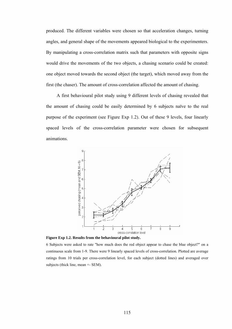
produced. The different variables were chosen so that acceleration changes, turning
angles, and general shape of the movements appeared biological to the experimenters.
By manipulating a cross-correlation matrix such that parameters with opposite signs
would drive the movements of the two objects, a chasing scenario could be created:
one object moved towards the second object (the target), which moved away from the
first (the chaser). The amount of cross-correlation affected the amount of chasing.
A first behavioural pilot study using 9 different levels of chasing revealed that
the amount of chasing could be easily determined by 6 subjects naïve to the real
purpose of the experiment (see Figure Exp 1.2). Out of these 9 levels, four linearly
spaced levels of the cross-correlation parameter were chosen for subsequent
animations.
Figure Exp 1.2. Results from the behavioural pilot study.
6 Subjects were asked to rate "how much does the red object appear to chase the blue object?" on a
continuous scale from 1-9. There were 9 linearly spaced levels of cross-correlation. Plotted are average
ratings from 10 trials per cross-correlation level, for each subject (dotted lines) and averaged over
subjects (thick line, mean +- SEM).
115

Speed
After this initial phase, it was observed that an increase in the cross-correlation
parameter did not only increase the percept of chasing, but also increased speed of
both objects (speed depends on value of the cross-correlation parameter, see Figure
Exp 1.3). This is due to a higher a cross-correlation coefficient resulting in greater
dependence between the object’s movements, making the chasing object follow the
target more clearly, and the target to move away from the chaser more clearly. These
counteracting effects of increasing the attraction/repulsion behaviour increased the
movements of the objects, in terms of speed and area of the screen covered.
Figure Exp 1.3. Speed.
Speed changes of the object's movements due to variations in cross-correlation level. Left panel:
Average speed (mean and standard error of all trials) of both objects in the 8 different types of films
used in the experiment. Speed increased with the level of the cross-correlation parameter, but was
almost identical in the Interactive and Control trials. Right panel: Average speed of both objects over
time in all trials of each level of the cross-correlation parameter, and in their Control conditions: the
speed profile over time was very similar in the Interactive and the Control trials.
To control for this potential confound, it was decided to create matched control
animations, where the speed and general movement characteristics where conserved
but the correlation between the objects' motion was destroyed. To this end, the
116
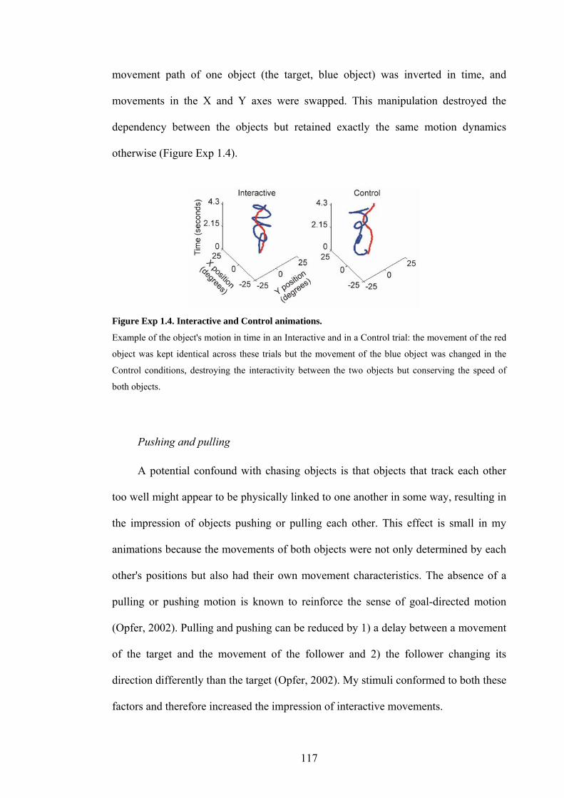
movement path of one object (the target, blue object) was inverted in time, and
movements in the X and Y axes were swapped. This manipulation destroyed the
dependency between the objects but retained exactly the same motion dynamics
otherwise (Figure Exp 1.4).
Figure Exp 1.4. Interactive and Control animations.
Example of the object's motion in time in an Interactive and in a Control trial: the movement of the red
object was kept identical across these trials but the movement of the blue object was changed in the
Control conditions, destroying the interactivity between the two objects but conserving the speed of
both objects.
Pushing and pulling
A potential confound with chasing objects is that objects that track each other
too well might appear to be physically linked to one another in some way, resulting in
the impression of objects pushing or pulling each other. This effect is small in my
animations because the movements of both objects were not only determined by each
other's positions but also had their own movement characteristics. The absence of a
pulling or pushing motion is known to reinforce the sense of goal-directed motion
(Opfer, 2002). Pulling and pushing can be reduced by 1) a delay between a movement
of the target and the movement of the follower and 2) the follower changing its
direction differently than the target (Opfer, 2002). My stimuli conformed to both these
factors and therefore increased the impression of interactive movements.
117
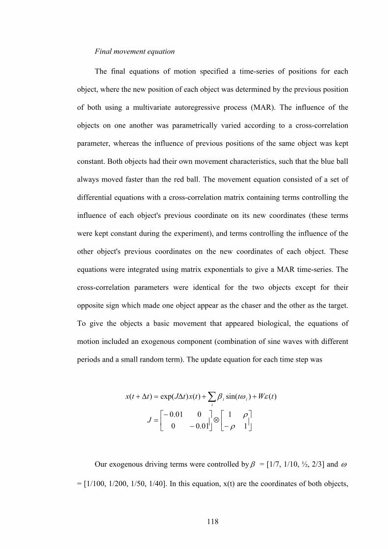
Final movement equation
The final equations of motion specified a time-series of positions for each
object, where the new position of each object was determined by the previous position
of both using a multivariate autoregressive process (MAR). The influence of the
objects on one another was parametrically varied according to a cross-correlation
parameter, whereas the influence of previous positions of the same object was kept
constant. Both objects had their own movement characteristics, such that the blue ball
always moved faster than the red ball. The movement equation consisted of a set of
differential equations with a cross-correlation matrix containing terms controlling the
influence of each object's previous coordinate on its new coordinates (these terms
were kept constant during the experiment), and terms controlling the influence of the
other object's previous coordinates on the new coordinates of each object. These
equations were integrated using matrix exponentials to give a MAR time-series. The
cross-correlation parameters were identical for the two objects except for their
opposite sign which made one object appear as the chaser and the other as the target.
To give the objects a basic movement that appeared biological, the equations of
motion included an exogenous component (combination of sine waves with different
periods and a small random term). The update equation for each time step was
⎥⎦
⎤⎢⎣
⎡−
⊗⎥⎦
⎤⎢⎣
⎡−
−=
++∆=∆+ ∑
11
01.00001.0
)()sin()()exp()(
ρρ
εωβ
J
tWttxtJttxi
ii
Our exogenous driving terms were controlled by β = [1/7, 1/10, ½, 2/3] and ω
= [1/100, 1/200, 1/50, 1/40]. In this equation, x(t) are the coordinates of both objects,
118

x(t-1) are their previous coordinates, J is the system’s Jacobian controlling the
dependencies, W is a constant that scales the random term ),0(~)( tNt ∆ε and t∆ is
the time step between two successive positions. J was based on a cross-correlation
matrix containing the cross-correlation coefficient ρ , which was modified across
conditions and increased the amount of interactivity. This resulted in the impression
of the red ball chasing the blue ball, in a parametric fashion.
Animacy ratings
Our parameter controlling the amount of animacy in the observed animations
being relatively abstract and the stimulus development process being long and
involving many steps, it was necessary to make sure that the final animations still
appeared animate. To this end, I asked 12 volunteers (6 male and 6 female, aged 23-
36), who were unaware of the different experimental conditions, the parameters used
and of the aim of the experiment, to judge how "alive" the objects appeared, on a
scale of 1-4. Results (Figure Exp 1.5) indicated that animations in the Interact
conditions appeared more animate than the Control animations (F(1,10) = 11.3, p <
0.01; repeated-measures 2-way ANOVA, within-subject factors Cross-correlation
level and Interact vs. Control, between-subjects factor gender), and importantly that
animacy ratings increased more with the cross-correlation level in the Interact
conditions compared to the Control conditions (interaction, F(1,10) = 24.3, p < 0.001).
There was no difference due to gender (F(1,10) = 0.001, p > 0.5), and no influence of
gender on either contrast (interaction Interactive vs. Control by gender: F(1,10) = 0.1,
p > 0.5; 3-way interaction: F(1,10) = 1.2, p > 0.2)4. Behavioural reports (see Appendix
4 The design of this experiment was identical to the fMRI protocol: there were eight experimental conditions, 10 repetitions each. Subjects were shown one example of each condition before the experiment, in random order and without explanation. Ratings were averaged over trials and then
119
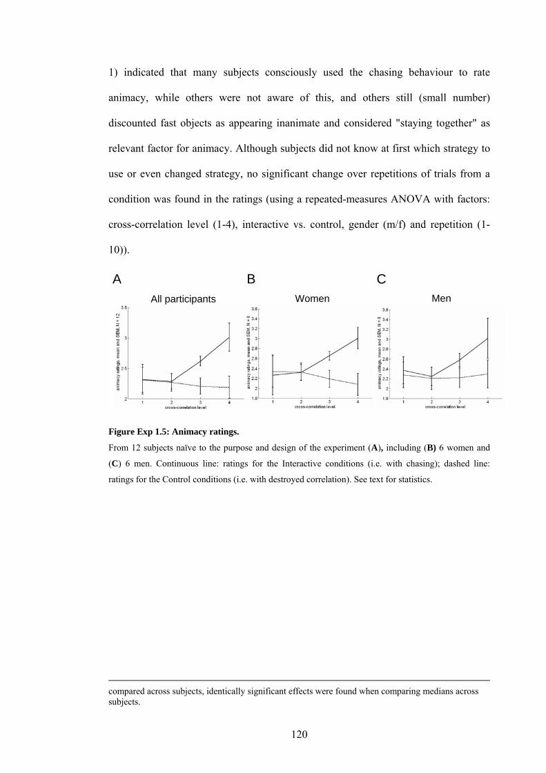
1) indicated that many subjects consciously used the chasing behaviour to rate
animacy, while others were not aware of this, and others still (small number)
discounted fast objects as appearing inanimate and considered "staying together" as
relevant factor for animacy. Although subjects did not know at first which strategy to
use or even changed strategy, no significant change over repetitions of trials from a
condition was found in the ratings (using a repeated-measures ANOVA with factors:
cross-correlation level (1-4), interactive vs. control, gender (m/f) and repetition (1-
10)).
A B C All participants Women Men
Figure Exp 1.5: Animacy ratings.
From 12 subjects naïve to the purpose and design of the experiment (A), including (B) 6 women and
(C) 6 men. Continuous line: ratings for the Interactive conditions (i.e. with chasing); dashed line:
ratings for the Control conditions (i.e. with destroyed correlation). See text for statistics.
compared across subjects, identically significant effects were found when comparing medians across subjects.
120

Methods
Design and conditions
In this experiment I wanted to identify regions whose responses increased in
relationship with the amount of interactivity between two objects, a movement
characteristic which correlates with attribution of animacy to these objects. I also
wanted to test whether these activation changes happen only when subjects pay
attention to the interactions between the objects or also when they perform another,
incidental task, for which animacy is irrelevant. As increases in interactivity are
associated with an increase in the speed of both objects, I used matched control
conditions with identical speed and similar, but uncorrelated movements. I therefore
used a factorial design with the following three factors: 1) four, linearly increasing
levels of interactive motion, 2) two task levels: an interactivity- and a speed-rating
task, and 3) two condition levels: Control and Interactive. Combinations of all these
factors resulted in 16 different trial types (Figure Exp 1.6), each repeated 10 times
during the experiment. The four levels of interactivity were created by manipulating a
cross-correlation parameter, which controlled the dependence between object
movements. Increasing this parameter in increased the objective and subjective
interactivity and their speed. I therefore used a matched Control condition for each
interactivity level, in which speed was identical but interactivity was destroyed
(Figure Exp 1.4). Subjects performed the two tasks on the same stimuli: in the
interactivity-rating task, subjects were asked: "how much does the red object follow
the blue object, one being the minimum and four the maximum?” In the speed-rating
task, the instructions were "how fast do the objects move, one being the minimum and
four the maximum?". They responded by pressing one of four buttons on a keypad
with the corresponding finger of the right hand. Ratings, response times, eye
121

movements and brain activation were recorded simultaneously during the experiment.
On these four types of dependent variable I assessed the effects of interactivity, and
the interaction between task and interactivity.
Figure Exp 1.6. Example of the 8 conditions.
Example of movements in the eight different animations used in both tasks of the experiment, yielding
16 different experimental conditions. Plotted are X and Y positions of the red disk (the Chaser) and the
blue disk (the Target) in visual degrees. The extent of the screen covered and the movement shapes can
be seen, but as the time dimension is lost on these 2-dimensional plots, interactivity cannot be judged.
Stimuli and films used
In the scanner, subjects watched short films (4.3 sec. per film) in which two
moving disks appeared to be either interacting or to move independently from each
other. The moving objects were two disks or balls, 2 degrees wide on the screen
(Figure Exp 1.1). One was red, the other blue, and the background was black. The
movement trajectories of the two disks were determined prior to the experiment by an
equation of motion implemented in Matlab (The MathWorks, Inc, Natick, MA) and
detailed below. This equation made the objects move in a way that appeared
biological and, in addition, allowed parametric control of the interactivity between the
122

two objects by varying the correlation between their movements. The visual display
was controlled by in-house presentation software (Cogent 2000,
http://www.vislab.ucl.ac.uk/Cogent2000/index.html) implemented in Matlab. Films
were projected onto an opaque surface in the scanner by an LCD projector, subjects
viewed them through a set of mirrors mounted on the headcoil.
Eye movements
Subjects were allowed to move their eyes freely during the experiment. To
evaluate potential confounds due to eye movement differences between conditions, I
recorded eye movements using an infrared eye-tracking system recording at 60 Hz
(ASL Model 504, Applied Science Laboratories, Bedford, MA), with remote, custom-
adapted optics for use in the scanner. Reliable eye tracking data throughout the whole
scanning session were only available in nine out of twelve subjects as the remaining
three subjects wore contact lenses, which created artefacts in the eye-tracker
recordings. Eye blinks were removed by eliminating all differences in successive time
points more than 3 standard deviations away from the mean difference, which were
replaced by the average of the positions immediately before and after the replaced
time point. The data in X and Y were mean-corrected, squared, and then summed to
yield a measure of total eye-movements (saccades and smooth pursuits were not
distinguished) for each trial. The results of this procedure were analysed with the
same tests as those used in the analysis of the subjects' ratings.
FMRI analysis: SPM whole-brain analysis
The analysis followed the standard steps as described in the Materials and
Methods section of the present thesis. Realignment was standard, no slice-time
123

correction was used, normalisation was done with resampling to a voxel size of 3
mm3. To enhance the signal to noise ratio and enable intersubject functional
anatomical comparison the images were smoothed by convolution with a 6-mm full
width at half maximum (FWHM) Gaussian kernel. A high-pass filter (using a cut-off
of 128 sec) and a correction for temporal auto-correlation in the data (AR 1 + white
noise) were applied to accommodate serial correlations. The design matrix of the first-
level (single subject) analysis comprised 32 regressors of interest, resulting from the
crossing of the following factors: 1) task (interactivity-rating or speed-rating), 2)
value of cross-correlation parameter [1, 2, 3 or 4] and 3) condition (Interactive or
Control trials), which yields 16 effects of interest; each was modelled using the
canonical hemodynamic response function (HRF) and the first temporal derivative.
Two covariates of no interest were included, made by convolving the average speed
of both objects during each trial with the HRF and its temporal derivative. This
enabled us to model separately the effect of the average object speed on the brain
activation, which was not an effect of interest in the analysis. Differential realignment
parameters were modelled as additional regressors of no interest to model movement-
related artifacts. Second-level analyses were performed as T-tests, after smoothing the
contrast images from the first-level analysis with an 8-mm FWHM kernel. The
threshold used for statistical significance was P<0.05 corrected for multiple
comparisons across the whole brain with inference at the cluster level.
FMRI analysis: regional analyses
In addition to the classical whole-brain voxel-wise analysis (see Methods
section), I performed regional analyses in the following regions of a priori interest: the
posterior superior temporal sulci and fusiform gyri of both hemispheres. These
124

regions of interest (ROI) were based on the average structural image from the
participants (see below), determined by reference to a brain atlas (Duvernoy, 1999)
and drawn with MRIcro software (Rorden & Brett, 2000)
(http://www.cla.sc.edu/psyc/faculty/rorden/mricro.html). They encompassed
coordinates of activation clusters found in several previous studies (see below). The
region of interest in the left fusiform gyrus extended from –52 to –17 mm in the X
dimension, from -72 to –24 mm in the Y dimension, and from –32 to –7 mm in the Z
dimension. The region of interest in the right fusiform gyrus extended from 29 to 48
mm in X, from -59 to -29 mm in Y, and from -31 to -7 mm in Z. Volumes were 8856
mm3 (left) and 8821 mm3 (right). The region of interest around the left posterior
superior temporal sulcus extended from –68 to –53 mm in X, from –66 to –34 in Y,
and from –29 to 0 in Z. The region of interest in the right superior temporal sulcus
extended from 50 to 70 mm in X, from -65 to -37 mm in Y, and from -29 to -2 mm in
Z. Volumes were 9234 mm3 (left) and 10422 mm3 (right). These regions of interest are
displayed in Figure Exp 1.11. Parameter estimates corresponding to the voxels in the
ROI were selected with in-house software based on the SPM2 software package.
These data were then averaged over all voxels within the ROI and tested for the
comparisons of interest (effects of a linear increase in goal-directedness, and task
effects on an increase in goal-directedness) using a repeated-measures 3-way
ANOVA in a statistics package (SPSS 11.0, SPSS Inc., Chicago, Illinois USA).
125

Results
Behavioural results
Ratings
Analysis of participants' ratings from the interactivity-rating task and the speed-
rating task performed in the scanner revealed the following effects (Figure Exp 1.7).
In the Interactive trials (where the movements of the two objects were correlated),
subjects rated the objects as increasingly interactive with increasing cross-correlation
between their movements (repeated-measures ANOVA, F(1,11) = 502.7, P<0.001).
However, subjects also considered the objects’ speed to be increasing with increasing
cross-correlation (F(1,11) = 446.8, P<0.001). Matched control films, in which the
correlation between the objects' movements was destroyed but the speed remained
almost identical, elicited no significant difference in measured object speed relative to
the Interactive trials: F(1,11) = 0.001, p>0.9. Figure Exp 1.3). The Control films were
rated by the subjects as displaying much less interactivity than the Interactive films
(interaction between cross-correlation level and Interactive vs. Control films, F(1,11)
= 29.4, P<0.001) but as having the same speed (F(1,11) = 1.0, p>0.3). These data
suggest that increasing the dependency between the movements of our two objects
increased the percept of interaction.
126

Figure Exp 1.7. Interactivity and Speed ratings.
Subjects’ ratings in the interactivity-rating task (left), and in the speed-rating task (right). On the X axis
are the different levels of the cross-correlation parameter that controlled the interactivity between the
objects' movements from 1 (minimum) to 4 (maximum), repeated for each task. Broken and unbroken
lines correspond respectively to Interactive conditions (with interactive motion) and to their matched
Control conditions (where the interactivity between the movements was destroyed, see Methods). On
the Y axis are the mean and standard errors (over subjects) of the subjects' ratings: 1 corresponds to the
minimum interactivity or speed, 4 corresponds to the maximum.
Response times
Response times are shown in Table Exp 1.1 and Figure Exp 1.8). Response
times were shorter in the Speed-rating task than in the Interactivity-rating task
(T(1,11) = 3.971, p <0.005). As subjects were free to answer at any point of the trial, I
suppose that this is because subjects needed to observe the animations only for a short
amount of time to determine the speed of the objects, as the speed did not vary too
much during the time course of the animation. For the Interactivity task, a number of
cues had to be collected before answering, and these were not distributed equally
during the course of the animation. These plots also revealed a "bell-shaped" variation
of the response times in the Speed-rating task when plotted with respect to the level of
127

contingency (see Figure Exp 1.8). I attribute this to the fact that the trials with extreme
cross-correlation values were easier to categorise in terms of speed than the "middle"
ones, for which subjects had to make a longer decision to attribute the trial to one of
the middle categories.
Speed-rating response times increased with cross-correlation level (linear:
F(1,11) = 34, P<0.001) but were not different in Interactive or Control trials (P>0.5),
and there was no interaction effect between the 2 factors. Plotted, these data show that
the significant effect results mainly from the responses in conditions with the smallest
cross-correlation level being markedly slower than the others. I suggest that this is
because animations with a slow speed were very easy to differentiate from animations
with a higher speed (because in contrast to animations with a higher speed,
movements were limited to only a relatively small part of the screen, see Figure Exp
1.6), but to differentiate animations with cross-correlation levels 2-4 from each other,
subjects had to spend some time to appreciate the movement details before making
their response.
Interactivity-rating response times did not vary linearly with cross-correlation
level (p>0.5), and were not different in Interactive or Control trials (P>0.5), but
responses times in the Control trials increased more with cross-correlation than in
Interactive trials (interaction between linear increase in cross-correlation and
Interactive vs. Control trials: F(1,11) = 25.8, P<0.001). Again, the plotted data reveal
that it is particularly one condition that contributes mostly to the effect: the very fast
response in Interactive trials with maximal cross-correlation (Figure Exp 1.8:
interactivity-rating, condition 4e). This could very well be explained by the following:
when objects moved fast, there were more interactive "events" (due to clear
correlation between movements), which would have helped subjects make their mind
128

up quickly when they saw them. In the matching control trials, subjects did not see
these events, and spent some time searching for them before giving a low interactivity
rating. With respect to analysis of the fMRI data, in which I used this interaction as
contrast of interest, this suggests that the effects I observed might have been slightly
different if the subjects had spent the same amount of time on all trials. But as I
pooled over both tasks (in the 2-way interaction) or compared between them (3-way
interaction), I believe the different effects in the response times of the two tasks
compensate each other mostly, thereby reducing false-positives and false-negatives.
Table Exp 1.1: Response times (12 subjects)
Response times (seconds) Level 1 Level 2 Level 3 Level 4
1) Explicit task
Interactive 3.4(.22) 3.4(.24) 3.5(.16) 2.8(.13)
Control 3.3(.27) 3.3(.21) 3.6(.23) 3.6(.20)
2) Implicit task
Interactive 2.2(.12) 3.0(.11) 3.2(.18) 2.7(.14)
Control 2.2(.13) 3.0(.19) 3.2(.15) 2.9(.11)
129

1e 1c 2e 2c 3e 3c 4e 4c 1e 1c 2e 2c 3e 3c 4e 4c
Interactivity-rating task Speed-rating task
Sec
onds
Sec
onds
Figure Exp 1.8. Response times.
From 12 subjects in all conditions of both tasks, acquired during the fMRI experiment. 1e to 4e
correspond to Interactive trials with cross-correlation levels 1 to 4, 1c to 4c correspond to the matched
control conditions.
Eye movements
As subjects were free to move their eyes while watching the animations, I
recorded their eye movements to test for differences between conditions. To this
effect, I recorded excursion in X and Y and computed the sum-of-squares of total
excursion for each condition, for each subject (see Table Exp 1.2 and Figure Exp 1.9).
The degree of cross-correlation between the objects' movements did not influence eye
movements (repeated-measures 3-way ANOVA, F(1,8) = 0.7, p > 0.4), and there were
no differences in eye movements between Interactive and Control trials (F(1,8) = 2.0,
P=0.2). The only factor that significantly influenced eye movements was the task
performed by the subjects: subjects moved their eyes less in the speed-rating task than
in the interactivity-rating task (interactivity-rating task vs. speed-rating task: F(1,8) =
5.8, P=0.04). As I did not compare directly the two tasks, this difference in eye
movements did not influence the interpretation of the brain activation analysis.
130

Table Exp 1.2: Eye movements (summed over time, 9 subjects)
Eye movements (arb. units) Level 1 Level 2 Level 3 Level 4
1) Explicit task
Interactive 9.3(1.9) 12(2) 10(2) 9.9(1.7)
Control 9.4(1.5) 9.3(1.6) 8.8(1.3) 10(2.3)
2) Implicit task
Interactive 7.3(1.1) 7.5(1.2) 7.7(1.4) 6.6(0.8)
Control 8.0(1.3) 6.4(1.2) 7.4(1.3) 6.2(0.9)
1e 1c 2e 2c 3e 3c 4e 4c 1e 1c 2e 2c 3e 3c 4e 4c
Interactivity-rating task Speed-rating task
Tota
l mov
emen
ts (a
rbitr
ary
units
)
Tota
l mov
emen
ts (a
rbitr
ary
units
)
Figure Exp 1.9. Eye movements.
From all conditions of both tasks for 9 subjects, acquired during the fMRI experiment. 1e to 4e
correspond to Interactive trials with cross-correlation levels 1 to 4, 1c to 4c correspond to the matched
control conditions.
131

Imaging results
Whole-brain
I used the parametric, factorial design of this experiment to test for activation
increases in relation to increases in interactivity displayed by the two objects, and to
test how these interaction-dependent responses were affected by the attentional task
the subject performed. To discount speed effects I assessed interaction-sensitive
activations as the difference between the regression of brain responses on interaction
level and the equivalent speed control trials.
- Main effects
I initially tested for regions with a linear increase in activation correlating with
an increase in the cross-correlation parameter. The regions that showed such an effect
were the fusiform gyrus and the medial occipital cortex, in both hemispheres (see
Table Exp 1.3). I also looked for regions with a greater activation in Interactive
conditions than in Controls, and found significant activation increases in the left
medial occipital cortex (Table Exp 1.3). Both of these main effects did not interest us
particularly, as the comparison of interest was the interaction between Cross-
correlation level and Interactive vs. Control conditions.
Table Exp 1.3: Brain activation data, whole-brain voxel-wise analysis. Significant clusters in the
comparisons of interest, surviving a threshold of P<0.05 corrected for multiple comparisons across the
whole brain (see Methods), at cluster level (*) or voxel level (FWE, **).
Structure Coordinates Size
(mm3)
Z score Brodmann
area
Contingency (linear increase)
L fusiform -24 -75 -12 7929 5.90** 19
132

L medial occipital cortex -6 -84 3 5.39** 17
R medial occipital cortex 3 -87 18 5.14* 18
R fusiform gyrus 27 -48 -6 3.95* 19
Interactive > control
L medial occipital cortex -9 -84 -6 263 3.58 * 18
-21 -93 -9 3.34 * 17
-18 -84 -18 3.33 * 18
- Effects of a linear increase of interactive motion
The following regions showed response increases with the increase in
correlation between the objects' movements in the Interactive condition but no
increases in the Control conditions: the right posterior, ascending branch of the
superior temporal sulcus in the inferior parietal cortex, the left superior temporal
gyrus, the medial occipital cortex in both hemispheres, the right fusiform gyrus, the
caudal part of the anterior cingulate gyrus, and the posterior paracingulate gyrus
(Figure Exp 1.10, Table Exp 1.4). This was tested formally with a two-way interaction
between a linear increase in the cross-correlation factor and the Interactive vs. Control
factor.
Table Exp 1.4: Interaction. Threshold used was identical to main effect (Table Exp 1.3).
Structure Coordinates Size
(mm3)
Z score Brodmann
area
Greater linear increase with contingency in the experimental stimuli
L medial occipital cortex -6 -69 0 549 4.79 *(**) 18
133

R medial occipital cortex 15 -72 -9 4.26 * 18
R fusiform gyrus 30 -63 -6 4.18 * 19
R asc. STS / inferior parietal 39 -57 27 147 4.51 * 39
R post cingulate 3 0 45 186 4.02 * 24
Medial occipital cortex 0 -66 18 243 3.98 * 23
L STG -60 -27 9 120 3.63* 41
L inf parietal / angular gyrus -45 -72 36 78 3.52 ~* 39
Post paracingulate 0 -48 39 117 3.48 * 31
134
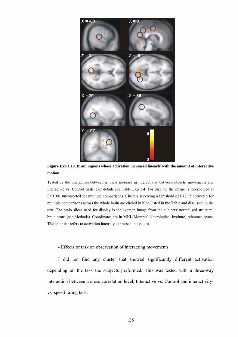
Figure Exp 1.10. Brain regions whose activation increased linearly with the amount of interactive
motion.
Tested by the interaction between a linear increase in interactivity between objects' movements and
Interactive vs. Control trials. For details see Table Exp 1.4. For display, the image is thresholded at
P<0.001 uncorrected for multiple comparisons. Clusters surviving a threshold of P<0.05 corrected for
multiple comparisons across the whole brain are circled in blue, listed in the Table and discussed in the
text. The brain slices used for display is the average image from the subjects' normalised structural
brain scans (see Methods). Coordinates are in MNI (Montreal Neurological Institute) reference space.
The color bar refers to activation intensity expressed in t values.
- Effects of task on observation of interacting movements
I did not find any cluster that showed significantly different activation
depending on the task the subjects performed. This was tested with a three-way
interaction between a cross-correlation level, Interactive vs. Control and interactivity-
vs. speed-rating task.
135

Anatomical regions of interest
As discussed in Part 1, Section 3.1.2, previous studies using similar types of
animations to those used in the present study showed activation in the posterior
superior temporal sulcus, the fusiform gyrus, the temporal poles and the media
prefrontal cortex (Castelli, Happe, Frith, & Frith, 2000; Blakemore et al., 2003;
Schultz et al., 2003). To complement my voxel-wise analysis on the whole brain with
analyses based on regions where I might expect activations, I subsequently examined
activation in pre-defined anatomical regions of interest: the posterior superior
temporal gyrus and fusiform gyrus in both hemispheres (for definition of these areas,
see Methods: fMRI analysis, Figure Exp 1.11 and Table Exp 1.5).
Table Exp 1.5: Regions of interest, characteristic and significant statistics
Region Extent X Extent Y Extent Z Volume
(mm3)
Fusiform L -52 to -17 -72 to -24 -32 to –7.2 1104
Fusiform R 29 to 48 -51 to -29 -31 to –7.2 1080
Posterior STS L -68 to -53 -66 to -34 0 to 29 1053
Posterior STS R 50 to 70 -65 to -37 2 to 29 1212
Activation in the left fusiform region of interest varied with the amount of goal-
directed motion displayed by the two objects (quadratic increase with respect to goal-
directedness and no increase in the control conditions, tested by interaction between
goal-directedness and Interactive vs. control trials: F(1,11) = 15, P=0.003). Activation
in the region of interest in the right fusiform gyrus did not vary significantly with the
amount of goal-directedness exhibited by the moving objects. Activation in the region
136

of interest in the left posterior superior temporal sulcus increased with goal-
directedness, but only when subjects rated the interactivity between the objects, not
when they rated their speed (3-way interaction between task, contingency level, and
Interactive vs. control trials: F(1,11)=9.6, P<0.01). Activation in the right posterior
superior temporal gyrus region of interest was not significantly different across
conditions or interactions between them.
Figure Exp 1.11. VOI anatomy and data.
Volumes of interest in the posterior superior temporal sulcus. Encompassing previous studies of
animate- and intentional-looking moving abstract objects. Yellow dots represent the maximum
intensities of activations found in following two structures: 1) superior temporal sulcus (lettered from a
to g, more than one coordinate per study): Blakemore et al, 2001; Blakemore et al, 2003; Schultz et al,
2003; Castelli et al, 2000; 2) Fusiform gyrus (lettered from a to d): Winston et al, 2002; Blakemore et
al, 2003; Schultz et al, 2003.
Activation time-course
Activation time-courses in the superior temporal sulcus showed peaks 10-12
seconds after animation onset (Figure Exp 1.12 A and B, top panels). Activation
peaks were higher when the objects were interacting than during the control trials
(Mann-Whitney test, interactivity-rating task, cross-correlation level 1: U = 42, speed-
rating task, cross-correlation level 4: U = 29, both P<0.05 one-tailed). Graphic plots
demonstrated the parametric increase of activations in relation to cross-correlation
137
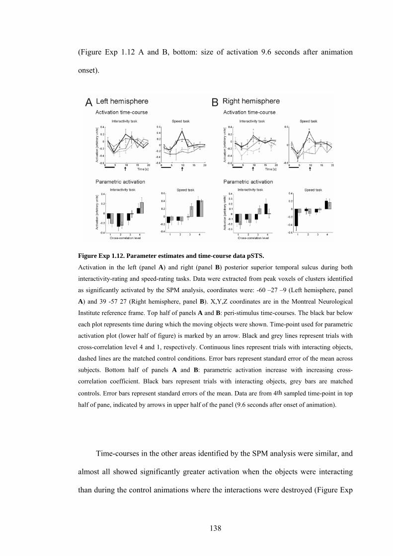
(Figure Exp 1.12 A and B, bottom: size of activation 9.6 seconds after animation
onset).
Figure Exp 1.12. Parameter estimates and time-course data pSTS.
Activation in the left (panel A) and right (panel B) posterior superior temporal sulcus during both
interactivity-rating and speed-rating tasks. Data were extracted from peak voxels of clusters identified
as significantly activated by the SPM analysis, coordinates were: -60 –27 –9 (Left hemisphere, panel
A) and 39 -57 27 (Right hemisphere, panel B). X,Y,Z coordinates are in the Montreal Neurological
Institute reference frame. Top half of panels A and B: peri-stimulus time-courses. The black bar below
each plot represents time during which the moving objects were shown. Time-point used for parametric
activation plot (lower half of figure) is marked by an arrow. Black and grey lines represent trials with
cross-correlation level 4 and 1, respectively. Continuous lines represent trials with interacting objects,
dashed lines are the matched control conditions. Error bars represent standard error of the mean across
subjects. Bottom half of panels A and B: parametric activation increase with increasing cross-
correlation coefficient. Black bars represent trials with interacting objects, grey bars are matched
controls. Error bars represent standard errors of the mean. Data are from 4th sampled time-point in top
half of pane, indicated by arrows in upper half of the panel (9.6 seconds after onset of animation).
Time-courses in the other areas identified by the SPM analysis were similar, and
almost all showed significantly greater activation when the objects were interacting
than during the control animations where the interactions were destroyed (Figure Exp
138

1.13; posterior paracingulate U = 41, P<0.04; caudal part of the anterior cingulate U =
39, P=0.03; right fusiform gyrus: NS in this analysis but very similar; medial occipital
cortex U = 42, P<0.05; all tests one-tailed). As the haemodynamic response is
expected to peak about 6 seconds after a neuronal event and our animations lasted for
4.3 seconds (black bar in Figure Exp 1.12, top), activation should be highest between
6 to 10 seconds after animation onset. While activation in the right fusiform and in the
superior temporal gyrus of both hemispheres peaked at 10-12 seconds after animation
onset, the local maximum in the medial occipital gyrus peaked at 7-10 seconds. The
later peaks I observed in the superior temporal and fusiform gyri could correspond to
a cumulative increase in BOLD response during sustained neuronal activation while
the objects were moving. The later peaks may also reflect continued processing even
after the objects had disappeared, a process which might not be expressed in the
medial occipital cortex.
139

Left medial occipitalRight fusiformPosterior ACCPosterior paracingulate
Activ
atio
n (a
rbitr
ary
units
)
Time (s)
Figure Exp 1.13. Time-course of activation in other regions identified in the Interaction contrast
(see Table Exp 1.4).
Data from Interact task and Speed task, respectively in upper and lower row. Coordinates: posterior
paracingulate cortex: [0 -48 39], posterior part of anterior cingulate cortex (posterior ACC): [3 0 45],
right fusiform gyrus: [30 -63 -6], left medial occipital cortex: [-6 -69 0]. See text above for details and
statistics, and Figure Exp 1.12 for additional information.
Discussion
This study shows that activation in the superior temporal gyrus and in the cortex
surrounding the superior temporal sulcus increases linearly with the interactivity
between the movements of two abstract, self-propelled objects. Directing attention to
the interactions rather than to their speed had no significant impact on activation
increases. These cortical structures are known to respond during observation of
biological motion, such as point-light walkers (Allison, Puce, & McCarthy, 2000;
Pelphrey et al., 2003).
Does the cortex in the superior temporal sulcus and gyrus respond to objective
movement characteristics or to categorical percepts?
140

In previous studies, using abstract moving "agents", a human controlled the
movements of the abstract shapes to create the desired effects (Heider & Simmel,
1944; Castelli, Happe, Frith, & Frith, 2000; Blythe, Todd, & Miller, 1999; Schultz et
al., 2003; Blakemore et al., 2003). Observers can reliably identify which behaviour an
animator intends to give to the objects he controls (Blythe, Todd, & Miller, 1999).
Animated cartoons on television also speak to the capacity to reliably evoke the
impression of complex intentional behaviour in abstract moving shapes. Although it is
very interesting that such stimuli induce activation in the superior temporal region and
in the fusiform gyrus (Blakemore et al., 2001; Blakemore et al., 2003; Castelli, Happe,
Frith, & Frith, 2000; Schultz et al., 2003), it is not clear whether (or which of) these
structures respond to objective characteristics of the objects' movements that indicate
they might be alive (such as parameters of self-propelled or goal-directed motion), or
whether these regions simply respond to anything that appears animate. In my study, I
selectively increased the interactivity between the movements of two abstract objects.
My results show that activation in certain parts of the superior temporal gyrus and the
cortex surrounding the superior temporal sulcus increases in proportion to the amount
of interactivity. This suggests that the superior temporal sulcus region is involved in
processing movement characteristics that characterise living beings rather than simply
responding to the presence of living beings.
Superior temporal sulcus region and social information
Together with the amygdala, the fusiform gyrus, the orbital and the medial
frontal cortex, the superior temporal sulcus region is thought to be part of a network
forming the "social brain" (Brothers, 1990; Adolphs, 2003). Neurophysiological
recordings in the posterior superior temporal area of monkeys revealed cells that
141

respond during observation of biological motion such as a walking person, moving
parts of a face such as mouth and eyes, or whole faces (Desimone, 1991).
Neuroimaging studies of human volunteers yielded similar results (Puce & Perrett,
2003). Pictures and words referring to animals also induce activation increases in the
superior temporal area (Martin, Ungerleider, & Haxby, 2000; Kanwisher, McDermott,
& Chun, 1997). But this area of the brain is also associated with higher cognitive
functions, such as the attribution of mental states to other living organisms (Frith &
Frith, 1999; Frith & Frith, 2003; Saxe & Kanwisher, 2003) and even to moving
abstract shapes (Castelli, Happe, Frith, & Frith, 2000; Blakemore et al., 2001; Schultz
et al., 2003; Blakemore et al., 2003). Activity in the posterior superior temporal sulcus
region has previously been found during imitation of human actions (Iacoboni et al.,
2001), which is performed mostly by imitating an action's goals rather than the actual
movements (Wohlschlaeger, Gattis, & Bekkering, 2003; Koski, Iacoboni, Dubeau,
Woods, & Mazziotta, 2003). This brain region is also thought to extract intentional
cues from goal-oriented human behaviour (Toni, Thoenissen, & Zilles, 2001). My
results show that this region is engaged by the observation of objects moving in a self-
propelled and interactive way, which are movement characteristics associated with
animate beings. Therefore my results are compatible with a role of the superior
temporal region in identifying animate agents in the environment based on their
movement, a necessary step in the processing of social information.
Animacy and agency
142
Other interesting studies that it might be relevant to compare the present results
with are two recent studies of agency by Farrer and colleagues (Farrer & Frith, 2002;
Farrer et al., 2003), in which a region in the right angular gyrus responded when
subjects attributed actions they saw to a person different from themselves. These
activations are located only a few millimeters away from the activation I labelled right
pSTS. With respect to the idea that the angular gyrus is activated when subjects
attribute actions they see to an agent different than themselves, one might interpret
our current results in the sense that with increasing clarity of the observed interaction
between the moving objects, the perception of another agent also increased, therefore
increasing the activation in the area thought to be involved in attribution of agency.
Other regions engaged by interacting objects: fusiform gyrus and medial
occipital cortex
Significant activation increases corresponding to increasing interactivity were
also found in the fusiform gyrus and the medial occipital cortex. Previous studies
showed activation increases in the fusiform gyrus during presentation of pictures of
faces (Haxby et al., 1994; Puce, Allison, Gore, & McCarthy, 1995; Kanwisher,
McDermott, & Chun, 1997) and of living beings (Chao, Martin, & Haxby, 1999;
Chao, Haxby, & Martin, 1999), and during observation of moving, abstract "agents"
(Castelli, Happe, Frith, & Frith, 2000; Schultz et al., 2003). Activation in the medial
occipital cortex is also known to increase during observation and naming of pictures
of animals but not tools (Perani et al., 1995; Martin, Wiggs, Ungerleider, & Haxby,
1996; Damasio, Grabowski, Tranel, Hichwa, & Damasio, 1996; Martin, Ungerleider,
& Haxby, 2000). Lesions of the medial occipital lobe (particularly in the left
hemisphere) are associated with a specific semantic knowledge deficit for animals
143

(Nielsen, 1958; Tranel, Damasio, & Damasio, 1997). As reviewed above, goal-
directed motion and self-propelled movement appear to be the main cues for the
attribution of animacy to abstract objects. It is therefore possible that activity in the
fusiform gyrus and the medial occipital cortex increased with increasing interactivity
because the moving objects appeared increasingly animate and animal-like.
144

Chapter 2. Experiment 2: Study of goal-attribution in a chasing paradigm
Methods
Design, conditions and tasks
In this experiment, I wanted to test whether moving objects elicit stronger
activation in the posterior superior temporal sulcus and gyrus when they appear to
attribute goals to a target object than when they appear to chase their target without
attributing goals to it. I used a chasing situation where one object tried to catch
another object by using one of two strategies. The chasing object appeared to attribute
a goal to the target object by heading towards the end-point of the target's position, as
if it had predicted where the target would go ("Predict" strategy), or simply followed
it (no goal attribution, "Follow" strategy). I used matched control conditions without
chasing for each experimental condition to control for differences in object motion
across conditions.
I also wanted to test whether activation changes happen only when subjects pay
attention to the strategy used by the objects or also when they perform another,
incidental task. To this end, I crossed the Strategy factor described above with an
Outcome factor: the chasing object reached the target object in only 50% of cases,
allowing the performance of an incidental task using the same animations. I therefore
had two tasks, as follows: 1) in the Strategy task, subjects categorised the chaser's
strategy into "following" or "using prediction of the trajectory endpoint"; 2) in the
Outcome task (the incidental task), they had to decide whether the chaser caught the
target at the end of the trial or not. During the control trials, subjects were asked to
press one of the two response buttons, at random. I therefore used a factorial design
with the following four factors: 1) two strategies for the chasing object: predicting or
145

following, 2) two outcome levels: target caught or missed, 3) two task levels: judging
strategy or judging outcome, and 4) two condition levels: chase and control (no
chase). For examples see Figure Exp 2.1. Combinations of all these factors resulted in
16 different trial types, each repeated 24 times during the experiment, for a total
scanning time of 19.5 minutes. Subjects responded by pressing one of two buttons on
a keypad with the corresponding finger of the right hand. Ratings, response times and
brain activation were recorded simultaneously during the experiment. On these two
types of dependent variable I assessed the effects of strategy and the interaction
between task and strategy.
Figure Exp 2.1. Design and stimuli.
The open red arrow indicates time at which the blue object started to move. Four experimental
conditions come from the combination of two factors in the experiment: Predict strategy vs. Follow
strategy and Catch outcome vs. No catch outcome. Below on the left is the specific control condition
for the Follow Catch condition; such specific control conditions existed for all four experimental
conditions, resulting in 8 conditions in total. Subjects categorised either the strategy or the outcome of
the blue object’s movement in separate tasks on the same animations, therefore the total number of
conditions x task combinations in the experiment is 16. The objects moved to any of the four corners of
the screen, with equal frequency.
146

Stimuli
Participants in the scanner watched short animations (4.2 sec. per animation) in
which two moving disks appeared to be either interacting or to move independently
from each other (Figure Exp 2.1). The moving objects were two disks or balls, with a
width on the screen in the scanner of 2 degrees of visual angle (Figure Exp 2.1, top
left). One was coloured bright red, the other bright blue, and the background was
black. The movement trajectories of the two disks were determined prior to the
experiment by an equation of motion implemented in Matlab (The MathWorks, Inc,
Natick, MA). The visual display was controlled by in-house presentation software
(Cogent 2000, http://www.vislab.ucl.ac.uk/Cogent2000/index.html) implemented in
Matlab. Animations were projected onto an opaque surface in the scanner by an LCD
projector, subjects viewed them through a mirror mounted on the headcoil.
In all the animations, the red object (the target) started to move first, describing
an arc from the center of the screen that ended in one of the four corners of the screen
(the example shown in Figure Exp 2.1 ends in the bottom left corner; during the
experiment, an equal number of trials with endpoint in each corner were shown). The
blue object (the chaser) started 0.93 seconds after the red object, and in half the trials
it tried to catch it (Chasing trials), in the other half of the trials it moved in the
opposite direction from the red object (Control trials). When trying to catch the red
object, the blue object either simply followed the target's trajectory or it went directly
to the endpoint of the target's movement, as if it had known the end-point of the red
object's trajectory. In half of the Chasing trials the blue object caught the red object, in
the other half it missed it by 6 degrees.
147

FMRI analysis
The analysis followed the standard steps as described in the Materials and
Methods section of the present thesis. Realignment was standard, no slice-time
correction was used, and normalisation was done with resampling to a voxel size of 2
mm3. To enhance the signal to noise ratio and enable intersubject functional
anatomical comparison the images were smoothed by convolution with a 6-mm full
width at half maximum (FWHM) Gaussian kernel. A high-pass filter (using a cut-off
of 128 sec) and a correction for temporal auto-correlation in the data (AR 1 + white
noise) were applied to accommodate serial correlations. The design matrix of the first-
level (single subject) analysis comprised 32 regressors of interest, resulting from the
crossing of the following factors: i) following or predicting strategy of the chaser, ii)
successful or unsuccessful outcome iii) task (strategy-categorisation or outcome-
categorisation), or iv) animations with chasing or controls, which yields 16 effects of
interest; each was modelled using the canonical hemodynamic response function
(HRF) and the first temporal derivative. Differential realignment parameters were
modelled as regressors of no interest to model movement-related artifacts. Second-
level analyses were performed as T-tests, after smoothing the contrast images from
the first-level analysis with an 8-mm FWHM kernel. For the analysis of task effects, I
was particularly interested in identifying regions that showed greater activation in the
explicit task than in the implicit task. To assess these effects, I used both a 3-way
interaction between all factors in the experiment, and an exclusive masking procedure,
which enables more sensitive comparisons between contrasts of interest by allowing
variable thresholding of the contrast used as a mask. Both analyses are standard
procedures implemented in SPM2. For the exclusive masking analysis, I first assessed
separately the interaction between predictive vs. following strategy and chase vs.
148

control trials in the strategy task and in the outcome task. I then masked the results of
this contrast in the Strategy task with the results of the same contrast in the Outcome,
thresholding the mask at P=0.05 uncorrected (the default in SPM2 for masking
procedures). To identify regions more active in the implicit than the explicit task, I
reversed the mask and contrast of interest, also thresholding the mask at P=0.05
uncorrected. Similar results in both masking procedures were obtained with all
thresholds below P=0.15, discussed and presented in Figure Exp 2.5 are analyses
using the mask thresholded at P=0.05.
Results
Behavioural results
Ratings
Analysis of participants' ratings from the Strategy task and the Outcome task
performed in the scanner showed that participants performed well in both the Strategy
and the Outcome task. Subjects had to perform the tasks only when a chase between
the two objects took place, and they correctly identified the strategy of the chasing
object in 94 percent of Predict trials and 91 percent of Follow trials (+- 3 percent SEM
over subjects). Positive outcome (i.e. a successful chase) was identified correctly in 89
percent of trials (+- 4) and negative outcome (i.e. an unsuccessful chase) was
identified correctly in 79 percent of trials (+- 5.6). There were no significant
differences between ratings of Predict and Follow trials (paired T-test, p>0.05), but
Positive Outcome trials were more often identified correctly than Negative Outcome
trials (paired T-test, P<0.05). This difference probably stems from subjects
responding too quickly and mis-categorising some Negative Outcome trials as
149

Positive Outcome trials. This difference does not influence the interpretation of the
brain activation analysis because trials with different outcomes were not compared
with each other and only served as an implicit task.
Strategy Outcome
*
Predict Follow
Perc
enta
g e c
orre
ct100
95
90
85
80
75
70
100
95
90
85
80
75
70Catch Miss
* p<0.05
Figure Exp 2.2. Ratings in the Strategy and the Outcome task.
Acquired during the fMRI experiment. N = 14, error bars represent S.E.M.
Response times
Response times (Figure Exp 2.3) were shorter in the Strategy task than in the
Outcome task (t (1,13) = -4.684, P<0.001; paired T-test). This was to be expected:
subjects were allowed to respond as soon as they had made up their mind in the trial,
which would take longer in the Outcome task. In this task, they had to wait until the
end of the trial to see it the Chaser reached the Target, while in the Strategy task, the
Chaser's strategy was apparent from early on.
In the Strategy task, response times were slightly longer in the Predict than in
the Follow trials (F(1,13) = 8.3, P<0.05; repeated-measures 2-way ANOVA, factors =
Predict vs. Follow, and Experimental vs. Control), but there was no difference
between Experimental and Control trials (p>0.5), and no interaction between
Experimental vs. Control and Predict vs. Follow factors (p>0.2). Planned post-hoc
150

tests showed no difference between Predict experimental vs. Predict control or
between Follow experimental vs. Follow control (all p>0.05). The difference between
Predict and Follow trials could be explained by a difference in difficulty (subjects had
a tendency to be better at correctly identifying Predict than Follow trials, see Figure
Exp 2.2). As there is no significant interaction between factors, these results do not
affect the interpretation of my fMRI data, as I focused my analysis precisely on the
interaction contrast (see fMRI results below).
In the Outcome task, response times were similar in the Catch and Miss trials,
(p>0.05; repeated-measures 2-way ANOVA, factors = Catch vs. Miss, and
Experimental vs. Control), but there was a difference between Experimental and
Control trials (F(1,13) = 21.1, P=0.01), and a significant interaction between the 2
factors (F(1,13) = 9.0, P<0.05). Planned post-hoc tests showed that response times in
Catch Experimental were longer than in Catch Control (F(1,13) = 21.8, P<0.001); as
was the case in the comparison Miss Experimental vs. Miss Control (F(1,13) = 30.0
P<0.001). That subjects took longer to respond in the Experimental than the Control
trials for both Catch and Miss trials is most probably related to the fact mentioned
above: in the Experimental trials, they had to wait until almost the end of the trial
before deciding whether the Target was reached, but not so in the Control trials were
objects were not chasing each other. The interaction can be explained by the same
effect as seen in the task performance data: subjects probably found it more difficult
to identify Catch than Miss trials (they miscategorised more Miss as Catch trials than
the opposite), which can explain why they spent more time before making up their
mind during Experimental Miss trials. Again, these effects did not interest us because
they do not influence the interpretation of the brain activation analysis because trials
151
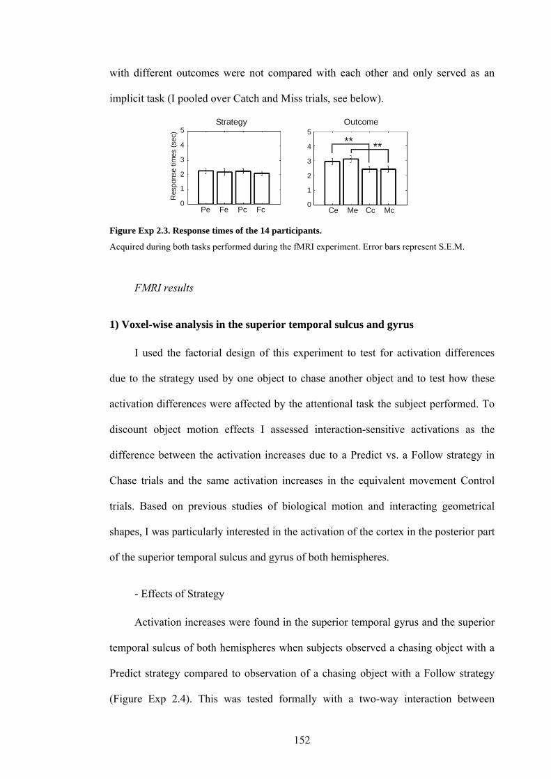
with different outcomes were not compared with each other and only served as an
implicit task (I pooled over Catch and Miss trials, see below).
** **
5
0
4
3
2
1
Res
pons
e tim
es (s
ec) 5
4
3
2
1
Pe Fe Pc Fc Ce Me Cc Mc
Strategy Outcome
0
Figure Exp 2.3. Response times of the 14 participants.
Acquired during both tasks performed during the fMRI experiment. Error bars represent S.E.M.
FMRI results
1) Voxel-wise analysis in the superior temporal sulcus and gyrus
I used the factorial design of this experiment to test for activation differences
due to the strategy used by one object to chase another object and to test how these
activation differences were affected by the attentional task the subject performed. To
discount object motion effects I assessed interaction-sensitive activations as the
difference between the activation increases due to a Predict vs. a Follow strategy in
Chase trials and the same activation increases in the equivalent movement Control
trials. Based on previous studies of biological motion and interacting geometrical
shapes, I was particularly interested in the activation of the cortex in the posterior part
of the superior temporal sulcus and gyrus of both hemispheres.
- Effects of Strategy
Activation increases were found in the superior temporal gyrus and the superior
temporal sulcus of both hemispheres when subjects observed a chasing object with a
Predict strategy compared to observation of a chasing object with a Follow strategy
(Figure Exp 2.4). This was tested formally with a two-way interaction between
152

Predict vs. Follow strategies and Chase vs. Control trials to discount differences in
object movement. The opposite contrast yielded no significant activation increases in
the superior temporal sulcus or gyrus.
Table Exp 2.1. FMRI data, voxel-wise analysis. Significant clusters in the comparisons of interest,
surviving a threshold of P<0.05 corrected for multiple comparisons across voxels in the superior
temporal sulcus or gyrus (see Methods).
Structure Coordinates Size (mm3) Z score
Predict > Follow strategy * Chase vs. Control
R superior temporal gyrus 48 -44 12 600 4.48
L middle temporal gyrus -60 -56 4 176 3.83
L superior temporal gyrus -56 -30 4 584 3.58
Predict > Follow strategy * Chase vs. Control, Strategy task only
L superior temporal gyrus -54 -34 4 936 4.2
Predict > Follow strategy * Chase vs. Control, Strategy task masked by Outcome task
L superior temporal gyrus -54 -34 4 784 4.2
153

Figure Exp 2.4. Brain areas responding more to Predict than Follow trials, irrespective of task.
Clusters in the superior temporal sulcus and gyrus whose activation was significantly greater during
observation of a Predict strategy than a Follow strategy. Tested formally with a two-way interaction
between Predict vs. Follow strategies and Chase vs. Control trials. For details see Table Exp 3.1. For
display, the image is thresholded at P<0.001 uncorrected for multiple comparisons. Clusters surviving a
threshold of P<0.05 corrected for multiple comparisons across anatomical search regions are circled in
blue, listed in Table Exp 3.1 and discussed in the text. The brain slices used for display is the average
image from the subjects' normalised structural brain scans (see Methods). Coordinates are in MNI
(Montreal Neurological Institute) reference space. The color bar refers to activation intensity expressed
in t values.
- Effects of task on strategy
When tested as interactions between Strategy and Task, no cluster showed
significantly different activation depending on the task the subjects performed. This
was tested by the following three-way interactions: interaction between Task
(Strategy vs. Outcome), Strategy (Predict vs. Follow) and Chase vs. Control trial, and
interaction between Task (Outcome vs. Strategy), Strategy (Predict vs. Follow) and
Chase vs. Control trial. However, a cluster of voxels in the left posterior superior
154

temporal sulcus was found whose activation was greater during observation of a
Predict vs. a Follow strategy when subjects performed the Strategy categorisation
task, but was not significantly activated when subjects performed the Outcome task.
When I masked (exclusive masking) the results of the comparison in the Strategy task
with those of the Outcome task, the cluster in the left posterior superior temporal
sulcus identified in the Strategy task remained significantly activated (Figure Exp 2.5
a). This suggests that voxels in this cluster respond only when subjects performed the
Strategy task. Masking the results of the comparison in the Outcome task with those
of the Strategy task yielded a cluster of voxels in the right middle temporal gyrus,
located caudal and ventral to the cluster of activation in the posterior superior
temporal sulcus mentioned above. This cluster was not located within the superior
temporal sulcus and gyrus search regions (Figure Exp 2.5 b).
155

Strategy Outcome
pred foll pred foll
Strategy Outcome
pred foll pred foll
pred foll pred foll
Strategy Outcome
pred foll pred foll
Strategy Outcome
Figure Exp 2.5. Brain regions: task effects.
Clusters in the superior temporal sulcus and gyrus whose activation was significantly greater during
observation of a Predict strategy than a Follow strategy and was affected by the subjects' task. A:
activations in the Strategy task masked with activation in the Outcome task (exclusive mask, threshold
of mask: P=0.05 uncorrected). B: activations in the Outcome task masked with activation in the
Strategy task (exclusive mask, threshold of mask: P=0.05 uncorrected). The cluster circled in blue
survived a threshold of P<0.05 corrected for multiple comparisons across anatomical search regions,
was listed in Table Exp 3.1 and discussed in the text. The cluster circled in red was activated to a
similar degree but was not located within the search regions. The brain slices, coordinate system and
color bar index are similar to Figure Exp 2.4.
2) Activation time-courses
Activation time-courses in the voxels of the superior temporal sulcus and
gyrus with strongest activation increase during observation of a Predict vs. a Follow
strategy confirmed the results of the voxel-wise analysis (Figure Exp 2.6). In the
superior temporal sulcus in the left hemisphere, activation during the initial phase of
the trial was higher in Predict trials than in Follow trials, but only when subjects
performed the Strategy task (repeated-measures 2-way ANOVA: interaction between
156
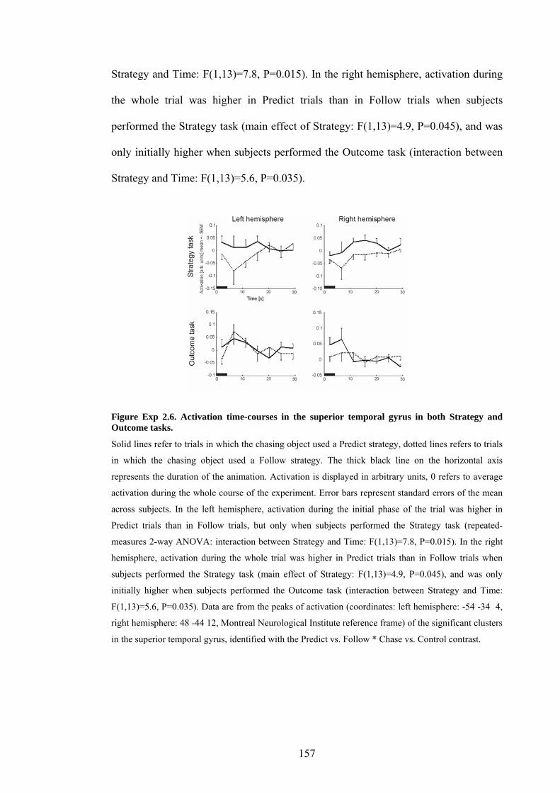
Strategy and Time: F(1,13)=7.8, P=0.015). In the right hemisphere, activation during
the whole trial was higher in Predict trials than in Follow trials when subjects
performed the Strategy task (main effect of Strategy: F(1,13)=4.9, P=0.045), and was
only initially higher when subjects performed the Outcome task (interaction between
Strategy and Time: F(1,13)=5.6, P=0.035).
Figure Exp 2.6. Activation time-courses in the superior temporal gyrus in both Strategy and Outcome tasks.
Solid lines refer to trials in which the chasing object used a Predict strategy, dotted lines refers to trials
in which the chasing object used a Follow strategy. The thick black line on the horizontal axis
represents the duration of the animation. Activation is displayed in arbitrary units, 0 refers to average
activation during the whole course of the experiment. Error bars represent standard errors of the mean
across subjects. In the left hemisphere, activation during the initial phase of the trial was higher in
Predict trials than in Follow trials, but only when subjects performed the Strategy task (repeated-
measures 2-way ANOVA: interaction between Strategy and Time: F(1,13)=7.8, P=0.015). In the right
hemisphere, activation during the whole trial was higher in Predict trials than in Follow trials when
subjects performed the Strategy task (main effect of Strategy: F(1,13)=4.9, P=0.045), and was only
initially higher when subjects performed the Outcome task (interaction between Strategy and Time:
F(1,13)=5.6, P=0.035). Data are from the peaks of activation (coordinates: left hemisphere: -54 -34 4,
right hemisphere: 48 -44 12, Montreal Neurological Institute reference frame) of the significant clusters
in the superior temporal gyrus, identified with the Predict vs. Follow * Chase vs. Control contrast.
157

Discussion
This study shows that activation in the superior temporal gyrus and in the cortex
surrounding the superior temporal sulcus increases when a simple object appears to
chase another object by understanding the target's goal and predicting its movement
instead of simply following it. Directing attention to the object's strategy rather than to
the outcome of its chase further increased activation in the left superior temporal
gyrus.
Attribution of intentions and goal-directed movements and the superior
temporal sulcus and gyrus
A number of studies have shown activation increases in the posterior part of the
superior temporal sulcus and gyrus (among other regions) during attribution of mental
states to other agents, also called mentalising or theory-of-mind (Frith & Frith, 2003).
The exact mechanism on which this ability is based is not fully determined yet, but
intuitively, a necessary first step towards the attribution of mental states to an agent
could be the attribution of goals to this agent. When moving objects appear to follow
goals, they are more likely to be considered animate by human observers (Opfer,
2002; Csibra, 2003). Previous studies show that activation in the superior temporal
sulcus and gyrus increases during observation of geometrical shapes if their
movements appear goal-directed or to have intentions (Castelli, Happe, Frith, & Frith,
2000). These data suggest that this part of the cortex is sensitive to movement
characteristics associated with living entities, especially goal-directed movements. In
my present study, participants observed an object understanding the goals of another
object and predicting its movements or simply following it in order to catch it. As
predicting the movements of a target object is a characteristic associated with a
potential agent, the greater activation in the superior temporal sulcus and gyrus when
158

the chasing object predicted the movements of the target object compared to simply
following it suggests that this brain region plays a role in the identification of
intentional agents (i.e. agents that appear to have intentions).
Attention effects
The cluster identified in the left superior temporal gyrus showed greater
activation increases in Predict trials compared to Follow trials when subjects tried to
identify the strategy of the chaser than when they assessed the outcome of the chase
(Figure Exp 2.5a). As discussed in the introduction, activation increases in the
superior temporal gyrus area have been reported in the right hemisphere when
attending to the emotion or the trustworthiness of faces, and in the left hemisphere
when attending to contingency between the movements of abstract objects
(Blakemore et al., 2003). Blakemore and colleagues suggested that the attention
effects they observed could be due to top-down effects related to the search for agents,
rather than bottom-up detection on the basis of visual cues. In my case, attending to
the strategy of the moving agents, while not being exactly equivalent to searching for
agents, cued subjects into paying attention to characteristics of living organisms,
rather than simply comparing the positions of the chaser to the position of the target at
the end of the trial in the Outcome task. I suggest that the active process of searching
either for agents or for characteristics of their movements can increase or prolong
neural responses in areas already sensitive to entities displaying potentially goal-
directed behaviour.
159

Time-course: early activations vs. constant
In the left hemisphere, activation during the initial phase of the trial was higher
in Predict trials than in Follow trials when subjects performed the Strategy task; in the
right hemisphere, a similar pattern was observed during the Outcome task (Figure Exp
2.6). Activation in the right hemisphere was higher in Predict trials than in Follow
trials during the whole trial when subjects performed the Strategy task. In the two
cases where the activation was only higher in Predict than Follow trials at the
beginning of the trial, plots of the data show that the activation difference was present
at 6.75 seconds after trial onset (Figure Exp 2.6, top left and bottom right), which is
compatible with a neural event occurring at the onset of the trial. At the next sampled
time point (11.25 seconds), the activation difference in the left hemisphere was less
important and it disappeared in the right hemisphere. I suggest that this reflects an
initial and automatic response of the cortex to our animations, which is increased
and/or prolonged when subjects pay attention to a socially relevant dimension of the
objects’ movement. The time-course analyses suggest that this response is stronger in
the right hemisphere, but as the SPM analyses do not confirm this tendency, I cannot
argue strongly in favour of hemispheric specialisation in this task.
No activation increases in the medial prefrontal cortex
It is interesting to note that I did not find activation increases in the medial
prefrontal cortex or the temporal poles in all the contrasts I tested, areas which,
together with the superior temporal sulcus, are thought to be involved in the
attribution of mental states (Frith & Frith, 2003). Previous PET and fMRI studies
using complex animations leading to attribution of mental states have shown
activation in these areas (Castelli, Happe, Frith, & Frith, 2000; Schultz et al., 2003),
but studies using simple animations have not (Blakemore et al., 2001; Blakemore et
160

al., 2003). The latter studies used animations with objects interacting causally,
appearing animate, and/or interacting contingently, which are not thought to lead to
attribution of mental states. In my present study, observers watching an object
attributing a goal to another object did not show a significantly greater activation in
the medial prefrontal cortex than when watching an object following another object,
regardless of the task the subjects were performing. I suggest that subjects solved this
simple task with only a small mentalising effort or none at all, which was not
sufficient to induce activation in the medial prefrontal cortex. While activation
increase in the superior temporal sulcus could be related to mentalising, I would rather
suggest that it is due to detection of entities displaying goal-directed behaviour or
appearing to have intentions, which could explain why activation in the medial
prefrontal cortex was not increased. This view is supported by informal reports from
my subjects, and is consistent with results and interpretation of a previous study
(Blakemore et al., 2003).
Differences in movement paths
A possible alternative explanation of activation differences caused by the
observation of objects with different movement paths could be the difference in
complexity between the paths. In the present experiment, this was controlled by
matched control conditions in which the paths of both chasing and target objects were
identical to the paths in the chasing trials except for the fact that the chasing object did
not move towards the target object but in the opposite direction. This control
condition only affected the contingency between the objects’ paths and not the
movement paths themselves. Objects moving in opposite directions could be seen to
be avoiding instead of approaching each other, and therefore represent another form
of social interaction, which could also induce activation in the superior temporal
161

sulcus. Although this is a very valid possibility, I observed activation increases in the
superior temporal sulcus and gyrus not only in the interaction between Chase vs.
Control and Predict vs. Follow strategy, but also in the comparison of all Chase with
all Control trials (data not shown). Also, within Chase trials, the activation was
stronger when the chasing object appeared to attribute a goal to the target object than
when it just followed it. All these data support my interpretation suggesting that goal
attribution by the Chaser induces activation increases in the superior temporal sulcus
and gyrus, and that attention to the object’s strategy further increases the activation.
Specialisation along the superior temporal sulcus?
The cortex in the superior temporal sulcus and gyrus responds to i) the
attribution of mental states to others, ii) the identification of biological motion and iii)
the response to animate-looking, moving geometrical shapes. Whether different parts
of the cortex in the sulcus and gyrus are specialised for each of these functions or not
is not yet known, although recent unpublished results from a study by Saxe, Xiao,
Kovacs, Perrett and Kanwisher suggest that different parts of the pSTS area respond
to biological motion or to false belief stories (discussed in Saxe & Kanwisher, 2003).
Against the idea of specialisation is the fact that activation increases during
observation of biological motion have been found all along the superior temporal
sulcus (Puce & Perrett, 2003) and do not appear organised in a particular way.
Instead, a number of studies of all three types of processes listed above have yielded
activation in the same area of the superior temporal gyrus and sulcus (Figure Exp 2.7).
The activation increases found in the present study are also located in the pSTS
area. This suggests that this region of the cortex could be involved in a common
aspect of these three tasks, for example the response to goal-directed movements,
whether expressed by moving abstract shapes or human beings.
162

-
Figure Exp 2.7. Comparison between STS/STG activation in all 3 experiments.
Experiments 3 in yellow with red contour, experiment 1 in green and experiment 2 in blue and red,
respectively for task-dependent and non-task-dependent regions, and previous studies (in black) in
following topics: mentalising (Fletcher et al., 1995; Goel, Grafman, & Hallett, 1995; Gallagher et al.,
2000; Brunet, Sarfati, Hardy-Bayle, & Decety, 2000; Vogeley et al., 2001), observation of animate
agents (Castelli, Happe, Frith, & Frith, 2000; Schultz et al., 2003), biological motion (Bonda, Petrides,
Ostry, & Evans, 1996; Grossman et al., 2000; Puce, Allison, Bentin, Gore, & McCarthy, 1998; Grezes
& Costes, 1998; Grezes et al., 2001; Wicker, Michel, Henaff, & Decety, 1998; Hoffman & Haxby,
2000; Campbell et al., 2001) and detection of change (Downar, Crawley, Mikulis, & Davis, 2000).
Displayed on Maximum Intensity Projection glass brain with SPM2. Full coordinates used: Experiment
1: [-60 –27 9; 39 –57 27], experiment 2: [-60 –56 4; -54 –34 4; 48 –44 12], Fletcher 1995: [-44 -58 20;
42 -50 20], Goel 1995: [-42 -70 20; -44 -64 20], Gallagher 2000: [-54 -66 22; 60 -48 22], Castelli 2000:
[-58 -48 4; 60 -56 12], Brunet 2000: [-64 -42 2; 52 -46 0], Vogeley 2001: [-46 -44 22; 58 -56 12],
Schultz 2003: [-57 -61 19; 51 -57 15], Grezes 2001: [-35 -75 10], Bonda 1996: [-48 -61 17; 56 -54 8],
Grossman 2000: [-54 -60 13; 44 -60 19], Puce 1998: [-48 -51 4; 49 -51 5], Campbell 2001: [-58 -44 15;
52 -36 9], Grezes 1998: [-60 -46 10; 64 -38 16], Wicker 1998: [-44 -68 4; 42 -58 8], Hoffman 2000: [-
48 -60 8; 48 -60 8], Downar 2000: [-54 -48 10].
163

Chapter 3. Experiment 3: action observation
3.1. Introduction and overview
When someone mimes or pretends to perform an action, her or his movements
are not directed towards a present object, and are thought to be based on stored
representations of the target object rather than perceptual input (Goodale, Jakobson, &
Keillor, 1994). The following data suggest that mimed movements are controlled by
different processes from object-directed actions. Mimed movements can have
different kinematics from object-directed or actual movements (Goodale, Jakobson, &
Keillor, 1994) and are much less affected by visual illusions (Westwood, Chapman, &
Roy, 2000) than are actual movements (Aglioti, DeSouza, & Goodale, 1995;
Haffenden & Goodale, 1998; Ellis, Flanagan, & Lederman, 1999; Flanagan &
Beltzner, 2000). These data, and the report of a patient with ventral visual stream
lesions (James, Culham, Humphrey, Milner, & Goodale, 2003) who is unable to
perform mimed movements based on perceptual cues (Goodale, Jakobson, & Keillor,
1994), suggest that neural structures underlying control of mimed actions are located
in the ventral rather than the dorsal visual stream (Milner & Goodale, 1995;
Westwood, Chapman, & Roy, 2000). Thus, the fact that actual and mimed movements
differ based on their object-directedness might determine their control by different
visual streams.
Observing and imitating object-directed actions engages ventral and dorsal
parietal, ventral and dorsal premotor, and superior temporal areas (Iacoboni et al.,
2001; Buccino et al., 2001; Koski, Iacoboni, Dubeau, Woods, & Mazziotta, 2003;
Grezes & Decety, 2001; Grezes, Armony, Rowe, & Passingham, 2003). A growing
wealth of data suggests that the superior temporal sulcus is involved in the processing
164

of information relevant to social communication and interaction, including goal-
directed behaviour (displayed as humans reaching and grasping for objects or as
interacting abstract objects), biological motion, body parts, faces, eye gaze (for
reviews, see: Puce & Perrett, 2003; Haxby, Hoffman, & Gobbini, 2002; Adolphs,
2003). While all these types of information involve some kind of motion, this region
of the brain is also involved in more cognitive tasks, such as mentalising (Frith &
Frith, 2003). The inferior premotor area responds also during observation of object-
directed movements made by humans, and also during their execution (Iacoboni et al.,
1999; Koski, Iacoboni, Dubeau, Woods, & Mazziotta, 2003). In monkeys, neurons
that respond during observation of reaching and grasping actions have been found in
the superior temporal sulcus (Jellema, Baker, Wicker, & Perrett, 2000), and neurons
responding both during observation and execution of object-directed action (Mirror
Neurons) have been described in the ventral premotor (di Pellegrino, Fadiga, Fogassi,
Gallese, & Rizzolatti, 1992; Gallese, Fadiga, Fogassi, & Rizzolatti, 1996; Umilta et
al., 2001) and parietal cortices (Fogassi, Gallese, Fadiga, & Rizzolatti, 1998; Gallese,
Fogassi, Fadiga, & Rizzolatti, 2002). Recent data suggest that the Mirror Neurons
respond differently to object-directed and non-object-directed actions (i.e. a mimed
movement) on the basis of the movement kinematics rather than the object presence
(Umilta et al., 2001, see Figure Intro.9).
To test the relative importance of the object and the movement kinematics for
the identification of object-directed movement, I presented healthy volunteers lying in
a functional Magnetic Resonance Imaging (fMRI) scanner with four different object
manipulations. I presented Mimed and Actual versions of these manipulations, both
presented either with or without an object being manipulated. Subjects had to
discriminate between Mimed and Actual kinematics of different hand and arm
165

movements, irrespective of the presence of an object. I varied the difficulty of the task
by using manipulations with more or less important kinematic differences between
their Mimed and Actual versions.
To independently manipulate the types of kinematics and the object presence in
order to assess their respective effects, I created high-quality animations based on the
recorded movements of an actor, in which I could present either type of kinematics
(Mimed or Actual) either with or without object. This design offered in addition the
opportunity to test responses during artificial or realistic combinations of object and
movement kinematics.
3.2. The fMRI experiment
Methods
Subjects
Fourteen right-handed human volunteers (eleven Males and three Females, aged
21 to 40, average age 29.2 years) participated in the study. Two participants had
problems understanding the instructions and were excluded from the study. All
participants gave full written informed consent and the study was approved by the
local ethics committee.
I independently manipulated the two main factors that could provide information
during the observation of object-directed movements: the kinematics of the movement
and the object presence. I presented two types of movements – Actual manipulation of
an object and Mimed manipulation of the same object – which differed exclusively in
movement kinematics. I also manipulated the visual presence of the target object in
the animation – object present or absent – which was manipulated completely
independently of movement kinematics. I combined these two factors to create a (2x2)
166

design resulting in four experimental conditions (Figure Exp 3.1A). For example, one
of the four possible conditions showed an Actual manipulation of an object (based on
the recorded movement kinematics of an actor really manipulating an object, see
details below) but presented without object (object was visually absent from the
animation scene) (Figure Exp 3.1A bottom right). Obviously, only two of the four
animations represented realistic situations, namely the kinematics of an actual
manipulation movement presented with an object, and the kinematics of a mimed
manipulation movement presented without object. The two other conditions
represented artificial situations that cannot be encountered in reality. The control
condition was a static image of the same actor without an object (Figure Exp 3.1A far
right). I used in-house stimulus presentation software (Cogent 2000,
http://www.vislab.ucl.ac.uk/Cogent2000/index.html) implemented in Matlab (The
MathWorks Inc, Natick, MA) to present the animations and record button presses.
167

MimedMimed
Figure Exp 3.1. Experimental design and stimuli.
A The experimental conditions were determined by a 2x2 factorial design, with factors: movement
kinematics (X axis of the Figure, Mimed or Actual) and object presence (Y axis), resulting in four
experimental conditions. Two of these conditions represented realistic situations (Figure Exp 3.2 C),
the other two represented artificial situations. The Control condition was a static actor without object,
not changing during the whole trial. B Objects manipulated by the actor were a heavy book, a ketchup
bottle, a stack of two blocks and a telephone, shown here together with the virtual actor's hand for size
comparison. The animations were presented in colour in the scanner.
Recording of the movement kinematics
Before the experiment, and in the absence of the subjects, I digitised (see
Motion capture system, below) the movement kinematics of an actor performing (1) a
manipulation of an object that was physically present and visible to the actor (an
actual manipulation), and (2) a movement as similar as possible to (1) but executed in
the absence of any target object (a mimed manipulation). The recorded movement
kinematics were then imported into an animation program (Poser 4, Curious Labs Inc,
Santa Cruz, California, www.curiouslabs.com) to animate a virtual actor.
Motion capture system
168

The movements of the actor's arm and hand were recorded using a magnetic
motion tracking system sampled at 100 Hz (miniBird sensors, Ascension Technology
Corp., Burlington, Vermont, www.ascension-tech.com). A total of eight sensors were
used, attached to the terminal segments of each digit, the dorsal hand surface, the
forearm and the upper arm (see Figure Exp 3.2 A). The miniBird system reports 6
degrees of freedom per sensor (3 translations and 3 rotations) by detecting currents
induced in the sensors by a magnetic field generated by a stationary transmitter. The
joint angles of the digits were calculated from the positions and rotations of the
terminal segments of each digit relative to the hand. The joint angles of the arm were
calculated directly from the rotations of the associated arm sensors. The system was
calibrated before each recording session by the actor adopting a series of standard
postures. Movement recordings consisting of sequential frames of rotation angles for
all joints of the hand and arm were exported offline into Poser to animate a virtual
actor.
169

“mimed”“mimed”
Figure Exp 3.2. Movement kinematics and animation.
A Movement capture sensors were placed at the fingertips and dorsal aspect of the hand, and on the
lower and upper arm. B Examples of reconstructed joint angle time-series from two different joints and
for two different manipulation types, with recordings in Mimed and Actual conditions (respectively
dotted and continuous lines, 4 traces each, to create 8 different animations per manipulation type).
These time-courses were reconstructed from the positions and rotations of the movement captors, using
an inverse model algorithm using the anatomical characteristics of the virtual actor. 31 joint measures
were acquired this way. Vertical line at time 0 and short lines respectively represent initial contact and
final release of the object(s) by the recorded actor. C Corresponding screen-shots from animations with
Actual kinematics presented with object (top) and Mimed kinematics presented without object
(bottom).
Differences between Mimed and Actual movement kinematics
Actual and mimed movements of the actor differed from each other by the
movements of some of the joints of the arm and hand. I assessed the differences in
movement kinematics between Mimed and Actual versions for each of the
manipulation types, both during the period in which the actor’s hand was in contact
170

with the manipulated object and the whole duration of the manipulation, for the 30
joint variables for which I had movement data. For each of the four object
manipulations, joint movements in the four "mimed" and the four "actual" trials (area
under the curve of the movement of each joint in "actual" and "mimed" object
manipulations, see example in Figure Exp 3.2 B) were ranked and compared in
multivariate ANOVAs5 in SPSS (one ANOVA per object, fixed factors: actual vs.
mimed manipulation, four recordings each. Dependent variables were 30 joint
movement measures: X, Y and Z rotations where applicable to the following joints:
shoulder, forearm, phalanges 1 and 2 of the thumb, and all 3 phalanges of index,
middle, ring and little fingers).
The number of joints with significantly different movement profiles (P<0.05)
varied depending on the time window within which the movement kinematics were
analysed, and depending on the manipulation type (Table Exp 3.1). The number of
joint variables (out of 30) and percentage that showed significant differences between
Mimed and Actual movements both during the period in which the object was in
contact with the actor’s hand and also for the whole length of the movement was 15
for Book manipulations (50%), 0 for Bottle, 3 for Phone (10%) and 10 for Boxes
(33%). In addition, 5 joint variables (17%) showed differences only when the object
was in contact with the actor's hand in Book manipulations, while this number was 10
for Bottle manipulations (33%), 0 for Phone and 5 (17%) for Boxes. Only Phone
manipulations showed significant differences in one joint variable when analysed
during the whole movement but not when analysed during the contact phase between
object and hand.
5 To account for possible non-normality in the joint movement data, a non-parametric based approach was used, consisting of rank-transforming the data before applying parametric tests (Conover & Iman, 1981).
171

Table Exp 3.1. Joint differences in the different movements. Number of joint measures which show
significant differences between Mimed and Actual kinematics, for all four types of object
manipulation. Dark grey boxes represent significant differences in the manipulation phase (M), the
light grey box represents significant differences when analysed for the whole movement (W), and black
boxes represent significant differences when analysed during both periods.
Four conditions
To complete the animations for the four conditions of the (2x2) design, I added
the object to half of the actual manipulations and to half of the mimed manipulations.
In all animations presented with object, the object was attached to the hand whenever
the actor was supposed to hold the object, resulting in a very realistic, fluid action.
The general aspects of the virtual actor, his clothing, the lighting, and the camera view
angle were identical across conditions; the proportion of the actor visible on the
screen was almost the same for all trial types and included hand, arm and torso, but
not mouth or face. 3D rendering was performed using Poser's built-in algorithms. All
actions started with a closed resting hand at a particular position on a table in front of
the subject (this was the position adopted by the virtual actor in the control trials),
then the hand opened and moved towards the object situated closer to the actor, the
hand manipulated it and finally returned to the resting position it started from. The
actions executed by the actor were (Figure Exp 3.1 B): dialing a seven-digit number
on a desk telephone; lifting a heavy book and placing it on a transparent shelf on the
left of the actor; grasping, reversing and shaking a ketchup bottle, then putting it back
to the starting position; lifting successively each of two stacked boxes and stacking
them to the left of the actor.
172

Each animation lasted about 4 sec and was presented at 30 frames/sec. The
animations were projected onto a screen in the scanner, situated about 10 cm away
from the subjects’ face. 12 trials of each condition per manipulation type were
presented, for a total of 192 trials. In addition there were 64 control trials (static
person without object, as described above). All subjects saw all animations, in
different randomised orders. One experimental session lasted 26 min.
Task
Prior to the experiment, subjects were told about the task and how the stimuli
were made, then shown examples of all conditions of the experiment. In a two-
alternative forced-choice decision task, the participants were asked to categorise each
trial into actual or mimed manipulation, depending on the movement kinematics
displayed in the animation, without paying attention to the presence of the object.
Participants pressed with their right hand one button of a response box if they thought
the movement was an actual manipulation, and another button if they considered it to
be a mimed manipulation. They were instructed to respond as soon as they had
reached their decision. No responses were given during the control trials.
Image acquisition
A Siemens VISION System (Siemens, Erlangen, Germany), operating at 2
Tesla, acquired both T1-weighted anatomical images and gradient-echo echoplanar
T2*-weighted MRI images with blood oxygenation level dependent (BOLD) contrast.
The scanning sequence was a trajectory-based reconstruction sequence with repetition
time of 2736 ms and echo time of 35 ms. Each volume, positioned to cover the whole
brain, comprised 36 sagittal slices, with an isotropic in-plane resolution of 3 mm, a
slice thickness of 3 mm and a 1 mm interval between slices. For each subject, 648
173

volumes were acquired in one session of 29 minutes, including 5 subsequently
discarded “dummy” volumes at the start of the session to allow for T1 equilibration
effects. A structural MR image was acquired for each subject (modified MP RAGE
sequence; (Deichmann, Good, Josephs, Ashburner, & Turner, 2000); parameters
were: TR = 11ms, TE = 4 ms, Flip Angle 12 degrees, image matrix 256 pixels (Read)
x 224 pixels (Phase), voxel size 1 x 1 x 1 mm, 176 slices per volume).
fMRI data analysis
The (2x2) design with factors movement type (based on kinematics) and visual
object presence was used for the fMRI data analysis to assess the main effects of
object presence and of type of kinematics, and the interaction between the two. These
effects were calculated for each type of manipulation (i.e. Book, Bottle, Phone and
Boxes).
The analysis followed the standard steps as described in the Materials and
Methods section of the present thesis. Realignment was standard, no slice-time
correction was used, normalisation was done with resampling to a voxel size of 3
mm3. To enhance the signal to noise ratio and enable intersubject functional
anatomical comparison the images were smoothed by convolution with a 6-mm full
width at half maximum (FWHM) Gaussian kernel. A high-pass filter (using a cut-off
of 128 sec) and a correction for temporal auto-correlation in the data (AR 1 + white
noise) were applied to accommodate serial correlations. My model included two
regressors for each of the four trial types: one regressor for correctly categorised and
one regressor for incorrectly categorised trials. Thus the design matrix contained
twelve regressors of interest: actual or mimed manipulation, each presented with or
without object, for each manipulation type. Each of these conditions was modelled as
a variable-length epoch from stimulus presentation onset to the subject's button press
174

by a series of delta (“stick”) functions. Each was modelled using the canonical
hemodynamic response function (HRF). The control trials and intertrial intervals were
modelled implicitly in the session effect regressor. Differential realignment
parameters were modelled as regressors of no interest to model movement-related
artifacts. Second-level analyses were performed in a 3-way ANOVA (Henson and
Penny, in preparation), using correction for unequal between- and within-subject
variance components (non-sphericity correction), after smoothing the contrast images
from the first-level analysis with an 8-mm FWHM kernel. The threshold used for
statistical significance was P<0.05 corrected for multiple correction across the whole
brain, with inferences at the voxel or cluster level (based on a P<0.001 uncorrected
map).
Image used for display
The mean image used for display in the Figures was calculated by averaging the
twelve subjects’ structural images that were previously coregistered with the mean
functional image of the same subject and normalised to the standard Montreal
Neurological Institute (MNI) space. Anatomical structures were identified with brain
atlases by Duvernoy (Duvernoy, 1999).
Results
Behaviour
Using the (2x2) experimental design, I assessed participants’ discrimination
between Actual and Mimed manipulation movements in the animations. I also
assessed the effects of Object presence and the interaction between Object presence
and type of Movement dynamics, separately for each of the 4 types of object
175

manipulation presented (Book, Bottle, Phone and Boxes; see Figure Exp 3.3).
Subjects could correctly categorise Mimed and Actual versions of Book and Boxes
manipulations (repeated-measures 2-way ANOVA (Mimed vs. Actual, Object vs. No
object), F(1,11) = 7, P<0.05 for Book and F(1,11) = 11.6., P<0.01 for Boxes) For the
Bottle manipulations, they could discriminate between Mimed and Actual versions
(F(1,11) = 6.2, P<0.05), but systematically mistook Mimed for Actual and Actual for
Mimed. They could not discriminate between Mimed and Actual versions of the
Phone manipulations at all (F(1,11) = 0.6, p>0.5). The presence of an object increased
categorisation performance only for the Boxes manipulation (F(1,11) = 9.2, P<0.05).
Book Bottle
Phone Boxes
Figure Exp 3.3. Behavioural results.
Continuous and broken lines represent data from conditions with Actual and Mimed kinematics
respectively. On X axis are trials with and without objects, on Y axis are subject's average
categorisation ratings (+- SEM). Stars to the left of a plot indicate significant main effects of movement
kinematics (2-way repeated measures ANOVA F(1,11), P<0.05), which indicate that subjects could
discriminate the objects. The star at the middle of the bottom right plot indicates a significant
interaction between Object presence and Kinematics: effectively, subjects' categorisation was better
when the animation contained an object.
Comparing these results to the statistics on objective differences in the
movement kinematics allows interesting interpretations. The object manipulation
176

types that were correctly categorised (Book and Boxes) were those that had the most
joint variables with significant differences in movement kinematics (respectively 66
and 57% of joint variables showed significant differences when analysed during
manipulation). Bottle manipulations, in which subjects could discriminate between
Mimed and Actual versions but categorised them incorrectly, had only 33 % of joint
measures that showed significant differences in movement kinematics, and in contrast
with all other manipulation types, these differences were only found in the object-
hand contact period. Phone manipulations, which subjects could not discriminate at
all, had the smallest number of joint variables with significant differences between
Mimed and Actual movement versions (13%). One could therefore conclude that 1)
over 33% of joint variables with significant differences between Mimed and Actual
allowed discrimination, but correct categorisation was possible only when over 50%
of joint variables showed differences, or when these were found both during the
whole movement and the contact phase between object and hand, not only the latter.
Behavioural results have a (non-significant) tendency to correlate with the number of
joint variables with significant differences between movement versions, when
analysed for the contact period between object and hand (see Figure Exp 3.4).
177

Figure Exp 3.4. Correlation between kinematic information and behaviour.
Correlation between kinematic information in all four objects (measured as number of joint variables
that showed significant difference between Mimed and Actual kinematics during object manipulation)
and behaviour (measured by F values from categorisation task, normalised data): not significant but a
trend is visible.
fMRI analysis: 1) whole-brain analysis
For this analysis, all animations of each manipulation type were compared
against the resting actor, then both the main effects of movement type and the main
effects of object presence were tested, and then the interaction between them.
All animations vs. resting actor
Animations of all types of manipulation vs. a resting actor without object
showed significant activation increases (all at P<0.05 corrected for multiple
comparisons across all voxels of the brain) in dorsal and ventral parietal cortices,
fusiform gyrus, lateral occipital areas, posterior part of the superior temporal sulcus,
ventral and dorsal premotor cortices, SMA/pre-SMA, and thalamus, all in both
hemispheres (Figure Exp 3.4 A, Table Exp 3.2). Book and Boxes induced
significantly greater activation than Bottle and Phone (all at P<0.05 corrected for
multiple comparisons across all voxels of the brain) in medial occipital cortex,
fusiform gyrus, lateral occipital cortex, and dorsal parietal cortex, all in both
178

hemispheres (Figure Exp 3.4 B, Table Exp 3.3). Bottle and Phone induced greater
activation than Book and Boxes in bilateral pSTS (Figure Exp 3.4 C, Table Exp 3.4)
(only at the slightly lower thresholds of P<0.0001 uncorrected, P<0.06 corrected for
multiple comparisons across the whole brain, P<0.01 corrected for multiple
comparisons across all voxels of the posterior STS as defined anatomically).
Figure Exp 3.5. Brain activation during all conditions with a moving actor vs. the resting actor image.
A For each manipulation type, areas commonly activated by all manipulation types are in green-brown.
Image thresholded at P<0.05 corrected for multiple comparisons across the whole brain. B Areas more
activated by manipulation types Book and Boxes (correctly categorised by the participants) than Phone
and Bottle (incorrectly categorised). Threshold: P<0.05 corrected (whole brain) C Greater activation in
the posterior superior temporal sulcus during observation of Phone and Bottle than Book and Boxes
manipulations. No other regions showed significant activation increase in this contrast. Threshold:
P<0.05 corrected (pSTS search region; P=0.054 corrected for whole brain).
179
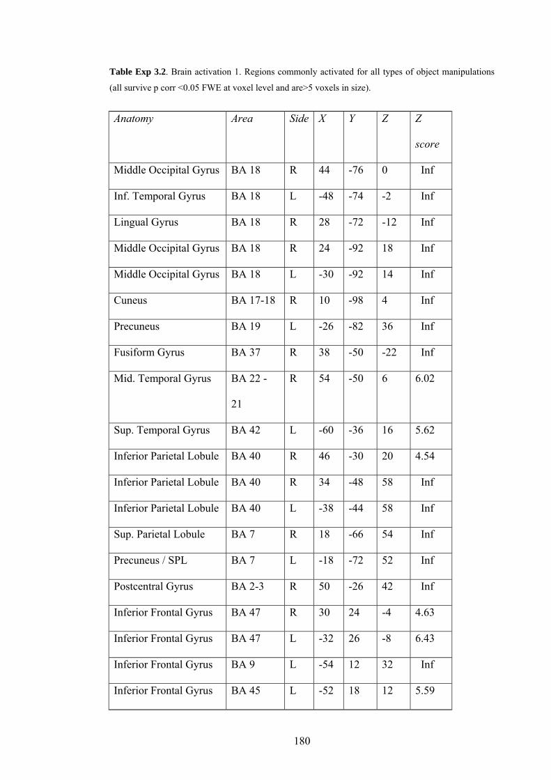
Table Exp 3.2. Brain activation 1. Regions commonly activated for all types of object manipulations
(all survive p corr <0.05 FWE at voxel level and are>5 voxels in size).
Anatomy Area Side X Y Z Z
score
Middle Occipital Gyrus BA 18 R 44 -76 0 Inf
Inf. Temporal Gyrus BA 18 L -48 -74 -2 Inf
Lingual Gyrus BA 18 R 28 -72 -12 Inf
Middle Occipital Gyrus BA 18 R 24 -92 18 Inf
Middle Occipital Gyrus BA 18 L -30 -92 14 Inf
Cuneus BA 17-18 R 10 -98 4 Inf
Precuneus BA 19 L -26 -82 36 Inf
Fusiform Gyrus BA 37 R 38 -50 -22 Inf
Mid. Temporal Gyrus BA 22 -
21
R 54 -50 6 6.02
Sup. Temporal Gyrus BA 42 L -60 -36 16 5.62
Inferior Parietal Lobule BA 40 R 46 -30 20 4.54
Inferior Parietal Lobule BA 40 R 34 -48 58 Inf
Inferior Parietal Lobule BA 40 L -38 -44 58 Inf
Sup. Parietal Lobule BA 7 R 18 -66 54 Inf
Precuneus / SPL BA 7 L -18 -72 52 Inf
Postcentral Gyrus BA 2-3 R 50 -26 42 Inf
Inferior Frontal Gyrus BA 47 R 30 24 -4 4.63
Inferior Frontal Gyrus BA 47 L -32 26 -8 6.43
Inferior Frontal Gyrus BA 9 L -54 12 32 Inf
Inferior Frontal Gyrus BA 45 L -52 18 12 5.59
180

Inferior Frontal Gyrus BA 46 L -52 42 8 4.49
Medial Frontal Gyrus BA 6 R 4 2 56 7.36
Middle Frontal Gyrus BA 6 R 24 -6 58 Inf
Precentral Gyrus BA 6 or 9 R 50 2 40 Inf
Precentral Gyrus BA 6 L -64 -14 40 5.77
Middle Frontal Gyrus BA 6 L -32 -2 52 Inf
Pulvinar L -20 -28 2 5.34
Thalamus R 14 -20 4 4.89
Cerebellum L -14 -76 -50 4.93
Cerebellum L -20 -78 -18 5.9
Cerebellum R 4 -76 -36 5.31
Table Exp 3.3. Brain activation 2. Regions showing activation differences depending on the type of
manipulation (during all conditions; all survive p corr <0.05 FWE corrected for voxels of the whole
brain or the pSTS).
1) Greater activation during Book and Boxes displays than Bottle and Phone.
Anatomy Area Side X Y Z
Z
score
Cuneus BA 17 L -12 -82 10 Inf
Fusiform Gyrus BA 19 L -28 -56 -10 7.28
Middle Occipital Gyrus BA 19 L -36 -86 12 6.69
Middle Occipital Gyrus BA 19 R 34 -86 14 7.12
Precuneus / SPL BA 7 R 12 -64 62 6.69
Sup. Parietal Lobule BA 7 L -22 -60 64 6.45
Postcentral Gyrus BA 2 R 36 -32 42 5.01
181

Cerebellum: Declive R 24 -58 -16 4.68
2) Greater activation during Bottle and Phone displays than Book and Boxes.
Anatomy Area Side X Y Z Z score
Posterior STS BA 22 R 54 -42 12 4.42
Posterior STS BA 22 L -64 -34 4 4
Main effect 1: Effect of movement type
There were no significant activation differences between animations with
Mimed and Actual movement kinematics, for any type of manipulation.
Main effect 2: Effect of object presence on manipulation of all types
Observation of animations where the manipulation movement was shown with
an object compared to animations without an object (Figure Exp 3.5 A) induced
stronger activation in fusiform (left hemisphere for all manipulations, only at
uncorrected threshold for Phone; right hemisphere for all, with Boxes and Phone at
uncorrected threshold only), dorsal parietal cortex (left hemisphere: Phone and Boxes,
right hemisphere: Phone, Boxes at uncorrected threshold) and middle occipital gyrus
(left and right hemisphere, both for Phone and Boxes, Bottle at P<0.001 uncorrected
threshold only). In Book there was a significant activation in left posterior medial
occipital cortex.
Animations where the manipulation movement was shown without an object
induced significantly stronger activation only for the Book and Boxes manipulations
when compared with animations presented with an object (Figure Exp 3.5 B). The
activated regions were mostly different between Boxes and Book; the only common
182

region with significantly increased activation was right posterior STG. Other regions
significantly active in Book where: left dorsal premotor cortex, medial superior
frontal cortex and the body of the caudate nucleus. Other regions significantly active
in Boxes where: left posterior STS and STG, posterior cingulate and medial occipital
cortex.
Figure Exp 3.6. Effects of object presence.
A Brain regions more active in conditions with objects than conditions without objects, for each type of
object manipulation. Overlaps are found in the fusiform gyrus and the lateral occipital cortex. Image
thresholded at P<0.05 corrected for multiple comparisons across the whole brain. B Brain regions with
higher activation in trials without objects compared to trials with objects, for all object manipulation
types. Only Book and Boxes showed significant activation in this contrast. Threshold: P<0.05 corrected
(cluster level, identified at P<0.001 uncorrected).
183

Table Exp 3.4. Brain activation 3. Regions showing activation differences during conditions with
Object and Without object (* survive P<0.05 whole-brain corrected at voxel level, other survive
correction at cluster level only). H refers to Hemisphere, BA to Brodmann Area.
1) Greater activation during Object than No object displays.
Anatomy H BA X +- Y +- Z +- Objects (Z score)
R fusiform R 19/3
7
36 (9) 58 (10) -15 (5) Bk(5.7)* Bt(4.4)*
Ph(3.4) Bx(3.5)
L fusiform L 19/3
7
-30 (2) -53 (8) -13 (3) Bk(7.1)* Bt(4.9)*
Ph(3.9) Bx(4.7)
R mOccG R 19 31 (5) -90 (2) 15 (1) Bt(3.2) Ph(5.3)*
Bx(3.9)
L mOccG L 19 -33 (3) -90 (4) 16 (2) Bt(3.4) Ph(3.6)
Bx(4.5)
R SPL R 7 24 (6) -61 (11) 55 (11) Ph(4.7)* Bx(3.5)
L SPL L 7 -25 (1) -66 (0) 46 (4) Ph(4.3)* Bx(4.3)
R IPL R 40 60 (0) -34 (0) 44 (0) Ph(3.1)
2) Greater activation during No object than Object displays.
Anatomy H
BA X +- Y +- Z +-
Objects (Z
score)
Mid.-Sup. Front Gyr. L 8-9 -36 26 50 Bk(4)*
Inf. Parietal Lobule R 40 54 -40 24 Bk(3.9)*
Sup. Temporal Gyrus R 22 49 (5) -47 (1) 14 (2) Bk(3.7)*
Bx(4.3)*
Inferior Frontal Gyrus L 47 -56 34 0 Bk(3.8)
184

Middle Frontal Gyrus R 9 44 16 34 Bk(3.6)
Middle Frontal Gyrus R 10 40 40 26 Bk(3.3)
Middle Frontal Gyrus L 6 -56 2 46 Bx(3.2)
Inferior Frontal Gyrus R 46-45 44 26 14 Bk(3.4)
Inferior Frontal Gyrus L 45-44 -48 18 16 Bk(3.6)
Precentral Gyrus L 6 -38 -8 60 Bk(3.2)
Medial Frontal Gyrus R 6 4 16 52 Bx(3.7)*
Mid. Temporal Gyrus L 21 -62 -38 -4 Bx(4.5)*
Mid. Temporal Gyrus R 21 60 -30 -8 Bx(4.1)
Sup. Temporal Gyrus L 38 -58 14 -4 Bx(3.5)
Interaction: Regions responding differently to artificial or realistic
movement-object combinations.
Combinations of movement and object that represent realistic situations (i.e. a
Mimed movement presented without object and an Actual movement presented with
an object) induced greater activation (Figure Exp 3.6, Table Exp 3.5) than artificial
situations (Mimed movement with object and Actual movement without object) in a
number of regions, but again only for Book and Boxes manipulations (although
activation for Book manipulations only survived a threshold of P<0.0001
uncorrected). The regions with increased activation were again mostly different
between Boxes and Book; the only region with overlapping activation increases was
located in the inferior left precentral gyrus. Other regions were significantly active for
the Boxes manipulations: bilateral pSTG extending into pSTS on the left, posterior
cingulate, posterior medial occipital cortex, superior medial parietal cortex and left
superior precentral gyrus.
185

There were no regions with significantly higher activation in the opposite
contrast.
Figure Exp 3.7. Congruence effects.
Brain regions with higher activation during Realistic than Artificial movement / object combinations.
This contrast compared conditions Mimed No object and Real Object with Mimed Object and Real No
object. Only Boxes showed significant activations in this contrast. Threshold: P<0.05 corrected (cluster
level, based on P<0.001 uncorrected).
Table Exp 3.5. Brain activation 4. Regions showing greater activation during Realistic (congruent) vs.
Artificial (incongruent) movement / object combinations. Threshold used was P<0.05 corrected at the
cluster level (based on P<0.001 uncorrected map).
Anatomy Side Area X Y Z Object (Z score)
Putamen L -14 16 -4 Bx(3.2)
Thalamus L -16 -30 6 Bx(3.7)
Insula R BA 13 46 0 4 Bx(3.5)
Precentral Gyrus L BA 4 -32 -34 62 Bx(3.4)
Precentral Gyrus L BA 4 -60 -2 18 Bk(3.9)**
186

Precentral Gyrus R BA 4 58 -6 20 Bk(3.7)*
Middle Frontal Gyrus L BA 6 -42 0 54 Bx(3.4)
Superior Frontal Gyrus L BA 6 -22 -8 72 Bx(3.3)
Superior Frontal Gyrus R BA 6 16 -14 72 Bx(3.2)
Postcentral Gyrus R BA 43 58 -18 14 Bx(3.3)
Postcentral Gyrus R BA 7 4 -48 70 Bx(4.1)
Postcentral Gyrus L BA 7 -8 -52 70 Bx(3.9)
Postcentral Gyrus L BA 3 -44 -28 60 Bx(3.4)
Postcentral Gyrus R BA 3 28 -32 70 Bx(3.4)
Inferior Parietal Lobule L BA 40 -50 -30 26 Bx(3.5)
Sup. Temporal Gyrus R BA 22 60 -42 8 Bx(3.7)
Mid. Temporal Gyrus L BA 39 -34 -64 22 Bk(4.4)
Mid. Temporal Gyrus R BA 39 44 -48 6 Bx(3.7)
fMRI analysis 2: Correlation with kinematic information
As discrimination performance showed a trend to correlate with kinematic
information (Figure Exp 3.4), and given that correctly categorised animations induced
different activation than incorrectly categorised animations (Figure Exp 3.5 B and C),
correlation between kinematic information and brain activation was tested directly in
SPM. Regression analyses were performed on contrast images from each
manipulation type comparing animations across all experimental conditions with the
resting actor, using as explanatory variables the number of joint variables with
significant differences in kinematics between Mimed and Actual movement versions
(Table Exp 3.1). I looked for voxels which showed positive or negative correlation
187
with kinematic information. A positive correlation was found in occipital and parietal
regions, including posterior medial occipital cortex, lateral occipital areas, fusiform
gyrus, and medial superior dorsal parietal cortex. The only brain regions that showed
a significant inverse correlation with kinematic information were clusters in bilateral
pSTS, and a small cluster in left medial occipital cortex (Figure Exp 3.8 A).
Given the evidence on the role of the pSTS during action observation and
biological motion and given the results of the experiments on observation of animate-
looking, moving objects, I decided to complement the analysis performed within SPM
with a region-of-interest analysis on the pSTS. I averaged the parameter estimates
from the clusters of voxels in the pSTS responding more strongly to incorrectly
discriminated than correctly discriminated animations (see Figure Exp 3.5 C) and
correlated this average activation with the number of joints showing significant
differences in kinematic information between Mimed and Actual manipulation
versions (Figure Exp 3.8 B). Activation in the pSTS of both hemispheres showed a
strong inverse correlation with kinematic information (Left: R2 = 0.96, p = 0.02;
Right: R2 = 0.94, p = 0.03), thereby confirming the results of the regression analysis
performed directly in SPM.
188

Figure Exp 3.8. Correlation between stimuli, and pSTS activation.
A Correlation between kinematic information in all four objects (measured as number of joint variables
that showed significant difference between Mimed and Actual kinematics during object manipulation)
and brain activation performed in SPM. Left part: positive correlation (Thresholded at p<0.05 corrected
for multiple comparisons across all the voxels of the brain). Right part: negative correlation. Only the
pSTS and a small cluster in left medial occipital cortex (not shown) showed significant inverse
correlation (Threshold: p<0.001 uncorrected). B Correlation between kinematic information in all four
objects and the activation in posterior STS (normalised data, Right hemisphere in red, left hemisphere
in blue).
Discussion
This study yielded the following behavioural and brain activation results.
Analysis of behaviour showed that subjects were able to correctly categorise half of
the presented manipulations observed into Mimed and Actual movements on the basis
of their kinematics, while the presence of an object irrelevant to the task increased
their performance in one occasion. This categorisation was made correctly only when
1) more than a third of the movement parameters show significant objective
differences between Mimed and Actual kinematics and 2) when these differences are
present during more than just the contact phase between hand and object.
Discrimination performance showed a correlative trend with kinematic information.
189

Analysis of brain activation showed that comparing all types of manipulation
with a resting actor reveals significant activation increases in brain regions known to
be involved in action observation (premotor cortex, STS) and in primary and
secondary visual regions (including fusiform gyrus, LOC, STS and dorsal parietal
cortex). Activation in some of these areas was dependent on kinematic information
available for the task at hand: activation was greater when there was more kinematic
information in many visual areas (posterior medial occipital cortex, fusiform gyrus,
LOC, dorsal parietal cortex), and activation in the pSTS correlated inversely with
kinematic information. When subjects observed object manipulations they could
categorise correctly into Mimed and Actual, activation in the right posterior STG was
increased when no object was presented in the animation compared to when an object
was present.
I propose that these data suggest a role of the superior temporal sulcus in the
extraction of task-relevant biological motion information from observed human
movements, with activation compensating for task difficulty (caused in the present
study by less kinematic differences between the movements to categorise or no
information about the movement of the object during the manipulation). This is in
opposition to other brain areas responding during action observation, such as the other
parts of the visual system whose activation appears to increase with kinematic
information, and to the premotor cortex whose activity does not seem to be influenced
by kinematic information.
Stimuli
Our behavioural results show that when 33% or more joint variables showed
differences between Mimed and Actual movements, subjects were able to
discriminate between these two movement versions, but only the manipulations with
190

57 % or more joint variables showing significant differences were discriminated
correctly. In the correctly discriminated manipulations, three-quarters or more of the
variables showed significant differences between Mimed and Actual versions during
the whole duration of the movement. Therefore, necessary for a correct categorisation
were significant differences between Mimed and Actual movements in a certain
number of joints, during a period longer than just the contact phase between the object
and the actor’s hand. Interestingly, the presence of the object had only a mild effect on
performance: it increased only for Boxes, where categorisation was already successful
in their absence. Previous studies showed that movement kinematics of Mimed and
Actual movements can be different (Goodale, Jakobson, & Keillor, 1994), and also
show less variation between replications of a particular movement by one person than
do Actual movements (Mason, Gomez, & Ebner, 2001). Studies also showed that
observers can tell when people experience violations of their expectations about target
objects, such as a box being lighter or heavier than expected when picked up
(Runeson & Frykholm, 1983; Grezes, Frith, & Passingham, 2004). The present study
shows that subtle differences in movement kinematics, probably resulting from the
effect of the object on the movement, can be picked up by subjects, if they exist in a
sufficient number of the joints involved in the movement and if they last a certain
time. The importance of the part of the movement before contact with the object (the
reaching part) as a defining part of the action can be seen from studies which showed
that two principal components can explain more than 80 percent of the variation of
hand and finger joints during a variety of reaching and grasp actions (Santello,
Flanders, & Soechting, 1998; Mason, Gomez, & Ebner, 2001), with the second of
these components corresponding to the reaching and preshaping phase of the hand. It
is therefore not surprising that differences between kinematics of Mimed and Actual
191

movements need to be found also in this part of the movement for correct
identification by observers.
Convincing pantomime artists, who create the impression that they are
manipulating an object that is not actually present, probably base their performance on
their capacity to minimise the kinematic differences between their mime and the
actual manipulations of an object, resulting for observers in the impression that the
object actually had an effect on the pantomime’s movements. Recording movement
kinematics from such artists could therefore be an interesting extension to my
behavioural results.
STS and processing of biological motion
As discussed in Part 1, Chapter 3, Section 3.2.2, there is a growing amount of
data suggesting that the region surrounding the posterior STS and STG is implicated
in action observation and observation of biological motion. The present study shows
that activity in this area increases with task difficulty during an action-observation
task (note: this cannot simply reflect a time-on-task effect, as activity was modelled
only from stimulus onset to subject’s response). Particularly, I found the following
effects. First, activation was greater during observation of manipulations for which
Mimed and Actual versions show little kinematic differences, which were also the
movements that subjects had more trouble categorising into Mimed or Actual. This
suggests that the pSTS “works harder” to extract the movement information contained
in the stimuli and relevant to the task.
Second, during observation of the two types of manipulation that subjects could
discriminate well, activation in the pSTS was greater when there was no object
present in the animation. The behavioural data show that the presence of an object
increased subjects’ Mimed vs. Actual discrimination of the Boxes manipulations.
192

Animations without objects appear to be more difficult to categorise than animations
with objects, which might be due to the absence of information available about the
way the object moved in response to the actor’s movement; this information might
have informed subjects about the type of movement they were observing. These two
facts suggest that the more information was available to the subjects to solve their
task, the better they succeeded and the less pSTS recruitment was necessary (Figure
Exp 3.9). This is in opposition to the responses of many other visual areas also
responding to animations of human actions, whose activation increased with available
kinematic information. This might suggest that these regions, being less specialised in
the analysis of biological motion than the pSTS, simply respond more when there are
more visual information available to solve the task.
I believe that these data consolidate the link between processing of biological
movements and the pSTS (for a recent review see Puce & Perrett, 2003).
Discriminating between subtly different movements could be useful for the detection
of non-verbal communication cues and the identification of mental states of the
observed people. This would conform to the role of the pSTS in the processing of
biological motion on the basis of which we make social attributions (Adolphs, 2003).
How certain regions could be specialised for the processing of moving or changing
aspects of a particular type of socially relevant stimuli has been described in detail in
a recent model of face perception (Haxby, Hoffman, & Gobbini, 2002).
STS and attribution of mental states
A function possibly related to action observation and the processing of
biological motion and associated with the pSTS is the attribution of mental states to
other agents, as seen in a variety of tasks and substrates (for a review, see Frith &
193

Frith, 2003). A recent study combining mental state attribution and action observation
showed that activity in the pSTS and parts of the premotor and orbitofrontal cortex
was greater when subjects thought that a person whose action they observed had a
false belief about the object he or she was manipulating (Grezes, Frith, & Passingham,
2004). In the present experiment, I did not find differences in activation between
observation of Mimed or Actual movements. Although this could be seen as clashing
with the results of the study by Grezes and colleagues, I believe that the task and
stimuli I used did not cue subjects into thinking about the mental states of the actor
executing the action. In my experiment, subjects were not told that the actor had
particular beliefs about the object he was to manipulate: they were only told that the
actor had mimed an action while observing the object, or had really performed the
action with the object. By contrast, in the study by Grezes et al, participants were told
that the person they were observing had sometimes been misled as to the weight of the
object they had to lift. This difference could have led subjects in my experiment to
pay more attention to the differences in kinematics than to differences in mental states
of the actor. This could explain why I found that activation in the pSTS varied
depending on the amount of kinematic differences between Mimed and Actual
movements the subjects had to observe, but not depending on the type of movement
kinematics themselves (Mimed or Actual).
STS and Goal-directed action
The posterior STS or TPJ is also known to be activated during execution and
imitation of goal-directed actions in humans (Winstein, Grafton, & Pohl, 1997;
Iacoboni et al., 2001). In monkeys also, neurons responding during observation of
actions have been found in this brain region (Jellema, Baker, Wicker, & Perrett,
194

2000). As discussed above, I did not find regions with activation differences between
Mimed and Actual conditions.
I do not think that this absence of an effect necessarily reduces the association
between pSTS and goal-directed action. Goals are defined differently by different
authors, as described in a recent study by Koski and colleagues (Koski, Iacoboni,
Dubeau, Woods, & Mazziotta, 2003). The goal-directed theory of imitation
(Bekkering, Wohlschlaeger, & Gattis, 2002) defines goals as physical objects that can
be targets for reaching and grasping movements, but also as a representation of the
goal in neural codes, in a “functional mechanism necessary to initiate an imitative
action” (Koski, Iacoboni, Dubeau, Woods, & Mazziotta, 2003). Tomasello separates
action goals from the means to achieve them (Tomasello, 1999), and Travis defines a
goal as a “mental state representing a desired state of affairs in the world” (Travis,
1997). Therefore, depending on the definition of goal, a mimed movement can be
considered goal-directed or not. If one considers a mimed movement as goal-directed
(after all, one recognises the object or the goal of a mimed action, which is really what
makes the mimed movement a mime, or one could say that the end-state of the mimed
movement is the goal of the movement), then the absence of an activation difference
in the pSTS between mimed and actual movements could be due to the fact that there
was a goal in both types of movements. If one defines mimed movements as not goal-
directed however, the present data can be seen to show the role of the pSTS in
extracting information from observed movements in order to discriminate them in
terms of goal-directedness. This would be supported by greater activation in the pSTS
during observation of movements that contained less information necessary for the
discrimination into Mimed and Actual. Whether a mimed movement is a goal-directed
movement or not does not influence the interpretation of the correlation between
195

activation and kinematic information very much: in both cases, the pSTS could be
involved in the extraction of task-relevant kinematic information. This result is
certainly more interesting than the absence of an activation difference between
observation of mimed and actual movements.
Ventral vs. dorsal stream
As discussed in the introduction, the ventral stream is thought to control mimed
movements, at least to observed objects, while object-directed actions are thought to
be controlled by the dorsal stream. Therefore it could have been possible that the two
visual streams would be activated differently also during observation of Mimed and
Actual actions. My results show that there was no significant difference in activation
in these two streams between observation of Mimed and Actual movements. Rather,
there was stronger activation in parts of both visual streams during observation of
correctly discriminated object manipulations and manipulations with more kinematic
information available for the task (medial dorsal parietal cortex, lateral and medial
occipital cortex, and fusiform gyri, all bilaterally, see Figures Exp 3.5 B and Exp 3.9
A), and stronger activation in bilateral pSTS during observation of less well
discriminated object manipulations or manipulation with less kinematic information
available for the task (Figures Exp 3.5 C and Exp 3.9 A). Therefore, it appears that the
difference in visual stream involvement that is found for the execution of mimed or
actual actions is not found for observation of these actions.
Activation of action-observation areas with animations
196

Although a previous study suggested that only the observation of physically
present actors can lead to activation of all parts of the action observation system
(Perani et al., 2001), I did find activation in all the areas classically associated with
action observation. Although there was some variation in activation in some of these
areas depending on the manipulation observed (Figure Exp 3.4 A and B), strong and
very similar activation patterns in these areas were found for all four types of
manipulation.
In contrast with my hypothesis, regions of the ventral premotor cortex that could
be analogues of monkey area F5 and could contain Mirror Neurons (Areas 44 and/or
45, see for example Grezes, Armony, Rowe, & Passingham, 2003) did not show
significant differences between observation of Mimed or Actual movement versions
or conditions with Object present or absent, for either type of object manipulation.
This is in contrast with neurophysiological findings (Umilta et al., 2001, see Figure
Intro.8) which show that Mirror Neurons respond during object-directed actions even
when the grasped object and the contact between the hand and object are out of view.
This was taken to imply that Mirror Neurons might identify the object-directedness of
the movement though analysis of the movement kinematics, rather than attending to
object presence. My present results could suggest either that 1) in humans, the
response of Mirror Neuron-analogue cells is different than in monkeys, or 2) cells
responding like monkey Mirror Neurons are mixed among other cells responding
differently and the resolution of fMRI cannot distinguish them, or 3) there is
inhibitory neural activity in the tissue surrounding Mirror Neuron-like cells during
conditions with Mimed kinematics, which would also lead to an increase in metabolic
rate and therefore to an increase in BOLD signal (Logothetis & Wandell, 2004), and
therefore no difference was found. Alternatively, Mirror Neuron-like cells are more
197
sensitive to the realism of the stimuli than other structures involved in action
observation in the human, and are therefore not triggered by my virtual reality
animations.
Realistic vs. artificial movement-object combinations
Only for animations depicting the manipulation of the two boxes, observation of
Realistic object and movement kinematics combinations resulted in higher activation
than during artificial combinations in the following brain regions: in the pSTS of both
hemispheres, anterior superior STS/inferior frontal gyrus (BA 22/44/BA6), anterior
dorsal (BA 1 and BA 3) and medial dorsal parietal cortex, posterior cingulate gyrus
and medial occipital cortex. Realistic vs. Artificial combinations correspond to the
effect of object presence on Mimed vs. Actual difference. Animations of the boxes
manipulations were the only type of manipulation in which the object presence had a
significant effect on Mimed/Actual discrimination performance (see Figure Exp 3.3).
Therefore the effect of object presence on brain activation is paralleled in the subjects'
performance. Whether these two observations are causally correlated, and in which
way, could not be determined in this study.
198

Part 5. General discussion
1. So what is this thesis all about?
The purpose of this thesis was to study the neural correlates of a basic cognitive
component of the mentalising ability, also known as Theory-of-Mind or ToM. ToM is
defined as the process of representing and manipulating mental states of the self or
other people. Previous neuroimaging studies from different research groups have
shown that 3 brain structures seem to be commonly activated in different tasks
involving mentalising: the medial prefrontal cortex (mPFC), the temporal pole,
particularly in the left hemisphere (TMPp) and the posterior superior temporal sulcus
(STS or pSTS). Although all 3 regions are known to show activation also during other
processes that could be related to or involved in mentalising, the relative contributions
of these regions to mentalising are speculative. The work in this thesis was
concentrated on a first step judged to be essential for ToM: the identification of living
entities in the environment.
This process was approached by studying types of movement particular to living
entities, because on the basis of developmental psychology studies, actions of living
entities appear to be very characteristic for living organisms and important for the
attribution of animacy. Also, arguably the most important use for mentalising is the
explanation and prediction of the behaviour of agents, which adds relevance to the
study of animate movement rather than shape, which was not studied in this
experiment series.
I suggest that a neural system specialised in the determination of what is animate
and what is not could rely on the identification of common characteristics in all types
of animate motion, which could be hard-wired and present at birth or acquired by
199

generalisation from the observation of diverse animate entities during development. A
neural system involved in detecting animate things should thus respond to these
characteristics. Developmental studies suggest that goal-directed and self-propelled
(also called internally-generated, see Premack and Premack, 1995) movements are the
most important movement characteristics for the attribution of animacy.
As one of the 3 neural structures commonly activated during mentalising tasks,
the superior temporal sulcus, is known to respond during the observation of biological
motion, I set out to test how the posterior STS responds to simple animations
displaying characteristics of animate motion, and see if this response is dependent of
attention or not. To this end, I first developed an algorithm for the motion of two
interacting, simple and abstract shapes, in which the variation of one parameter could
affect attribution of animacy by naïve observers.
Then, in an fMRI experiment, I looked for brain regions showing activation
correlating with the value of this parameter: they included the pSTS and other regions
associated with processing of living entities: the fusiform gyrus and the medial
occipital cortex, and two regions in the cingulate and posterior paracingulate cortex.
My results further suggest that this response is independent of the subject's focus of
attention. In a second experiment, in which I introduced a more complex parameter
also characteristic of animate movements and related to mentalising, I showed that the
pSTS responded again when the abstract object displayed more animate behaviour,
but this time, this response was increased when subjects paid attention to the
parameter associated with animacy. This suggests that simple and fundamental
movement characteristics of animate motion may activate the pSTS automatically,
while this structure responds to more complex characteristics, indicative of
representations of goals, only when subjects look for them. While the former response
200

is sensible for an automatic system detecting animate entities, the latter corresponds
more to a mentalising-like process, which is most probably a conscious process.
Both these experiments used moving, abstract objects. However, mentalising is
performed mostly in contact with other complex living animals, mostly human beings.
I therefore extended the study of biological movement to human actions in another
fMRI experiment, which showed that pSTS responds not only to biological
movements of different kinds, but also analyses these movements in order to
understand them by "extracting" kinematic information from observed human
movements. The subject's task was to determine if the observed movement was
mimed or not, which can be interpreted as goal-directed or not, depending on the
definition of goals one uses (see Introduction). The results showed that activity in
pSTS was higher when less kinematic information was present, suggesting that the
activity compensated for task difficulty. Evidently, evaluating whether an observed
movement is goal-directed or not is important for the identification of the mental state
of the agent performing the action, and therefore activity in the pSTS appears again to
be related to the mentalising process.
2. Activation in pSTS during observation of abstract and human movements
I would like to propose that pSTS activation in all experiments can be explained
by a role of this region in detection and analysis of biological motion. PSTS 1) shows
higher activation when observed movements look more animate (i.e. more goal-
directed vs. less goal-directed moving abstract shapes, shapes appearing to attribute
goals to target objects vs. simply following, human actions vs. resting actor), and 2)
when observing different movements of equal animacy, pSTS activation adapts to
demands of a task that requires extraction of information from the observed
201

movements. These roles are compatible with previous studies of biological motion
and as a part of the neural circuitry involved in mentalising.
With respect to goal-directedness, we cannot say that pSTS activation is always
greater when goals are present. While in both experiments involving abstract objects
pSTS activation was greater when objects showed more goal-directedness or appeared
to use representations of goals of a target object, in the action observation experiment,
pSTS activation was greater when there was less difference in kinematics between the
goal-directed and the non-goal-directed movement versions, but showed no
systematic activation difference depending on goal-directedness itself. While these
responses are slightly different, both suggest detection and processing of movement
cues indicative of goal-directed movements. I suggested above that this could be due
to different processes both performed by the pSTS: 1) detection and 2) analysis of
biological movements. The difference could also be due to different levels of
expertise of pSTS in abstract moving object movements and human actions, leading to
slightly different functions depending on the type of biological movements presented
and the task to be performed on them.
This pattern of response by the pSTS reminds of the response of the Visual
Word Form Area (VWFA) in the left fusiform gyrus (Cohen et al., 2000; Cohen et al.,
2002), whose activity is greater during observation of words rather than consonant
strings. But similarly to the activity in the pSTS found in action-categorisation
experiment of this thesis, activity in the VWFA is higher when pseudowords or words
of low frequency rather than high-frequency words are presented (Kronbichler et al.,
2004). This could reflect a similar increase in "effort" of the neuronal population in
trying to identify which word is presented, once the stimulus is close enough to a real
word (i.e. it contains vowels and consonants). I would argue that this response is
202
similar to that of the pSTS in the action-categorisation task of the thesis, in which,
once a visual stimulus has been identified as biological with reasonable certainty, a
greater "effort" is required when less kinematic information is available to solve the
task to be performed on the biological movements.
The involvement of pSTS in social cognition, including analysis of changeable
parts of faces and moving human bodies, suggests that this structure is specialised for
processing of socially relevant motion data. That it responds also when abstract
objects appear to interact in a fashion evocative of human- or animal-like behaviour is
interesting, but one should not forget that such animations are abstract and thus not
encountered often in normal life, and that therefore the pSTS has not much direct
experience in viewing it. If pSTS has evolved as a structure enabling recognition and
processing of social information in conspecifics (such as face and body movements),
then it might be expected to respond differently to such information rather than to
moving abstract, artificial stimuli, however sophisticated they are. Particularly,
observing human actions and trying to find which goal they seek to attain is an
everyday process for us, and the pSTS, being so specialised for human motion, could
adapt its involvement depending on task difficulty in order to supply other brain areas
specialised in action observation (such as the premotor cortex or intraparietal sulcus)
with enough information so that they can process the action. Detecting animate
motion cues in all types of movements (even abstract) is another process, with a
different purpose: more like a warning system ("there is maybe something living over
there, let's keep an eye on it"), which would be expected to respond differently to
clear human motion compared to abstract motion.
These differences in expertise might also lead to a different effect and role of
goals. Goal-directed motion might be a cue for attribution of animacy in the motion of
203

simple-looking entities (such as small insects, animals very far away), where almost
no information about animacy is given by their aspect. But in clearly human
movements located very close to the observer, goal-directed movements are more
subtle cues about the intentions of the person, or even substrates for imitation (see role
of goals in imitation discussed in Part 1, Chapter 3, Section 3.2.1). While there are
obvious links to intentionality in both cases, the different information content and use
of it could lead to different involvement of the pSTS. In this view, it can even appear
surprising and interesting that pSTS activation is connected to goals in both cases,
albeit slightly differently.
It needs to be clarified that there are two categories of visual cues that can carry
information about animacy: the movement and the aspect of an object (Gelman,
Durgin, & Kaufman, 1995; Opfer, 2002; Johnson, 2003), of which only movement
was studied in the experiments of this thesis. While I suggest that my data show a role
of the pSTS in detection and analysis of movement characteristics of animate entities,
it is possible that cues for animacy in the aspect of objects are processed by a different
area, for example the fusiform gyrus, other parts of the LOC (such as the extra-striate
body area, see Downing, Jiang, Shuman, & Kanwisher, 2001) or the medial occipital
cortex. This network might have distributed specialisations, as has been proposed for
processing of faces: static aspects might be processed in the lateral fusiform gyrus,
whereas changing aspects by the STS (Haxby, Hoffman, & Gobbini, 2002). This
interesting question needs to be addressed in further studies.
3. Other functions of the superior temporal sulcus and gyrus
In addition to mentalising, processing of animate motion, faces and other
socially relevant stimuli, the posterior part of the STS responds also to changes in
204

visual, auditory or sensory stimulation (Downar, Crawley, Mikulis, & Davis, 2000)
independently of the context of the task (Downar, Crawley, Mikulis, & Davis, 2001),
and even when there is no task to perform (Downar, Crawley, Mikulis, & Davis,
2002). It seems to be also involved in detection of visual oddballs (differing in form,
location, or both) (Marois, Leung, & Gore, 2000), and could be involved in
reorienting to visual events appearing at unexpected locations (Corbetta, Kincade,
Ollinger, McAvoy, & Shulman, 2000).
I do not think that novelty or the presence of a change in the stimuli presented in
the experiments of this thesis can explain the pSTS activations that were observed. In
all studies, an equal number of trials were presented in the conditions that were
compared to one another, and these were fully randomised (differently for each
subject). Also, in experiments 1 and 2, a closely-matched control condition was used
for each experimental condition, and always included in the contrasts of interest.
These control conditions were presented as frequently as the experimental conditions,
were also randomised, and could be considered to differ in terms of “novelty” and
“change” only with great difficulty6. In experiment 2, one region showed different
responses depending on the task performed, which should not affect novelty or change
in stimulation. In experiments 1 and 3, novelty or change could hardly explain the
correlations observed between stimulus characteristics and STS activation. I feel
therefore confident to propose a different role for the pSTS than novelty or change
6 An interesting idea would be to suggest that animate motion appears is characterised by being more novel or to contain more motion oddballs than inanimate motion. Indeed, one might consider that the aspects of the motion that cannot be explained by simple physical laws (see Part 1, Chapter 3, Section 3.1.1) or that are otherwise unexpected are critical for the movement to appear biological, and one might therefore consider these events as motion oddballs or novel trajectories. While this idea deserves further study, it is difficult to think that novelty is the only characteristic of biological motion given the numerous psychophysical studies demonstrating advanced identification of complex biological motion with very minimal stimuli (see Part 1, Chapter 3, Section 3.1.2), which very strongly suggests that some form of knowledge about biological motion is accessed and therefore stored in some form. The existence of such stored knowledge of biological movements appears difficult to reconcile with the postulated importance of novelty.
205

detection. Evidently, this structure might very well be involved in both types of
processes, and maybe more.
The pSTS might also be involved in integration of sensory information within
and between modalities (auditory and visual) about complex objects, such as pictures
and sounds of animals and tools and videos of moving tools (Beauchamp, Lee, Argall,
& Martin, 2004). Integrating such multisensory information could correspond to the
role of the superior temporal polysensory area (STP) of the monkey (see
Introduction). In all my experiments, only visual stimulation was used in all
conditions, and conditions all involved complex motion. As some data suggest that
biological motion could be a different kind of motion (see Part 1, Chapter 3, Section
3.1.2), one could argue that more types of visual information are contained in my
experimental than my control stimuli. Therefore the activation increases would also
have happened if another type of information would have been added to the control
conditions instead of biological motion. But the parametric response observed in the
pSTS in experiment 3 was inversely correlated to differences in objective motion
information, and there was no evident difference in “biologicalness” between the
objects used. Therefore, again, I think that the interpretation advanced for my
activations, based on the role of the STS in processing of biological motion, is fitting
the data best.
A recent study suggests that the pSTS is implicated in the learning of a tracking
task (Maquet, Schwartz, Passingham, & Frith, 2003). In this study, a group of
volunteers were asked perform a pursuit task in which they had to track a disk on a
computer screen with a joystick. Unbeknownst to them, the motion trajectory of this
disk in the horizontal axis was predictable, but the movements in the vertical axis
were unpredictable. Half the participants were sleep-deprived in the following night,
206
whereas the other half was allowed to sleep normally. When the sleep-deprived
participants had recovered from their lack of sleep 3 days later, both groups were
retested on the tracking task using movements with trajectories predictable in the
learned axis or the other (unlearned) axis. In the group that was allowed to sleep,
performance and activation in right STS showed a greater difference between the
learned and the new trajectories than in the sleep-deprived participants, and greater
connectivity was observed between the cerebellum and the right STS and between the
supplementary eye field and the frontal eye field.
As the STS activation observed by Maquet and colleagues falls in the same
range as the activations related to observation of animate and biological motion
described in this thesis, one could suggest a common process between learning of the
pursuit task and expertise in biological motion. One way in which the STS could be
involved in the acquisition of complex tracking skills is to extract predictable patterns
from observed motion. As the trajectories used in the study by Maquet and colleagues
had not been observed before, activation increased after exposition to them. As
humans are exposed to biological motion during all their life and pay great attention
to it because it is so relevant for their life, one could suggest that the STS has had
plenty of time to extract and store its invariant characteristics. These stored
characteristics could be activated during exposure to biological motion, and explain
activation increases and psychophysical performance when point-light displays are
compared to other types of complex but unlearned visual motion. In this view, the
STS could be considered as a module specialised in the analysis of complex visual
motion and storage of their characteristics, which has been highly trained on
biological movements during the life of the individual.
207

4. Potential confounding factors
While great care was taken in all experiments to minimise confounding factors
or their effects in all experiments, some might of course remain. Here is a review of
some of these confounds, their origin and possible solutions for follow-up studies.
In Experiment 1, the cross-correlation level affected speed (Figure Exp 1.3),
movement quantity and the extent of the screen covered by the moving objects
(Figure Exp 1.7). These factors were taken account of in the design by creating
matched Control conditions, in which these variables were almost identical to those in
the Interactive conditions, while the chasing behaviour between the objects was
destroyed. But despite this fact, interactivity ratings of the control animations (Figure
Exp 1.8) showed that some cues leading to attribution of goal-directed behaviour
might have remained present (ratings of interactivity increased with the cross-
correlation coefficient, even when the interaction between the objects was destroyed),
while animacy ratings of the same control animations revealed that these controls did
not appear animate (Figure Exp 1.6). This suggests that other influences might also
have affected the percept of animacy. Behavioural reports suggest that the number of
crossings of the objects could have made the objects appear goal-directed, and
crossings were more frequent when the objects moved more (i.e. higher cross-
correlation level). In addition, some subjects in the animacy attribution experiment
reported that objects in fast-moving animations did not appear animate, which further
implicates that the effect of speed should have been controlled better. Nevertheless,
on the whole, behavioural and fMRI results are significant and consistent with the
original hypotheses, which suggests that, although normalisation of the animations for
speed would have been ideal, the role of speed was probably controlled reasonably
well.
208
In Experiment 2, the movements of the objects were very simple, following arcs
or straight lines. This was chosen to make the path differences clearer and to simplify
subjects' tasks, but might have made the objects appear less animate than those of
Experiment 1. One could have used a modified version of the algorithm developed in
Experiment 1, but this appeared quite complex during stimulus development and
testing, and for the sakes of simplicity and speed it was decided to concentrate on the
main factor of Strategy. The movements of the objects nevertheless contained
characteristics associated with animacy (see Figure Exp 2.1): one appeared to chase
the other, the distance between them was not constant and the movements did not start
at the same time, thereby minimising the impression of pushing and pulling, and the
chaser stopped its movement when reaching the target, further increasing the
impression of goal-directed movements (Opfer, 2002).
In Experiment 3, the choice of object manipulation types could have been
different, to maximise differences between mimed and actual movement kinematics,
which might have isolated brain regions responding more during observation of
mimed or actual actions. Alternatively, the number of object manipulations used could
have been increased, to reinforce the correlation between brain kinematics
information, behaviour and brain activation. Either possibility would have involved
the recording of more movements and the analysis of their kinematics, which was
technically very complex and time-consuming. The technical set-up was quite fragile,
and an intrinsic design fault in the commercial recording equipment led Daniel
Wolpert's group to discontinue development of the recording system. The
consequences of the design fault were corrected by manual editing of the kinematic
recordings for the experiment described in this thesis, which was again time-
consuming but led to acceptable results. Therefore, instead of recording and analysing
209

many movements, preliminary tests using video recordings of mimed and actual
versions of different object manipulation movements were performed, using a number
of different objects, including: a cup (lifting, drinking, filling)), a jar (lifting), a
hammer and nail, different boxes (various lifting and placing movements), paper
(folding and tearing), a pen (writing on paper), and the four objects and manipulations
used in the experiment. The movements digitised and used in the experiment were
chosen following the assumption that they would show the most differences in
kinematics between mimed and actual versions and were the easiest to categorise for
observers. All these factors led to the design and stimuli used here, which nevertheless
yielded interesting data. Other alternatives might be used to avoid the technical
difficulties of this digitising equipment: use of an Opto-Track system (set of cameras
recording the 3-D position of captors in space, at high speed), but this has the
disadvantage that some of the captors might be hidden from the camera in parts of the
movements. Or using video recordings, a manipulated object could be edited out from
an Actual recording, but adding the object realistically into a Mimed recording
appears very difficult. The use of a high-speed video camera together with a
movement digitising system recently led to an interesting idea in Daniel Wolpert's
laboratory: one could resample the frames of a high-frequency video recording using
the digitised movements, thereby systematically altering the movement kinematics of
a recorded movement. These methods are currently explored in Daniel Wolpert's
laboratory and might lead to fMRI studies in the future.
5. How the 3 mentalising regions could interact
The three brain regions systematically involved in mentalising are the medial
prefrontal cortex (mPFC), the superior temporal sulcus (STS) and the temporal pole
210

(TMPp). The mPFC is connected both to STS and TPMp (Bachevalier, 1997). Results
from the studies performed in this thesis support the role of the pSTS in the detection
and analysis of goal-directed movements, which are characteristics of animate agents.
This is important for identification of animate entities, whose behaviour can often be
explained by mental states, whether they actually have them or not. This is also
important because understanding of the movements of animate entities is an essential
component of the mentalising ability itself.
I propose that the pSTS, once it has identified a potentially living organism,
observes its actions (together with the ventral premotor cortex, vPM) and extracts
from its movements cues as to which goal underlies the organism's actions. It then
communicates this information to the TMPp (either directly or via the mPFC), which
tries to find a matching behavioural script for the successive actions observed. This is
useful in predicting what the organism might do next. If information continues to be
received from the pSTS, the actual behaviour of the entity can be compared to the
behaviour predicted by the script (either in the pSTS, the TMPp or in another brain
structure), and the mismatches registered. Both the script information from TMPp and
the movement and animacy information from pSTS would then be transmitted to the
mPFC and/or the STS7, which would try to identify possible mental states explaining
the behaviour (see description of the A-M module in Part 1, Chapter 2, Section 2.3).
The hypothetical mental state would then be compared to the desired mental state of
the agent if there is one. Based on the discrepancy, an action could be selected to
change the mental state (again using the A-M model), dependent on the script
currently in context (TMPp, see just above). The selected action would then be
7 See the latest studies suggesting that STS and not MPFC is essential for mentalising: neuroimaging (Saxe & Kanwisher, 2003) and lesion studies (Bird, Castelli, Malik, Frith, & Husain, 2004; Samson, Apperly, Chiavarino, & Humphreys, 2004).
211

planned and executed by the various components of the motor system (Figure
Discussion.1)
Animacydetector
Observation of other’s action
Other’smental state
Desiredmental state
of otherDifferencein mental
stateRequiredactionof self
Execution
of action
Visualinput
Actionof self
Self
A - M
A - M
pSTS
vPMpSTS - TMPp
mPFC / pSTS
mPFC / pSTS
mPFC / pSTS
TMPp - mPFC
Motorsystem
mPFC / pSTSOther
Figure Discussion.1. Possible neural correlates of ToM cognitive components.
The cognitive model was described in the Introduction. See text for details.
6. What comes next? Future directions
Many open questions remain at the end of this thesis, both about perception of
animacy and mentalising. Future experiments should increase understanding of the
necessary and sufficient characteristics leading to attribution of animacy, through
psychophysical experiments using tightly controlled stimuli, varying both movement
and aspect of the objects, and assessing the relative importance of movement and
aspect in different situations. Also, it is not clear how much of these characteristics
are common across different human cultures, or between humans and diverse other
animals. While neurophysiological and fMRI data suggest that the STS of rhesus
macaques also responds during observation of moving and immobile animals
(Logothetis, Guggenberger, Peled, & Pauls, 1999), what about other animals? Do they
212

differentiate between animate and inanimate entities like we do, is that as evolutionary
useful as we might think?
The ontogenic development of animacy attribution is potentially very
interesting: are characteristics of animate motion hard-coded in brain structures (such
as the pSTS), or do they crystallise after repeated expositions during development?
Then would restricted exposure to particular types of animate motion affect animacy
attribution, and maybe also brain activity? How about the connections to development
of other cognitive processes, such as executive function? Does the cultural definition
of what is alive and what is not affect animacy attribution? If yes, does it influence
psychophysical performance in experiments with very simple stimuli such as used in
this thesis? If computers become more complex, one can wonder if they will one day
be considered really animate, beyond the colloquial use of mentalistic terms to
describe their behaviour ("My computer really wants to annoy me today: look what it
did to my thesis file!"). But this might rely upon a different process than the
observation of human actions for example, whose “biologicalness” appears to play a
decisive role in activating the action representation system (Castiello, 2003).
Regarding the neural basis of mentalising, future developments could involve
better cognitive models of mentalising, from which one could derive tasks that engage
only some of the mentalising areas. This could lead to better understanding of the
relative contributions of all 3 areas known to be involved in mentalising, and maybe
show how other regions involved in social cognition (orbitofrontal cortex, amygdala),
complex visual processing (elements of the lateral occipital complex) or action
observation (premotor cortex, intraparietal sulcus) can contribute in particular
situations. The recent development of more sophisticated neuroimaging data analysis
tools and techniques, such as combined EEG and fMRI or connectivity software tools
213

such as SPM’s Dynamic Causal Modelling (DCM), could also reveal the time-course
of engagement of mentalising areas and how these regions interact with each other in
the normal brain.
7. Conclusion
In conclusion of the thesis, I propose that: 1) detecting animate movements is
important for mentalising; 2) as the movement of animate entities is characterised (at
least partly) by being goal-directed, brain regions involved in identification of such
animate entities could be detecting and analysing goal-directed motion, 3) my results
confirm this hypothesis: the pSTS is involved during detection and analysis of goal-
directed motion, whether during observation of movements of abstract objects or
humans. As this brain region is a part of the mentalising network proposed by some
authors, this might explain how it contributes to the whole process of mentalising.
214

Part 6. Appendices
Appendix 1: Behavioural reports of animacy testing in Experiment 1.
Question asked was: "how alive do the objects look to you? Please rate on a scale
from 1-4." It was suggested that subjects might feel a bit lost at the beginning of the
experiment, but they should not worry and do the task anyway. They were encouraged
to use the whole scale. Animations and experimental protocols used were the same as
in the fMRI experiment, but subjects were different. Participants were neither aware
of the experimental conditions nor of the purpose or the critical parameters of the
experiment. One participant had participated in a very early stage pilot study.
Subject 1
Looked at the objects chasing each other, the more they chased each other, the more
alive they appeared.
Subject 2
Was a bit lost at the beginning of the experiment; was using chasing at the end of the
experiment; quantity of movement was also a clue for animacy: the more they moved,
the more animate they appeared.
Subject 3
Objects appeared more alive when one object appeared to interact with the other; this
happened when, after the objects crossed each other, at least one of them came back
towards the other object. This was more apparent when the objects were slow, and
they therefore appeared more animate.
215

Subject 4
Was looking at chasing at the end of the experiment as a criterion.
Subject 5
Did not really know what rule to follow. NOTE: ratings nevertheless showed a
(small) difference between Interactive and Control.
Subject 6
Found the task difficult, was paying attention independently to the movements of both
objects at the beginning but very soon focused on their interaction. Used chasing
behaviour as critical parameter.
Subject 7
Considered that objects appeared more alive when they stayed close to each other,
which showed that they cared for each other.
Subject 8
Found it difficult, was looking at chasing at the end of the experiment to determine
animacy.
Subject 9
Found it difficult, used 3 different strategies: first he focussed on playfulness of both
objects, then on how much one follows the other, then on both factors together.
Subject 10
216

Found it difficult, none looked particularly animate, used spatial extent as criterion for
animacy and other complex mathematical measures. Note: used only the two middle
values of the scale.
Subject 11
PARTICIPATED IN PILOTS
Found that the faster the objects moved, the easier it was to rate their animacy (thinks
his response times are lower with higher speed). Used how much they followed each
other and playfulness and jerkiness as criteria for animacy. Note: he remembered
having participated in the pilot experiment. His ratings were very much like the
average plot, as were some of the other subjects' ratings, suggesting not much
influence of having seen the stimuli before.
Subject 12
Discounted fast objects as appearing too artificial; otherwise used following and
playfulness as index of animacy. The animations made him think of a mother and a
child interacting: the child runs around the mother, strays around a bit but always
comes back.
Appendix 2: kinematic recordings of object manipulations used in Experiment 3
Blue traces represent Actual movement kinematics (4 recordings per manipulation
type), red traces represent Mimed movement kinematics (4 recordings per
manipulation type). Plots of joint variables with significant differences in kinematics
between Actual and Mimed as analysed during the whole movement only have a dark
blue background, those with significant differences as analysed during the
217

manipulation phase of the movement have a green background, and those with
significant differences in both forms of analysis have a light blue background.
Book
Bottle
218

Phone
Boxes
219

Part 7. References
Reference List
Adolphs, R. (1999). Social cognition and the human brain. Trends Cogn Sci.,
3, 469-479.
Adolphs, R. (2003). Cognitive neuroscience of human social behaviour.
Nat.Rev.Neurosci., 4, 165-178.
Aggleton, J. P., Burton, M. J., & Passingham, R. E. (1980). Cortical and
subcortical afferents to the amygdala of the rhesus monkey (Macaca mulatta). Brain
Res., 190, 347-368.
Aglioti, S., DeSouza, J. F., & Goodale, M. A. (1995). Size-contrast illusions
deceive the eye but not the hand. Curr.Biol., 5, 679-685.
Allison, T., Puce, A., & McCarthy, G. (2000). Social perception from visual
cues: role of the STS region. Trends Cogn Sci., 4, 267-278.
Allman, J. M., Hakeem, A., Erwin, J. M., Nimchinsky, E. A., & Hof, P.
(2001). The anterior cingulate cortex - the evolution of an interface between emotion
and cognition. Unity Know.Converg.Nat.Hum.Sci., 953, 107-117.
Ashburner, J. & Friston, K. J. (1999). Nonlinear spatial normalisation using
basis functions. Human Brain Mapping, 7, 254-266.
Ashburner, J., Neelin, P., Collins, D. L., Evans, A. C., & Friston, K. J. (1997).
Incorporating prior knowledge into image registration. Neuroimage., 6, 344-352.
220

Aziz-Zadeh, L., Iacoboni, M., Zaidel, E., Wilson, S., & Mazziotta, J. (2004).
Left hemisphere motor facilitation in response to manual action sounds.
Eur.J.Neurosci., 19, 2609-2612.
Aziz-Zadeh, L., Maeda, F., Zaidel, E., Mazziotta, J., & Iacoboni, M. (2002).
Lateralization in motor facilitation during action observation: a TMS study. Exp.Brain
Res., 144, 127-131.
Bachevalier, J., Meunier, M., Lu, M. X., & Ungerleider, L. G. (1997).
Thalamic and temporal cortex input to medial prefrontal cortex in rhesus monkeys.
Exp.Brain Res., 115, 430-444.
Barbas, H. (1988). Anatomic organization of basoventral and mediodorsal
visual recipient prefrontal regions in the rhesus monkey. J.Comp Neurol., 276, 313-
342.
Baron-Cohen, S. (1995). Mindblindness: an essay on autism and theory of
mind. Cambridge, MA: MIT Press.
Baron-Cohen, S., Jolliffe, T., Mortimore, C., & Robertson, M. (1997). Another
advanced test of theory of mind: evidence form very high functioning adults with
autism or Asperger syndrome. J.Child Psychol.Psychiat., 38, 813-822.
Baron-Cohen, S., Leslie, A., & Frith, U. (1985). Does the autistic child have a
theory of mind? Cognition, 21, 37-46.
Barresi, J. & Moore, C. (1996). Intentional relations and social understanding.
Behavioural and Brain Sciences, 19, 107-122.
221

Bassili, J. N. (1976). Temporal and spatial contingencies in the perception of
social events. Journal of personality and social psychology, 33, 680-685.
Beauchamp, M. S., Lee, K. E., Argall, B. D., & Martin, A. (2004). Integration
of auditory and visual information about objects in superior temporal sulcus. Neuron,
41, 809-823.
Beauchamp, M. S., Lee, K. E., Haxby, J. V., & Martin, A. (2002). Parallel
visual motion processing streams for manipulable objects and human movements.
Neuron, 34, 149-159.
Beauchamp, M. S., Lee, K. E., Haxby, J. V., & Martin, A. (2003). FMRI
responses to video and point-light displays of moving humans and manipulable
objects. J.Cogn Neurosci., 15, 991-1001.
Bekkering, H., Wohlschlaeger, A., & Gattis, M. (2002). Imitation of gestures
in children is goal-directed. Quarterly Journal of Experimental Psychology, 53, 153-
164.
Belin, P., Zatorre, R. J., Lafaille, P., Ahad, P., & Pike, B. (2000). Voice-
selective areas in human auditory cortex. Nature, 403, 309-312.
Bennett, J. (1978). Some remarks about concepts. Behavioural and Brain
Sciences, 1, 557-560.
Berthoz, S., Armony, J. L., Blair, J. L. R., & Dolan, R. J. (2002). An fMRI
study of intentional and unintentional (embarassing) violations of social norms. Brain,
125, 1696-1708.
222

Bird, C., Castelli, F., Malik, O., Frith, U., & Husain, M. (2004). The impact of
extensive medial frontal lobe damage on 'Theory of Mind' and cognition. Brain, 127,
914-928.
Blakemore, S. J., Boyer, P., Pachot-Clouard, M., Meltzoff, A., Segebarth, C.,
& Decety, J. (2003). The detection of contingency and animacy from simple
animations in the human brain. Cereb.Cortex, 13, 837-844.
Blakemore, S. J. & Decety, J. (2001). From the perception of action to the
understanding of intention. Nat.Rev Neurosci., 2, 561-567.
Blakemore, S. J., Fonlupt, P., Pachot-Clouard, M., Darmon, C., Boyer, P.,
Meltzoff, A. N., Segebarth, C., & Decety, J. (2001). How the brain perceives
causality: an event-related fMRI study. Neuroreport, 12, 3741-3746.
Blakemore, S. J., Frith, C. D., & Wolpert, D. M. (1999). Spatio-temporal
prediction modulates the perception of self-produced stimuli. J.Cogn Neurosci., 11,
551-559.
Bloch, F., Hansen, W. W., & Packard, M. (1946). The nuclear induction
experiment. Phys.Rev., 70, 474-485.
Blythe, P. W., Todd, P. M., & Miller, G. F. (1999). How motion reveals
intention: categorising social interactions. In G.Gigerenzer & P. M. Todd (Eds.),
Simple heuristics that make us smart (pp. 257-285). Oxford: Oxford University Press.
Bonda, E., Petrides, M., Ostry, D., & Evans, A. (1996). Specific involvement
of human parietal systems and the amygdala in the perception of biological motion.
J.Neurosci., 16, 3737-3744.
223
Boussaoud, D., Ungerleider, L. G., & Desimone, R. (1990). Pathways for
motion analysis: cortical connections of the medial superior temporal and fundus of
the superior temporal visual areas in the macaque. J.Comp Neurol., 296, 462-495.
Brass, M., Bekkering, H., & Prinz, W. (2001). Movement observation affects
movement execution in a simple response task. Acta Psychologica, 106, 3-22.
Brothers, L. (1990). The social brain: a project for integrating primate
behaviour and neurophysiology in a new domain. Concepts in Neuroscience, 1, 27-
151.
Brunet, E., Sarfati, Y., Hardy-Bayle, M.-C., & Decety, J. (2000). A PET
investigation of the attribution of mental intentions with a nonverbal task.
Neuroimage., 11, 157-166.
Buccino, G., Binkofski, F., Fink, G. R., Fadiga, L., Fogassi, L., Gallese, V.,
Seitz, R. J., Zilles, K., Rizzolatti, G., & Freund, H. J. (2001). Action observation
activates premotor and parietal areas in a somatotopic manner: an fMRI study.
Eur.J.Neurosci., 13, 400-404.
Bush, G., Luu, P., & Posner, M. I. (2000). Cognitive and emotional influences
in the anterior cingulate cortex. Trends Cogn Sci., 4, 215-222.
Campbell, R., MacSweeney, M., Surguladze, S., Calvert, G., McGuire, P.,
Suckling, J., Brammer, M. J., & David, A. S. (2001). Cortical substrates for the
perception of face actions: an fMRI study of the specificity of activation for seen
speech and for meaningless lower-face acts (gurning). Brain Res.Cogn Brain Res., 12,
233-243.
224

Castelli, F., Happe, F., Frith, U., & Frith, C. (2000). Movement and mind: a
functional imaging study of perception and interpretation of complex intentional
movement patterns. Neuroimage., 12, 314-325.
Castiello, U. (2003). Understanding other people's actions: intention and
attention. J.Exp.Psychol.Hum.Percept.Perform., 29, 416-430.
Cavada, C. & Goldman-Rakic, P. S. (1989). Posterior parietal cortex in rhesus
monkey: II. Evidence for segregated corticocortical networks linking sensory and
limbic areas with the frontal lobe. J.Comp Neurol., 287, 422-445.
Chao, L. L., Haxby, J. V., & Martin, A. (1999). Attribute-based neural
substrates in temporal cortex for perceiving and knowing about objects.
Nat.Neurosci., 2, 913-919.
Chao, L. L., Martin, A., & Haxby, J. V. (1999). Are face-responsive regions
selective only for faces? Neuroreport, 10, 2945-2950.
Cohen, L., Dehaene, S., Naccache, L., Lehericy, S., Dehaene-Lambertz, G.,
Henaff, M. A., & Michel, F. (2000). The visual word form area: spatial and temporal
characterization of an initial stage of reading in normal subjects and posterior split-
brain patients. Brain, 123 ( Pt 2), 291-307.
Cohen, L., Lehericy, S., Chochon, F., Lemer, C., Rivaud, S., & Dehaene, S.
(2002). Language-specific tuning of visual cortex? Functional properties of the Visual
Word Form Area. Brain, 125, 1054-1069.
Conover, W. J. & Iman, R. L. (1981). Rank tranformation as a bridge between
parametric and nonparametric statistics. The American Statistician, 35, 124-129.
225

Corbetta, M., Kincade, J. M., Ollinger, J. M., McAvoy, M. P., & Shulman, G.
L. (2000). Voluntary orienting is dissociated from target detection in human posterior
parietal cortex. Nat.Neurosci., 3, 292-297.
Csibra, G. (2003). Teleological and referential understanding of action in
infancy. Philos.Trans.R.Soc.Lond B Biol.Sci., 358, 447-458.
Csibra, G., Gergely, G., Biro, S., Koos, O., & Brockbank, M. (1999). Goal
attribution without agency cues: the perception of 'pure reason' in infancy. Cognition,
72, 237-267.
Damasio, H., Grabowski, T. J., Tranel, D., Hichwa, R. D., & Damasio, A. R.
(1996). A neural basis for lexical retrieval. Nature, 380, 499-505.
Deichmann, R., Good, C. D., Josephs, O., Ashburner, J., & Turner, R. (2000).
Optimization of 3-D MP-RAGE sequences for structural brain imaging. Neuroimage.,
12, 112-127.
Dennett, D. (1978). Beliefs about beliefs. Behavioural and Brain Sciences, 1,
568-570.
Desimone, R. (1991). Face-selective cells in the temporal cortex of the
monkey. J.Cogn Neurosci., 3, 1-8.
Desmond, J. E. & Glover, G. H. (2002). Estimating sample size in functional
MRI (fMRI) neuroimaging studies: statistical power analyses. J Neurosci Methods,
118, 115-128.
Devinsky, O., Morrell, M. J., & Vogt, B. A. (1995). Contribution of anterior
cingulate cortex to behaviour. Brain, 118, 279-306.
226

di Pellegrino, G., Fadiga, L., Fogassi, L., Gallese, V., & Rizzolatti, G. (1992).
Understanding motor events: a neurophysiological study. Exp.Brain Res., 91, 176-
180.
Dickinson, A. R. & Balleine, B. W. (2000). Causal cognition and goal-directed
action. In C.Heyes & L. Huber (Eds.), Evolution of Cognition (pp. 185-204).
Cambridge, MA: MIT Press.
Dittrich, W. H. & Lea, S. E. (1994). Visual perception of intentional motion.
Perception, 23, 253-268.
Downar, J., Crawley, A. P., Mikulis, D. J., & Davis, K. D. (2000). A
multimodal cortical network for the detection of changes in the sensory environment.
Nat.Neurosci., 3, 277-283.
Downar, J., Crawley, A. P., Mikulis, D. J., & Davis, K. D. (2001). The effect
of task-relevance on the cortical response to changes in visual and auditory stimuli: an
event-related fMRI study. Neuroimage., 14, 1256-1267.
Downar, J., Crawley, A. P., Mikulis, D. J., & Davis, K. D. (2002). A cortical
network sensitive to stimulus salience in a neutral behavioural context across multiple
sensory modalities. J Neurophysiol., 87, 615-620.
Downing, P. E., Jiang, Y., Shuman, M., & Kanwisher, N. (2001). A cortical
area selective for visual processing of the human body. Science, 293, 2470-2473.
Duelli, R. & Kuschinsky, W. (1993). Changes in brain capillary diameter
during hypocapnia and hypercapnia. J Cereb Blood Flow Metab, 13, 1025-1028.
Duvernoy, H. M. (1999). The Human Brain. Vienna: Springer Verlag.
227
Eifuku, S., De Souza, W. C., Tamura, R., Nishijo, H., & Ono, T. (2004).
Neuronal correlates of face identification in the monkey anterior temporal cortical
areas. J Neurophysiol., 91, 358-371.
Ellis, R. R., Flanagan, J. R., & Lederman, S. J. (1999). The influence of visual
illusions on grasp position. Exp.Brain Res., 125, 109-114.
Fadiga, L., Fogassi, L., Pavesi, G., & Rizzolatti, G. (1995). Motor facilitation
during action observation: a magnetic stimulation study. J.Neurophysiol., 73, 2608-
2611.
Fagg, A. H. & Arbib, M. A. (1998). Modeling parietal-premotor interactions
in primate control of grasping. Neural Netw., 11, 1277-1303.
Farrer, C., Franck, N., Georgieff, N., Frith, C. D., Decety, J., & Jeannerod, M.
(2003). Modulating the experience of agency: a positron emission tomography study.
Neuroimage., 18, 324-333.
Farrer, C. & Frith, C. D. (2002). Experiencing oneself vs another person as
being the cause of an action: the neural correlates of the experience of agency.
Neuroimage., 15, 596-603.
Felleman, D. J. & Van Essen, D. C. (1991). Distributed hierarchical
processing in primate cerebral cortex. Cereb.Cortex, 1, 1-47.
Ferrari, P. F., Gallese, V., Rizzolatti, G., & Fogassi, L. (2003). Mirror neurons
responding to the observation of ingestive and communicative mouth actions in the
monkey ventral premotor cortex. Eur.J.Neurosci., 17, 1703-1714.
228

Ferstl, E. C. & von Cramon, D. Y. (2002). What does the frontomedian cortex
contribute to language processing: coherence or theory of mind? Neuroimage., 17,
1599-1612.
Flanagan, J. R. & Beltzner, M. A. (2000). Independence of perceptual and
sensorimotor predictions in the size-weight illusion. Nat.Neurosci., 3, 737-741.
Fletcher, P. C., Happe, F., Frith, U., Baker, S. C., Dolan, R. J., Frackowiak, R.
S., & Frith, C. D. (1995). Other minds in the brain: a functional imaging study of
'theory of mind' in story comprehension. Cognition, 44, 283-296.
Fogassi, L., Gallese, V., Fadiga, L., & Rizzolatti, G. (1998). Neurons
responding to the sight of goal-directed hand/arm actions in the parietal area PF7b of
the macaque monkey. Soc.Neurosci.Abs. 24, 257.5.
Ref Type: Abstract
Friston, K. J., Ashburner, J., Frith, C. D., Poline, J. B., Heather, J. D., &
Frackowiak, R. S. (1995). Spatial registration and normalisation of images.
Hum.Brain Mapp., 2, 165-189.
Friston, K. J., Fletcher, P. C., Josephs, O., Holmes, A. P., Rugg, M. D., &
Turner, R. (1998). Event-related fMRI: Characterizing differential responses.
Neuroimage., 7, 30-40.
Friston, K. J., Josephs, O., Rees, G., & Turner, R. (1998). Nonlinear event-
related responses in fMRI. Mag Res Med, 39, 41-52.
Frith, C. D. & Frith, U. (1999). Interacting minds--a biological basis. Science,
286, 1692-1695.
229

Frith, U. (2001). Mind blindness and the brain in autism. Neuron, 32, 969-979.
Frith, U. & Frith, C. D. (2003). Development and neurophysiology of
mentalizing. Philos.Trans.R.Soc.Lond B Biol.Sci., 358, 459-473.
Gallagher, H. L. & Frith, C. D. (2003). Functional imaging of 'theory of mind'.
Trends Cogn Sci., 7, 77-83.
Gallagher, H. L., Happe, F., Brunswick, N., Fletcher, P. C., Frith, U., & Frith,
C. D. (2000). Reading the mind in cartoons and stories: an fMRI study of 'theory of
mind' in verbal and nonverbal tasks. Neuropsychologia, 38, 11-21.
Gallagher, H. L., Jack, A. I., Roepstorff, A., & Frith, C. D. (2002). Imaging
the intentional stance in a competitive game. Neuroimage., 16, 814-821.
Gallese, V., Fadiga, L., Fogassi, L., & Rizzolatti, G. (1996). Action
recognition in the premotor cortex. Brain, 119 ( Pt 2), 593-609.
Gallese, V., Fogassi, L., Fadiga, L., & Rizzolatti, G. (2002). Action
representation in the inferior parietal lobule. In W.Prinz & B. Hommel (Eds.),
Attention and Performance (XIX ed., Oxford: Oxford University Press.
Gallese, V. & Goldman, A. (1998). Mirror neurons and the simulation theory
of mind-reading. Trends Cogn Sci., 2, 493-501.
Gelman, R., Durgin, F., & Kaufman, L. (1995). Distinguishing between
animates and inanimates: not by motion alone. In S.Sperber, D. Premack, & A.
Premack (Eds.), Causal cogniition: a multi-disciplinary debate ( Cambridge: Oxford
University Press.
230
Gergely, G., Bekkering, H., & Kiraly, I. (2002). Rational imitation in
preverbal infants. Nature, 415, 755.
Gergely, G. & Csibra, G. (1997). Teleological reasoning in infancy: the
infant's naive theory of rational action. A reply to Premack and Premack. Cognition,
63, 227-233.
Gergely, G. & Csibra, G. (2003). Teleological reasoning in infancy: the naive
theory of rational action. Trends Cogn Sci., 7, 287-292.
Gergely, G., Nadasdy, Z., Csibra, G., & Biro, S. (1995). Taking the intentional
stance at 12 months of age. Cognition, 56, 165-193.
Giese, M. A. & Poggio, T. (2003). Neural mechanisms for the recognition of
biological movements. Nat.Rev.Neurosci., 4, 179-192.
Godschalk, M., Lemon, R. N., Kuypers, H. G., & Ronday, H. K. (1984).
Cortical afferents and efferents of monkey postarcuate area: an anatomical and
electrophysiological study. Exp.Brain Res., 56, 410-424.
Goel, V., Grafman, J. N. S., & Hallett, M. (1995). Modelling other minds.
Neuroreport, 6, 1741-1746.
Goldman, A. (1989). Interpretation psychologized. Mind Lang, 4, 161-185.
Goldman, A. (1993). Consciousness, folk psychology and cognitive science.
Consciousness and Cognition, 2, 364-382.
231

Goodale, M. A., Jakobson, L. S., & Keillor, J. M. (1994). Differences in the
visual control of pantomimed and natural grasping movements. Neuropsychologia,
32, 1159-1178.
Gopnik, A. (1993). How we know our minds: the illusion of first-person
knowledge of intentionality. Behav.Brain Res., 16, 1-14.
Gopnik, A. & Astington, J. W. (1988). Children's understanding of
representational change and its relation to the understanding of false belief and the
appearance-reality distinction. Child Dev., 59, 26-37.
Greene, J. D., Sommerville, R. B., Nystrom, L. E., Darley, J. M., & Cohen, J.
D. (2001). An fMRI investigation of emotional engagement in moral judgement.
Science, 293, 2105-2108.
Greenwald, A. G. (1970). Sensory feedback mechanisms in performance
control: with special reference to the ideomotor principle. Psychol.Rev., 77, 73-99.
Gregory, C., Lough, S., Stone, V. E., Erzinclioglu, S., Martin, L., Baron-
Cohen, S., & Hodges, J. R. (2002). Theory of mind in patients with frontal variant
frontotemporal dementia and Alzheimer's desease: theoretical and practical
implications. Brain, 125, 752-764.
Grezes, J., Armony, J. L., Rowe, J., & Passingham, R. E. (2003). Activations
related to "mirror" and "canonical" neurones in the human brain: an fMRI study.
Neuroimage., 18, 928-937.
Grezes, J. & Costes, N. (1998). Top-down effect of strategy on the perception
of biological motion: a PET investigation. Cognitive Neuropsychology, 15, 582.
232

Grezes, J. & Decety, J. (2001). Functional anatomy of execution, mental
simulation, observation, and verb generation of actions: a meta-analysis. Hum.Brain
Mapp., 12, 1-19.
Grezes, J., Fonlupt, P., Bertenthal, B., Delon-Martin, C., Segebarth, C., &
Decety, J. (2001). Does perception of biological motion rely on specific brain
regions? Neuroimage., 13, 775-785.
Grezes, J., Frith, C. D., & Passingham, R. E. (2004). Inferring false beliefs
from the actions of oneself and others: an fMRI study. Neuroimage., 21, 744-750.
Grossman, E., Donnelly, M., Price, R., Pickens, D., Morgan, V., Neighbor, G.,
& Blake, R. (2000). Brain areas involved in perception of biological motion. J.Cogn
Neurosci., 12, 711-720.
Haffenden, A. M. & Goodale, M. A. (1998). The effect of pictorial illusion on
prehension and perception. J.Cogn Neurosci., 10, 122-136.
Harman, G. (1978). Studying the chimpanzee's theory of mind. Behavioural
and Brain Sciences, 1, 591.
Haruno, M., Wolpert, D. M., & Kawato, M. (2001). Mosaic model for
sensorimotor learning and control. Neural Comput., 13, 2201-2220.
Haxby, J. V., Hoffman, E. A., & Gobbini, M. I. (2002). Human neural systems
for face recognition and social communication. Biol.Psychiatry, 51, 59-67.
Haxby, J. V., Horwitz, B., Ungerleider, L. G., Maisog, J. M., Pietrini, P., &
Grady, C. L. (1994). The functional organization of human extrastriate cortex: a PET-
rCBF study of selective attention to faces and locations. J.Neurosci., 14, 6336-6353.
233

Heider, F. & Simmel, M. (1944). An experimental study of apparent
behaviour. Am.J.Psychol., 57, 243-259.
Hoffman, E. A. & Haxby, J. V. (2000). Distinct representations of gaze and
identity in the distributed human neural system for face perception. Nat.Neurosci., 3,
80-84.
Howard, R. J., Brammer, M., Wright, I., Woodruff, P. W., Bullmore, E. T., &
Zeki, S. (1996). A direct demonstration of functional specialization within motion-
related visual and auditory cortex of the human brain. Curr.Biol., 6, 1015-1019.
Hyder, F., Rothman, D. L., Mason, G. F., Rangarajan, A., Behar, K. L., &
Shulman, R. G. (1997). Oxidative glucose metabolism in rat brain during single
forepaw stimulation: a spatially localised 1H[13C] nuclear magnetic resonance
imaging study. J Cereb Blood Flow Metab, 17, 1040-1047.
Iacoboni, M., Koski, L. M., Brass, M., Bekkering, H., Woods, R. P., Dubeau,
M. C., Mazziotta, J. C., & Rizzolatti, G. (2001). Reafferent copies of imitated actions
in the right superior temporal cortex. Proc.Natl.Acad.Sci.U.S.A, 98, 13995-13999.
Iacoboni, M., Lieberman, M. D., Knowlton, B. J., Molnar-Szakacs, I., Moritz,
M., Throop, C. J., & Fiske, A. P. (2004). Watching social interactions produces
dorsomedial prefrontal and medial parietal BOLD fMRI signal increases compared to
a resting baseline. Neuroimage., 21, 1167-1173.
Iacoboni, M., Woods, R. P., Brass, M., Bekkering, H., Mazziotta, J. C., &
Rizzolatti, G. (1999). Cortical mechanisms of human imitation. Science, 286, 2526-
2528.
234

James, T. W., Culham, J., Humphrey, G. K., Milner, A. D., & Goodale, M. A.
(2003). Ventral occipital lesions impair object recognition but not object-directed
grasping: an fMRI study. Brain, 126, 2463-2475.
James, W. (1890). The principles of psychology. (Paperback, 1983 ed.)
Harvard University Press.
Jellema, T., Baker, C. I., Wicker, B., & Perrett, D. I. (2000). Neural
representation for the perception of the intentionality of actions. Brain Cogn, 44, 280-
302.
Jellema, T. & Perrett, D. I. (2003a). Cells in monkey STS responsive to
articulated body motions and consequent static posture: a case of implied motion?
Neuropsychologia, 41, 1728-1737.
Jellema, T. & Perrett, D. I. (2003b). Perceptual history influences neural
responses to face and body postures. J.Cogn Neurosci., 15, 961-971.
Johansson, G. (1973). Visual perception of biological motion and a model for
its analysis. Perception and Psychophysics, 14, 201-211.
Johnson, S. C. (2003). Detecting agents. Philos.Trans.R.Soc.Lond B Biol.Sci.,
358, 549-559.
Josephs, O. & Henson, R. N. A. (1999). Event-related functional magnetic
resonance imaging: modelling, inference and optimization. Proc.R.Soc.Lond B
Biol.Sci., 354, 1228.
235

Kanwisher, N., McDermott, J., & Chun, M. M. (1997). The fusiform face area:
a module in human extrastriate cortex specialized for face perception. J.Neurosci., 17,
4302-4311.
Karnath, H.-O. (2001). New insights into the functions of the superior
temporal cortex. Nat.Rev Neurosci., 2, 568-576.
Kerzel, D., Hecht, H., & Kim, N.-G. (1999). Image velocity, not Tau, explains
arrival-time judgments from global optical flow.
J.Exp.Psychol.Hum.Percept.Perform., 25, 1540-1555.
Kilner, J. M., Paulignan, Y., & Blakemore, S. J. (2003). An interference effect
of observed biological movement on action. Curr.Biol., 13, 522-525.
Kohler, E., Keysers, C., Umilta, M. A., Fogassi, L., Gallese, V., & Rizzolatti,
G. (2002). Hearing sounds, understanding actions: action representation in mirror
neurons. Science, 297, 846-848.
Koski, L., Iacoboni, M., Dubeau, M. C., Woods, R. P., & Mazziotta, J. C.
(2003). Modulation of cortical activity during different imitative behaviors.
J.Neurophysiol., 89, 460-471.
Koski, L., Wohlschlager, A., Bekkering, H., Woods, R. P., Dubeau, M. C.,
Mazziotta, J. C., & Iacoboni, M. (2002). Modulation of motor and premotor activity
during imitation of target-directed actions. Cereb.Cortex, 12, 847-855.
Kronbichler, M., Hutzler, F., Wimmer, H., Mair, A., Staffen, W., & Ladurner,
G. (2004). The visual word form area and the frequency with which words are
encountered: evidence from a parametric fMRI study. Neuroimage, 21, 946-953.
236

Kurata, K. (1991). Corticocortical inputs to the dorsal and ventral aspects of
the premotor cortex of macaque monkeys. Neurosci.Res., 12, 263-280.
Kwong, K. K., Belliveau, J. W., Chesler, D. A., Goldberg, I. E., Weiskopf, R.
M., Poncelet, B. P., Kennedy, D. N., Hoppel, B. E., Cohen, M. S., Turner, R., Cheng,
H.-M., Brady, T. J., & Rosen, B. R. (1992). Dynamic magnetic resonance imaging of
human brain activity during primary sensory stimulation. Proc.Natl.Acad.Sci.U.S.A,
89, 5675-5679.
Lacquaniti, F., Terzuolo, C., & Viviani, P. (1983). The law relating kinematic
and figural aspects of drawing movements. Acta Psychologica, 54, 115-130.
Lauterbur, P. C. (1973). Image formation by induced local interactions:
examples employing the nuclear magnetic resonance. Nature, 242, 190-191.
Leslie, A. & Thaiss, L. (1992). Domain specificity in conceptual development:
Neuropsychological evidence from autism. Cognition, 43, 225-251.
Logothetis, N. K., Guggenberger, H., Peled, S., & Pauls, J. (1999). Functional
imaging of the monkey brain. Nat.Neurosci., 2, 555-562.
Logothetis, N. K. & Wandell, B. A. (2004). Interpreting the BOLD signal.
Annu Rev Physiol, 66, 735-769.
Lotze, R. H. (1852). Medizinische Psychologie oder Physiologie der Seele.
(reprint 1966 ed.) Amsterdam: E.J. Bonset.
Luppino, G., Murata, A., Govoni, P., & Matelli, M. (1999). Largely segregated
parietofrontal connections linking rostral intraparietal cortex (areas AIP and VIP) and
the ventral premotor cortex (areas F5 and F4). Exp.Brain Res., 128, 181-187.
237

Mandeville, J. B., Marota, J. J., Ayata, C., Zaharchuk, G., Moskovitz, M. A.,
Rosen, B. R., & Weiskopf, R. M. (1999). Evidence of a cerebrovascular postarteriole
windkessel with delayed compliance. J Cereb Blood Flow Metab, 19, 679-689.
Mansfield, P. (1977). Multi-planar image formation using NMR spin echoes.
J.Physiol, 10, 349-352.
Maquet, P., Schwartz, S., Passingham, R., & Frith, C. (2003). Sleep-related
consolidation of a visuomotor skill: brain mechanisms as assessed by functional
magnetic resonance imaging. J.Neurosci., 23, 1432-1440.
Marois, R., Leung, H. C., & Gore, J. C. (2000). A stimulus-driven approach to
object identity and location processing in the human brain. Neuron, 25, 717-728.
Martin, A., Ungerleider, L. G., & Haxby, J. V. (2000). Category specificity
and the brain: the sensory/motor model of semantic representations of objects. In
M.S.Gazzaniga (Ed.), The new cognitive neurosciences (second edition ed., pp. 1023-
1036). Cambridge, MA: MIT Press.
Martin, A., Wiggs, C. L., Ungerleider, L. G., & Haxby, J. V. (1996). Neural
correlates of category-specific knowledge. Nature, 379, 649-652.
Mason, C. R., Gomez, J. E., & Ebner, T. J. (2001). Hand synergies during
reach-to-grasp. J Neurophysiol., 86, 2896-2910.
McLeod, P., Dittrich, W. H., Driver, J., Perrett, D., & Zihl, J. (1996).
Preserved and impaired detection of structure from motion by a "motion-blind"
patient. Visual Cognition, 3, 363-391.
238

Meltzoff, A. (1995). Understanding the intentions of others: re-enactment of
intended acts by 18-month-old children. Developmental Psychology, 31, 838-850.
Meltzoff, A. & Moore, C. (1977). Imitation of facial and manual gestures by
human neonates. Science, 198, 75-78.
Meltzoff, A. & Moore, M. K. (1997). Explaining facial imitation: a theoretical
model. Early Dev.Parenting, 6, 179-192.
Michotte, A. (1946). La perception de la causalite, etudes psychologiques VI.
Louvain, France: Institut superieur de philosophie.
Milner, A. D. & Goodale, M. A. (1995). The visual brain in action. Oxford:
Oxford University Press.
Moran, M. A., Mufson, E. J., & Mesulam, M. M. (1985). Neural inputs into
the temporopolar cortex of the rhesus monkey. Journal of Computational Neurology,
256, 88-103.
Murata, A., Fadiga, L., Fogassi, L., Gallese, V., Raos, V., & Rizzolatti, G.
(1997). Object representation in the ventral premotor cortex (area F5) of the monkey.
J.Neurophysiol., 78, 2226-2230.
Murata, A., Gallese, V., Luppino, G., Kaseda, M., & Sakata, H. (2000).
Selectivity for the shape, size, and orientation of objects for grasping in neurons of
monkey parietal area AIP. J.Neurophysiol., 83, 2580-2601.
Narumoto, J., Okada, T., Sadato, N., Fukui, K., & Yonekura, Y. (2001).
Attention to emotion modulates fMRI activity in human right superior temporal
sulcus. Brain Res.Cogn Brain Res., 12, 225-231.
239

Neri, P., Morrone, M. C., & Burr, D. C. (1998). Seeing biological motion.
Nature, 395, 894-896.
Nielsen, J. M. (1958). Memory and amnesia. Los Angeles: San Lucas Press.
Nimchinsky, E. A., Gilissen, E., Allman, J. M., Perl, D. P., Erwin, J. M., &
Hof, P. (1999). A neuronal morphologic type unique to humans and great apes.
Proc.Natl.Acad.Sci.U.S.A, 96, 5268-5273.
O'Doherty, J., Dayan, P., Schultz, J., Deichmann, R., Friston, K. J., & Dolan,
R. J. (2004). Dissociable roles of ventral and dorsal striatum in instrumental
conditioning. Science, 304, 452-454.
Ogawa, S. & Lee, T.-M. (1990). Magnetic resonance imaging of blood vessels
at high fields: in vivo and in vitro measurements and image simulation. Mag Res Med,
16, 9-18.
Ogawa, S., Lee, T.-M., Nayak, A. S., & Glynn, P. (1990). Oxygenation-
sensitive contrast in magnetic resonance image of rodent brain at high magnetic fields.
Mag Res Med, 14, 68-78.
Ogawa, S., Tank, D. W., Menon, R., Ellerman, J. M., Kim, S.-G., Merkle, H.,
& Ugurbil, K. (1992). Brain magnetic resonance imaging with contrast dependent on
blood oxygenation. Proc.Natl.Acad.Sci.U.S.A, 89, 5951-5955.
Opfer, J. E. (2002). Identifying living and sentient kinds from dynamic
information: the case of goal-directed versus aimless autonomous movement in
conceptual change. Cognition, 86, 97-122.
240

Pauling, L. & Coryall, C. D. (1936). The magnetic properties and structure of
hemoglobin, oxyhemoglobin and carbonoxyhemoglobin. Proc.Natl.Acad.Sci.U.S.A,
22, 210-216.
Pelphrey, K. A., Mitchell, T. V., McKeown, M. J., Goldstein, J., Allison, T., &
McCarthy, G. (2003). Brain Activity Evoked by the Perception of Human Walking:
Controlling for Meaningful Coherent Motion. Journal of Neuroscience, 23, 6819-
6825.
Perani, D., Cappa, S. F., Bettinardi, V., Bressi, S., Gorno-Tempini, M.,
Matarrese, M., & Fazio, F. (1995). Different neural systems for the recognition of
animals and man-made tools. Neuroreport, 6, 1637-1641.
Perani, D., Fazio, F., Borghese, N. A., Tettamanti, M., Ferrari, S., Decety, J.,
& Gilardi, M. C. (2001). Different brain correlates for watching real and virtual hand
actions. Neuroimage., 14, 749-758.
Perner, J. & Lang, B. (1999). Development of theory of mind and executive
control. Trends Cogn Sci., 3, 337-343.
Perrett, D. I., Harries, M. H., Bevan, R., Thomas, S., Benson, P. J., Mistlin, A.
J., Chitty, A. J., Hietanen, J. K., & Ortega, J. E. (1989). Frameworks of analysis for
the neural representation of animate objects and actions. J.Exp.Biol. 146, 87-113.
Ref Type: Abstract
Perrett, D. I., Harries, M. H., Mistlin, A. J., Hietanen, J. K., Benson, P. J.,
Bevan, R., Thomas, S., Oram, M. W., Ortega, J. E., & Brierly, K. (1990). Social
signals analyzed at the single cell level: Someone is looking at me, something touched
me, something moved. Int.J.Comp.Psychol. 4, 25-55.
241

Ref Type: Abstract
Picard, N. & Strick, P. L. (2001). Imaging the premotor areas.
Curr.Opin.Neurobiol., 11, 663-672.
Povinelli, D. J. & Vonk, J. (2003). Chimpanzee minds: suspiciously human?
Trends Cogn Sci., 7, 157-160.
Premack, D. (1990). The infant's theory of self-propelled objects. Cognition,
36, 1-16.
Premack, D. & Premack, A. J. (1995). Origins of human social competence. In
M.S.Gazzaniga (Ed.), The Cognitive Neurosciences (pp. 205-218). Cambridge, MA:
MIT Press/ Bradford Books.
Premack, D. & Woodruff, G. (1978). Does the chimpanzee have a theory of
mind? Behavioural and Brain Sciences, 4, 515-526.
Prinz, W. (2002). Experimental approaches to imitation. In A.Meltzoff & W.
Prinz (Eds.), The imitative mind: Development, evolution, and brain bases (pp. 143-
162). Cambridge,UK: Cambridge University Press.
Puce, A., Allison, T., Bentin, S., Gore, J. C., & McCarthy, G. (1998).
Temporal cortex activation in humans viewing eye and mouth movements.
J.Neurosci., 18, 2188-2199.
Puce, A., Allison, T., Gore, J. C., & McCarthy, G. (1995). Face-sensitive
regions in human extrastriate cortex studied by functional MRI. J.Neurophysiol., 74,
1192-1199.
242

Puce, A. & Perrett, D. (2003). Electrophysiology and brain imaging of
biological motion. Philos.Trans.R.Soc.Lond B Biol.Sci., 358, 435-445.
Purcell, E. M., Torrey, H. C., & Pound, R. V. (1945). Resonance absorption
by nuclear magnetic moment. Phys.Rev., 69, 37-38.
Rilling, J., Gutman, D., Zeh, T., Pagnoni, G., Berns, G., & Kilts, C. (2002). A
neural basis for social cooperation. Neuron, 35, 395-405.
Rizzolatti, G., Camarda, R., Fogassi, L., Gentilucci, M., Luppino, G., &
Matelli, M. (1988). Functional organization of inferior area 6 in the macaque monkey.
II. Area F5 and the control of distal movements. Exp.Brain Res., 71, 491-507.
Rizzolatti, G., Fadiga, L., Gallese, V., & Fogassi, L. (1996). Premotor cortex
and the recognition of motor actions. Brain Res.Cogn Brain Res., 3, 131-141.
Rizzolatti, G., Fogassi, L., & Gallese, V. (2001). Neurophysiological
mechanisms underlying the understanding and imitation of action. Nat.Rev.Neurosci.,
2, 661-670.
Rorden, C. & Brett, M. (2000). Stereotaxic display of brain lesions.
Behav.Neurol., 12, 191-200.
Rowe, A. D., Bullock, P. R., Polkey, C. E., & Morris, R. G. (2001). 'Theroy of
mind' impairments and their relationship to executive functioning following frontal
lobe excisions. Brain, 124, 600-616.
Runeson, S. & Frykholm, G. (1983). Kinematic specification of dynamics as
an informational basis for person and action perception: Expectation, gender
recognition, and deceptive intention. J.Exp.Psychol.Gen., 112, 585-615.
243

Sakata, H., Taira, M., Kusunoki, M., Murata, A., & Tanaka, Y. (1997). The
TINS Lecture. The parietal association cortex in depth perception and visual control
of hand action. Trends Neurosci., 20, 350-357.
Sakata, H., Taira, M., Murata, A., & Mine, S. (1995). Neural mechanisms of
visual guidance of hand action in the parietal cortex of the monkey. Cereb.Cortex, 5,
429-438.
Samson, D., Apperly, I. A., Chiavarino, C., & Humphreys, G. W. (2004). Left
temporoparietal junction is necessary for representing someone else's belief.
Nat.Neurosci., Advance online publication, 1-2.
Santello, M., Flanders, M., & Soechting, J. F. (1998). Postural hand synergies
for tool use. J.Neurosci., 18, 10105-10115.
Saxe, R. & Kanwisher, N. (2003). People thinking about thinking people. The
role of the temporo-parietal junction in "theory of mind". Neuroimage., 19, 1835-
1842.
Schenk, T. & Zihl, J. (1997). Visual motion perception after brain damage: II.
Deficits in form-from-motion perception. Neuropsychologia, 35, 1299-1310.
Scholl, B. J. & Tremoulet, P. D. (2000). Perceptual causality and animacy.
Trends Cogn Sci., 4, 299-309.
Schultz, R. T., Grelotti, D. J., Klin, A., Kleinman, J., Van der, G. C., Marois,
R., & Skudlarski, P. (2003). The role of the fusiform face area in social cognition:
implications for the pathobiology of autism. Philos.Trans.R.Soc.Lond B Biol.Sci., 358,
415-427.
244

Searle, J. R. (1983). Intentionality: an essay in the philosophy of mind.
Cambridge: Cambridge University Press.
Shidara, M., Kawano, K., Gomi, H., & Kawato, M. (1993). Inverse-dynamics
encoding of eye movement by Purkinje cells in the cerebellum. Nature, 365, 50-52.
Siegal, M. & Varley, R. (2002). Neural systems involved in theory of mind.
Nat.Rev.Neurosci., 3, 462-471.
Singer, T., Kiebel, S. J., Winston, J. S., Dolan, R. J., & Frith, C. D. (2004).
Brain responses to the acquired moral status of faces. Neuron, 41, 653-662.
Sperber, D. & Wilson, D. (1986). Relevance: communication and cognition.
Oxford: Blackwell.
Stone, V. E., Baron-Cohen, S., & Knight, R. T. (1998). Frontal contribution
sot Theory of Mind. J.Cogn Neurosci., 10, 640-656.
Strafella, A. P. & Paus, T. (2000). Modulation of cortical excitability during
action observation: a transcranial magnetic stimulation study. Neuroreport, 11, 2289-
2292.
Stuss, D. T., Gallup, G. G. Jr., & Alexander, M. P. (2001). The frontal lobes
are necessary for 'theory of mind'. Brain, 124, 279-286.
Tai, Y. F., Scherfler, C., Brooks, D. J., Sawamoto, N., & Castiello, U. (2004).
The human premotor cortex is 'mirror' only for biological actions. Curr.Biol., 14, 117-
120.
245

Thornton, I. M., Rensink, R. A., & Shiffrar, M. (2002). Active versus passive
processing of biological motion. Perception, 31, 837-853.
Tomasello, M. (1999). The cultural origins of human cognition. Cambridge,
MA: Harvard University Press.
Tomasello, M., Call, J., & Hare, B. (2003). Chimpanzees understand
psychological states - the question is which ones and to what extent. Trends Cogn
Sci., 7, 153-156.
Toni, I., Thoenissen, D., & Zilles, K. (2001). Movement preparation and
motor intention. Neuroimage., 14, S110-S117.
Tranel, D., Damasio, H., & Damasio, A. R. (1997). A neural basis for the
retrieval of conceptual knowledge. Neuropsychologia, 35, 1319-1327.
Travis, L. L. (1997). Goal-based organization of event memory in toddlers. In
P.W.van den Broek, P. J. Bauer, & T. Bourg (Eds.), Developmental spans in event
comprehension and representation: bridging fictional and actual events (pp. 111-
138). Mahwah, NJ: Erlbaum.
Tremoulet, P. D. & Feldman, J. (2000). Perception of animacy from the
motion of a single object. Perception, 29, 943-951.
Turner, R., Le Bihan, D., Moonen, C. T., Despres, D., & Frank, J. (1991). Mag
Res Med, 22, 159-166.
Umilta, M. A., Kohler, E., Gallese, V., Fogassi, L., Fadiga, L., Keysers, C., &
Rizzolatti, G. (2001). I know what you are doing: a neurophysiological study. Neuron,
31, 155-165.
246

Vaina, L. M., Lemay, M., Bienfang, D. C., Choi, A. Y., & Nakayama, K.
(1990). Intact "biological motion" and "structure from motion" perception in a patient
with impaired motion mechanisms: a case study. Vis.Neurosci., 5, 353-369.
Vaina, L. M., Solomon, J., Chowdhury, S., Sinha, P., & Belliveau, J. W.
(2001). Functional neuroanatomy of biological motion perception in humans.
Proc.Natl.Acad.Sci.U.S.A, 98, 11656-11661.
Vanzetta, I. & Grinvald, A. (1999). Increased cortical oxidative metabolism
due to sensory stimulation: implications for functional brain imaging. Science, 286,
1555-1558.
Villringer, A. & Dirnagl, U. (1995). Coupling of brain activity and cerebral
blood flow: basis of functional neuroimaging. Cerebrovasc Brain Metab Rev, 7, 240-
276.
Vogeley, K., Bussfeld, P., Newen, A., Hermann, S., Happe, F., Falkai, P.,
Maier, W., Shah, N. J., Fink, G. R., & Zilles, K. (2001). Mind reading: neural
mechanisms of theory of mind and self-perspective. Neuroimage., 14, 181.
von Kriegstein, K., Eger, E., Kleinschmidt, A., & Giraud, A.-L. (2003).
Modulation of neural responses to speech by directing attention to voices or verbal
content. Cognit.Brain Res., 17, 48-55.
Wachsmuth, E., Oram, M. W., & Perrett, D. I. (1994). Recognition of objects
and their component parts: responses of single units in the temporal cortex of the
macaque. Cereb Cortex, 4, 509-522.
247

Warren, J. D., Uppenkamp, S., Patterson, R. D., & Griffiths, T. D. (2003).
Separating pitch chroma and pitch height in the human brain.
Proc.Natl.Acad.Sci.U.S.A, 100, 10038-10042.
Weiskrantz, L., Elliott, J., & Darlington, C. (1971). Preliminary observations
on tickling oneself. Nature, 230, 598-599.
Wellman, H. M., Cross, D., & Watson, J. (2001). Meta-analysis of theory-of-
mind development: the truth about false belief. Child Development, 5, 655-684.
Westwood, D. A., Chapman, C. D., & Roy, E. A. (2000). Pantomimed actions
may be controlled by the ventral visual stream. Exp.Brain Res., 130, 545-548.
Wicker, B., Michel, F., Henaff, M., & Decety, J. (1998). Brain regions
involved in the perception of gaze: a PET study. Neuroimage., 8, 221-227.
Wimmer, H. & Perner, J. (1983). Beliefs about beliefs: Representation and
constraining function of wrong beliefs in young children's understanding of deception.
Cognition, 13, 103-128.
Winstein, C. J., Grafton, S. T., & Pohl, P. S. (1997). Motor task difficulty and
brain activity: investigation of goal-directed reciprocal aiming using positron emission
tomography. J.Neurophysiol., 77, 1581-1594.
Winston, J. S., Strange, B. A., O'Doherty, J., & Dolan, R. J. (2002). Automatic
and intentional brain responses during evaluation of trustworthiness of faces.
Nat.Neurosci., 5, 277-283.
248

Wohlschlaeger, A., Gattis, M., & Bekkering, H. (2003). Action generation and
action perception in imitation: an instance of the ideomotor principle.
Philos.Trans.R.Soc.Lond B Biol.Sci., 358, 501-515.
Wolpert, D. M., Doya, K., & Kawato, M. (2003). A unifying computational
framework for motor control and social interaction. Philos.Trans.R.Soc.Lond B
Biol.Sci., 358, 593-602.
Wolpert, D. M., Ghahramani, Z., & Flanagan, J. R. (2001). Perspectives and
problems in motor learning. Trends Cogn Sci., 5, 487-494.
Wolpert, D. M. & Kawato, M. (1998). Multiple paired forward and inverse
models for motor control. Neural Netw., 11, 1317-1329.
Woodward, A. L. (1998). Inafants selectively encode the goal object of an
actor's reach. Cognition, 69, 1-34.
Woodward, A. L. & Sommerville, J. A. (2000). Twelve-month-old infants
interpret action in context. Psychological Science, 11, 73-77.
Worsley, K. J., Marrett, P., Neelin, P., Friston, K. J., & Evans, A. C. (1996). A
unified statistical approach for determining significant signals in images of cerebral
activation. Hum.Brain Mapp., 4, 58-73.
Zatorre, R. J., Bouffard, M., & Belin, P. (2004). Sensitivity to auditory object
features in human temporal neocortex. J.Neurosci., 24, 3637-3642.
249