Inflammatory Joint Diseases and Tumors of Bones and...
36
Inflammatory Joint Diseases and Inflammatory Joint Diseases and Tumors of Bones and Joints Tumors of Bones and Joints Tumors of Bones and Joints Tumors of Bones and Joints Alfonso López Alfonso López Atlantic Veterinary College Atlantic Veterinary College University of Prince Edward Island University of Prince Edward Island University of Prince Edward Island University of Prince Edward Island January 13, 2010 January 13, 2010
-
Upload
nguyenkhuong -
Category
Documents
-
view
228 -
download
1
Transcript of Inflammatory Joint Diseases and Tumors of Bones and...
Inflammatory Joint Diseases and Inflammatory Joint Diseases and Tumors of Bones and JointsTumors of Bones and JointsTumors of Bones and JointsTumors of Bones and Joints
Alfonso LópezAlfonso LópezAtlantic Veterinary CollegeAtlantic Veterinary College
University of Prince Edward IslandUniversity of Prince Edward IslandUniversity of Prince Edward IslandUniversity of Prince Edward Island
January 13, 2010 January 13, 2010
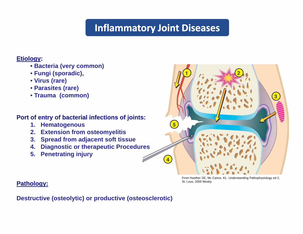
Inflammatory Joint DiseasesInflammatory Joint Diseases
EtiologyEtiology::•• Bacteria (very common)• Fungi (sporadic)• Fungi (sporadic), • Virus (rare)• Parasites (rare)• Trauma (common)
Port of Port of entry of bacterial infections of joints:entry of bacterial infections of joints:1. Hematogenous2 Extension from osteomyelitis2. Extension from osteomyelitis3. Spread from adjacent soft tissue4. Diagnostic or therapeutic Procedures5. Penetrating injury
Pathology:Pathology:From Huether SE, Mc Cance, KL. Understanding Pathophysiology ed 2, St. Lous, 2000 Mosby
Destructive (osteolytic) or productive (osteosclerotic)

Inflammatory Joint Diseases Inflammatory Joint Diseases Arthritis and Arthritis and SynovitisSynovitis
•• Infectious etiologyInfectious etiologyMos common in farm animals– Mos common in farm animals
– Less common in dogs/cats
•• Pathogenesis:Pathogenesis:Pathogenesis:Pathogenesis:– Hematogenous bacteria (+++)– Omphalitis, sepsis, FPT– Several joints (polyarthritis)
• LesionsLesions– Synovial effusion
E d t ( ti fib i t )– Exudate (suppurative, fibrinous, etc.)
Most common bacteria causingMost common bacteria causing arthritisarthritis
A pyogenes E rhusiopathiae E coliA. pyogenes E. rhusiopathiae E. coli
S. suis II H. parasuis (Glasser’s H. somni
R. equi M. hyosynoviae M. bovis
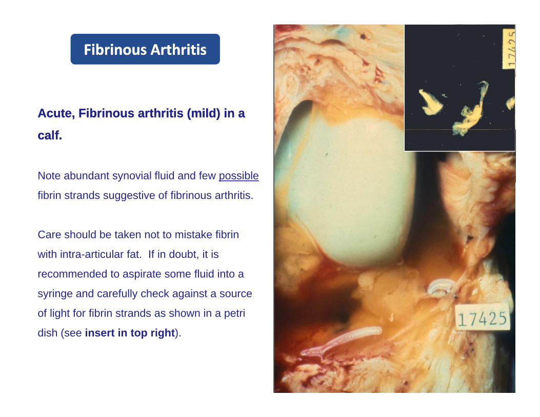
Fibrinous ArthritisFibrinous Arthritis
Acute, Fibrinous arthritis (mildAcute, Fibrinous arthritis (mild) in a ) in a
calf.calf.
Note abundant synovial fluid and few possible
fibrin strands suggestive of fibrinous arthritis.
Care should be taken not to mistake fibrinCare should be taken not to mistake fibrin
with intra-articular fat. If in doubt, it is
recommended to aspirate some fluid into a
syringe and carefully check against a source
of light for fibrin strands as shown in a petri
dish (see insert in top right).
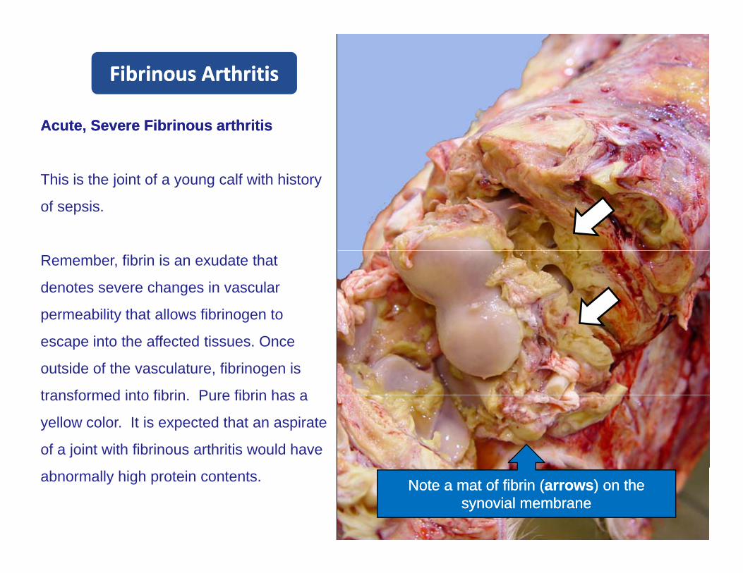
Fibrinous ArthritisFibrinous Arthritis
Acute, Acute, Severe Fibrinous arthritisSevere Fibrinous arthritis
This is the joint of a young calf with historyThis is the joint of a young calf with history
of sepsis.
Remember, fibrin is an exudate that
denotes severe changes in vascular
permeability that allows fibrinogen to
escape into the affected tissues. Once
outside of the vasculature, fibrinogen is
transformed into fibrin Pure fibrin has atransformed into fibrin. Pure fibrin has a
yellow color. It is expected that an aspirate
of a joint with fibrinous arthritis would have
abnormally high protein contents. Note a mat of fibrin (Note a mat of fibrin (arrowsarrows) on the ) on the synovial membranesynovial membrane
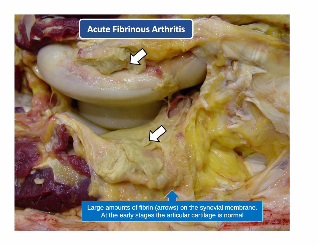
Acute Fibrinous ArthritisAcute Fibrinous Arthritis
Large amounts of fibrin (arrows) on the synovial membrane. Large amounts of fibrin (arrows) on the synovial membrane. At the early stages the articular cartilage is normalAt the early stages the articular cartilage is normal
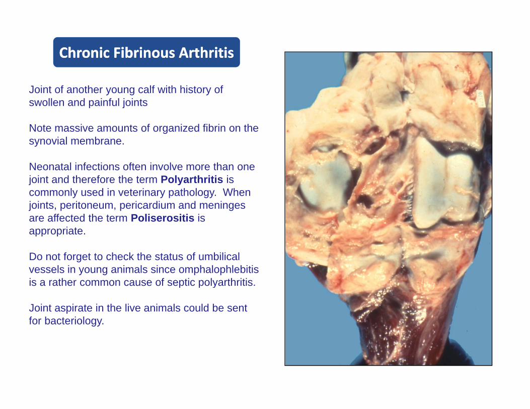
Chronic Fibrinous ArthritisChronic Fibrinous Arthritis
Joint of another young calf with history of swollen and painful joints
Note massive amounts of organized fibrin on theNote massive amounts of organized fibrin on the synovial membrane.
Neonatal infections often involve more than one joint and therefore the term Polyarthritis isjoint and therefore the term Polyarthritis is commonly used in veterinary pathology. When joints, peritoneum, pericardium and meningesare affected the term Poliserositis is appropriateappropriate.
Do not forget to check the status of umbilical vessels in young animals since omphalophlebitisis a rather common cause of septic polyarthritisis a rather common cause of septic polyarthritis.
Joint aspirate in the live animals could be sent for bacteriology.
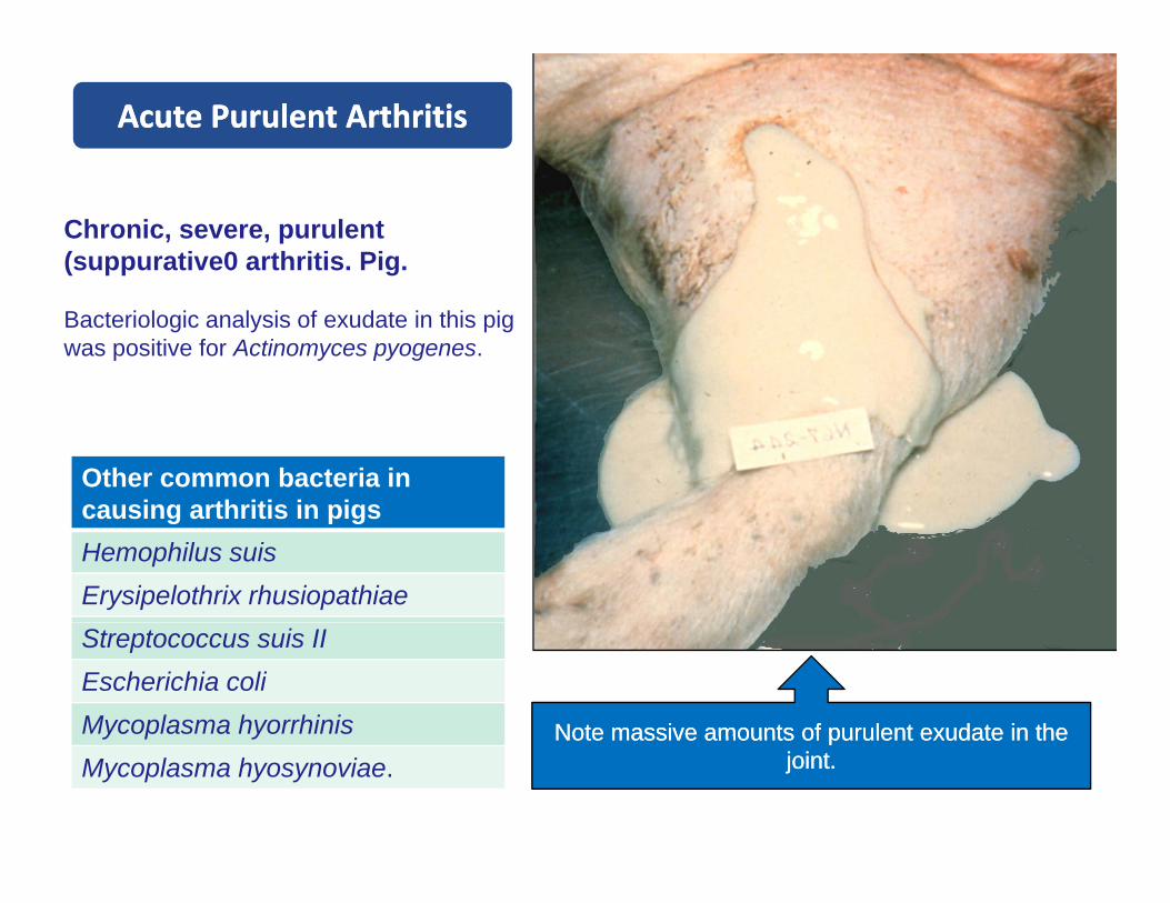
Acute Purulent ArthritisAcute Purulent Arthritis
Chronic, severe, purulent (suppurative0 arthritis. Pig.
Bacteriologic analysis of exudate in this pig was positive for Actinomyces pyogenes.
Other common bacteria in causing arthritis in pigscausing arthritis in pigsHemophilus suisErysipelothrix rhusiopathiae
Note massive amounts of purulent exudate in theNote massive amounts of purulent exudate in the
Streptococcus suis IIEscherichia coliMycoplasma hyorrhinis Note massive amounts of purulent exudate in the Note massive amounts of purulent exudate in the
joint. joint. y p y
Mycoplasma hyosynoviae.

Chronic Fibrinous Arthritis and Chronic Fibrinous Arthritis and OsteomyelitisOsteomyelitis
Chronic, severe, suppurative arthritis-osteomyelitis (severe).
When cartilage becomes ulcerated in septic arthritis, offending organisms may reach subchondral bone and bone marrow resulting in osteomyelitis.
On the other hand, organisms may also reach the joint structures from a primary underlying osteomyelitis. Some times it is difficult to tell what structure was first affected such as in this slide.
Note abundant exudate, mark thickening of joint (arrows) and large bone abscesses in the epiphyses of long bones (asterisks).

Chronic Arthritis / Porcine Chronic Arthritis / Porcine ErysipelaErysipela
Chronic, severe, Chronic, severe, arthritis arthritis (End(End--stage stage jointjoint) / Macerated bone) / Macerated bone
Note severe osseous and articular deformation due to extensive formation of osteophytes (end-stage joint).
Similar to what happens with chronic degenerative joints disease, long-standing arthritis may lead to extensive deformation due toarthritis may lead to extensive deformation due to osteophytes proliferation resulting from chronic inflammation.
This pig survived for several months and infectionThis pig survived for several months and infection with Erysipelothirx rhusiopathiae.

BursitisBursitis
B hi fill d ith i lB hi fill d ith i l fl idfl id t t i llt t i ll l t d d t dl t d d t dBursas are cushions filled with synovialBursas are cushions filled with synovial--fluid fluid strategically strategically located around some tendons located around some tendons vulnerable to vulnerable to frictionfriction--injury. Bursas are lined by a synovial membrane and injury. Bursas are lined by a synovial membrane and undergo undergo inflammation inflammation in response in response to injury to injury or hematogenous infections as synovial joints.or hematogenous infections as synovial joints.
•• Etiology of BursitisEtiology of Bursitis– Infectious (hematogenous)– Traumatic– Undetermined
•• Bursitis in HorsesBursitis in Horses– Fistulous Withers (supraspinus bursa)Fistulous Withers (supraspinus bursa)– Poll Evil (Atlanto-occipital bursa)
• Bursitis in Ruminants– Caprine Arthritis Encephalitis (CAE)– Brucellosis
•• Lesions:Lesions:Lesions:Lesions:– Swollen bursa– Synovial effusion
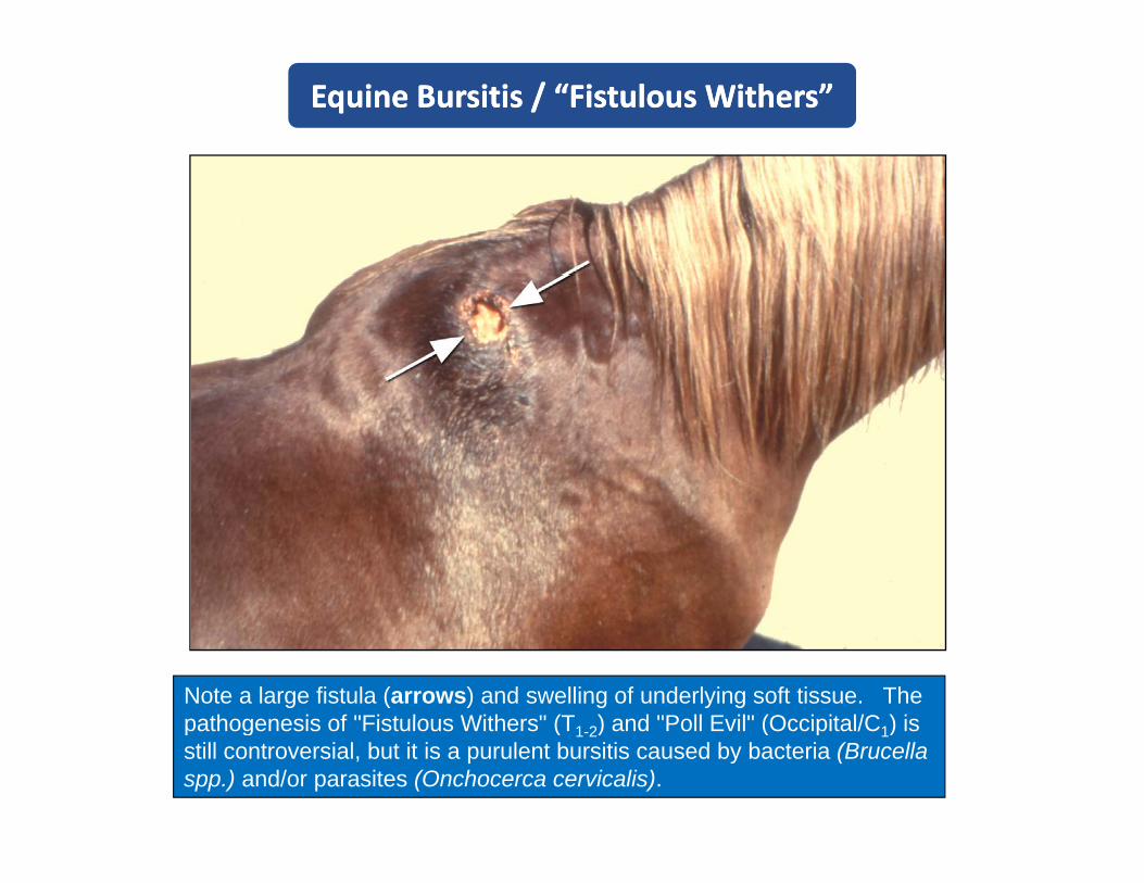
Equine Bursitis / “Fistulous Withers”Equine Bursitis / “Fistulous Withers”
Note a large fistula (arrows) and swelling of underlying soft tissue. The pathogenesis of "Fistulous Withers" (T ) and "Poll Evil" (Occipital/C ) ispathogenesis of "Fistulous Withers" (T1-2) and "Poll Evil" (Occipital/C1) is still controversial, but it is a purulent bursitis caused by bacteria (Brucellaspp.) and/or parasites (Onchocerca cervicalis).

Carpal Bursitis / BovineCarpal Bursitis / Bovine
Note swelling in the carpal region
(arrows).
The term hygroma refers to a cystic structure filled with fluid often mixed with blood which is surrounded by a fibrous capsule. Hygromas are severe form of serous bursitis in which synovial fluid accumulates over time. Hygromas are generally associated to infectious diseases such as yg g ybrucellosis (Brucella abortus) and Caprine Arthritis-encephalitis (retrovirus). There are also acquired “false bursa” in giant breeds of dogs that develops over bony protuberances at pressure points such as the lateral elbow, the greater trochanter of the femur and the tuber coxae.
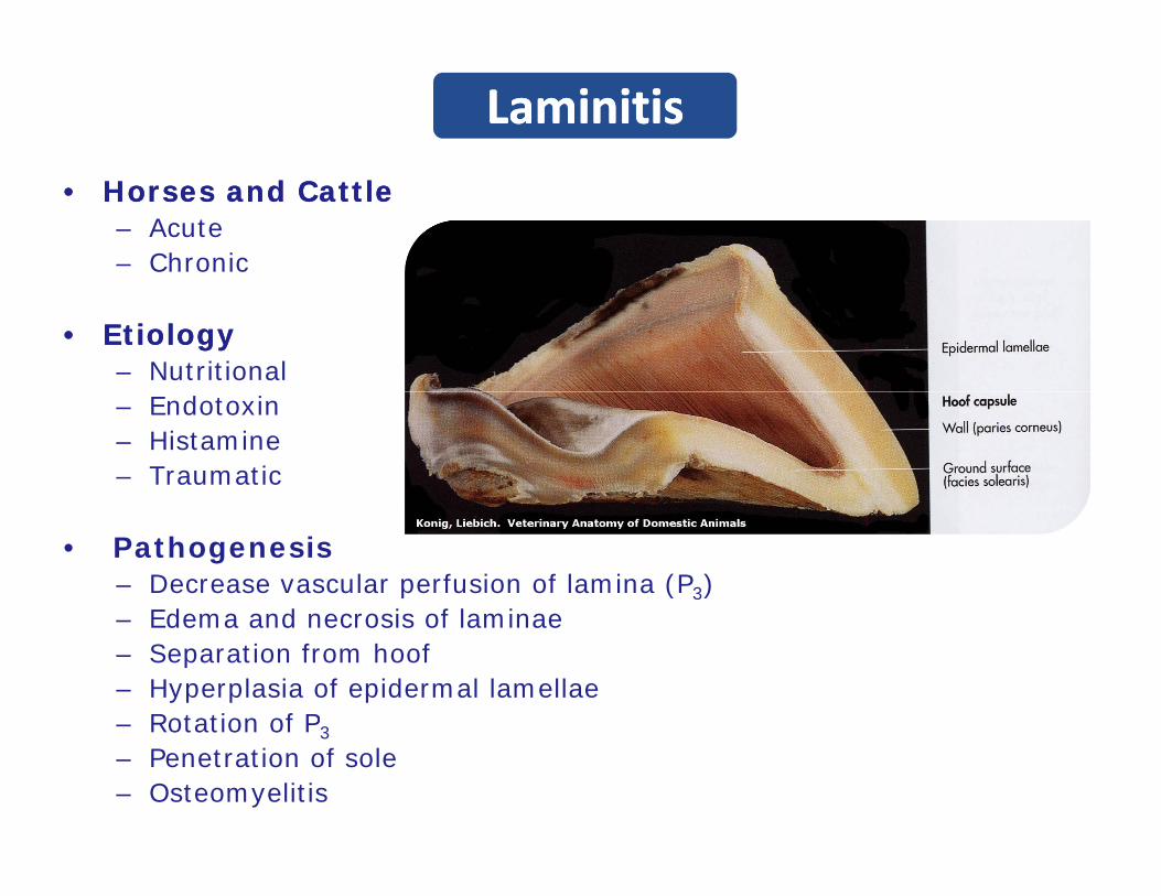
LaminitisLaminitis
•• Horses and CattleHorses and Cattle– Acute– Chronic
•• EtiologyEtiology– Nutritional– Endotoxin– Histamine– Traumatic
• Pathogenesis– Decrease vascular perfusion of lamina (P3)
Edema and necrosis of laminae– Edema and necrosis of laminae– Separation from hoof– Hyperplasia of epidermal lamellae– Rotation of P3Rotation of P3
– Penetration of sole– Osteomyelitis
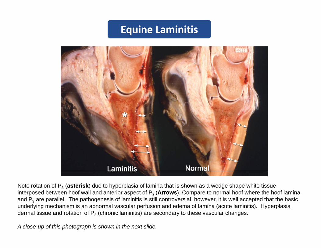
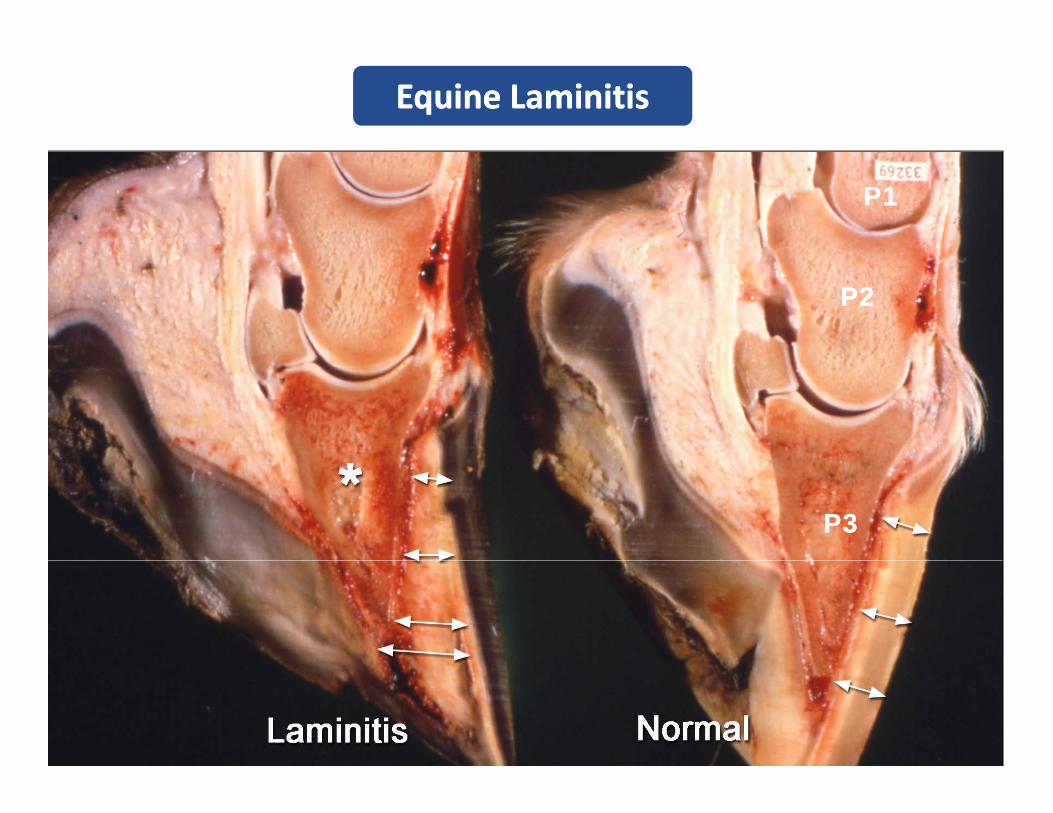
Equine LaminitisEquine Laminitis
Note rotation of P3 (asteriskasterisk) due to hyperplasia of lamina that is shown as a wedge shape white tissue interposed between hoof wall and anterior aspect of P3 (ArrowsArrows). Compare to normal hoof where the hoof lamina and P3 are parallel. The pathogenesis of laminitis is still controversial, however, it is well accepted that the basic 3 p p g punderlying mechanism is an abnormal vascular perfusion and edema of lamina (acute laminitis). Hyperplasia dermal tissue and rotation of P3 (chronic laminitis) are secondary to these vascular changes.
A close-up of this photograph is shown in the next slide.
Equine LaminitisEquine Laminitis
P1P1
P2P2
P3P3
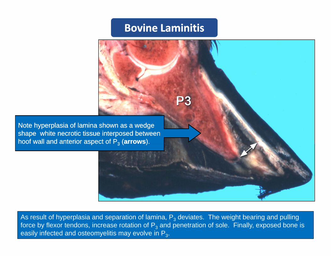
Bovine LaminitisBovine Laminitis
Note hyperplasia of lamina shown as a wedge Note hyperplasia of lamina shown as a wedge shape white necrotic tissue interposed between shape white necrotic tissue interposed between hoof wall and anterior aspect of Phoof wall and anterior aspect of P33 ((arrowsarrows).).pp 33 (( ))
As result of hyperplasia and separation of lamina, P3 deviates. The weight bearing and pulling force by flexor tendons, increase rotation of P3 and penetration of sole. Finally, exposed bone is easily infected and osteomyelitis may evolve in P3.
GoutGout
Articular Gout:Articular Gout:– Animals and humans– High intake of protein– Chickens genetic impaired secretion of uric
acid– Lesions:
• Crystals and granulomas in synovium• Bone destruction
Visceral Gout:Visceral Gout:–– Primary kidney failurePrimary kidney failure
UrateUrate depositsdeposits–– UrateUrate depositsdeposits•• KidneyKidney•• HeartHeart•• Serosal surfaceSerosal surface
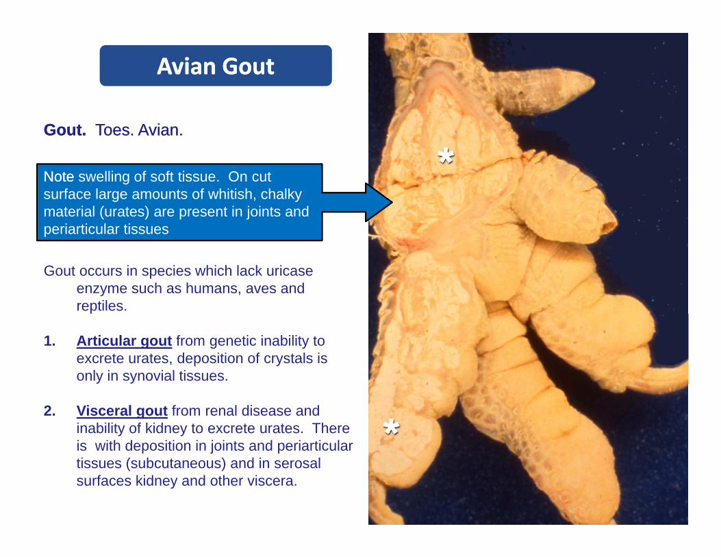
Avian GoutAvian Gout
Gout.Gout. Toes. Avian.Toes. Avian.
N tN t lli f ft ti O tNote Note swelling of soft tissue. On cut surface large amounts of whitish, chalky material (urates) are present in joints and periarticular tissues
Gout occurs in species which lack uricaseenzyme such as humans, aves and reptiles.
1. Articular gout from genetic inability to excrete urates, deposition of crystals is only in synovial tissues.
2. Visceral gout from renal disease and inability of kidney to excrete urates. There is with deposition in joints and periarticulartissues (subcutaneous) and in serosal surfaces kidney and other viscera.
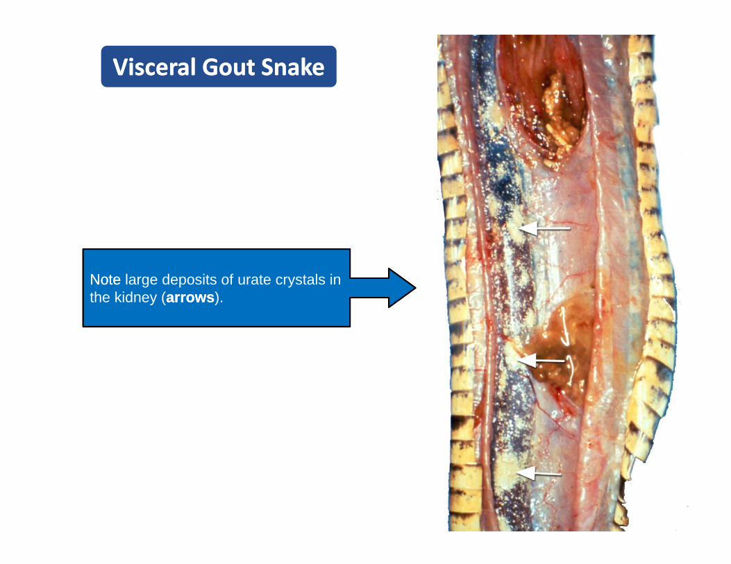
Visceral Gout SnakeVisceral Gout Snake
Note Note large deposits of urate crystals in the kidney (arrowsarrows).
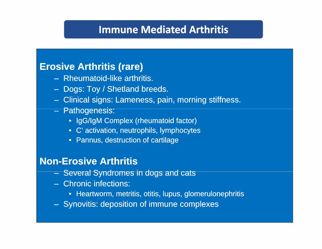
Immune Mediated ArthritisImmune Mediated Arthritis
Erosive Arthritis (rare)Erosive Arthritis (rare)RheumatoidRheumatoid like arthritislike arthritis–– RheumatoidRheumatoid--like arthritis.like arthritis.
–– Dogs: Toy / Shetland breeds.Dogs: Toy / Shetland breeds.–– Clinical signs: Lameness, pain, morning stiffness.Clinical signs: Lameness, pain, morning stiffness.
P th iP th i–– Pathogenesis:Pathogenesis:•• IgGIgG//IgMIgM Complex (rheumatoid factor)Complex (rheumatoid factor)•• C’ activation, neutrophils, lymphocytesC’ activation, neutrophils, lymphocytes•• Pannus destruction of cartilagePannus destruction of cartilage•• Pannus, destruction of cartilagePannus, destruction of cartilage
NonNon--Erosive ArthritisErosive ArthritisSeveral Syndromes in dogs and catsSeveral Syndromes in dogs and cats–– Several Syndromes in dogs and catsSeveral Syndromes in dogs and cats
–– Chronic infections:Chronic infections:•• Heartworm, Heartworm, metritismetritis, otitis, lupus, , otitis, lupus, glomerulonephritisglomerulonephritis
SynovitisSynovitis: deposition of immune complexes: deposition of immune complexes–– SynovitisSynovitis: deposition of immune complexes: deposition of immune complexes

RRheumtoidheumtoid‐‐like Arthritis . Doglike Arthritis . Dog
Histopathology. Synovial membrane. Histopathology. Synovial membrane.
NoteNote large number of plasma cells andNote Note large number of plasma cells and lymphocytes infiltration around congested blood vessels (asteriskasterisk). Plasma cells (arrowsarrows) have an eccentric nucleus, abundant eosinophilic cytoplasm and aabundant eosinophilic cytoplasm and a discrete white discoloration next to nuclear zones (Golgi).
Rheumatoid-like arthritis in dogs is similar but not identical to human rheumatoid arthritis. Only . 25% of affected dogs are positive for the rheumatoid factor (IgG/IgMcomplex).
Systemic lupus erythematosus and other allergic diseases must be ruled out before a diagnosis of Rheumatoid-like arthritis is made.
Tumors of Bones and JointsTumors of Bones and Joints
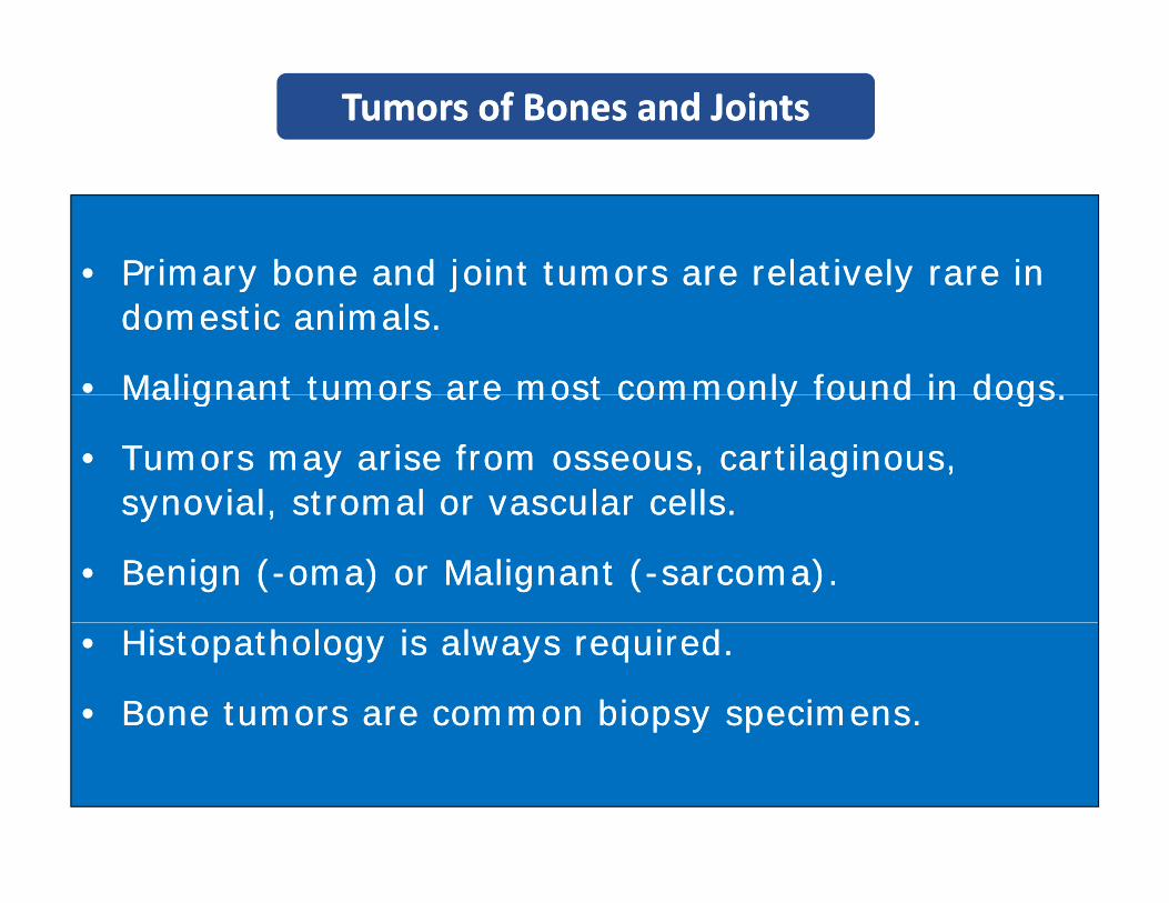
•• Primary bone and joint tumors are relatively rare in Primary bone and joint tumors are relatively rare in Primary bone and joint tumors are relatively rare in Primary bone and joint tumors are relatively rare in domestic animals.domestic animals.
•• Malignant tumors are most commonly found in dogs.Malignant tumors are most commonly found in dogs.Malignant tumors are most commonly found in dogs.Malignant tumors are most commonly found in dogs.
•• Tumors may arise from osseous, cartilaginous, Tumors may arise from osseous, cartilaginous, synovial stromal or vascular cellssynovial stromal or vascular cellssynovial, stromal or vascular cells.synovial, stromal or vascular cells.
•• Benign (Benign (--omaoma) or Malignant () or Malignant (--sarcoma).sarcoma).
•• Histopathology is always required.Histopathology is always required.
•• Bone tumors are common biopsy specimens.Bone tumors are common biopsy specimens.
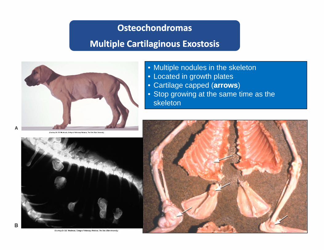
OsteochondromasOsteochondromas
Multiple CartilaginousMultiple Cartilaginous ExostosisExostosis
• Multiple nodules in the skeleton• Located in growth plates
Multiple Cartilaginous Multiple Cartilaginous ExostosisExostosis
Located in growth plates• Cartilage capped (arrowsarrows)• Stop growing at the same time as the
skeleton

OsteochondromasOsteochondromas
Multiple Cartilaginous Multiple Cartilaginous ExostosisExostosis
Osteochondromas also known as Multiple Cartilaginous Exostoses areMultiple Cartilaginous Exostoses are cartilage-capped bony protuberances which stop growing when the rest of the skeleton does.
It is still arguable if osteochondromas are multiple polyostotic tumors or dysplasia affecting the growth of cartilages. It is most commonly seen in dogs and horsescommonly seen in dogs and horses. Grossly, appear as multiple bony nodules near the growth plates. These unique tumors are most commonly seen in dogs, cats and sheepcats and sheep.
Note large osseous masses in the vertebral processes (top) and ribs (bottom).
OsteogenicOsteogenic Sarcoma (Osteosarcoma)Sarcoma (Osteosarcoma)
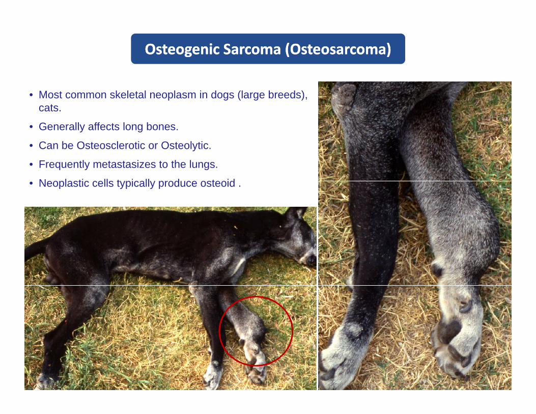
• Most common skeletal neoplasm in dogs (large breeds), cats.
• Generally affects long bonesGenerally affects long bones.
• Can be Osteosclerotic or Osteolytic.
• Frequently metastasizes to the lungs.
Neoplastic cells t picall prod ce osteoid• Neoplastic cells typically produce osteoid .
OsteogenicOsteogenic Sarcoma (Osteosarcoma)Sarcoma (Osteosarcoma)
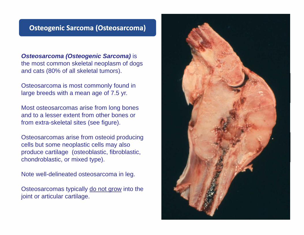
Osteosarcoma (Osteogenic Sarcoma) is the most common skeletal neoplasm of dogs and cats (80% of all skeletal tumors). ( )
Osteosarcoma is most commonly found in large breeds with a mean age of 7.5 yr.
Most osteosarcomas arise from long bones and to a lesser extent from other bones or from extra-skeletal sites (see figure).
Osteosarcomas arise from osteoid producing cells but some neoplastic cells may also produce cartilage (osteoblastic, fibroblastic, chondroblastic, or mixed type)., yp )
Note well-delineated osteosarcoma in leg.
Osteosarcomas typically do not grow into the yp y gjoint or articular cartilage.

OsteogenicOsteogenic Sarcoma / Dog Sarcoma / Dog
The leg was amputated and microscopic examination confirmed the diagnosis of osteosarcomaosteosarcoma.
It is documented that osteosarcomas may arise from sites of previous fractures or from sites with metal pins used to reduce f tfractures.
Note a metal pin adjacent to theNote a metal pin adjacent to the tumoral mass.

TypesTypes of Osteosarcoma / of Osteosarcoma / RadiographsRadiographs
OsteoscleroticOsteosclerotic typetype OsteolyticOsteolytic typetype

OsteogenicOsteogenic Sarcoma Sarcoma Lung MetastasisLung Metastasis
Osteosarcomas frequently metastasize to other organs, particularly to the lung.
gg
Note numerous metastatic tumors Note numerous metastatic tumors scattered throughout the pulmonary scattered throughout the pulmonary parenchyma (arrows)parenchyma (arrows)
Si t ft t t i t th
parenchyma (arrows). parenchyma (arrows).
Since osteosarcomas often metastasize to the lung, radiographic examination of lungs is always recommended.
Metastasis / Lung HE section/ Low MagnificationMetastasis / Lung HE section/ Low Magnification
N tN t t l d l (t l d l ( t i kt i k) i th l) i th lNote Note tumoral nodule (tumoral nodule (asteriskasterisk) in the pulmonary ) in the pulmonary parenchymaparenchyma
Osteosarcoma. Osteosarcoma. HemotoxylinHemotoxylin--eosin eosin OsteogenicOsteogenic Sarcoma / Dog Sarcoma / Dog
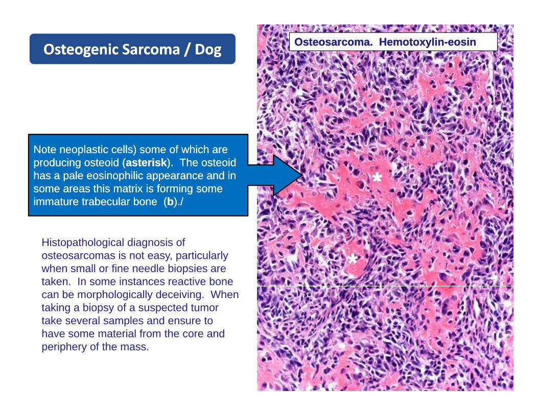
Note neoplastic Note neoplastic cells) cells) some of which are some of which are producing producing osteoidosteoid ((asteriskasterisk). The ). The osteoidosteoidhas a pale has a pale eosinophiliceosinophilic appearance and in appearance and in **some areas this matrix is forming some some areas this matrix is forming some immature immature trabeculartrabecular bone (bone (bb)./)./
Histopathological diagnosis of osteosarcomas is not easy, particularly when small or fine needle biopsies are taken. In some instances reactive bone
**can be morphologically deceiving. When taking a biopsy of a suspected tumor take several samples and ensure to have some material from the core and periphery of the mass.
ChondrosarcomaChondrosarcoma
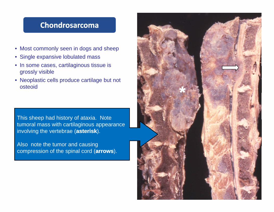
• Most commonly seen in dogs and sheep• Single expansive lobulated mass• In some cases cartilaginous tissue isIn some cases, cartilaginous tissue is
grossly visible• Neoplastic cells produce cartilage but not
osteoid **This sheep had history of ataxia. NoteThis sheep had history of ataxia. Note tumoral mass with cartilaginous appearance involving the vertebrae (asteriskasterisk).
Also note the tumor and causingAlso note the tumor and causing compression of the spinal cord (arrowsarrows).
ChondrosarcomaChondrosarcoma
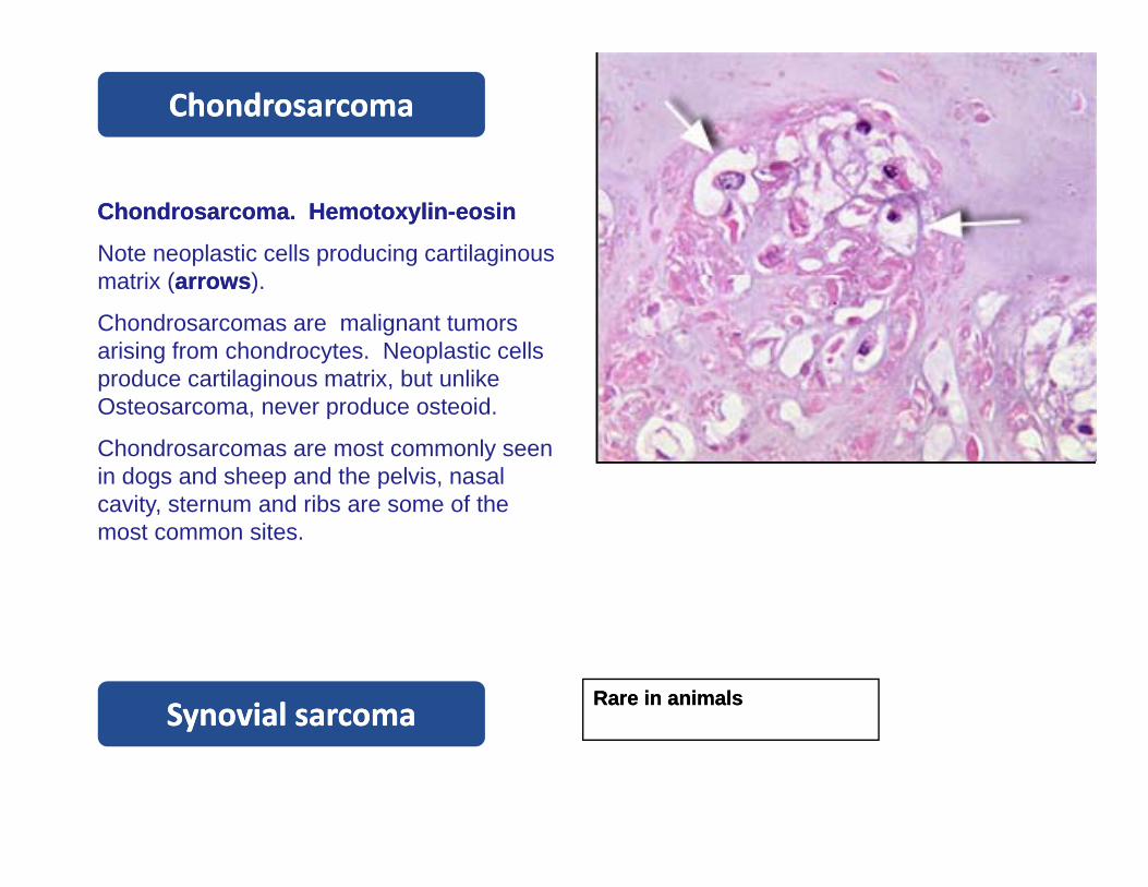
Chondrosarcoma. Chondrosarcoma. HemotoxylinHemotoxylin--eosineosin
Note neoplastic cells producing cartilaginous t i ( )matrix (arrowsarrows).
Chondrosarcomas are malignant tumorsarising from chondrocytes. Neoplastic cells produce cartilaginous matrix, but unlike p g ,Osteosarcoma, never produce osteoid.
Chondrosarcomas are most commonly seen in dogs and sheep and the pelvis, nasal ca it stern m and ribs are some of thecavity, sternum and ribs are some of the most common sites.
Rare Rare in animalsin animalsSynovial sarcomaSynovial sarcomaSynovial sarcomaSynovial sarcoma

Canine Spinal Cord MyelomaCanine Spinal Cord Myeloma
T i t b l b dT i t b l b dTumor in vertebral body Tumor in vertebral body causing compression of causing compression of spinal cord in a cat (arrows). spinal cord in a cat (arrows).
Courtesy of Drs. S. Martinson and L Pack
Some images were acquired from veterinary colleges of Some images were acquired from veterinary colleges of Canada, United States and Mexico and the names of Canada, United States and Mexico and the names of pathologists who contributed with some slides are pathologists who contributed with some slides are known. Their valuable contribution is sincerely known. Their valuable contribution is sincerely acknowledged.acknowledged.
I would like to thank Adriana López, University of I would like to thank Adriana López, University of Western Ontario, and Eileen Kinch for editorial Western Ontario, and Eileen Kinch for editorial assistance; Dr María Forzán Atlantic Veterinaryassistance; Dr María Forzán Atlantic Veterinaryassistance; Dr. María Forzán, Atlantic Veterinary assistance; Dr. María Forzán, Atlantic Veterinary College, for critically reviewing these modules.College, for critically reviewing these modules.

Inflammatory Joint Diseases and Inflammatory Joint Diseases and Tumors of Bones and JointsTumors of Bones and Joints
Inflammatory Joint Diseases and Inflammatory Joint Diseases and Tumors of Bones and JointsTumors of Bones and Joints
If you have any comments, criticisms or suggestions about this or any If you have any comments, criticisms or suggestions about this or any
other tutorial module please let me know other tutorial module please let me know other tutorial module please let me know.other tutorial module please let me know.
Also, if you find any errors or typos please let me know too. Thanks ! Also, if you find any errors or typos please let me know too. Thanks ! , y y yp p, y y yp p
[email protected]@upei.ca