Infectious papillomavirus in the vapor of warts treated ...
9
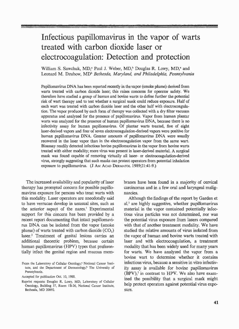
Infectious papillomavirus in the vapor of warts treated with carbon dioxide laser or electrocoagulation: Detection and protection William S. Sawchuk, MD,a Paul J. Weber, MD,b Douglas R. Lowy, MD,a and Leonard M. Dzubow, MDb Bethesda, Maryland, and Philadelphia, Pennsylvania Papillomavirus DNA has been reported recently in the vapor (smoke plume) derived from warts treated with carbon dioxide laser; this raises concerns for operator safety. We therefore have studied a group of human and bovine warts to define further the potential risk of wart therapy and to test whether a surgical mask could reduce exposure. Half of each wart was treated with carbon dioxide laser and the other half with electrocoagula- tion. The vapor produced by each form of therapy was collected with a dry filter vacuum apparatus and analyzed for the presence of papillomavirus. Vapor from human plantar warts was analyzed for the presence of human papillomavirus DNA, because there is no infectivity assay for human papillomavirus. Of plantar warts treated, five of eight laser-derived vapors and four of seven electrocoagulation-derived vapors were positive for human papillomavirus DNA. Greater amounts of papillomavirus DNA were usually recovered in the laser vapor than in the electrocoagulation vapor from the same wart. Bioassay readily detected infectious bovine papillomavirus in the vapor from bovine warts treated with either modality; more virus was present in laser-derived materiaL A surgical mask was found capable of removing virtually all laser- or electrocoagulation-derived virus, strongly suggesting that such masks can protect operators from potential inhalation exposure to papillomavirus. (J AM ACAD DERMAToL 1989;21:41-9.) The increased availability and popularity of laser therapy has prompted concern for possible papillo- mavirus exposure for persons who treat warts with this modality. Laser operators are anecdotally said to have verrucae develop in unusual sites, such as the anterior aspect of the nares. I Experimental support for this concern has been provided by a recent report documenting that intact papillomavi- rus DNA can be isolated from the vapor (smoke plume) of warts treated with carbon dioxide (C0 2 ) laser. 2 Treatment of genital lesions carries an additional theoretic problem, because certain human papillomavirus (HPV) types that preferen- tially infect the genital region and mucous mem- From the Laboratory of Cellular Oncology," National Cancer Insti- tute, and the Department of Dermatology,b The University of Pennsylvania. Accepted for publication Oct. 10, 1988. Reprint requests: Douglas R. Lowy, MD, Laboratory of Cellular Oncology, Building 37, Room IB-26, National Cancer Institute, Bethesda, MD 20892. branes have been found in a majority of cervical carcinomas and in a few oral and laryngeal malig- nancies. Although the findings of the report by Garden et al. 2 are highly suggestive, whether papillomavirus material in the vapor contained potentially infec- tious virus particles was not detennined, nor was the potential virus exposure from lasers compared with that of another treatment modality. We have studied the relative amounts of virus isolated from the vapor of human and bovine warts treated with laser and with electrocoagulation, a treatment modality that has been widely used for many years for warts. We have analyzed the vapor from a bovine wart to determine whether it contains infectious virus, because a sensitive in vitro infectiv- ity assay is available for bovine papillomavirus (BPV),3 in contrast to HPV. We also have exam- ined the possibility that a surgical mask might help protect operators against potential virus expo- sure. 41
Transcript of Infectious papillomavirus in the vapor of warts treated ...
Infectious papillomavirus in the vapor of wartstreated with carbon dioxide laser orelectrocoagulation: Detection and protectionWilliam S. Sawchuk, MD,a Paul J. Weber, MD,b Douglas R. Lowy, MD,a andLeonard M. Dzubow, MDb Bethesda, Maryland, and Philadelphia, Pennsylvania
Papillomavirus DNA has been reported recently in the vapor (smoke plume) derived fromwarts treated with carbon dioxide laser; this raises concerns for operator safety. Wetherefore have studied a group of human and bovine warts to define further the potentialrisk of wart therapy and to test whether a surgical mask could reduce exposure. Half ofeach wart was treated with carbon dioxide laser and the other half with electrocoagulation. The vapor produced by each form of therapy was collected with a dry filter vacuumapparatus and analyzed for the presence of papillomavirus. Vapor from human plantarwarts was analyzed for the presence of human papillomavirus DNA, because there is noinfectivity assay for human papillomavirus. Of plantar warts treated, five of eightlaser-derived vapors and four of seven electrocoagulation-derived vapors were positive forhuman papillomavirus DNA. Greater amounts of papillomavirus DNA were usuallyrecovered in the laser vapor than in the electrocoagulation vapor from the same wart.Bioassay readily detected infectious bovine papillomavirus in the vapor from bovine wartstreated with either modality; more virus was present in laser-derived materiaL A surgicalmask was found capable of removing virtually all laser- or electrocoagulation-derivedvirus, strongly suggesting that such masks can protect operators from potential inhalationexposure to papillomavirus. (J AM ACAD DERMAToL 1989;21:41-9.)
The increased availability and popularity of lasertherapy has prompted concern for possible papillomavirus exposure for persons who treat warts withthis modality. Laser operators are anecdotally saidto have verrucae develop in unusual sites, such asthe anterior aspect of the nares. I Experimentalsupport for this concern has been provided by arecent report documenting that intact papillomavirus DNA can be isolated from the vapor (smokeplume) of warts treated with carbon dioxide (C02)
laser.2 Treatment of genital lesions carries anadditional theoretic problem, because certainhuman papillomavirus (HPV) types that preferentially infect the genital region and mucous mem-
From the Laboratory of Cellular Oncology," National Cancer Institute, and the Department of Dermatology,b The University ofPennsylvania.
Accepted for publication Oct. 10, 1988.
Reprint requests: Douglas R. Lowy, MD, Laboratory of CellularOncology, Building 37, Room IB-26, National Cancer Institute,Bethesda, MD 20892.
branes have been found in a majority of cervicalcarcinomas and in a few oral and laryngeal malignancies.
Although the findings of the report by Garden etal. 2 are highly suggestive, whether papillomavirusmaterial in the vapor contained potentially infectious virus particles was not detennined, nor wasthe potential virus exposure from lasers comparedwith that of another treatment modality. We havestudied the relative amounts of virus isolated fromthe vapor of human and bovine warts treated withlaser and with electrocoagulation, a treatmentmodality that has been widely used for many yearsfor warts. We have analyzed the vapor from abovine wart to determine whether it containsinfectious virus, because a sensitive in vitro infectivity assay is available for bovine papillomavirus(BPV),3 in contrast to HPV. We also have examined the possibility that a surgical mask mighthelp protect operators against potential virus exposure.
41

42 Sawchuk et al.
Journal of theAmerican Academy of
Dermatology
1
R
5 7 Table I. HPV DNA in vapor from humanplantar warts
HPV DNA in vapor material
High positive, Visualized after 3·hour exposure; Low Positive. visual·ized after 12·hour exposure.*The DNA in this sample was lost during extraction and could not beassayed.
Patient HPV type Laser I Electrocoagulation
Low positiveNot done*NegativeNegativeLow positiveNegativeHigh positiveLow positive
Low positiveHigh positiveNegativeNegativeHigh positiveNegativeHigh positiveLow positive
21221212
12345678
0-
Fig. 1. Dot-blot analysis of DNA extracted from laserplume (upper set) and from electrocoagulation plume(lower set) of human plantar warts. Warts from subjects1,3, and 8 were hybridized with labeled HPV type 2 andsubjects 5 and 7 with HPV type 1. Blots were washedunder stringent conditions. Subject 3 is considerednegative, subjects 1 and 8 are low positive, and subjects5 and 7 are high positive.
MATERIAL AND METHODS
Treatment of warts
Eight persons with plantar warts that were morphologically flat and uniform were selected for study,because the amount of virus and viral DNA in plantarwarts is usually large compared with that in otherclinical types.4-8 All treatment was carried out in Philadelphia, Pa., by the same person (P. J. W.). The size ofeach wart was determined by measuring its surface areawith the use of a grid composed of transparent 1 romsquares. Lesions were anesthetized with equal parts of0.25% bupivicaine and 1% lidocaine without epinephrine(1 ml/cm2 of tissue). Before therapy, a small shavebiopsy specimen was taken (along the bisecting midlineof the specimen, so that equal portions of each treatmenthalf were included) from each wart for HPV typing.The specimen was placed immediately in lysis buffer (10mmol/L TRIS-hydroxymethyl-amino methane, pH 8.0;10 mmoljL sodium chloride; 10 mmolfL ethylenediamine tetraacetic acid; 0.5% sodium dodecylsulfate; and 50,ugjml proteinase K) for storage and transport toBethesda', Md., at room temperature.
Half of each wart was treated with a Xanar CO2
laser, model No. XA-20 (Johnson & Johnson, NewBrunswick, N.J.), with the use of a uniform 1 rom spotsize at a setting of 10 W, a beam sweep speed of 3mm/sec, and a peak intensity of 1270 W/cm2
, until thesurface of the lesion and a 2 rom margin were completely charred. The char was vigorously removed with asaline-soaked sponge; then laser application and removalof char were repeated until there was no furtherbubbling of the wart tissue or until no excessive bleedingor friable dermis remained, which required an averageof four to eight passes with the laser. The other half ofeach wart was treated by electrocoagulation, with theuse of a Valleylab Surgistat (Boulder, Colo.). A 25gauge needle applied to a Bernsco adapter (Bemsco
Inc., Seattle, Wash.) was used as the uniform tip, whichwas inserted into a Valleylab disposable hand pencilunit, No. E2515. The coagulation level was maintainedat a setting of 6, and passes were made over the wart at arate of 2 mm/sec. The resulting char was removed witha curette; repeated electrocoagulation and curettagewere then performed for an average of three to fourcycles per lesion.
A previously excised bovine wart (kindly provided byDr. Carl Olsen, University of Wisconsin, Madison),which had been stored at -70 0 C in Bethesda, Md., washydrated for 12 hours in cold phosphate buffered saline,pH 7.4; frozen again at -70 0 C; and shipped on dry iceto Philadelphia. Thirty minutes before treatment withlaser or electrocoagulation, the bovine wart was placedin sterile saline and allowed to thaw at room temperature. Equal volumes of wart, determined byfiuiddisplacement, were treated with laser and electrocoagulation. Treatment of the bovine wart was carriedout as for the human warts, except that the bovinewart was rotated with fine forceps during lasertreatment, and curettage was performed with the wartheld against a saline-soaked gauze pad during electrocoagulation.
Collection of vapor samples
The vapor from either procedure was collected withthe use of a vacuum device (Stackhouse laser smokeevacuation unit [model No. LFA] with filter [modelNo. LFA-103]; Stackhouse Associates, El Segundo,Calif.). A 6-foot-long, corrugated, clear plastic hose(smallest internal diameter, 2 cm) was interposedbetween the collecting device and the LFS-103 filter.During collection of vapor from warts treated witheither modality, the end of the collection device near thewart was maintained 2 cm from the wart surface. The

Volume 21Number 1July 1989 Papillomavirus vapor of warts 43
Fig. 2. Focal transformation induced in C127 mouse cells by vapor from laser orelectrocoagulation treatment of a bovine wart. Dishes were stained 4 weeks after infection.Negative control (JVEG CTR) dish of e127 cells shows no background foci. Dish with laservapor-infected cells shows many foci. Dish with electrocoagulation vapor-infected cellsshows fewer foci compared with that seen in laser dish.
vacuum unit was kept at a constant power level of 10,which uniformly generated a 42 L/min airflow (determined with a Timeter Instrument [Lancaster, Pa.] IIIBmeter) without the interposed collecting device and a 40L/min airflow with the device "in-line."
A special "dry" collecting device was constructed totrap airborne papillomavirus without passage throughliquid. It was prepared to function by two mechanisms:direct gas/particle impact and Venturi effect. Thedevice was a 15 cm clear plastic tube, with an internaldiameter of 2 cm, to which a metal screw of 3 mm X 7.5cm had been affixed by metal wire to the exact center ofthe tube. A 1.6 cm metal washer waS welded to a boltand screwed to a fixed position along the screw. Thewasher was constructed to seat a piece of type HA filterpaper, 1.9 cm diameter, 0.45 }.Lm pore size (MilliporeCorp., Bedford, Mass.) With a 3 rom hole punched out ofits exaCt center. Finally, three 1 rom screws were placedin the plastic tube approximately 4 rom proximal to thelocation of the washer. A 1.2 X 7.0 cm strip of filterpaper (grade No. 613, catalog No. 28310-128; VWRScientific, Philadelphia, Pa.) was curled and insertedinto the collection tube until it rested in the upwindposition against the three screws. The unit was con·structed so that gas and particulate matter would firstreach the 1.9 cm filter disk. The airflow would increasebehind the disk by the Venturi effect and depositparticulate matter on the nearby, now cylindric, strip ofVWR Scientific filter paPer. Before each use, the aircollection apparatus was cleaned three timeS with alcohol swabs. One VWR Scientific filter strip and onecircular Millipore filter disk were used for each collection period; separate sterile instruments were used foreach procedure. For testing the ability of a surgicalmask to filter papillomavirus, a 4.4 cm disk cut from amask (catalog No. 1818; 3M Surgical Products, St.Paul, Minn.) was placed over the collecting device, infront of the collection filter, and secured with arubber band. After exposure to vapor, the mask and tiI-
ter were analyzed separately for the presence of viralDNA.
Filters and masks to be analyzed for papillomavirusDNA were placed in vials with lysis buffer and shippedto Bethesda at room temperature. When treatmentmaterial was to be used in the infectivity assay, vaporwas collected on a sterile VWR Scientific filter strip for60 seconds, transferred to sterile tubes for transportationto Bethesda, Md., on dry ice, and stored at '-70 0 C.
B:PV infectivity assay
When used for infectivity studies, vapor-derivedmaterial collected on a filter was solubilized in 1.9 nilDulbecco's modified Eagle's mediuni, with 50 U /nilpenicillin and 50 i.tg/ml streptomycin, and incubated 1hour at room temperature, with occasional vortexmg.The fluid was then t>i!,etted into Eppendorf tubes(Brinkmann Instruments; Westbury, NS.) and centri~
fuged at 2000 revolutions per minute in art Eppendorfcentrifuge (Brinkmann) for 5 minutes to remove thelarge debris.
The prepared extracts were assayed for· infectiousBPV by testing their ability to indticefocal transfotma~tion of mouse c1i7 cells, as deScribed.3 Briefly, 60 romdishes seeded the previous day with 165 cells wereinfected with 0.5 ml of filter extract (obtained asdescribed earlier) or 0.5 ml of a control BPV preparation (isolated directly from a bovine wart) known tocontain about 100 focus-forming units of virus. Vnd~r
the conditions used, infectious BPV particles will sc6reas positive, but naked viral DNA will be negative. After2 weeks' incubation, focal transformation of cells wasreadily detectable, and some dishes were fixed andstained (three parts i % methylene blue, two partsmethanol, and one part 1% carbolfuchsin).
To confirm that the focal transformation observedwas the result of infection with BPV, companioncultures were analyzed for the preSence of BPV DNAby selecting for morphologically transformed cells and

Journal of theAmerican Academy of
44 Sawchuk et al. Dermatology
A R
(+)00 OL ELECTR G
Fig. 3. Photomicrograph of individual foci induced by vapor from bovine wait.(-)CONTROL, C127 cells alone; (+) control, cells infected with known BPV extract; LASER.cells infected with extract from laser vapor; ELECTROCOAG, cells infected with extractfrom electrocoagulation vapor. (Magnification X 8, except for ELECTROCOAG. whichwas XI2.)
harvesting the cells for high-molecular-weight DNAextniction9 and Southern blot analysis. JO For each sample, 8 p.g of high-molecular-weight DNA was digestedwith 25 U1).its of Cla1 restriction endonuclease underconditions recommended by the supplier (New EnglandBiolabs, Beverly, Mass.), run on an 0.8% agarose gel toseparate the DNA fragments, and transferred by capillary action to a nylon filter (Schleicher & Schuell,Keene, N.H.). The filter was baked, prehybridized, andhybridized according to the manufacturer's directions(Nytran product information'; Schleicher & Schuell)with the use of cloried 32P-Iabeled BPV-1 DNA as aprobe. The BPV-I DNA had been nick translated11 with32P-dCTP (2000-3000 Cijmrnol; Amersham, ArlingtonHeights, Ill.); and 20 X 106 cpm (specific activity,2.2 X 107 cpmjp.g) were used in each hybridization.After hybridization at 42° C overnight, the blot was
washed according to the manufacturer's directions, witha final stringent wash in 0.1 X standard saline citrate(0.15 moljL sodium chloride, 0.Ql5 mol/L sodiumcitrate), 1% sodium dodecylsulfate at 65° C. The blotwas placed in a photographic cassette with Kodak XAR5 film and stored at -70° C until the film wasdeveloped.
Dot-blot analysis
To extract DNA from filters and masks on whichvapor had been collected, the lysis bliffer in each vialwas increased to 3 ml and incubated at 37° C for 6 to 12hours, with occasional vortexing. The solution was thendeproteinized with phenOl and chloroform-isoamyl alcohol, ethanol precipitated, washed sequentially with 70%and 95% ethanol, vacuum dried, and resuspended in 80fLl of 10 mmoljL TRIS-hydroxymethyl-amino methane,

Volume 21Number 1July 1989
pH 8.0, and 1 mmoljL ethylenediamine tetraacetic acid(TE). Thirty microliters of each resuspended DNA wasmixed with 20 1-£1 of TE and 50 1-£1 of 20 X standardsatirie citrate, denatured by boiling for 10 minutes, andblotted onto BA 83 nitrocellulose paper (Schleicher &Schuell) by use of a dot-blot apparatus. One microgramof high-molecular-weight human DNA from a personwithout warts was used as a negative controL
Before hybridization of the material described above,the small pretreatment biopsy specimens from each wartwere used to determine the HPV type in the wart. In thisprocedure cloned HPV DNAs were blotted onto nitrocellulose paper with a dot-blot apparatus, and DNAextracted from each wart biopsy specimen was nicktranslated as described earlier and used as a probe tohybridize against the known HPV types. Blots were thenwashed under nonstringent (4 X 20 minutes in 5 X standard saline citrate, 0.1% sodium dodecylsulfate at 65°C) or stringent (4 X 20 minutes in 0.1 X standard salinecitrate, 0.1% sodium dodecylsulfate at 65° C) conditionsand exposed as described earlier. This analysis verifiedthat the lesion contained papillomavirus DNA anddetermined the HPV DNA type in the wart. The DNAof the corresponding cloned HfiV type was then used asthe probe, after it was labeled by nick translation, todetect the minimal amounts of viral DNA expected tohave been isolated from the vapor-derived material.After hybridization, the blots were washed under stringent conditions and processed for autoradiography.
RESULTSHPV DNA in vapor from treated human warts
HPV type I was found in three warts; the otherfive had HPV type 2. HPV DNA was detected infive of eight laser-derived vapors and in four ofseven electrocoagulation-derived vapors (Table Iand Fig. 1). (The electrocoagulation vapor fromone patient could not be analyzed.) There was acomplete correlation between the two treatmentmodalities and the detection of viral DNA in thevapors. For either form of therapy, the vaporsderived from warts with HPV type I tended tohave significantly larger amounts of viral DNAthan those derived from warts with HPV type 2.The vapors from all three warts with HPV type I(from subjects 2, 5, and 7) were strongly positivefor viral DNA. In contrast, the vapors from onlytwo of the five warts with HPV type 2 (from subjects I and 8) had detectable levels of viral DNA.
Vapor contains infectious papillomavirus
The results obtained herein indicated that viralDNA was present in the vapors derived from
Papillomavirus vapor ofwarts 45
treatment with laser or electrocoagulation, but theydid not determine whether the papillomavirusmaterial in the vapor was infectious. The currentlack of a reproducible in vitro infectivity assay forHPV particles makes this assessment virtuallyimpossible to perform for HPV. This problem canbe circumvented, however, if bovine warts are used,because a sensitive, quantitative cell transformationassay is available for BPY.3 Portions of a bovinewart therefore were treated with laser or electrocoagulation, with the use of the same machines andsettings and vapor collection system as for thehuman warts. The material derived from thebovine wart vapor was then tested for the presenceof infectious BPV by assaying its ability to inducemorphologic transformation of mouse el27 cells.Foci of morphologically transformed cells werefound in material derived from both forms oftherapy; the products of the laser vapor inducedsignificantly more foci than those from electrocoagulation (Figs. 2 and 3).
To verify that the focal transformation had beeninduced by BPV, high-molecular-weight DNA wasextracted from the morphologically transformedcells induced by the laser-derived extract andanalyzed for the presence of BPV-l DNA by theSouthern blot technique, with the use of molecularly cloned BPV-I DNA as the detection probe. Thetransformed cells contained BPV-I DNA; Claldigestion of the test DNA yielded the three expected BPV DNA fragments of 5.9, 1.3, and 0.6 Kbthat hybridized with the BPV DNA probe andcomigrated with those of the positive BPV-l DNAcontrol (Fig. 4). The results indicate that infectiousBPV is present in the vapor-derived material.
Surgical mask blocks passage of virus
Having demonstrated that the vapor from bothforms of treatment contains infectious BPV, wethen tested whether placing a surgical mask in thevapor path could inhibit passage of the virus ontothe collection filter. Vapor products derived fromlaser or electrocoagulation treatment of the bovinewart were collected on a filter, as previouslydescribed. For each fonn of treatment there weretwo I5-second treatment periods, first without themask and then with the mask in place. The filtersfrom each treatment period were analyzed bydot-blot hybridization for the presence of BPVDNA (Fig. 5). As expected, viral DNA waspresent on the filters used to collect the electrocoa-

46 Sawchuk et al.
Journal of theAmerican Academy of
Dermatology
Fig. 4. Southern blot analysis of DNA from C127mouse cells morphologically transformed by laser vapor.H~gh-molecular-weight DNA was digested with CIal.Blot was probed wi.th labeled BPV-l DNA. NegativecOntrol (C12l cells alone) shows no BPV DNA present.Positive control ((+)CTR), C127 cells infected withknown BPV extract and C127 cells infected with laservapor products (LASER), show identical expected BPVDNA fragments (5.9, 1.3, and 0.6 Kb).
Laser smoke as an entity may represent atheoretic health hazard, because in rats exposed tolong-term inhalation of CO2 laser smoke, emphysema, bronchiolitis and congestive interstitial pneumonia developed. 12 In addition to the possibleadverse effects of the smoke alone, it is importantto know whether viable cells or microbial agentssurvive in the laser plume. Contradictory resultshave been reported. One group noted that S-91melanoma cells appeared to survive in the neadynium laser plume, 13 whereas other researchersshowed a lack of tumor cell viability of the sameS-91 melanoma line in the CO2 laser plume. 14
Efforts to detect viable cells in warts treated withCO2 laser were reported to be unsuccessful. I5 Inone studyl6 bacteria were shown to survive CO2
laser treatment, whereas in another studyl7 virtualdestruction of the bacteria was demonstrated. Asnoted in the introduction to this article, intactpapillomavirus DNA has been demonstrated in thelaser plume from bovine and human warts treatedwith the CO2 laser. 2
Results of our study have confirmed and extended those in the report by Garden et a1,2 in severalways. In addition to detecting papillomavirus DNAin the laser plume from treated human and bovinewarts, we also have documented, with a bioassay,that the plume from bovine warts contains infectious bovine papillomavirus particles (virions). Wealso have shown that papillomavirus, in somewhatlower concentrations, can be isolated from theplume resulting from treatment of bovine and human warts by electrocoagulation. Although ourstudies of infectious virus were limited to BPV, it isreasonable to infer that viable HPV virions mayalso persist in the plume derived from human material. Of perhaps greatest interest, we have shownthat papillomavirus material in the plume can beefficiently trapped by a simple surgical mask.
We believed it was important to document thatthe plume contained infectious papillomavirus particles. Although naked papillomavirus DNA can beinfectious,18-20 the specific infectivity of virions isgenerally several orders of magnitude higher thanthat for naked DNA, so virions represent a muchgreater theoretic hazard than naked viral DNA.For example, viral DNA from the closely relatedpolyomavirus (which is a member of the papovavirus group, as are papillomaviruses) is four to fiveorders of magnitude less infectious than virus
DISCUSSION
.3
.6
. 2.
(+)127
gulation and laser vapors produced before the maskwas put in pll:).ce. With either form of treatment,placing the· surgical mask in front of the filterprevented the vif\ls in the vapor from collecting onthe filter. DNA wa~ extracted from the masks toverify that the mask had indeed trapped the virusmaterial ~nd was found to contain substantialamounts of BPV DNA (Fig. 5). This result also~on:fir:nJed that viral material continued to bepresent in the vapor during the treatment periodwith the mask in place and that airflow had beenmaintained during this time.

Volume 21Number IJuly 1989 Papillomavirus vapor of warts 47
Fig. 5. Dot-blot analysis of BPV DNA from laser andelectrocoagulation vapor blocked by surgical mask.Blots were probed with labeled BPV-l DNA andwashed under stringent conditions. First column, DNAextracted from filters exposed to vapor before placementof mask; detectable viral DNA. Second column, DNAextracted from filters exposed to vapor with mask inplace; no detectable viral DNA. Third column, DNAextracted from masks used during exposure of filters insecond column; abundant viral DNA was trapped.
0-o
p
factor may indicate that HPV exposure associatedwith this procedure may not be particularly hazardous. ElectrosurgiCal devices are usually used incombination with curettage for treatment of warts,rather than continuously, as with the laser. Without the continuous vaporization and repeat passesexpected with laser treatment, electrocoagulationtreatment of warts results in less tissue destructionby the current. Laser plumes are dense, whereasthe plume from electorcoagulation is much morediffuse. Our collection device was placed immediately (2 cm) above the treatment site; the operator's face is usually not located there during theprocedure. The larger amounts of virus recoveredfrom the laser vapor suggests that laser treatmentmay pose a greater theoretic hazard than electrocoagulation.
HPV types differ regarding concentration ofvirions that are typically present in lesions, theirinfectivity for different regions of the body, andtheir malignant potential. Because HPV type 1lesions usually have the highest concentration ofvirus particles,4-8 lesions associated with this virustype would appear to pose the greatest theoreticrisk for infection. This potential hazard is balancedby the benign nature of lesions induced by thisHPV type. In addition, infection by HPV type 1 isusually restricted to the extremities. Therefore itstheoretic risk to the face should be low, andwearing gloves (and shoes) should drasticallyreduce biologically important exposure. HPV type2 lesions generally have a lower concentration of
particles when both are administered parenterallyto susceptible mice. 21 We therefore performed theinfectivity assay under conditions that would detectinfectious virions but not potentially infectious viralDNA. The positive results obtained with this assayindicate that a significant proportion of the viralDNA detected in the plume was present in virions.
For our analysis of human warts, we studiedplantar warts with HPV type I or HPV type 2. Wemonitored the presence of HPV material in theplume by assaying the viral DNA in the plume. Ingeneral we found more viral DNA in the plumefrom warts with HPV type I than from warts withHPV type 2. This result correlates with the observation that of all clinical wart types plantar wartscontaining HPV type I appear to be the richest inviral particles.4-8
These results, in conjunction with what is knownabout the biologic characteristics of various typesof papillomaviruses,22-25 may have some implications for the theoretic operator risk from the plume.(It is not likely that the plume material represents asignificant hazard to the patient for distant spread,because the patient is presumably exposed chronically to his or her own virus.) Operator risk isprincipally to exposed areas, which means that theface, including the upper airway, and hands represent the major regions of potential concern. Thepresence of infectious papillomavirus virions in theplume may appear initially to represent a significant theoretic hazard. We, however, believe theactual degree of risk is not very great and that itcan be reduced further by relatively simple precautions. It should be remembered that warts are nothighly contagious, adults are not likely to developnongenital warts, and genital warts are usuallytransmitted sexually. Although many health careworkers apparently examine warts without wearinggloves, the incidence of hand warts among thisgroup does not appear to be unusually high.
The clinical implications of the positive HPVresults obtained in the vapor from electrocoagulation may be subject to various interpretations.Compared with the laser vapor, the amount ofvirus recovered was significantly less. We do notbelieve that the vapor from electrocoagulationrepresents a major risk. Physicians do not appear tobe especially concerned about the risk of wartsdeveloping from this modality, and the lack ofreports implicating electrocoagulation as a risk
48 Sawchuk et al.
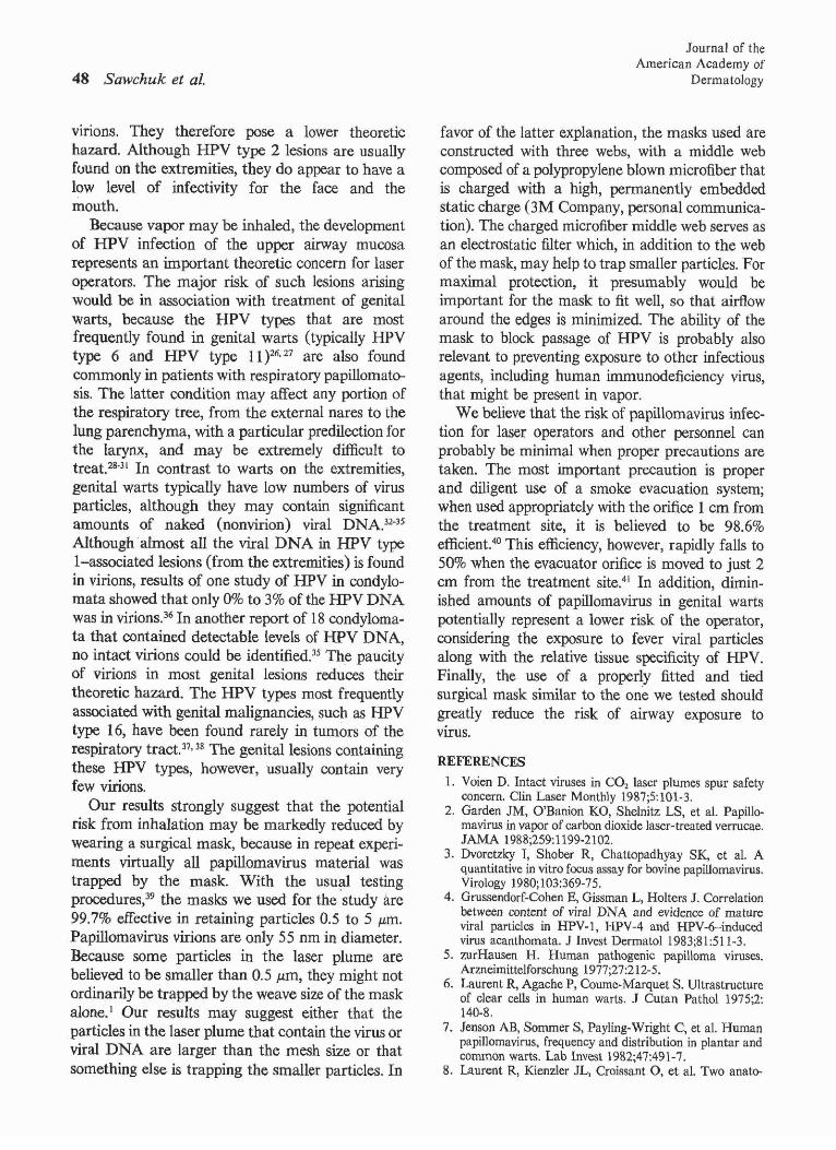
Vinons. They therefore pose a lower theoretichazard. Although HPV type 2 lesions are usuallyfound on the extremities, they do appear to have alow level of infectivity for the face and themouth.
Because vapor may be inhaled, the developmentof HPV infection of the upper airway mucosarepresents an important theoretic concern for laseroperators. The major risk of such lesions arisingwould be in association with treatment of genitalwarts, because the HPV types that are mostfrequently found in genital warts (typically HPVtype 6 and HPV type 11 )26. 27 are also foundcommonly in patients with respiratory papillomatosis. The latter condition may affect any portion ofthe respiratory tree, from the external nares to thelung parenchyma, with a particular predilection forthe larynx, and may be extremely difficult totreat.28-31 In contrast to warts on the extremities,genital warts typically have low numbers of virusparticles, although they may contain significantamounts of naked (nonvirion) viral DNA,32-35
Although almost all the viral DNA in HPV typeI-associated lesions (from the extremities) is foundin virions, results of one study of HPV in condylomata showed that only 0% to 3% of the HPV DNAwas in virions.36 In another report of 18 condylomata that contained detectable levels of HPV DNA,no intact virions could be identified.35 The paucityof virions in most genital lesions reduces theirtheoretic hazard. The HPV types most frequentlyassociated with genital malignancies, such as HPVtype 16, have been found rarely in tumors of therespiratory tract. 37,38 The genital lesions containingthese HPV types, however, usually contain veryfew virions.
Our results strongly suggest that the potentialrisk from inhalation may be markedly reduced bywearing a surgical mask, because in repeat experiments virtually all papillomavirus material wastrapped by the mask. With the usu;:l1 testingprocedures,39 the masks we used for the study are99.7% effective in retaining particles 0.5 to 5 pm.Papillomavirus virions are only 55 nm in diameter.Because some particles in the laser plume arebelieved to be smaller than 0.5 }.Lm, they might notordinarily be trapped by the weave size of the maskalone. 1 Our results may suggest either that theparticles in the laser plume that contain the virus orviral DNA are larger than the mesh size or thatsomething else is trapping the smaller particles. In
Journal of theAmerican Academy of
Dermatology
favor of the latter explanation, the masks used areconstructed with three webs, with a middle webcomposed of a polypropylene blown microfiber thatis charged with a high, permanently embeddedstatic charge (3M Company, personal communication). The charged microfiber middle web serves asan electrostatic filter which, in addition to the webof the mask, may help to trap smaller particles. Formaximal protection, it presumably would beimportant for the mask to fit well, so that airflowaround the edges is minimized. The ability of themask to block passage of HPV is probably alsorelevant to preventing exposure to other infectiousagents, including human immunodeficiency virus,that might be present in vapor.
We believe that the risk of papillomavirus infection for laser operators and other personnel canprobably be minimal when proper precautions aretaken. The most important precaution is properand diligent use of a smoke evacuation system;when used appropriately with the orifice 1 em fromthe treatment site, it is believed to be 98.6%efficient.40 This efficiency, however, rapidly falls to50% when the evacuator orifice is moved to just 2em from the treatment site.41 In addition, diminished amounts of papillomavirus in genital wartspotentially represent a lower risk of the operator,considering the exposure to fever viral particlesalong with the relative tissue specificity of HPv.Finally, the use of a properly fitted and tiedsurgical mask similar to the one we tested shouldgreatly reduce the risk of airway exposure tovirus.
REFERENCES
1. Voien D. Intact viruses in CO2 laser plumes spur safetyconcern. Clin Laser Monthly 1987;5:101-3.
2. Garden JM, O'Banion KO, Shelnitz LS, et al. Papillomavirus in vapor of carbon dioxide laser-treated verrucae.JAMA 1988;259:1199-2102.
3. Dvoretzky T, Shober R, Chattopadhyay SK, et aI. Aquantitative in vitro focus assay for bovine papillomavirus.Virology 1980;103:369-75.
4. Grussendorf-Cohen E, Gissman L, Halters J. Correlationbetween content of viral DNA and evidence of matureviral particles in HPV-l, HPV-4 and HPV-6-inducedvirus acanthomata. J Invest Dermatol 1983;81:511-3.
5. zurHausen H. Human pathogenic papilloma viruses.Arzneimittelforschung 1977;27:212-5.
6. Laurent R, Agache P, Coumc-Marquet S. Ultrastructureof clear cells in human warts. J Cutan Pathol 1975;2:14Q..8.
7. Jenson AB, Sommer S, Payling-Wright C, et aI. Humanpapillomavirus, frequency and distribution in plantar andcommon warts. Lab Invest 1982;47:491-7.
8. Laurent R, Kienzler JL, Croissant 0, et aI. Two anato-

Volume 21Number 1July 1989
micoclinical types of warts with plantar locations: specificcytopathogenic effects of papillomavirus. Arch DermatolRes 1982;274:101-11.
9. Lowy DR, Rands E, Scolnick EM. Helper independenttransformation by unintegrated Harvey sarcoma virusDNA. J Virol 1978;26:291-8.
10. Maniatis T, Fritsch EF, Sambrook J. Molecular cloning:a laboratory manual. Cold Spring Harbor, N.Y.: ColdSpring Harbor Laboratory, 1982.
11. Rigbly PWJ, Dieckmann M, Rbocles C, et al. Labelingdeoxyribonucleic acid to high specific activity in vitro bynick translation with DNA polymerase 1. J Mol Bioi1977;113:237-51.
12. Barrish MS, Elbakry M. The effects of laser smoke on thelung of rats. Am J Obstet Gynecol 1987;156:1260-5.
13. Hoye RC, Ketcham AS, Riggle GC. The airbornedissemination of viable tumor by high energy neodyniumlaser. Life Sci 1967;6:119-25.
14. Oosterhuis JW, Verschueren RCJ, Eibergen R, et al. Theviability of cells in the waste products of CO2-laserevaporation of Cloudman mouse melanomas. Cancer1982;49:61-7.
15. Bellina JH, Stjernholm RL, Kurpel JE. Analysis of plumeemissions after papovavirus irradiation with the carbondioxide laser. J Reprocl Med 1982;27:268-70.
16. Byrne PO, Sisson PR, Oliver PD, et al. Carbon dioxidelaser irradiation of bacterial targets in vitro. J Hosp Infect1987;9:265-73.
17. Mullarky MB, Norris CW, Goldberg ID. The efficacy ofthe CO2 laser in the sterilization of skin seeded withbacteria: survival at the skin surface and in the plumeemissions. Laryngoscope 1985;95:186-7.
18. Ito Y. A tumor producing factor extracted by phenol frompapillomatous tissue (Shope) of cottontail rabbits. Virology 1960;12:596-601.
19. Ito Y, Evans CA. Induction of tumors in domestic rabbitswith nucleic acid preparations from partially purifiedShope papillomavirus and from extracts of the papillomasof domestic and cottontail rabbits. J Exp Med 1961;114:485-500.
20. Lowy DR, Dvoretzky I, Shober R, et al. In vitrotumorigenic transformation by a defined sub-genomicfragment of bovine papillomavirus DNA. Nature 1980;287:72-4.
21. Israel MA, Chan HW, Hourhan SL, et al. Biologicalactivity of polyoma viral DNA in mice and hamsters. JVirol.1979;29:990-6.
22. Koller LD, Olson C. Attempted transmission of wartsfrom man, cattle and horses and deer fibroma to selectedhosts. J Invest Dermatol 1972;58:366-8.
23. Kreider JW, Howett MK, Stoler MH, et al. Susceptibilityof various human tissues to transformation in vivo withhuman papillomavirus type 11. Int J Cancer 1987;39:45965.
Papillomavirus vapor of warts 49
24. Kreider JW, Howett MK, Stoler MR, et al. Tissuespecific expression of human papillomavirus type 11. In:Steinberg BM, Brandsma JL, Taichman LB. Cancercells, vol 5. Cold Spring Harbor, N.Y.: Cold SpringHarbor Laboratory, 1987:215-21.
25. Pfister H. Papillomaviruses: general description, taxonomy, and classification. In: Salzman NP, Howley PM, eds.The papovaviridae: the papillomaviruses. New York:Plenum, 1987:1-38.
26. Mounts P, Shah KV, Kashima R. Viral etiology ofjuvenile- and adult-onset squamous cell papilloma of thelarynx. Proc Natl Acad Sci USA 1982;79:5425-9.
27. Gissman L, Wolnik L, Ikenberg H, et al. Humanpapillomavirus types 6 and 11 sequences in genital andlaryngeal papillomas and in some cervical cancers. ProcNatl Acad Sci USA 1983;80:560-3.
28. Strong MS, Vaughan CW, Healy GB, et al. Recurrentrespiratory papillomatosis management with the CO2
laser. Ann Otol Rhinol Laryngol 1976;85:508-16.29. Smith L, Gooding C. Pulmonary involvement in laryngeal
papillomatosis. Pediatr Radiol 1974;2:161-4.30. Steinberg BM, Abramson AL, Laryngeal papillomas.
Clin Dermatol 1985;3:130-43,31. Klos 1. Clinical course of larngeal papillomas in children.
Ann Otol Rhinol Larngol 1970;79:1132-8.32. Orth G, Favre M. Human papillomaviruses: biochemical
and biological properties. Clin Dermatol 1985;3:27-42.33. Oriel JD, Almeida JD. Demonstration ofvirus particles in
human genital warts. Br J Vener Dis 1970;46:37-42.34. Dun AEG, Ogilvie MM. Intranuclear virus particles in
human genital wart tissue: observations on the ultrastructure of the epidermal layer. J Ultrastruct Res 1968;22:282-95.
35. Krzyzek RA, Watts SL, Anderson DL, et al. Anogenitalwarts contain several distinct species of human papillomavirus. J Virol 1980;36:236-44.
36. Gissmann L, zurHausen H. Partial characterization ofviral DNA from human genital warts (condyloma accuminata). Int J Cancer 1980;25:605-9.
37. Ostrow RS, Manias DA, Fong WJ, et al. A survey ofhuman cancers for human papillomavirus DNA by filterhybridization. Cancer 1987;59:429-34.
38. Stemlau A, Gissmann L, Ikenberg H, et aL Humanpapillomavirus type 16 related DNA in an anaplasticcarcinoma of the lung. Cancer 1985;55:1737-40.
39. Greene VW, Vesley D. Methocl for evaluating effectiveness of surgical masks. J BacteriolI962;83:663-7.
40. Trevor M. Presence of virus in CO2 laser plumes raisesinfection concern. Hosp Infect Control 1987:166-7.
41. Mihashi S, Ueda S, Hirano M, et al. Some problemsabout condensates induced by CO2 laser irradiation.Presented at the Fourth International Society for LaserSurgery, Tokyo, Nov 1981.