INDICES IN PERIODONTOLOGY
47
INDICES IN PERIODONTOLOGY GUIDED BY PRESENTED BY Dr. AMIT GOEL KAVISHA KAPOOR MDS (PERIO) 2nd YR
Transcript of INDICES IN PERIODONTOLOGY
INDICES IN PERIODONTOLOGY
GUIDED BY PRESENTED BY Dr. AMIT GOEL KAVISHA KAPOOR MDS (PERIO) 2nd YR
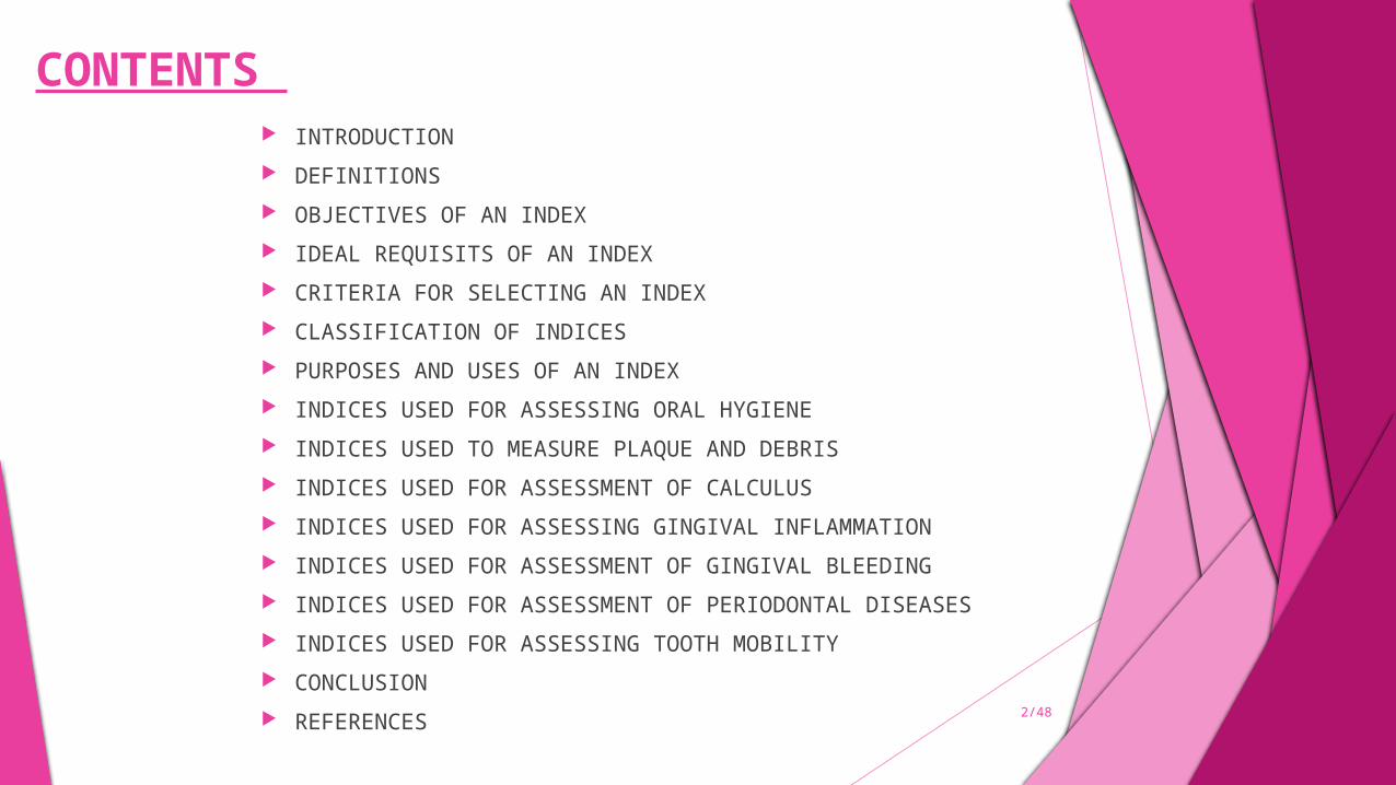
CONTENTS INTRODUCTION DEFINITIONS OBJECTIVES OF AN INDEX IDEAL REQUISITS OF AN INDEX CRITERIA FOR SELECTING AN INDEX CLASSIFICATION OF INDICES PURPOSES AND USES OF AN INDEX INDICES USED FOR ASSESSING ORAL HYGIENE INDICES USED TO MEASURE PLAQUE AND DEBRIS INDICES USED FOR ASSESSMENT OF CALCULUS INDICES USED FOR ASSESSING GINGIVAL INFLAMMATION INDICES USED FOR ASSESSMENT OF GINGIVAL BLEEDING INDICES USED FOR ASSESSMENT OF PERIODONTAL DISEASES INDICES USED FOR ASSESSING TOOTH MOBILITY CONCLUSION REFERENCES 2/48
INTRODUCTION
Dental index or indices are devices to find out the incidence, prevalence and severity of the disease, based on which preventive programs can be adopted.
An index is an expression of the clinical observation in a numerical value. It helps to describe the status of the individual or a group with respect to a condition being measured.
An index score can be more consistent and less subjective than a word description of that condition.
3/48
DEFINITIONS INDEX
A numerical value describing the relative status of a population on a graduated scale with definite upper and lower limits, which is designed to permit and facilitate comparison with other populations classified by the same criteria and methods. ’ – ‘’Russell. A. l – 1956’’
‘’Epidemiological indices are attempts to quantitate clinical conditions on a graduated scale, thereby facilitating comparison among populations examined by
same criteria and methods’’. - ‘’ Irving Glickman – 1950’’
4/48
An index is an expression of clinical observation in numeric values. It is used to describe the status of the individual or group with respect to a condition being measured. The use of numeric scale and a standardized method for interpreting observations of a condition results in an index score that is more consistent and less subjective than a word description of that condition. – ‘’Esther M Wilkins’’ - 1987
Oral indices are essentially set of values, usually numerical with maximum and minimum limits, used to describe the variables or a specific conditions on a graduated scale, which use the same criteria and method to compare a specific variable in individuals, samples or populations with that same variables as is found in other individuals, samples or populations. – ‘’George P Barnes’’ - 1985
OBJECTIVES OF AN INDEX
The main purpose or objective of using indices in
dental epidemiology is to increase understanding
of the disease process, thereby leading to method
of control and prevention. In addition it attempts to discover populations
at high and low risk and to define specific problem
under investigation.
5/48
IDEAL REQUISITES OF AN INDEX
Clarity, simplicity and objectivity Validity Reliability Quantifiability Sensitivity Acceptability
6/48
CRITERIA FOR SELECTING AN INDEX
Simple to use and calculate. Should permit the examination of many people in a short period of time. Should require minimum armamentarium and expenditure. Should be highly reproducible in assessing a clinical condition when used by one or
more examiners. Should not cause discomfort to the patient and should be acceptable to the patient. Should be free as possible from subjective interpretation Should be amenable to statistical analysis Should be strongly related numerically to the clinical stages of the specific disease
under investigation.
7/`48
CLASSIFICATION OF INDICES
BASED ON THE DIRECTION IN WHICH THEIR SCORES CAN FLUCTUATE
IRREVERSIBLE INDICES
REVERSIBLE INDICES
DEPENDING UPON THE EXTENT TO WHICH AREAS OF ORAL CAVITY ARE MEASURED
FULL MOUTH INDICES
SIMPLIFIED INDICES
8/48

BASED ON DISEASE ENTITY WHICH THEY MEASURE
Disease index Symptom index Treatment index
SPECIAL CATEGORIES AS -
Simple index
Cumulative index
9/48
USES OF AN INDEX
FOR INDIVIDUALS - Provide individual assessment to help patient recognize an oral problem.
Reveal degree of effectiveness of present oral hygiene practices.
Motivate the person in preventive and professional care for elimination and control of oral disease.
Evaluate the success of individual & professional treatment over a period of time by comparing index scores.
IN RESEARCH: Measure effectiveness of specific agents or devices for prevention/control/treatment
of oral conditions.
IN COMMUNITY HEALTH : Show prevalence and trends of incidences.
Assess needs of a community
10/48

INDICES FOR ASSESSING ORAL HYGIENE AND PLAQUE ORAL HYGIENE INDEX ( OHI)
Developed in 1960 by John C Greene & Jack R vermillion.
To classify and assess oral hygiene status.
OHI comprises of 2 components
DEBRIS INDEX(DI)
CALCULUS INDEX(CI)
METHODOLOGY
Mouth is divided into 6 segments:
11/48
Rules:1. Only fully erupted permanent teeth scored2. Third molars & incompletely erupted teeth are not scored3. The Buccal & lingual debris scores are both taken on the tooth in a segment having the greatest surface area covered by debris4. Similarly calculus scores are taken on the tooth in a segment having the greatest surface area covered by supra & sub-gingival calculus.5. Using no.5 explorer(shepherds’ hook) debris & calculus are estimated by running on Buccal/labial or lingual surface noting occlusal or incisal extent of the debris as it is removed from the tooth surface.6. Thus in this buccal/labial or lingual scores are not taken from same tooth

PROCEDURE – Each segment examined for debris or calculus.
From each segment one tooth is used for calculating the individual index for that segment.
The tooth used for the calculation must have the greatest area covered by either debris or calculus.
Buccal/labial and lingual surfaces.
DI – no:23 explorer (shepherd’s hook)
CI – no:5 explorer
CRITERIA OF SCORING (DI)AND (CI)
12/48
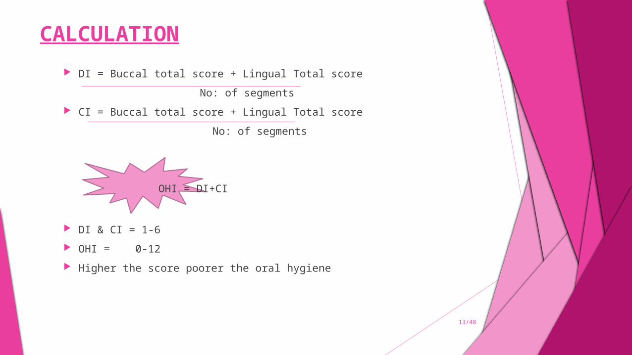
CALCULATION DI = Buccal total score + Lingual Total score
No: of segments
CI = Buccal total score + Lingual Total score
No: of segments
OHI = DI+CI
DI & CI = 1-6
OHI = 0-12
Higher the score poorer the oral hygiene
13/48
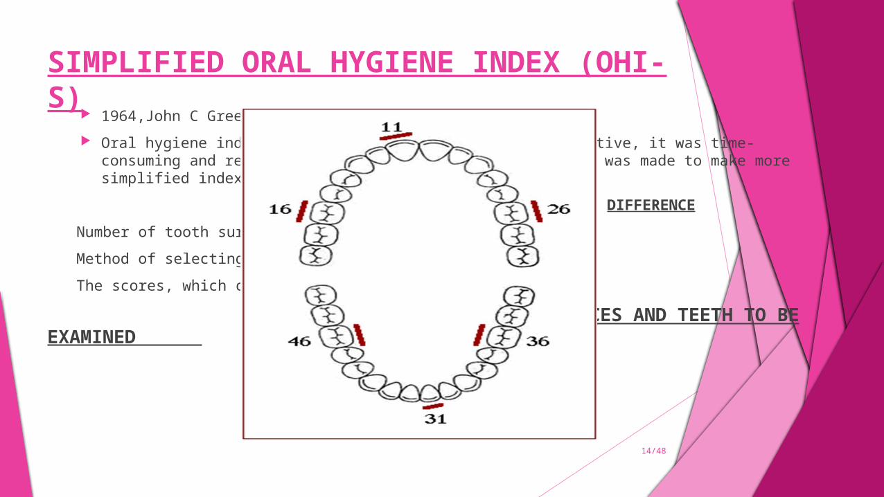
SIMPLIFIED ORAL HYGIENE INDEX (OHI-S) 1964,John C Greene & Jack R vermillion.
Oral hygiene index was determined to be simple and sensitive, it was time- consuming and required more decision – making. So effort was made to make more simplified index with equal sensitivity.
DIFFERENCE
Number of tooth surfaces scored 16 rather 12
Method of selecting the surface
The scores, which can be obtained
SURFACES AND TEETH TO BE EXAMINED
14/48
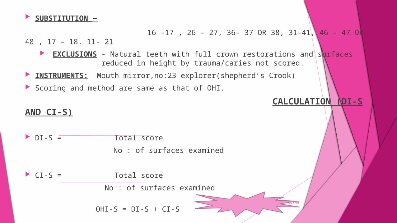
SUBSTITUTION – 16 -17 , 26 – 27, 36- 37 OR 38, 31-41, 46 – 47 OR 48 , 17 – 18. 11- 21
EXCLUSIONS - Natural teeth with full crown restorations and surfaces reduced in height by trauma/caries not scored.
INSTRUMENTS: Mouth mirror,no:23 explorer(shepherd’s Crook) Scoring and method are same as that of OHI.
CALCULATION (DI-S AND CI-S)
DI-S = Total score
No : of surfaces examined
CI-S = Total score
No : of surfaces examined
OHI-S = DI-S + CI-S15/48
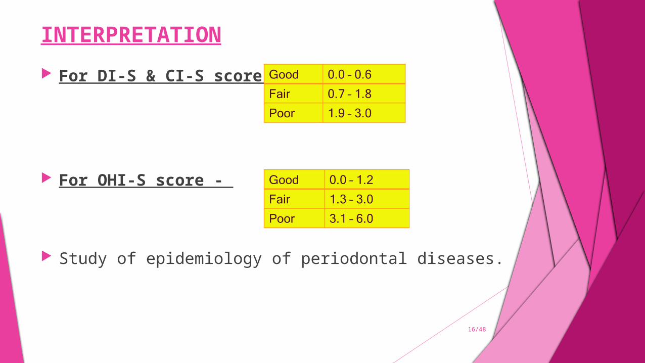
INTERPRETATION
For DI-S & CI-S score –
For OHI-S score -
Study of epidemiology of periodontal diseases.
16/48
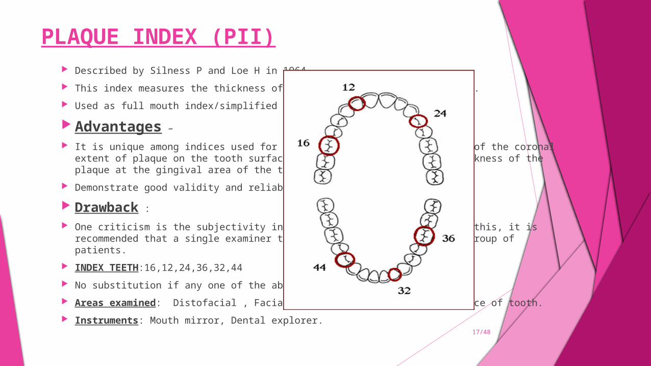
PLAQUE INDEX (PII) Described by Silness P and Loe H in 1964.
This index measures the thickness of plaque on the gingival one third.
Used as full mouth index/simplified index.
Advantages –
It is unique among indices used for the assessment of plaque because of the coronal extent of plaque on the tooth surface area and assesses only the thickness of the plaque at the gingival area of the tooth
Demonstrate good validity and reliability
Drawback :
One criticism is the subjectivity in estimating plaque. To overcome this, it is recommended that a single examiner to be trained and used with each group of patients.
INDEX TEETH:16,12,24,36,32,44
No substitution if any one of the above teeth are missing.
Areas examined: Distofacial , Facial, Mesio-facial and lingual surface of tooth.
Instruments: Mouth mirror, Dental explorer.17/48
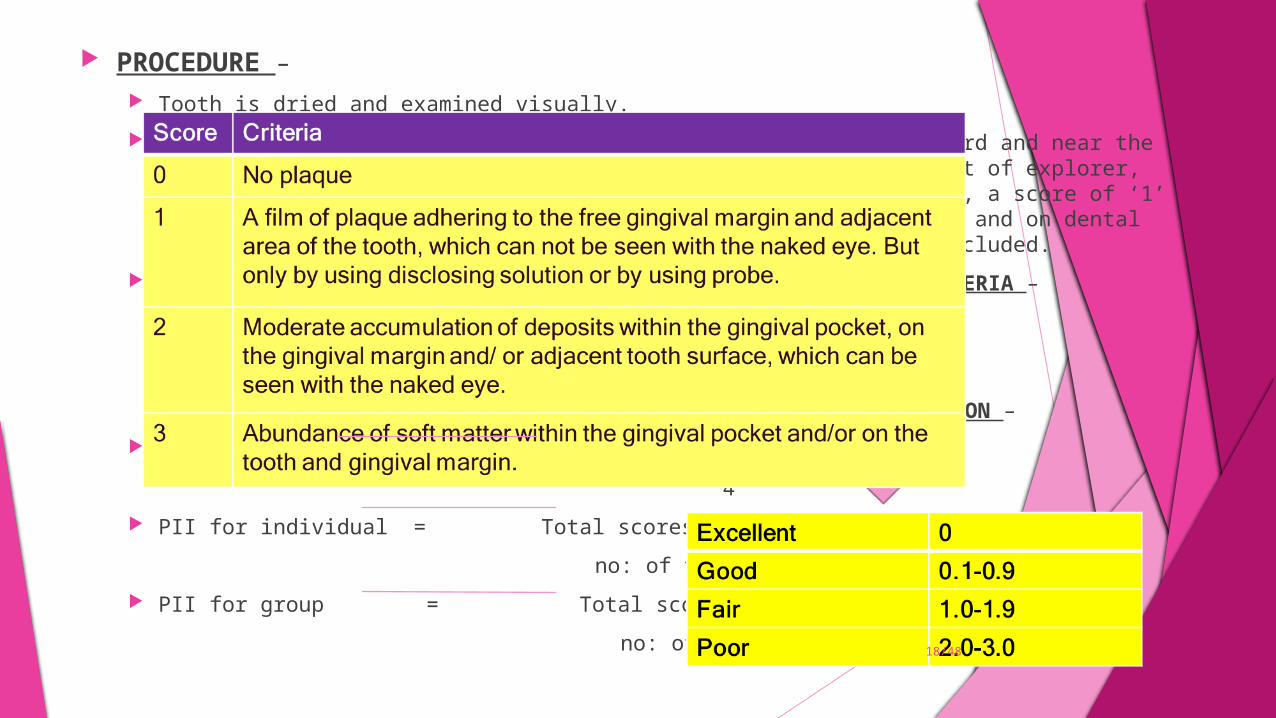
PROCEDURE – Tooth is dried and examined visually.
Explorer Is passed across the tooth surface in the cervical third and near the entrance of gingival sulcus. When no plaque adheres to the point of explorer, the area is considered to have a ‘0’ score. When plaque adheres, a score of ‘1’ is assigned. Plaque that is on the surface of calculus deposits and on dental restorations of all types in cervical third is evaluated and included.
SCORING CRITERIA –
CALCULATION AND INTERPRETATION – PII for a tooth = Scores of 4 areas 4 PII for individual = Total scores no: of teeth examined PII for group = Total score no: of individuals
18/48
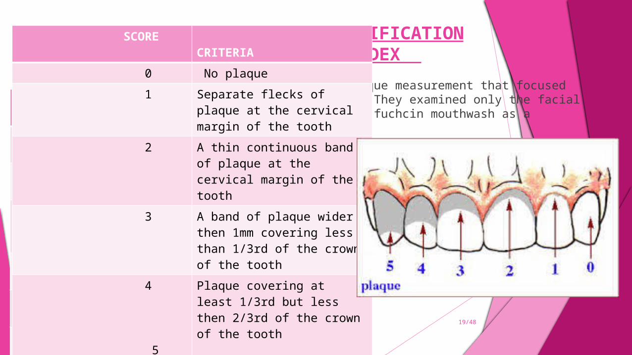
TURESKY – GILMORE- GLICKMAN MODIFICATION OF THE QUIGLEY – HEIN PLAQUE INDEX Quigley G. Hein . J in 1962, reported a plaque measurement that focused on the gingival third of the tooth
surface. They examined only the facial surfaces of the anterior teeth, using basic fuchcin mouthwash as a disclosing agent.
The Quigley – Hein plaque index was modified by Turesky S, Gilmore N.D and Glickman I in 1970. Modification was done by strengthening the objectivity of the original criteria. This system of scoring
plaque is relatively easy to use because of the objective definitions of each numerical score. Instruments used – Mouth mirror and Disclosing agent. Method – labial , buccal and lingual surfaces are assessed after using disclosing agent. Scoring criteria - Calculation and interpretation :
IS = TS/ No of surfaces examined
0-1 = low
>2 = High 19/48
Score Criteria 0 no plaque 1 flecks of stain of the
gingival margin 2 Definitive line of plaque
on gingival margin 3 Gingival third of surface 4 Two- thirds of surface 5 Greater then 2/3rd of
the surface
SCORE CRITERIA 0 No plaque 1 Separate flecks of plaque at
the cervical margin of the tooth
2 A thin continuous band of plaque at the cervical margin of the tooth
3 A band of plaque wider then 1mm covering less than 1/3rd of the crown of the tooth
4
5
Plaque covering at least 1/3rd but less then 2/3rd of the crown of the tooth
Plaque covering 2/3rd or more of the crown of the tooth
GLASS INDEX It was developed by GLASS R.L in 1965. This index assesses the presence and extent of debris accumulation , for evaluating tooth – brushing
efficacy.. CRITERIA – Code 0 – no visible debris Code 1 – debris visible at gingival margin but discontinuous less than1mm in height Code 2 – debris continuous at gingival margin – greater than 1mm in height. Code 3- debris involving entire gingival third of the tooth Code 4- debris generally scattered over tooth surface CALCULATION – Debris index score per person – total debris score of all the teeth examined / total no of teeth examined. Glass criteria of scoring places more emphasis on the gingival third of the tooth surface than does the
OHI- S, and so this index is useful in clinical trials of preventive and therapeutic agents. 20/48
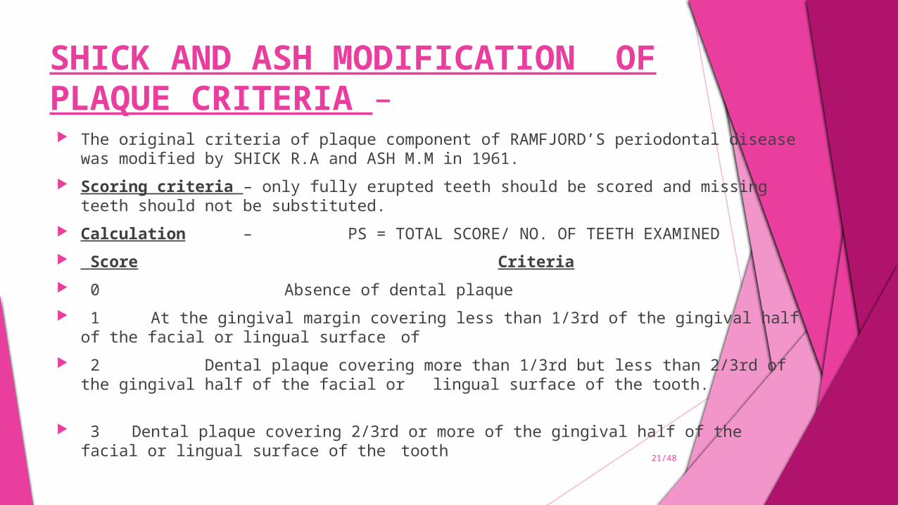
SHICK AND ASH MODIFICATION OF PLAQUE CRITERIA – The original criteria of plaque component of RAMFJORD’S periodontal disease was modified by SHICK
R.A and ASH M.M in 1961. Scoring criteria – only fully erupted teeth should be scored and missing teeth should not be substituted. Calculation – PS = TOTAL SCORE/ NO. OF TEETH EXAMINED Score Criteria 0 Absence of dental plaque 1 At the gingival margin covering less than 1/3rd of the gingival half of the facial or lingual
surface of 2 Dental plaque covering more than 1/3rd but less than 2/3rd of the gingival half of the facial or
lingual surface of the tooth. 3 Dental plaque covering 2/3rd or more of the gingival half of the facial or lingual surface of the
tooth
21/48

NAVY PLAQUE INDEX (NPI) The navy plaque index was developed by GROSSMAN F.D & FEDI P.F in 1970. This index was designed
to assess the plaque control status among naval personnels and to measure any subsequent changes. METHOD : The navy plaque index is obtained by scoring the amount of plaque found on six selected teeth (index teeth)
by using a disclosing solution. The teeth examined are. 16, 21,24,36,41,44 and surfaces are – facial and lingual of the each six teeth, the facial surfaces are divided
into three major areas as – Gingival Area (G), Mesial Proximal Area (M) and Distal Proximal Area (D). The stained plaque in contact with the gingival is scored as follows- Area M = 3 Area G = 2 Area D = 3 when plaque is found not in contact with gingival tissue but is found on any tooth surface, one
point is added to the facial or lingual score. Calculation – the highest for any of the six teeth scored is the patient’s NAVY plaque index score. All teeth
scores are added to give the total NPI score. 22/48
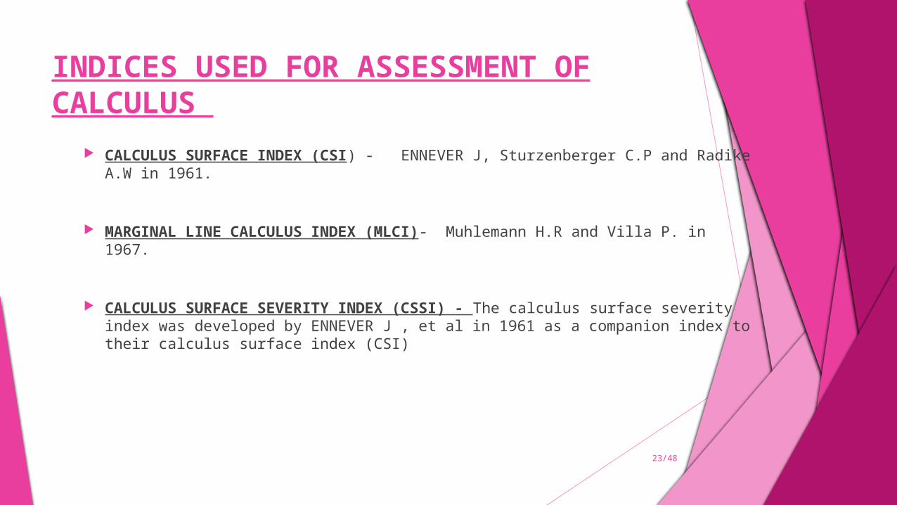
INDICES USED FOR ASSESSMENT OF CALCULUS
CALCULUS SURFACE INDEX (CSI) - ENNEVER J, Sturzenberger C.P and Radike A.W in 1961.
MARGINAL LINE CALCULUS INDEX (MLCI)- Muhlemann H.R and Villa P. in 1967.
CALCULUS SURFACE SEVERITY INDEX (CSSI) - The calculus surface severity index was developed by ENNEVER J , et al in 1961 as a companion index to their calculus surface index (CSI)
23/48
INDICES USED FOR ASSESSING GINGIVAL INFLAMMATION PAPILLARY – MARGINAL ATTACHMENT INDEX (PMA)- MAURY MASSLER AND SCHOUR .L 1944. No. of gingival units effected were counted rather then the severity of inflammation.
METHOD
A gingival unit was divided into three compartments –
Papillary gingiva, Marginal gingiva, Attached gingiva
Presence or absence of inflammation on each gingival unit is recorded and usually only maxillary and mandibular incisors, canines and premolars were examined.
SCORING CRITERIA
Papillary component (p) Marginal component (m) Attached component
score criteria 0 Normal 1 Might papillary
enlargement 2 Obvious increase
in size , BO Pressue
3 Excessive inc in size, spontaneous bleeding
4 5
Necrotic papilla
Atrophy and loss
score criteria 0 Normal 1 Engorgement, slight inc
in size, no bleeding 2 Obvious engorgement ,
bleeding on pressure
3 Swollen collar, spontaneous bleeding , beginning infiltration
4 Necrotic gingiva 5 Recession of the free
marginal gingiva below CEJ due to inflammatory changes.
score criteria 0 Normal 1 Slight engorgement with
loss of stippling, changes in color may or may not be present
2 3
Obvious engorgement with marked inc in redness and pocket formation
Advanced periodontitis and deep pockets.
24/48
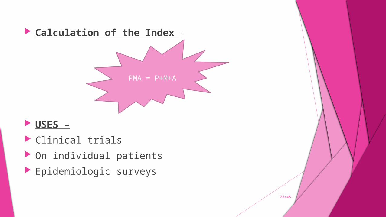
Calculation of the Index –
USES – Clinical trials On individual patients Epidemiologic surveys
PMA = P+M+A
25/48
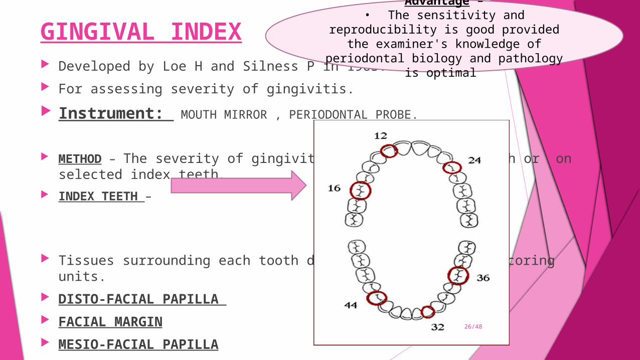
GINGIVAL INDEX Developed by Loe H and Silness P in 1963. For assessing severity of gingivitis.
Instrument: MOUTH MIRROR , PERIODONTAL PROBE.
METHOD – The severity of gingivitis is scored on all teeth or on selected index teeth. INDEX TEETH –
Tissues surrounding each tooth divided into 4 gingival scoring units. DISTO-FACIAL PAPILLA FACIAL MARGIN MESIO-FACIAL PAPILLA LINGUAL GINGIVAL MARGIN
26/48
Advantage –• The sensitivity and reproducibility is
good provided the examiner's knowledge of periodontal biology and pathology is optimal
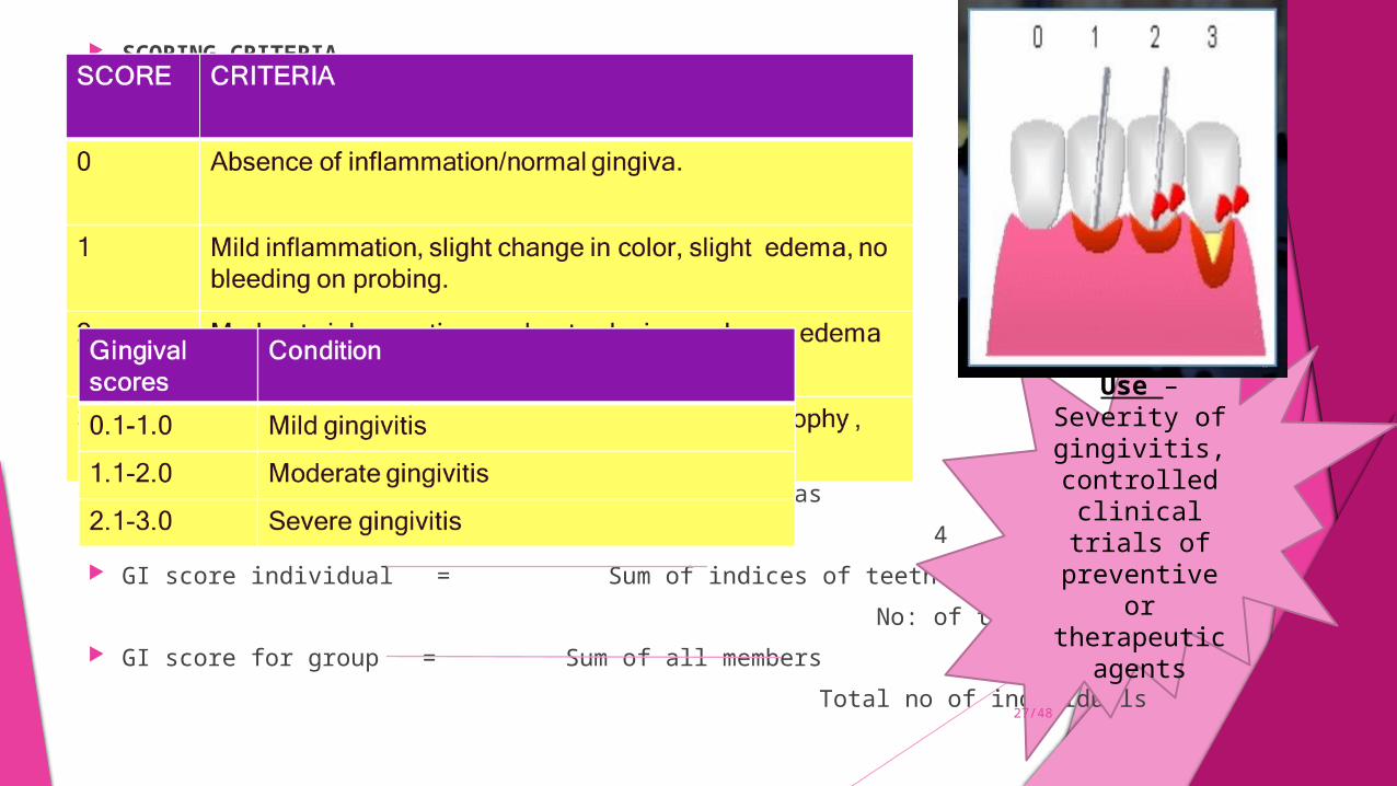
SCORING CRITERIA –
Calculation and interpretation – GI score for a tooth = Scores from 4 areas
4 GI score individual = Sum of indices of teeth
No: of teeth examined GI score for group = Sum of all members
Total no of individuals
Use –Severity of gingivitis, controlled
clinical trials of preventive or therapeutic
agents
27/48
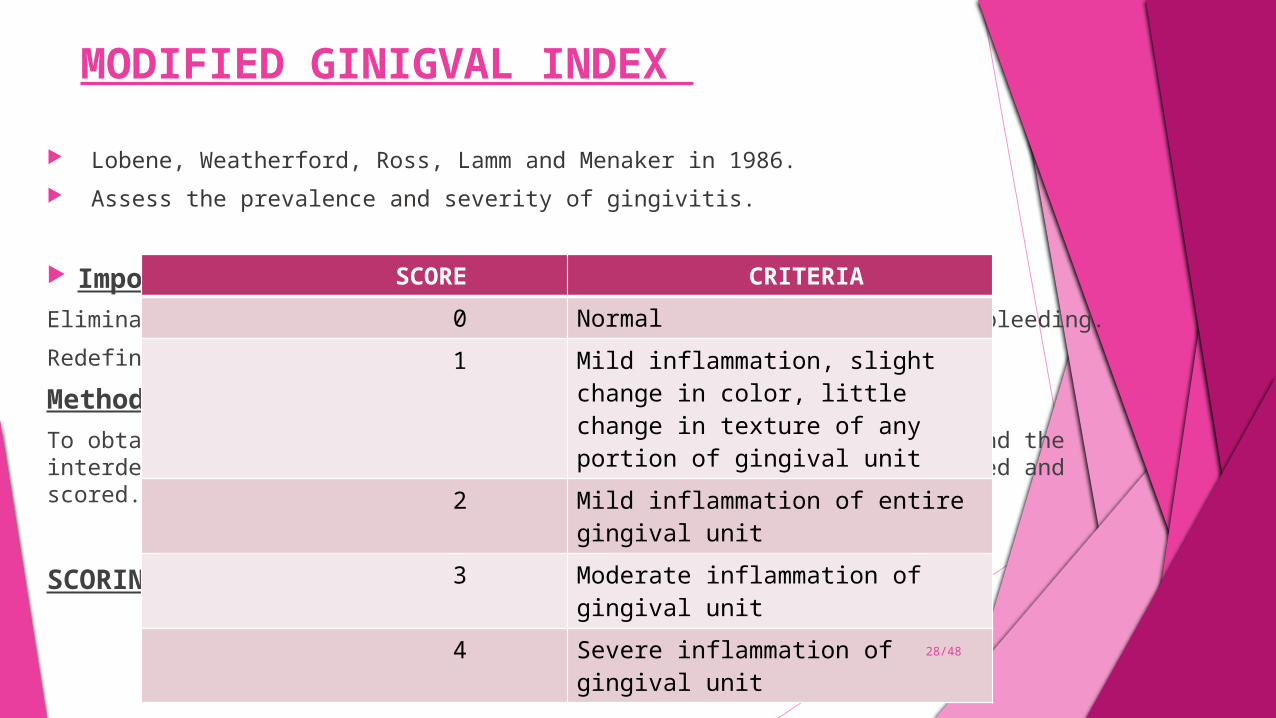
MODIFIED GINIGVAL INDEX
Lobene, Weatherford, Ross, Lamm and Menaker in 1986. Assess the prevalence and severity of gingivitis.
Important changes in GI –Elimination of gingival probing to assess the presence or absence of bleeding.
Redefinition of scoring system for mild and moderate inflammation.
Method -
To obtain MGI , labial and lingual surfaces of the gingival margins and the interdental papilla of all erupted teeth except 3rd molars are examined and scored.
SCORING –
SCORE CRITERIA 0 Normal
1 Mild inflammation, slight change in color, little change in texture of any portion of gingival unit
2 Mild inflammation of entire gingival unit
3 Moderate inflammation of gingival unit
4 Severe inflammation of gingival unit
28/48
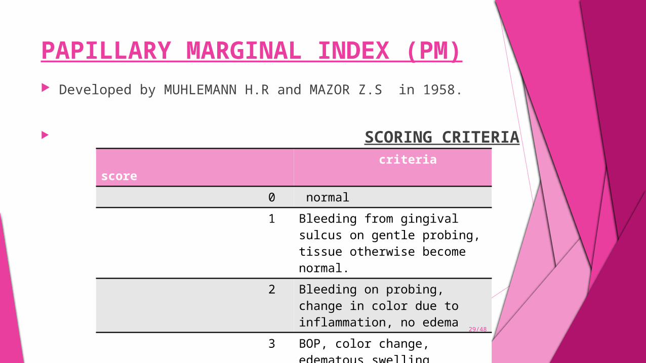
PAPILLARY MARGINAL INDEX (PM) Developed by MUHLEMANN H.R and MAZOR Z.S in 1958.
SCORING CRITERIA score criteria 0 normal 1 Bleeding from gingival sulcus
on gentle probing, tissue otherwise become normal.
2 Bleeding on probing, change in color due to inflammation, no edema
3 BOP, color change, edematous swelling
4 Ulceration with additional symptoms 29/48
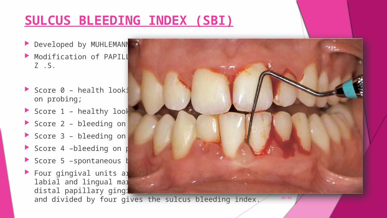
SULCUS BLEEDING INDEX (SBI)
Developed by MUHLEMANN H.R AND SEN.S in 1971. Modification of PAPILLARY – MARGINAL INDEX of MUHLEMANN and MAZOR Z .S.
SCORING CRITERIA Score 0 – health looking papillary and marginal gingiva no bleeding on probing; Score 1 – healthy looking gingiva, bleeding on probing; Score 2 – bleeding on probing, change in color, no edema; Score 3 – bleeding on probing, change in color, slight edema; Score 4 –bleeding on probing, change in color, obvious edema; Score 5 –spontaneous bleeding, change in color, marked edema. Four gingival units are scored systematically for each tooth: the labial and lingual marginal
gingival (M units) and the mesial and distal papillary gingival (P units). Scores for these units are added and divided by four gives the sulcus bleeding index.
30/48
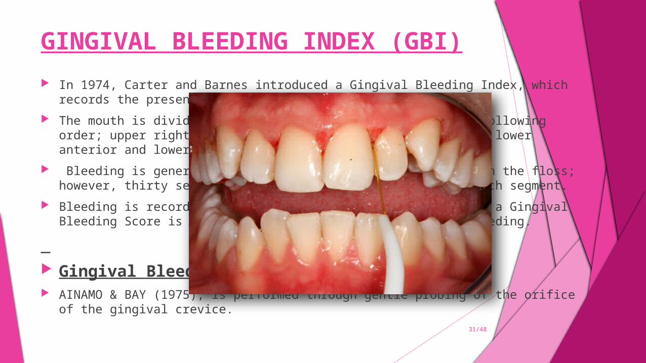
GINGIVAL BLEEDING INDEX (GBI) In 1974, Carter and Barnes introduced a Gingival Bleeding Index, which records the presence or absence of
gingival inflammation. The mouth is divided into six segments and flossed in the following order; upper right, upper anterior, upper
left, lower left, lower anterior and lower right. Bleeding is generally immediately evident in the area or on the floss; however, thirty seconds is allowed for
re- inspection of each segment. Bleeding is recorded as present or absent. For each patient a Gingival Bleeding Score is obtained by noting
the total units of bleeding.
Gingival Bleeding Index (GBI)- AINAMO & BAY (1975), is performed through gentle probing of the orifice of the gingival crevice.
31/48
PAPILLARY BLEEDING INDEX
Introduced by Saxer and Muehlemann (1975), as cited by Muehlemann (1977). A periodontal probe is inserted into the gingival sulcus at the base of the papilla on the
mesial aspect, and then moved coronally to the papilla tip. This is repeated on the distal aspect of the papilla.
The intensity of any bleeding is recorded as: Score 0 – no bleeding; Score 1 – A single discreet bleeding point; Score 2 – Several isolated bleeding points or a single line of blood appears; Score 3 – The interdental triangle fills with blood shortly after probing; Score 4 – Profuse bleeding occurs after probing; blood flows immediately into the marginal
sulcus.
32/48
EASTMAN INTERDENTAL BLEEDING INDEX
Caton & Polson (1985) developed the Eastman Interdental Bleeding Index (EIB). A wooden interdental cleaner is inserted between the teeth from the facial aspect, depressing
the interdental tissues 1 to 2 mm. This is repeated four times and the presence or absence of bleeding within 15 s is recorded.
Path on insertion should be parallel to occlusal surface. Insertion and removal of interdental cleaner is done 4 times and then moved on to next
interproximal area.
SCORE = no. of bleeding areas/total no. of areas Х 100
35/48
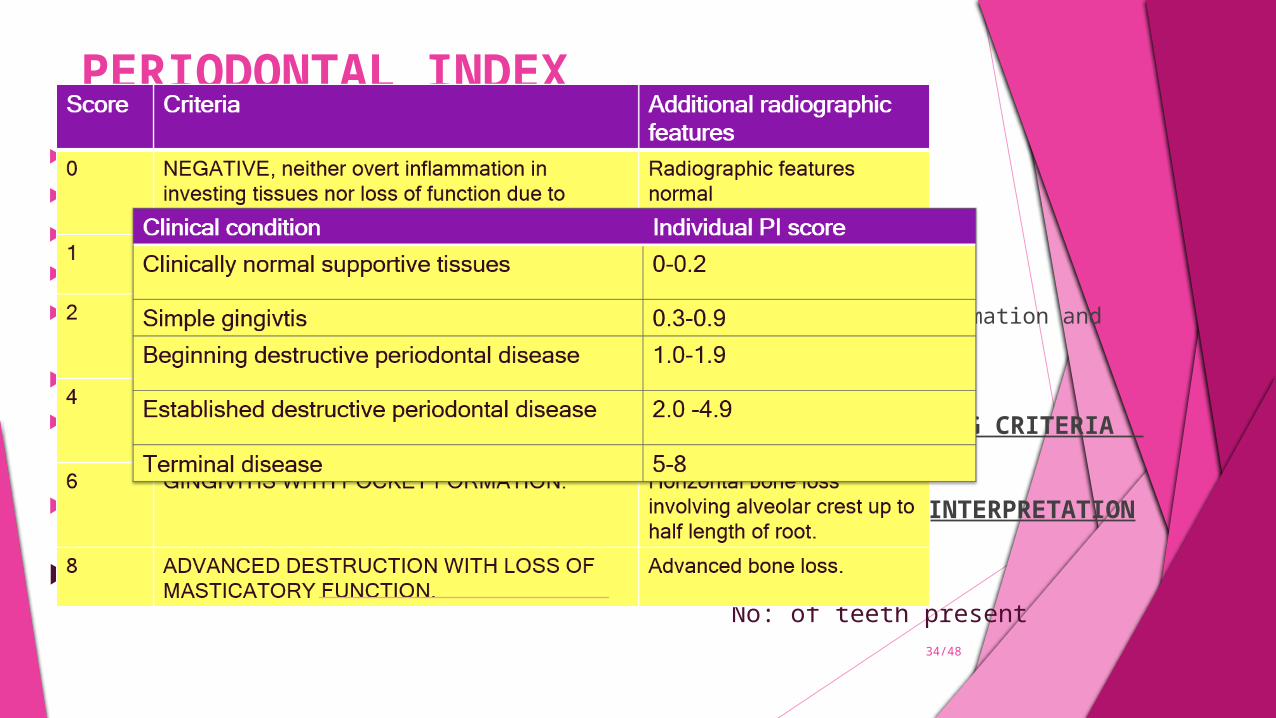
PERIODONTAL INDEX RUSSELL’S PERIODONTAL INDEX (RPI) – Developed by Russell A.L in 1956 To estimate deeper periodontal diseases. All teeth present examined. Gingival tissue surrounding each tooth assessed for gingival inflammation and periodontal involvement.
Instruments : Mouth Mirror, plain probe.
SCORING CRITERIA
CALCULATION AND INTERPRETATION
PI score per person = Sum of individual scores No: of teeth present
34/48
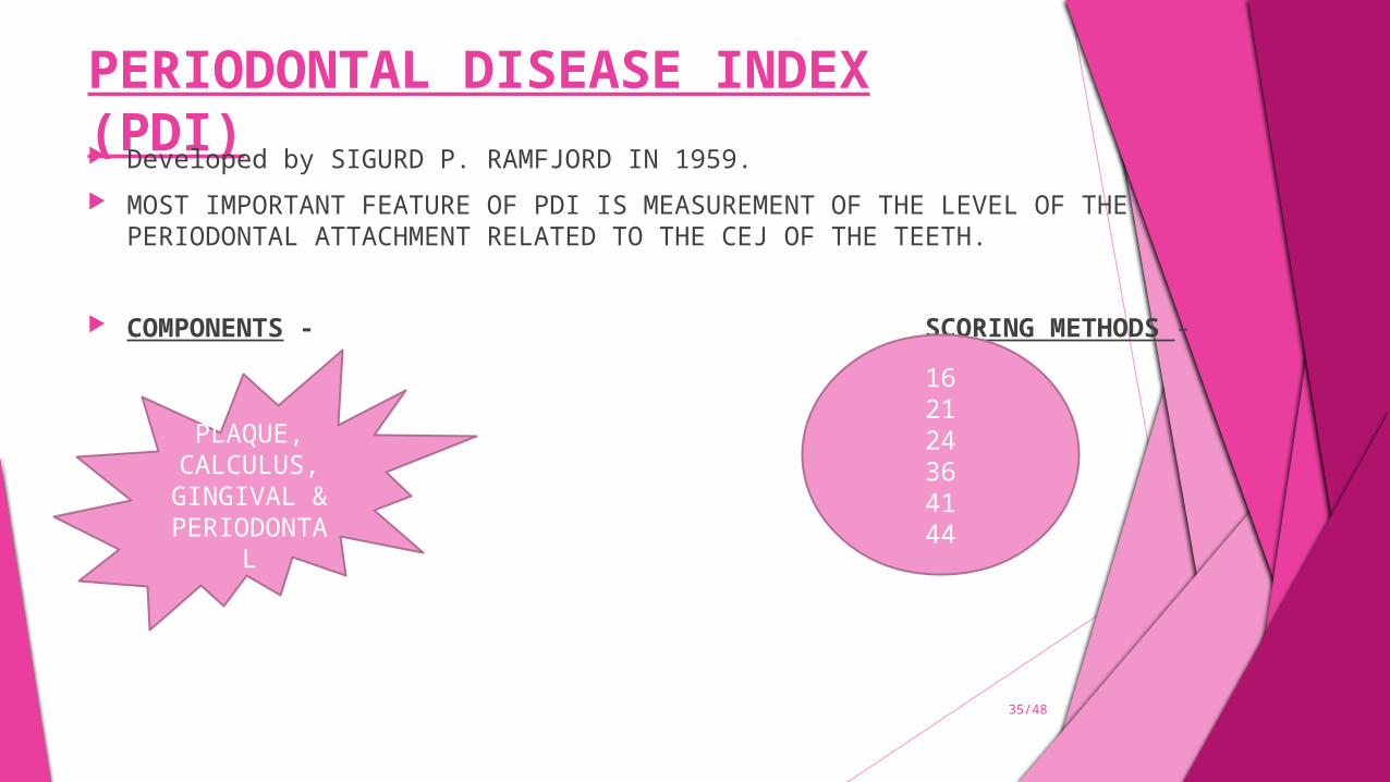
PERIODONTAL DISEASE INDEX (PDI) Developed by SIGURD P. RAMFJORD IN 1959. MOST IMPORTANT FEATURE OF PDI IS MEASUREMENT OF THE LEVEL OF THE
PERIODONTAL ATTACHMENT RELATED TO THE CEJ OF THE TEETH.
COMPONENTS - SCORING METHODS -
PLAQUE, CALCULUS, GINGIVAL & PERIODONT
AL
162124364144
35/48
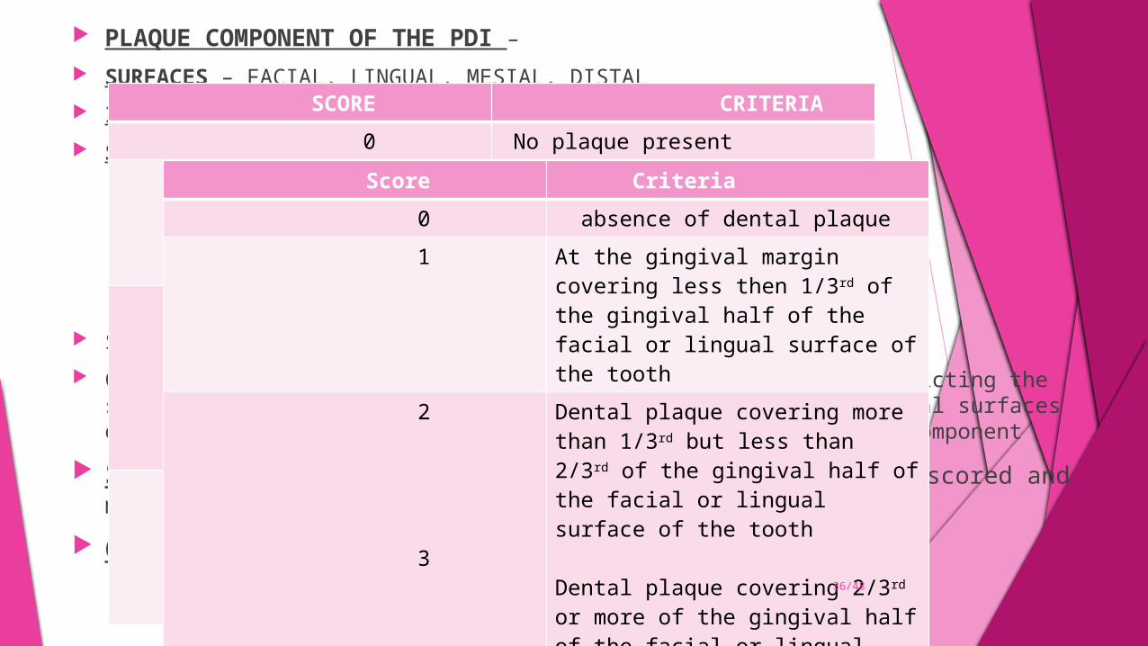
PLAQUE COMPONENT OF THE PDI – SURFACES – FACIAL, LINGUAL, MESIAL, DISTAL INSTRUMENTS- MOUTH MIRROR, DENTAL EXPLORER SCORING CRITERIA –
SHICK AND ASH modification of plaque criteria – Consist of six teeth excluding the interproximal area and restricting the scoring of plaque to the gingival half, of
the facial and lingual surfaces of the index teeth. Selected teeth are same as that of plaque component
Scoring criteria – only fully erupted teeth should be scored and missing teeth should not be substituted.
Calculation – PS = TOTAL SCORE/ NO.OF TEETH EXAMINED
SCORE CRITERIA 0 No plaque present 1 Plaque present but not on all
interproximal, buccal and lingual surfaces of the tooth
2 Plaque present on all interproximal, buccal and lingual surfaces of the tooth , but covering half than one half of these surfaces
3 Plaque extending over all interproximal , buccal and lingual surfaces, and covering more than one half of these surfaces
Score Criteria 0 absence of dental plaque 1 At the gingival margin covering
less then 1/3rd of the gingival half of the facial or lingual surface of the tooth
2
3
Dental plaque covering more than 1/3rd but less than 2/3rd of the gingival half of the facial or lingual surface of the tooth
Dental plaque covering 2/3rd or more of the gingival half of the facial or lingual surface of the tooth
36/48
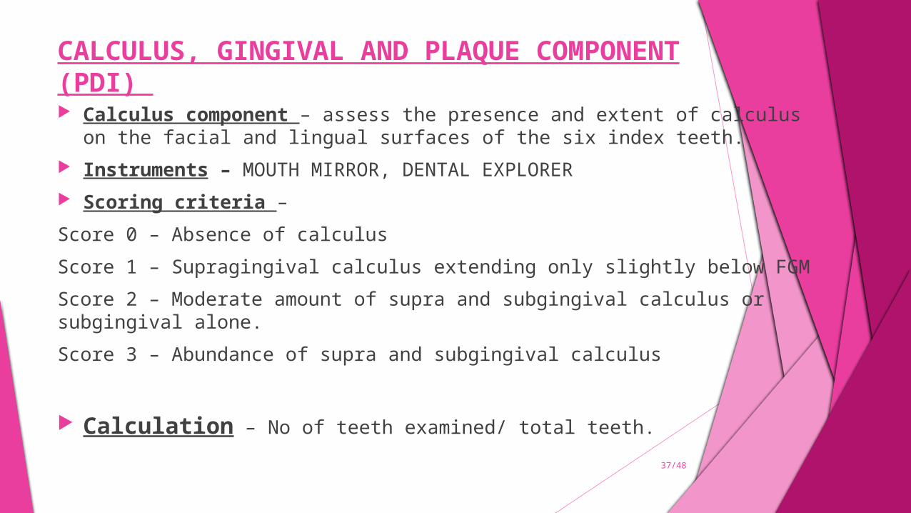
CALCULUS, GINGIVAL AND PLAQUE COMPONENT (PDI) Calculus component – assess the presence and extent of calculus on the facial and lingual
surfaces of the six index teeth. Instruments – MOUTH MIRROR, DENTAL EXPLORER Scoring criteria –
Score 0 – Absence of calculus
Score 1 – Supragingival calculus extending only slightly below FGM
Score 2 – Moderate amount of supra and subgingival calculus or subgingival alone.
Score 3 – Abundance of supra and subgingival calculus
Calculation – No of teeth examined/ total teeth.
37/48
GINGIVAL AND PERIODONTAL COMPONENT
Gingival status is scored first. Dried superficially by gently touching with absorbing cotton, and examined for color change, form ,
consistency and bleeding. Crevice depth is recorded in relation to CEJ. Instruments used – mouth mirror and university of Michigan probe number 0 probe.
Score 0 - Absence of signs of inflammation
Score 1 – mild to moderate inflammatory changes not extending around the tooth.
Score 2 - mild to moderately severe gingivitis extending around the tooth.
Score 3 – severe gingivitis characterized by marked redness , swelling , tendency to bleed , and ulceration.
Score 4 – gingival crevice in any of the four areas , extending apically to CEJ but not more then 3 mm.
Score 5 - gingival crevice in any of the four areas , extending apically to CEJ between 3-6mm.
Score 6 - gingival crevice in any of the four areas , extending apically more then 6 mm from CEJ.
Calculation – PDI – TOTAL OF INDIVIDUAL TOOTH SCORES/NUMBER OF
TEETH EXAMINED
SCORING CRITERIA
38/48
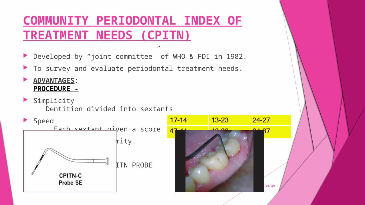
COMMUNITY PERIODONTAL INDEX OF TREATMENT NEEDS (CPITN) Developed by “joint committee” of WHO & FDI in 1982. To survey and evaluate periodontal treatment needs. ADVANTAGES: PROCEDURE - Simplicity Dentition divided into sextants Speed Each sextant given a score International uniformity.
INSTRUMENT USED - CPITN PROBE
39/48
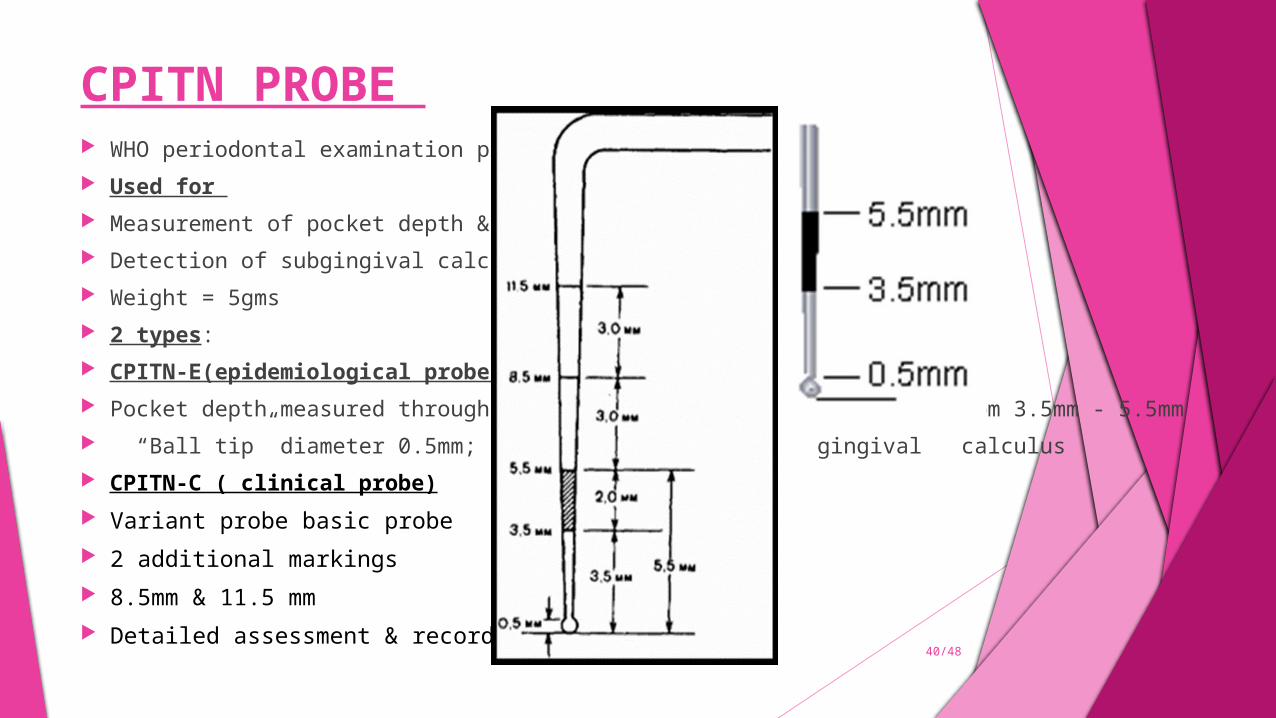
CPITN PROBE WHO periodontal examination probe. Used for Measurement of pocket depth & Detection of subgingival calculus. Weight = 5gms 2 types: CPITN-E(epidemiological probe) Pocket depth measured through color coding; black mark starting from 3.5mm - 5.5mm “Ball tip” diameter 0.5mm; easy detection of sub gingival calculus CPITN-C ( clinical probe) Variant probe basic probe 2 additional markings 8.5mm & 11.5 mm Detailed assessment & recording of deep pockets. 40/48
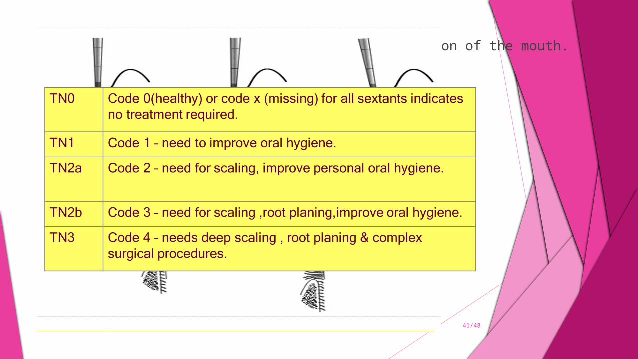
Best estimators of the worst periodontal condition of the mouth. >20 years
Molars examined in pairs & highest score recorded. Up to 19 years
CODING CRITERIA-
41/48

COMMUNITY PERIODONTAL INDEX(CPI)
This index is based on the modification of the earlier used community periodontal index of treatment needs (CPITN).
INCLUSION – MEASUREMENT OF ‘’LOSS OF ATTACHMENT ‘’ AND ELIMINATION OF THE ‘’TREATMENT NEEDS’’ category.
INSTRUMENTS USED – MOUTH MIRROR , THE CPITN – C PROBE. SCORING CRITERIA –
Score 0 – healthy.
Score 1 – bleeding observed, directly or by using mouth mirror, after probing.
Score 2- calculus detected during probing, but all of the black band on the probe visible.
Score 3 – pocket 4 – 5 mm ( gingival margin within the black band on the probe)
Score 4 – pocket 6 mm or more ( black band on the probe not visible)
X- excluded sextant
9 – not recorded
Loss of attachment – Criteria of scoring Code o – loss of attachment 0-3mm (CEJ not visible and CPI score 0-3 ).Code 1 – loss of attachment 4- 5 mm (CEJ within the black band).Code 2 – loss of attachment 6- 8mm (CEJ between the upper limit of the black band and the 8.5mm ring )Code 3 – loss of attachment 9- 11mm (CEJ between the 8.5mm and 11.5mm rings)Code 4- loss of attachment 12mm or more(CEJ beyond the 11.5mm rings ).X – excluded sextant (less than two teeth present )9 – not recorded ( CEJ neither visible nor detectable )
42/48
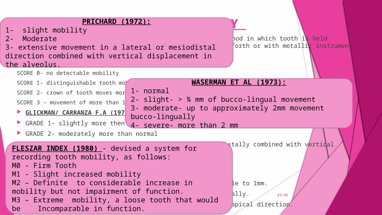
ASSESSMENT OF TOOTH MOBILITY MILLER(1985) – has described the most common clinical method in which tooth is held between handles of the two
instruments & moved back and forth or with metallic instrument and one finger. Criteria –
SCORE 0- no detectable mobility
SCORE 1- distinguishable tooth mobility
SCORE 2- crown of tooth moves more than 1mm in any direction
SCORE 3 – movement of more than 1mm in any direction.
GLICKMAN/ CARRANZA F.A (1972)– GRADE 1- slightly more then normal GRADE 2- moderately more than normal GRADE 3 – severe mobility faciolingually and or mesiodistally combined with vertical displacement.
GENCO R(1984).- assessed mobility as – DEGREE 1 – Horizontal mobility of crown is from detectable to 1mm. DEGREE 2 – mobility of crown ranges from 1-2 mm horizontally. DEGREE 3 – mobility of crown is observed in vertical or apical direction.
43/48
PRICHARD (1972):1- slight mobility2- Moderate3- extensive movement in a lateral or mesiodistal direction combined with vertical displacement in the alveolus.
WASERMAN ET AL (1973):1- normal 2- slight- > ¾ mm of bucco-lingual movement3- moderate- up to approximately 2mm movement bucco-lingually4- severe- more than 2 mm
FLESZAR INDEX (1980) - devised a system for recording tooth mobility, as follows:M0 - Firm Tooth M1 - Slight increased mobility M2 – Definite to considerable increase in mobility but not impairment of function.M3 – Extreme mobility, a loose tooth that would be Incomparable in function.
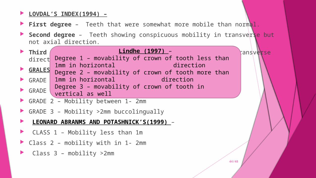
LOVDAL’S INDEX(1994) – First degree – Teeth that were somewhat more mobile than normal. Second degree – Teeth showing conspicuous mobility in transverse but not axial direction. Third degree – Teeth being mobile in axial as well as on transverse direction. GRALES AND SHALES(1999) – GRADE 0 – No apparent mobility GRADE 1- Mobility less than 1mm buccolingually GRADE 2 – Mobility between 1- 2mm GRADE 3 – Mobility >2mm buccolingually LEONARD ABRANMS AND POTASHNICK’S(1999) – CLASS 1 – Mobility less than 1m Class 2 – mobility with in 1- 2mm Class 3 – mobility >2mm
Lindhe (1997) –Degree 1 – movability of crown of tooth less than 1mm in horizontal
directionDegree 2 – movability of crown of tooth more than 1mm in horizontal
directionDegree 3 – movability of crown of tooth in vertical as well
44/48
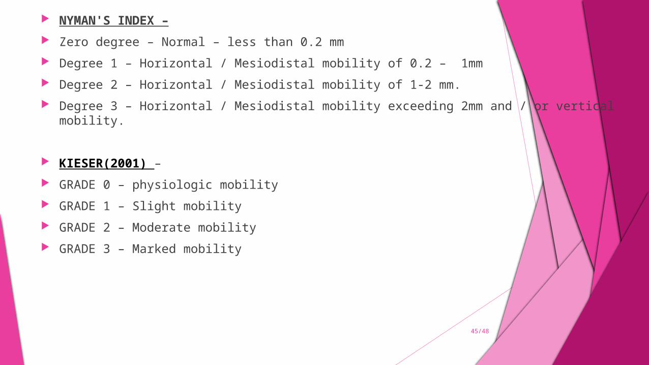
NYMAN'S INDEX – Zero degree – Normal – less than 0.2 mm Degree 1 – Horizontal / Mesiodistal mobility of 0.2 – 1mm Degree 2 – Horizontal / Mesiodistal mobility of 1-2 mm. Degree 3 – Horizontal / Mesiodistal mobility exceeding 2mm and / or vertical mobility.
KIESER(2001) – GRADE 0 – physiologic mobility GRADE 1 – Slight mobility GRADE 2 – Moderate mobility GRADE 3 – Marked mobility
45/48

CONCLUSION
Periodontal indices have contributed to identification, prevention and treatment of periodontal disease over the years since their inception. These indices are based on the prevailing understanding of the pathogenesis and progression of periodontal disease. Thus, with the better understanding of the periodontal disease process these indices have changed from the simple Russell’s
Periodontal Index to the current Moustakis’s Genetic Susceptibility Index. Each of these indices has its merits and limitations, so, an ideal index which detects the ongoing progressive periodontal destruction and also identifies the active and inactive sites of disease, is the need of the hour
46/48
REFERENCES
Soben Peter. Indices in dental epidemiology,4th edition Soben Peter. Indices in dental epidemiology , 3rd edition Essentials Of Preventive and Community Dentistry 3ed.123-231. Kinane DF, Lindhe J. Pathogenesis of periodontitis. Kunaal Dhingra and Kharidhi Laxman Vandana. Indices for measuring
periodontitis : A literature review, International Dental Journal 2011; 61: 76–84 In: Lindhe J, Karring T, Lang NP, Eds. Clinical Periodontology and Implant
Dentistry. Maria Augusta Bessa Rebelo and Adriana Corrêa de Queiroz, Federal
University of Amazonas Brazil. Gingival indices : state of art , Gingival Diseases – Their Aetiology, Prevention and Treatment
47/48