
Independent Study Guide The Innate Immune Response ...facweb.northseattle.edu/hfelise/BIO 260 Spring...
6
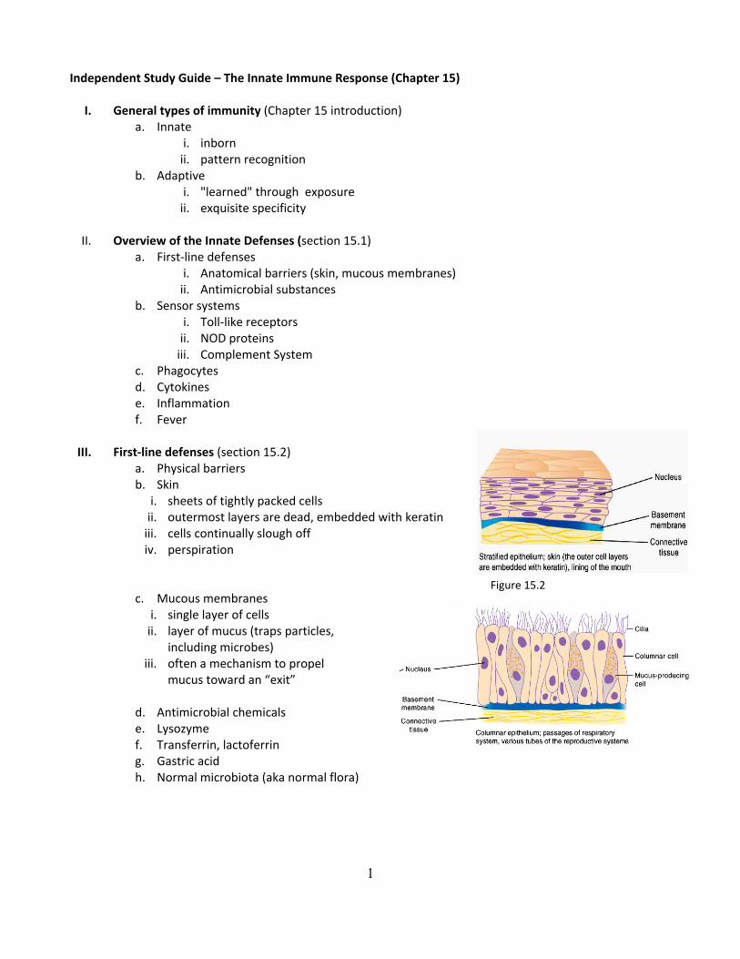
1 Independent Study Guide – The Innate Immune Response (Chapter 15) I. General types of immunity (Chapter 15 introduction) a. Innate i. inborn ii. pattern recognition b. Adaptive i. "learned" through exposure ii. exquisite specificity II. Overview of the Innate Defenses (section 15.1) a. First‐line defenses i. Anatomical barriers (skin, mucous membranes) ii. Antimicrobial substances b. Sensor systems i. Toll‐like receptors ii. NOD proteins iii. Complement System c. Phagocytes d. Cytokines e. Inflammation f. Fever III. First‐line defenses (section 15.2) a. Physical barriers b. Skin i. sheets of tightly packed cells ii. outermost layers are dead, embedded with keratin iii. cells continually slough off iv. perspiration c. Mucous membranes i. single layer of cells ii. layer of mucus (traps particles, including microbes) iii. often a mechanism to propel mucus toward an “exit” d. Antimicrobial chemicals e. Lysozyme f. Transferrin, lactoferrin g. Gastric acid h. Normal microbiota (aka normal flora) Figure 15.2
Transcript of Independent Study Guide The Innate Immune Response ...facweb.northseattle.edu/hfelise/BIO 260 Spring...
1
Independent Study Guide – The Innate Immune Response (Chapter 15)
I. General types of immunity (Chapter 15 introduction) a. Innate
i. inborn ii. pattern recognition
b. Adaptive i. "learned" through exposure ii. exquisite specificity
II. Overview of the Innate Defenses (section 15.1)
a. First‐line defenses i. Anatomical barriers (skin, mucous membranes) ii. Antimicrobial substances
b. Sensor systems i. Toll‐like receptors ii. NOD proteins iii. Complement System
c. Phagocytes d. Cytokines e. Inflammation f. Fever
III. First‐line defenses (section 15.2)
a. Physical barriers b. Skin
i. sheets of tightly packed cells ii. outermost layers are dead, embedded with keratin iii. cells continually slough off iv. perspiration
c. Mucous membranes i. single layer of cells ii. layer of mucus (traps particles,
including microbes) iii. often a mechanism to propel
mucus toward an “exit”
d. Antimicrobial chemicals e. Lysozyme f. Transferrin, lactoferrin g. Gastric acid h. Normal microbiota (aka normal flora)
Figure 15.2
2
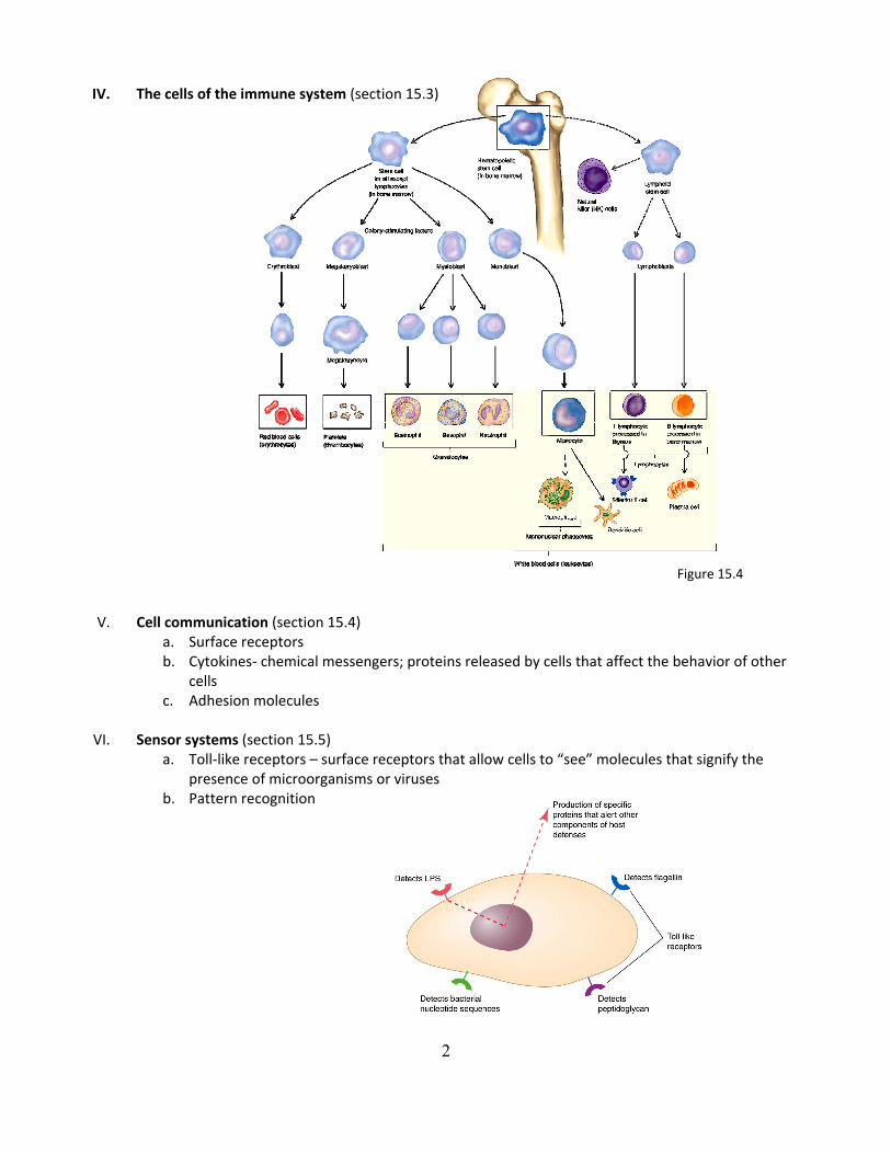
IV. The cells of the immune system (section 15.3) V. Cell communication (section 15.4)
a. Surface receptors b. Cytokines‐ chemical messengers; proteins released by cells that affect the behavior of other
cells c. Adhesion molecules
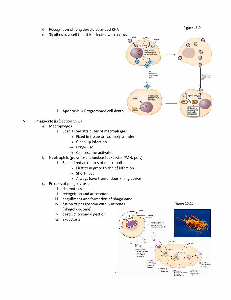
VI. Sensor systems (section 15.5)
a. Toll‐like receptors – surface receptors that allow cells to “see” molecules that signify the presence of microorganisms or viruses
b. Pattern recognition
Figure 15.4

3
c. The complement system – series of proteins that, when activated, result in the destruction/removal of foreign material; cascade reaction
Figure 15.6
Part of figure 17.9
Figure 15.7
4
d. Recognition of long double‐stranded RNA e. Signifies to a cell that it is infected with a virus
i. Apoptosis = Programmed cell death VII. Phagocytosis (section 15.6)
a. Macrophages i. Specialized attributes of macrophages
→ Fixed in tissue or routinely wander → Clean up infection → Long‐lived → Can become activated
b. Neutrophils (polymorphonuclear leukocyte, PMN, poly) i. Specialized attributes of neutrophils
→ First to migrate to site of infection → Short‐lived → Always have tremendous killing power
c. Process of phagocytosis i. chemotaxis ii. recognition and attachment iii. engulfment and formation of phagosome iv. fusion of phagosome with lysosomes
(phagolysosome) v. destruction and digestion vi. exocytosis
Figure 15.10
Figure 15.9

5
VIII. Inflammation – a coordinated response to invasion (section 15.7) a. Characteristics
i. Redness, pain, swelling heat ii. Purpose:
→ Contain a site of damage → Localize the response → Restore tissue function
iii. Factors that initiate the inflammatory response: → Microbial cell products detected by toll‐like receptors → Microbial surfaces (trigger the complement cascade)
iv. Tissue damage
b. The inflammatory process i. Pro‐inflammatory cytokines released ii. Dilation of small blood vessels
→ increased blood flow in the area iii. Leakage of fluids from vessels iv. Adherence of phagocytic cells to endothelial cell v. Diapedesis
Figure 15.11

6
IX. Study Questions 1. What are the first‐line defenses? 2. What is the function the sensor systems in innate immunity? 3. What are toll‐like receptors? 4. What is the complement system? 5. What is the role of phagocytes? 6. What are cytokines? 7. What triggers inflammation? 8. Describe the functions of lysozyme, transferrin, and gastric acid. 9. Describe how surface receptors and cytokines allow the cells of the immune system to communicate. 10. List three bacterial components recognized by toll‐like receptors. 11. Describe three outcomes of complement activation. 12. Describe the alternative pathway of complement activation. 13. What are the functions of C3b and C5a? 14. How can a cell recognize it is infected with a virus, and what does it do in response? 15. Compare and contrast monocytes, dendritic cells, macrophages, and neutrophils. 16. Describe the steps of the inflammatory process. 17. What is apoptosis?


