Increased demand, trophic stimulation Hyperplasia, … · 2008-07-03 · Reversible Cell Injury —...
18
Intracellular accumulations; calcifications 3. Metabolic alterations, genetic or acquired Cellular aging 4. Prolonged life span with cumulative sublethal injury Subcellular alterations in various organelles Mild chronic injury Irreversible injury cell death (necrosis, apoptosis) Progressive and severe (incl. DNA damage) Acute reversible injury Acute and self-limited Cell Injury 2. Reduced O2; Chemical Injury; Microbial Infection Metaplasia Chronic irritation (chemical or physical) Atrophy Decreased nutrients, stimulation Hyperplasia, hypertrophy Increased demand, trophic stimulation Cellular Adaptations 1. Altered Physiologic Stimuli Cellular Response Nature and Severity of Injurious Stimuli Figure 1-1
Transcript of Increased demand, trophic stimulation Hyperplasia, … · 2008-07-03 · Reversible Cell Injury —...

Intracellular accumulations; calcifications3. Metabolic alterations, genetic or acquired
Cellular aging4. Prolonged life span with cumulative sublethal injury
Subcellular alterations in various organellesMild chronic injury
Irreversible injury cell death (necrosis, apoptosis)Progressive and severe (incl. DNA damage)
Acute reversible injuryAcute and self-limited
Cell Injury2. Reduced O2; Chemical Injury; Microbial Infection
MetaplasiaChronic irritation (chemical or physical)
AtrophyDecreased nutrients, stimulation
Hyperplasia, hypertrophyIncreased demand, trophic stimulation
Cellular Adaptations1. Altered Physiologic Stimuli
Cellular ResponseNature and Severity of Injurious Stimuli
Figure 1-1
Hyperplasia1. Physiologic
• Hormonal • Compensatory
2. Pathologic • Excessive hormonal stimulation or GFs • Fertile soil for cancer
Hypertrophy1. Physiologic
• Functional demand • Specific hormonal stimulation
2. Pathologic3. Genes that induce hypertrophy
a. Genes that encode transcription factorsc-fos; c-jun
b. Growth factorsTGF-β; IGF-1; FGF
c. Vasoactive agentα-adrenergic agonist; endothelin-1;
angiotensin II
Atrophy (Physiologic or Pathologic)1. Decreased workload2. Loss of innervation3. Diminished blood supply4. Inadequate nutrition5. Loss of endocrine stimulation6. Aging (senile atrophy)7. Pressure
Metaplasia1. Reversible change2. Most common = columnar to squamous3. Vitamin A deficiency4. May induce malignant transformation
Figure 1-4

Reversible Cell Injury — the HALLMARK of reversible injury is: 1. Reduced oxidative phosphorylation2. ATP depletion3. Cellular swelling
Irreversible Injury and Cell Death1. Morphologic changes – cell death2. Necrosis – always pathologic3. Apoptosis – normal function; not necessarily assoc. with cell injury
Causes of Cell Injury1. Oxygen deprivation2. Physical agents3. Chemical agents and drugs4. Infectious agents5. Immunologic reactions6. Genetic derangements7. Nutritional imbalances
Figure 1-8
Figure 1-7
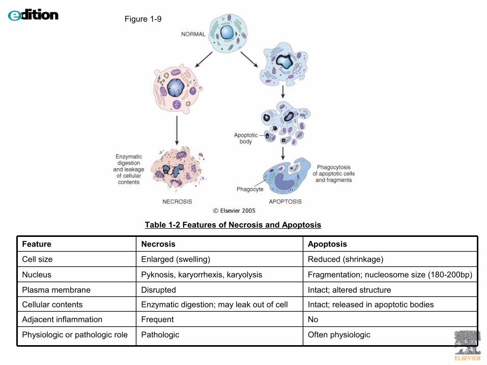
Often physiologicPathologicPhysiologic or pathologic role
NoFrequentAdjacent inflammation
Intact; released in apoptotic bodiesEnzymatic digestion; may leak out of cellCellular contents
Intact; altered structureDisruptedPlasma membrane
Fragmentation; nucleosome size (180-200bp)Pyknosis, karyorrhexis, karyolysisNucleus
Reduced (shrinkage)Enlarged (swelling)Cell size
ApoptosisNecrosisFeature
Table 1-2 Features of Necrosis and Apoptosis
Figure 1-9

Figure 1-10
1. Cellular response to injury depends on:- type of injury- its duration- its severity
2. Consequences of cell injury depend on:- cell type- cell state- cell adaptability
3. Cell injury results from functional / biochemical abnormalities in:- aerobic respiration- integrity of cell membranes- protein synthesis- the cytoskeleton- the integrity of the genetic apparatus
Mechanisms of Cell Injury:1. Depletion of ATP2. Mitochondrial damage3. Influx of intracellular Ca++; loss of Ca++ homeostasis4. Accumulation of O2-derived free radicals (oxidative stress)5. Defects in membrane permeability

Figure 1-11
Depletion of ATP
Depletion of ATP to <5-10% of normal levels has effects on many critical cellular systems:
1. Plasma membrane energy-dependent Na++ pump - Na++ accumulates intracellularly - K+ diffuses out of the cell - osmotic pull cell swelling
2. Cellular energy metabolism is altered - increased rate of anaerobic glycolysis - glycogen stores rapidly depleted - lactic acid accumulation dec. intracellular pH
3. Failure of the Ca++ pump influx of Ca++
4. Depletion of ATP structural disruption of the protein synthetic pathway detachment of ribosomes reduced protein synthesis
5. Irreversible damage to mitochondrial and lysosomal membranes
6. Proteins may become misfolded trigger the “unfolded protein response” that may lead to cell injury or death

Figure 1-12Mitochondria can be damaged by:1. Increased cytosolic Ca++2. Oxidative stress3. Breakdown of phospholipids - phospholipase A2 - sphingomyelin pathways - lipid breakdown products
Mitochondrial damage results in:1. Formation of high-conductance channel (mitochondrial permeability transition) (MPT)2. Reversible in the early stages3. MPT can become permanent with continued stimulus4. Irreversible MPT death blow to cell
Figure 1-13
1. Most intracellular Ca++ is sequestered in the mitochondria and ER
2. Ischemia and toxins increase cytosolic Ca++ - influx of Ca++ across the plasma membrane - release of Ca++ from the mitochondria and ER
3. Increased Ca++ activates a number of enzymes: - ATPases hasten ATP depletion - phospholipases membrane damage - proteases breakdown membrane and cytoskeletal proteins - endonucleases DNA and chromatin fragmentation

Figure 1-14
Accumulation of Oxygen-Derived Free Radicals(Oxidative Stress)
1. Absorption of radiant energy2. Enzymatic metabolism of exogenous chemicals or drugs3. Reduction-oxidation reactions - superoxide anion radical (O2-) - hydrogen peroxide (H2O2) - hydroxyl ions (OH)4. Transition metals5. Nitric Oxide (NO)
Effects of Reactive Species:1. Lipid peroxidation of membranes2. Oxidative modification of proteins3. Lesions in DNA
Mechanisms to Remove Free Radicals1. Antioxidants - Vitamin E - Vitamin A - Ascorbic acid - Glutathione
2. Storage and transport proteins - Transferrin - Ferritin - Lactoferrin - Ceruloplasmin
3. Enzymes - Catalase - Superoxide dismutase - Glutathione peroxidase

Figure 1-15
Defects in Membrane Permeability Result In:
1. Mitochondrial dysfunction2. Loss of membrane phospholipids3. Cytoskeletal abnormalities4. Reactive oxygen species5. Lipid breakdown products detergent effect on membranes
1. Injury to lysosomal membranes results inleakage of their enzymes into the cytoplasmand activation of these enzymes
2. Lysosomes contain: - RNases - DNases - proteases - phosphatases - glucosidases - cathepsins
3. Activation of enzymes leads to enzymatic digestion of cell components

Figure 1-17 Morphologic changes in reversible and irreversible cell injury. A, Electron micrograph of a normal epithelial cell of the proximal kidney tubule. Note abundant microvilli (mv) lining the lumen (L). N, nucleus; V, apical vacuoles (which are normal structures in this cell type).
B, Epithelial cell of the proximal tubule showing reversible ischemic changes. The microvilli (mv) are lost and have been incorporated in apical cytoplasm; blebs have formed and are extruded in the lumen (L). Mitochondria are slightly dilated. (Compare with A.)
C, Proximal tubular cell showing irreversible ischemic injury. Note the markedly swollen mitochondria containing amorphous densities, disrupted cell membranes, and dense pyknotic nucleus. (Courtesy of Dr. M.A. Venkatachalam, University of Texas, San Antonio, TX.)
Downloaded from: Robbins & Cotran Pathologic Basis of Disease (on 1 March 2005 09:25 PM)© 2005 Elsevier
Figure 1-17
Two phenomena consistently characterize irreversibility:1. Inability to reverse mitochondrial dysfunction2. Development of profound disturbances in membrane function
Histological Reversible Cell Injury:1. Cellular swelling2. Fatty change
Ultrastructural Reversible Cell Injury:1. Plasma membrane alterations - blebbing, blunting, distortion of microvilli, creation of myelin figures, loosening of intercellular attachments2. Mitochondrial changes - swelling, rarefaction, small amorphous densities3. Dilation of the ER - detachment and disaggregation of polysomes4. Nuclear alterations - disaggregation of granular and fibrillar elements

Types of Necrosis1. Coagulative necrosis – preservation of the basic outline of the coagulated cells2. Liquefactive necrosis – transformation of tissue into a viscous mass - Gangrenous necrosis – a limb that has lost its blood supply and undergone coagulative necrosis3. Caseous necrosis – a distinctive form of coagulative necrosis; most often a foci of tuberculous infection4. Fat necrosis – focal areas of fat destruction
Ischemic and Hypoxic Injury1. Ischemia tends to injure tissues faster than does hypoxia2. If O2 is restored, all of the disturbances are reversible3. If ischemia persists, irreversible injury and necrosis ensue
Ischemia-Reperfusion Injury1. Damage may be initiated during reoxygenation by increased generation of oxygen free radicals2. Reactive oxygen species can further promote the MTP may lead to cell death3. Ischemic injury is associated with inflammation; production of cytokines; increased expression of adhesion molecules4. Activation of Complement pathway
Figure 1-22

Chemical Injury1. Some chemical can act directly by combining with some critical molecular component or cellular organelle2. Others must be converted to reactive toxic metabolites - P45 mixed function oxidase in SER
Examples:Carbon tetrachloride CCl4 CCl3 + Cl-
Acetaminophen is detoxified by interaction with GSH
Figure 1-23 Figure 1-24

Causes of ApoptosisPhysiologic:1. The programmed destruction of cells during embrogenesis2. Hormone-dependent involution in the adult3. Cell deletion in proliferating cell populations4. Acute inflammatory reaction; immune response5. Elimination of potentially harmful self-reactive lymphocytes6. Cell death induced by cytotoxic T cells
Pathologic:1. Injurious stimuli – radiation, cytotoxic chemotherapy2. Viral diseases3. Pathologic atrophy in parenchymal organs after duct obstruction4. Cell death in tumors
Morphology of Apoptosis1. Cell shrinkage2. Chromatin condensation - the MOST CHARACTERISTIC feature of apoptosis is chromatin condensation with chromatin aggregrates at the periphery3. Cytoplasmic blebs and apoptotic bodies4. Phagocytosis of apoptotic cells or cell bodies
Biochemical Features of Apoptosis1. Protein cleavage - cysteine proteases (caspases)2. DNA breakdown - formation of oligonucleosomes (180-200 bp)3. Phagocytic recognition - apoptotic cells express phosphatidylerine in the outer layers of the membrane - cells may also express thrombospondin, an adhesive glycoprotein - these are recognized by MØ
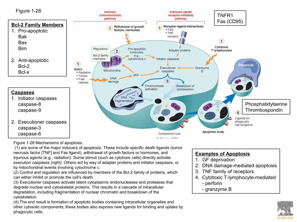
Figure 1-28
Figure 1-28 Mechanisms of apoptosis. (1) are some of the major inducers of apoptosis. These include specific death ligands (tumor necrosis factor [TNF] and Fas ligand), withdrawal of growth factors or hormones, and injurious agents (e.g., radiation). Some stimuli (such as cytotoxic cells) directly activate execution caspases (right). Others act by way of adapter proteins and initiator caspases, or by mitochondrial events involving cytochrome c. (2) Control and regulation are influenced by members of the Bcl-2 family of proteins, which can either inhibit or promote the cell's death. (3) Executioner caspases activate latent cytoplasmic endonucleases and proteases that degrade nuclear and cytoskeletal proteins. This results in a cascade of intracellular degradation, including fragmentation of nuclear chromatin and breakdown of the cytoskeleton. (4) The end result is formation of apoptotic bodies containing intracellular organelles and other cytosolic components; these bodies also express new ligands for binding and uptake by phagocytic cells.
PhosphatidylserineThrombospondin
TNFR1Fas (CD95)Bcl-2 Family Members
1. Pro-apoptotic Bak Bax Bim
2. Anti-apoptotic Bcl-2 Bcl-x
Caspases1. Initiator caspases caspase-8 caspase-9
2. Executioner caspases caspase-3 caspase-6
Examples of Apoptosis1. GF deprivation2. DNA damage-mediated apoptosis3. TNF family of receptors 4. Cytotoxic T-lymphocyte-mediated - perforin - granzyme B

Figure 1-29
Figure 1-30
The Extrinsic (Death Receptor-Initiated) PathwayFasL binds to Fas 3+ Fas come together and form a binding site foran adaptor protein (FADD- Fas-associated death domain) this thenbinds to the inactive caspase-8 multiple caspase-8 are brought together and they cleave one another active caspase-8 this thentriggers a cascade of caspase activation activation of executionercaspases
This pathway is INHIBITED by a protein FLIP which binds to pr-caspase-8But cannot cleave and activate it
The Intrinsic (Mitochondrial) PathwayIncreased mitochondrial permeability release ofpro-apoptotic molecules (AIF-apoptosis inducingfactor) and cytochrome c
Cytochrome c binds to Apaf-1 (apoptosis activatingfactor-1) this complex activates caspase-9
AIF enters the cytosol and binds to and neutralizesvarious inhibitors of apoptosis
Bcl-2 and Bcl-x directly INHIBIT Apaf-1 activation
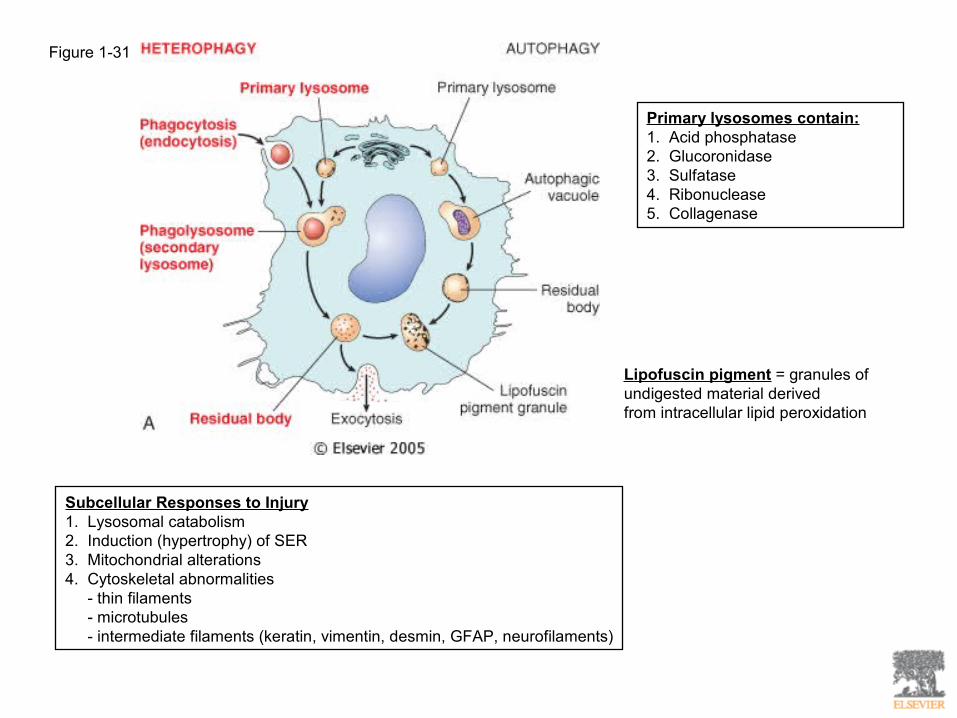
Figure 1-31
Primary lysosomes contain:1. Acid phosphatase2. Glucoronidase3. Sulfatase4. Ribonuclease5. Collagenase
Lipofuscin pigment = granules of undigested material derivedfrom intracellular lipid peroxidation
Subcellular Responses to Injury1. Lysosomal catabolism2. Induction (hypertrophy) of SER3. Mitochondrial alterations4. Cytoskeletal abnormalities - thin filaments - microtubules - intermediate filaments (keratin, vimentin, desmin, GFAP, neurofilaments)

Figure 1-35
Intracellular accumulation of abnormal amounts of substances:1. a normal cellular constituent accumulates2. an abnormal substance –exogenous or endogenous3. a pigment
Three types of abnormalities:1. normal endogenous substance; rate of metabolism is inadequate2. normal or abnormal endogenous substance accumulates; genetic or acquired defect in metabolism, packing, transport, secretion3. abnormal exogenous substance is deposited and accumulates
Types of Accumulations:1. Lipids - steatosis and fatty change (LIVER, HEART, MUSCLE, KIDNEY) - cholesterol and cholesterol esters (atherosclerosis, xanthomas, inflammation and necrosis, cholesterolosis, Niemann-Pick disease type C2. Proteins - reabsorption droplets in PCT kidneys - synthesis of excessive amounts - defects in protein folding may lead to “unfolded protein response”3. Hyaline change4. Glycogen - glycogen storage diseases - DM5. Pigments - exogenous pigments (carbon, tattoo) - endogenous pigments (lipofuscin, melanin, hemosiderin)
**Systemic overload of iron, hemosiderin is deposited in many organs and tissues; a condition called hemosiderosis and is seen with:1. increased absorption of dietary iron2. impaired use of iron3. hemolytic anemias4. transfusions

Figure 1-43
Pathologic Calcification1. Dystrophic calcification –occurs locally in dying tissues; NORMAL serum Ca++ levels
2. Metastatic calcification – occurs in normal tissues; almost always results from HYPERCALCEMIA - increased secretion of PTH - destruction of bone tissue - vitamin D-related disorders - renal failure
Metastatic calcification occurs in the following:1. Gastric mucosa2. Kidneys3. Lungs 4. Systemic arteries5. Pulmonary veins
Cellular Aging1. Telomere shortening utimately results in cell cycle arrest2. Telomerase = lengthens the telomeres by nucleotide addition