Incidence of Microbial Contamination of Lenses in Long-term · PDF fileIncidence of Microbial...
7
235 International Journal of Scientific Study | July 2016 | Vol 4 | Issue 4 Incidence of Microbial Contamination of Lenses in Long-term Soft Contact Lens Wearers Nita U Shanbhag 1 , Priyadarshini Cholera 2 , Ravi Karnam 3 , Nadim Khatib 4 1 Head and Professor, Department of Microbiology, D. Y. Patil Medical College, Navi Mumbai, Maharashtra, India, 2 Assistant Professor, Department of Ophthalmology, D. Y. Patil Hospital and Research Centre, D. Y. Patil Medical College, Navi Mumbai, Maharashtra, India, 3 Professor, Department of Microbiology, D. Y. Patil Hospital and Research Centre, D. Y. Patil Medical College, Nerul, Navi Mumbai, Maharashtra, India, 4 Resident, Department of Ophthalmology, D. Y. Patil Hospital and Research Centre, D. Y. Patil Medical College, Navi Mumbai, Maharashtra, India material, and modality of wear have a profound effect on the physiology of this tissue. Studies have shown that the physical presence of a CL, irrespective of oxygen transmissibility, disrupts corneal epithelial renewal mechanisms, producing a thinned, and stagnant epithelium. MATERIALS AND METHODS A study of 50 eyes in 30 patients was conducted at a tertiary care ophthalmic center for evaluating the incidence of microbial contamination of lenses in long-term soft CL wearers. On screening, a patient who fitted into the inclusion criteria (Table 1) were impressed on to hand over the lenses for the study to enable collection of data. These patients underwent a detailed eye examination as shown in Table 2. A thorough anterior segment examination was done to differentiate between corneal and conjunctival infection. INTRODUCTION Microbial keratitis (MK) is the most visually devastating complication associated with contact lens (CL) wear CL wear disrupts these protective mechanisms through breakdown of normal homeostatic surface renewal as well as damaging the corneal surface. Trauma, pre-existing ocular surface disease, and CL wear have been earmarked as the most common etiologies of microbial infection. CLs share an intimate relationship with the epithelial surface; all forms of CL wear, regardless of lens Original Article Abstract Introduction: Microbial keratitis is the most visually devastating complication associated with contact lens (CL) wear CL wear disrupts these protective mechanisms through breakdown of normal homeostatic surface renewal as well as damaging the corneal surface. Materials and Methods: A study of 50 eyes in 30 patients was conducted at a tertiary care ophthalmic center. There was a check on microbial contamination of lenses in long-term soft CL wearers. Patients who wanted to change their lenses after 1 year use or who wanted to discard their lenses due to redness, pain, watering, or blurring of vision were requested to give their lenses for smear culture antibiotic sensitivity and microbial culture. Results: The cultures were evaluated for bacterial, fungal, or Acanthamoeba growth. These were tabulated in a master chart and results documented. Conclusion: This study was conducted to impress the need to stop dispensing CLs at the optometrist counters where proper advice as to care of the lenses and their maintenance is not given. It also creates awareness about CL hygiene. Key words: Contact lens, Lens contamination, Microbial infection, Smear culture antibiotic sensitivity Access this article online www.ijss-sn.com Month of Submission : 05-2016 Month of Peer Review : 06-2016 Month of Acceptance : 07-2016 Month of Publishing : 07-2016 Corresponding Author: Dr. Nadim Khatib, Department of Ophthalmology D. Y. Patil Medical College, Navi Mumbai - 400 706, Maharashtra, India. Phone: +91-9920428272. E-mail: [email protected] Print ISSN: 2321-6379 Online ISSN: 2321-595X DOI: 10.17354/ijss/2016/413
Transcript of Incidence of Microbial Contamination of Lenses in Long-term · PDF fileIncidence of Microbial...
235 International Journal of Scientific Study | July 2016 | Vol 4 | Issue 4
Incidence of Microbial Contamination of Lenses in Long-term Soft Contact Lens WearersNita U Shanbhag1, Priyadarshini Cholera2, Ravi Karnam3, Nadim Khatib4
1Head and Professor, Department of Microbiology, D. Y. Patil Medical College, Navi Mumbai, Maharashtra, India, 2Assistant Professor, Department of Ophthalmology, D. Y. Patil Hospital and Research Centre, D. Y. Patil Medical College, Navi Mumbai, Maharashtra, India, 3Professor, Department of Microbiology, D. Y. Patil Hospital and Research Centre, D. Y. Patil Medical College, Nerul, Navi Mumbai, Maharashtra, India, 4Resident, Department of Ophthalmology, D. Y. Patil Hospital and Research Centre, D. Y. Patil Medical College, Navi Mumbai, Maharashtra, India
material, and modality of wear have a profound effect on the physiology of this tissue. Studies have shown that the physical presence of a CL, irrespective of oxygen transmissibility, disrupts corneal epithelial renewal mechanisms, producing a thinned, and stagnant epithelium.
MATERIALS AND METHODS
A study of 50 eyes in 30 patients was conducted at a tertiary care ophthalmic center for evaluating the incidence of microbial contamination of lenses in long-term soft CL wearers.
On screening, a patient who fitted into the inclusion criteria (Table 1) were impressed on to hand over the lenses for the study to enable collection of data. These patients underwent a detailed eye examination as shown in Table 2. A thorough anterior segment examination was done to differentiate between corneal and conjunctival infection.
INTRODUCTION
Microbial keratitis (MK) is the most visually devastating complication associated with contact lens (CL) wear CL wear disrupts these protective mechanisms through breakdown of normal homeostatic surface renewal as well as damaging the corneal surface.
Trauma, pre-existing ocular surface disease, and CL wear have been earmarked as the most common etiologies of microbial infection.
CLs share an intimate relationship with the epithelial surface; all forms of CL wear, regardless of lens
Original Article
AbstractIntroduction: Microbial keratitis is the most visually devastating complication associated with contact lens (CL) wear CL wear disrupts these protective mechanisms through breakdown of normal homeostatic surface renewal as well as damaging the corneal surface.
Materials and Methods: A study of 50 eyes in 30 patients was conducted at a tertiary care ophthalmic center. There was a check on microbial contamination of lenses in long-term soft CL wearers. Patients who wanted to change their lenses after 1 year use or who wanted to discard their lenses due to redness, pain, watering, or blurring of vision were requested to give their lenses for smear culture antibiotic sensitivity and microbial culture.
Results: The cultures were evaluated for bacterial, fungal, or Acanthamoeba growth. These were tabulated in a master chart and results documented.
Conclusion: This study was conducted to impress the need to stop dispensing CLs at the optometrist counters where proper advice as to care of the lenses and their maintenance is not given. It also creates awareness about CL hygiene.
Key words: Contact lens, Lens contamination, Microbial infection, Smear culture antibiotic sensitivity
Access this article online
www.ijss-sn.com
Month of Submission : 05-2016 Month of Peer Review : 06-2016 Month of Acceptance : 07-2016 Month of Publishing : 07-2016
Corresponding Author: Dr. Nadim Khatib, Department of Ophthalmology D. Y. Patil Medical College, Navi Mumbai - 400 706, Maharashtra, India. Phone: +91-9920428272. E-mail: [email protected]
Print ISSN: 2321-6379Online ISSN: 2321-595X
DOI: 10.17354/ijss/2016/413
Shanbhag, et al.: Incidence of Microbial Contamination of Lenses
236International Journal of Scientific Study | July 2016 | Vol 4 | Issue 4
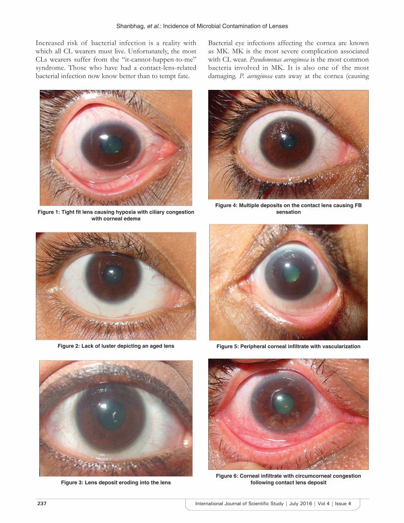
CLs documenting g rowth or eye les ions were photographed for prognosis (Figures 1-6). The lenses collected and were transported to the laboratory in a sterile autoclaved lens cleaning solution to avoid cross contamination.
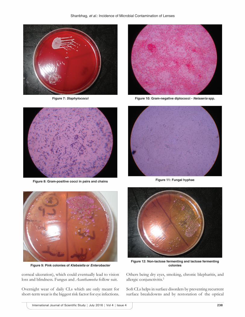
At the laboratory, the lens were transferred to glucose broth and incubated at 37° for 2 h. This broth was further cultured on blood agar, McConkey’s agar overnight at 37° and sabouraud’s at room temperature and 37° for 3 weeks. The growth observed was documented as bacterial, fungal and Acanthamoeba growth. Digital pictures were taken (Figures 7-12).
Results were tabulated and inference drawn (Tables 3 and 4).
The wear and tear of soft CL as it is creates microcracks. If handled roughly with nails then this forms earlier. Once the integrity of surface is lost, normal commensals or opportunist which will grow into the CL. During the wear of CL, if there is an inadvertent eye injury, these organisms will latch on the cornea causing corneal ulcer and subsequent loss of vision.
The cultures gave us a mixed basket of organisms with almost 1/3rd as no growth. Only those patients who had redness/conjunctival congestion or corneal defects were treated with antibiotics.
All were counseled as to CL hygiene (Table 5). They were reinforced on the corrective measures to be taken. In the case of a problem visit to the eye doctor was mandatory. Better to be safe than sorry.
DISCUSSION
The potential role of CL care solutions in MK has recently gained significant interest due to increased reports of fungal and Acanthamoeba keratitis. More recent reports support the view that corneal staining may be directly related to inflammation. This compromise includes the inhibition of apoptotic desquamation and a slowed renewal mechanism, producing a thinned, stagnant epithelial sheet. It appears that it is the cumulative breakdown of these collective processes that results in CL related MK and further illustrates the multifactorial nature of the disease process.1
Table 1: Inclusion and exclusion criteriaInclusion criteria Exclusion criteriaAge group: 15-35 years Age group: <15 or >35Lenses Lenses: RGP, bandage
soft CLSoft, yearly wear in power range of+5‑−20 which were discarded after the yearly use or prior to that due to redness pain watering or blurring of vision not permitting the wearer to use them
Conjunctivitis causing redness pain watering
Asymptomatic patient wearing CL for duration more than the prescribed schedule
Iridocyclitis causing redness pain wateringPatient not willing for any eye procedure
Asymptomatic patient wearing CL and never having got them cleaned for >3 months in yearly wear schedule
Patient not willing to come for regular follow-up
Symptomatic patient wearing CL with redness pain watering
RGP: Rigid gas permeable, CL: Contact lens
Table 2: Detailed eye examination and investigationsCL history Clinical examination InvestigationCL usage at what age Visual acuity on logmar chart CL sent for SCABS and KOHDuration of CL usage in a day BCVA: Spectacle power/CL power BUT/SchirmersType of CL Slit lamp examination Sac syringingCleaning procedure for the CL CL details IOP measurement by a non
contact tonometerAny faulting in the usage Soft/RGP If corneal lesion present corneal
scrape for SCABS and KOHSleeping with the lenses in the eye CL fit tight or loose CBC/ESRCleaning lenses with tap water Lack of lustre on CL indicating over-usage Urine: (Routine & Microscopy)Washing the eye with tap water with the lenses in the eye Deposits on CL ENT/Dental focus of infectionUsing OTC drugs - Pyrimon for any redness of the eye with the lenses on
Cornea
Continuing the usage of lenses in spite of getting a FB sensation redness watering or discharge in the eye
Corneal lesion
Not visiting a doctor but an optician for CL dispensing Fluorescent stainingImproper cleaning schedule for the lenses Photo of the lesion
Corneal sensationSac status to r/o chronic dacryocystitisIOP to r/o glaucomaBUT to r/o dry eyes
Shanbhag, et al.: Incidence of Microbial Contamination of Lenses
237 International Journal of Scientific Study | July 2016 | Vol 4 | Issue 4
Increased risk of bacterial infection is a reality with which all CL wearers must live. Unfortunately, the most CLs wearers suffer from the “it-cannot-happen-to-me” syndrome. Those who have had a contact-lens-related bacterial infection now know better than to tempt fate.
Bacterial eye infections affecting the cornea are known as MK. MK is the most severe complication associated with CL wear. Pseudomonas aeruginosa is the most common bacteria involved in MK. It is also one of the most damaging. P. aeruginosa eats away at the cornea (causing
Figure 1: Tight fit lens causing hypoxia with ciliary congestion with corneal edema
Figure 2: Lack of luster depicting an aged lens
Figure 3: Lens deposit eroding into the lens
Figure 4: Multiple deposits on the contact lens causing FB sensation
Figure 5: Peripheral corneal infiltrate with vascularization
Figure 6: Corneal infiltrate with circumcorneal congestion following contact lens deposit
Shanbhag, et al.: Incidence of Microbial Contamination of Lenses
238International Journal of Scientific Study | July 2016 | Vol 4 | Issue 4
corneal ulceration), which could eventually lead to vision loss and blindness. Fungus and Acanthamoeba follow suit.
Overnight wear of daily CLs which are only meant for short-term wear is the biggest risk factor for eye infections.
Others being dry eyes, smoking, chronic blepharitis, and allergic conjunctivitis.2
Soft CLs helps in surface disorders by preventing recurrent surface breakdowns and by restoration of the optical
Figure 7: Staphylococci
Figure 8: Gram-positive cocci in pairs and chains
Figure 9: Pink colonies of Klebsiella or Enterobacter
Figure 10: Gram-negative diplococci - Neisseria spp.
Figure 11: Fungal hyphae
Figure 12: Non-lactose fermenting and lactose fermenting colonies
Shanbhag, et al.: Incidence of Microbial Contamination of Lenses
239 International Journal of Scientific Study | July 2016 | Vol 4 | Issue 4
integrity of the surface. However in a tight lens syndrome, it might cause corneal edema with subsequent rupture of corneal bullae secondary infection can lead to MK.3
With the growth of soft CL wear, the incidence of CL-associated MK has increased up to 30% of all keratitis in developed countries. The microbes responsible for CL-associated keratitis include Gram-negative bacteria and rarely, Gram-positive bacteria and fungi, whereas Acanthamoeba predominated in the developed countries. Several CL-related and non-CL-related factors were attributed to the higher incidence of Acanthamoeba keratitis among CL wearers in developed nations. In contrast, bacteria were found to be the only pathogen for all CL-associated keratitis in this study. P. aeruginosa was reported to be the most common organism isolated from CL wearers in the developing world and similarly.
In developing countries like India, commonly used water is contaminated by gut commensals, especially Pseudomonas. A contact of CLs and CL storage cases with water can cause contamination by Pseudomonas, which survives well in the moist environment offered by CLs, CL storage cases, and lens care solutions. Contaminated CLs which were used by the patients, acted as a vector for transmitting the microbes from the CL storage cases to the patients’ conjunctiva and cornea by forming polysaccharide-containing bio-film on the posterior surface of soft CLs by bacterial adherence. Bacterial adherence to artificial surface is also thought to be mediated by hydrophobic bonding and relatively hydrophobic strains adhere very readily to CLs.4
A CL can act as a vector for micro-organisms to adhere to and transfer to the ocular surface. Commensal micro-organisms that uneventfully cohabitate on lid margins and conjunctivae and potential pathogens that are found transiently on the ocular surface can inoculate CLs in vivo. In the presence of reduced tissue resistance, these
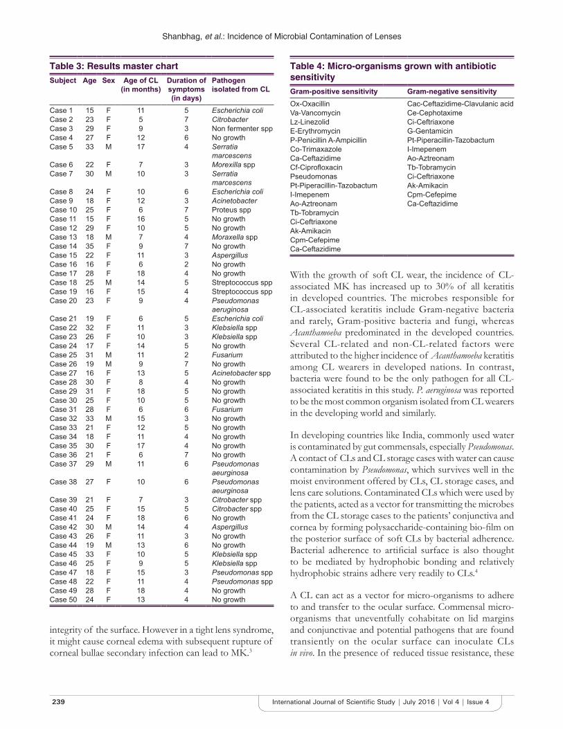
Table 3: Results master chartSubject Age Sex Age of CL
(in months)Duration of symptoms(in days)
Pathogen isolated from CL
Case 1 15 F 11 5 Escherichia coliCase 2 23 F 5 7 CitrobacterCase 3 29 F 9 3 Non fermenter sppCase 4 27 F 12 6 No growthCase 5 33 M 17 4 Serratia
marcescensCase 6 22 F 7 3 Morexilla sppCase 7 30 M 10 3 Serratia
marcescensCase 8 24 F 10 6 Escherichia coliCase 9 18 F 12 3 AcinetobacterCase 10 25 F 6 7 Proteus sppCase 11 15 F 16 5 No growthCase 12 29 F 10 5 No growthCase 13 18 M 7 4 Moraxella spp Case 14 35 F 9 7 No growthCase 15 22 F 11 3 AspergillusCase 16 16 F 6 2 No growthCase 17 28 F 18 4 No growthCase 18 25 M 14 5 Streptococcus sppCase 19 16 F 15 4 Streptococcus sppCase 20 23 F 9 4 Pseudomonas
aeruginosaCase 21 19 F 6 5 Escherichia coliCase 22 32 F 11 3 Klebsiella sppCase 23 26 F 10 3 Klebsiella sppCase 24 17 F 14 5 No growthCase 25 31 M 11 2 FusariumCase 26 19 M 9 7 No growthCase 27 16 F 13 5 Acinetobacter sppCase 28 30 F 8 4 No growthCase 29 31 F 18 5 No growth Case 30 25 F 10 5 No growthCase 31 28 F 6 6 FusariumCase 32 33 M 15 3 No growthCase 33 21 F 12 5 No growthCase 34 18 F 11 4 No growthCase 35 30 F 17 4 No growthCase 36 21 F 6 7 No growthCase 37 29 M 11 6 Pseudomonas
aeurginosaCase 38 27 F 10 6 Pseudomonas
aeurginosaCase 39 21 F 7 3 Citrobacter spp Case 40 25 F 15 5 Citrobacter sppCase 41 24 F 18 6 No growthCase 42 30 M 14 4 AspergillusCase 43 26 F 11 3 No growthCase 44 19 M 13 6 No growthCase 45 33 F 10 5 Klebsiella sppCase 46 25 F 9 5 Klebsiella sppCase 47 18 F 15 3 Pseudomonas spp Case 48 22 F 11 4 Pseudomonas sppCase 49 28 F 18 4 No growthCase 50 24 F 13 4 No growth
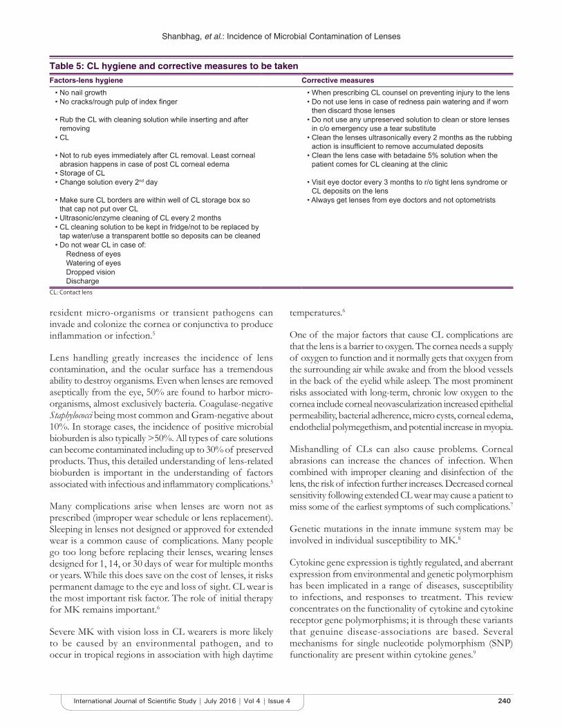
Table 4: Micro-organisms grown with antibiotic sensitivityGram-positive sensitivity Gram-negative sensitivityOx-Oxacillin Cac-Ceftazidime-Clavulanic acidVa-Vancomycin Ce-CephotaximeLz-Linezolid Ci-CeftriaxoneE-Erythromycin G-GentamicinP-Penicillin A-Ampicillin Pt-Piperacillin-TazobactumCo-Trimaxazole I-ImepenemCa-Ceftazidime Ao-AztreonamCf‑Ciprofloxacin Tb-TobramycinPseudomonas Ci-CeftriaxonePt-Piperacillin-Tazobactum Ak-AmikacinI-Imepenem Cpm-CefepimeAo-Aztreonam Ca-CeftazidimeTb-TobramycinCi-CeftriaxoneAk-AmikacinCpm-CefepimeCa-Ceftazidime
Shanbhag, et al.: Incidence of Microbial Contamination of Lenses
240International Journal of Scientific Study | July 2016 | Vol 4 | Issue 4
resident micro-organisms or transient pathogens can invade and colonize the cornea or conjunctiva to produce inflammation or infection.5
Lens handling greatly increases the incidence of lens contamination, and the ocular surface has a tremendous ability to destroy organisms. Even when lenses are removed aseptically from the eye, 50% are found to harbor micro-organisms, almost exclusively bacteria. Coagulase-negative Staphylococci being most common and Gram-negative about 10%. In storage cases, the incidence of positive microbial bioburden is also typically >50%. All types of care solutions can become contaminated including up to 30% of preserved products. Thus, this detailed understanding of lens-related bioburden is important in the understanding of factors associated with infectious and inflammatory complications.5
Many complications arise when lenses are worn not as prescribed (improper wear schedule or lens replacement). Sleeping in lenses not designed or approved for extended wear is a common cause of complications. Many people go too long before replacing their lenses, wearing lenses designed for 1, 14, or 30 days of wear for multiple months or years. While this does save on the cost of lenses, it risks permanent damage to the eye and loss of sight. CL wear is the most important risk factor. The role of initial therapy for MK remains important.6
Severe MK with vision loss in CL wearers is more likely to be caused by an environmental pathogen, and to occur in tropical regions in association with high daytime
temperatures.6
One of the major factors that cause CL complications are that the lens is a barrier to oxygen. The cornea needs a supply of oxygen to function and it normally gets that oxygen from the surrounding air while awake and from the blood vessels in the back of the eyelid while asleep. The most prominent risks associated with long-term, chronic low oxygen to the cornea include corneal neovascularization increased epithelial permeability, bacterial adherence, micro cysts, corneal edema, endothelial polymegethism, and potential increase in myopia.
Mishandling of CLs can also cause problems. Corneal abrasions can increase the chances of infection. When combined with improper cleaning and disinfection of the lens, the risk of infection further increases. Decreased corneal sensitivity following extended CL wear may cause a patient to miss some of the earliest symptoms of such complications.7
Genetic mutations in the innate immune system may be involved in individual susceptibility to MK.8
Cytokine gene expression is tightly regulated, and aberrant expression from environmental and genetic polymorphism has been implicated in a range of diseases, susceptibility to infections, and responses to treatment. This review concentrates on the functionality of cytokine and cytokine receptor gene polymorphisms; it is through these variants that genuine disease-associations are based. Several mechanisms for single nucleotide polymorphism (SNP) functionality are present within cytokine genes.9
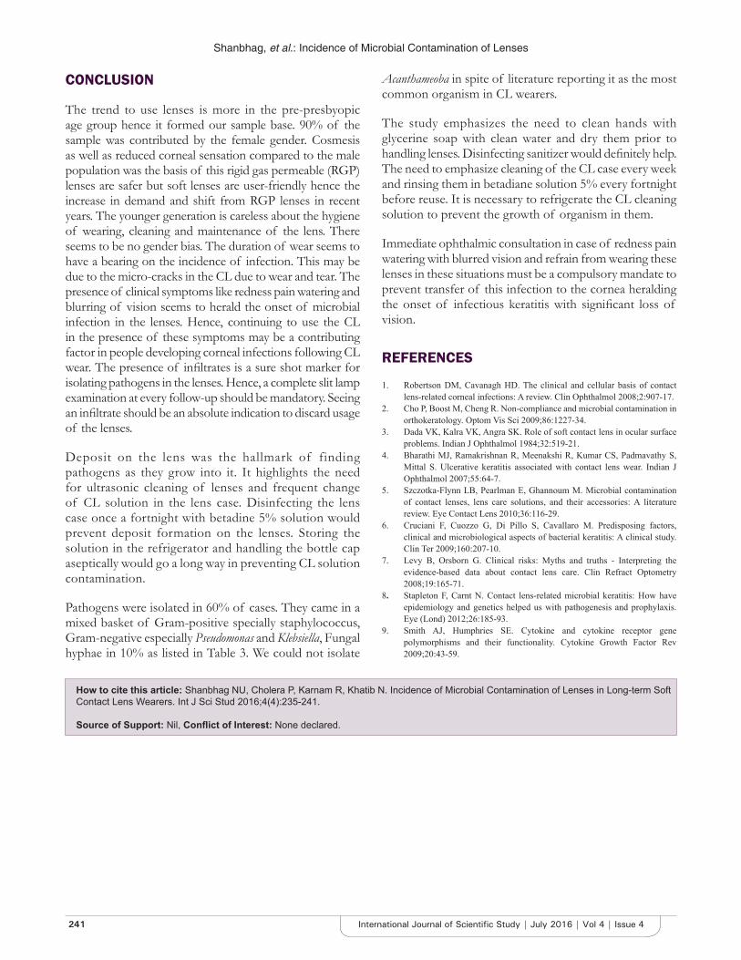
Table 5: CL hygiene and corrective measures to be takenFactors-lens hygiene Corrective measures
• No nail growth • When prescribing CL counsel on preventing injury to the lens• No cracks/rough pulp of index finger • Do not use lens in case of redness pain watering and if worn
then discard those lenses• Rub the CL with cleaning solution while inserting and after
removing• Do not use any unpreserved solution to clean or store lenses
in c/o emergency use a tear substitute• CL • Clean the lenses ultrasonically every 2 months as the rubbing
action is insufficient to remove accumulated deposits• Not to rub eyes immediately after CL removal. Least corneal
abrasion happens in case of post CL corneal edema• Storage of CL
• Clean the lens case with betadaine 5% solution when the patient comes for CL cleaning at the clinic
• Change solution every 2nd day • Visit eye doctor every 3 months to r/o tight lens syndrome or CL deposits on the lens
• Make sure CL borders are within well of CL storage box so that cap not put over CL
• Always get lenses from eye doctors and not optometrists
• Ultrasonic/enzyme cleaning of CL every 2 months• CL cleaning solution to be kept in fridge/not to be replaced by
tap water/use a transparent bottle so deposits can be cleaned• Do not wear CL in case of:
Redness of eyesWatering of eyesDropped visionDischarge
CL: Contact lens
Shanbhag, et al.: Incidence of Microbial Contamination of Lenses
241 International Journal of Scientific Study | July 2016 | Vol 4 | Issue 4
CONCLUSION
The trend to use lenses is more in the pre-presbyopic age group hence it formed our sample base. 90% of the sample was contributed by the female gender. Cosmesis as well as reduced corneal sensation compared to the male population was the basis of this rigid gas permeable (RGP) lenses are safer but soft lenses are user-friendly hence the increase in demand and shift from RGP lenses in recent years. The younger generation is careless about the hygiene of wearing, cleaning and maintenance of the lens. There seems to be no gender bias. The duration of wear seems to have a bearing on the incidence of infection. This may be due to the micro-cracks in the CL due to wear and tear. The presence of clinical symptoms like redness pain watering and blurring of vision seems to herald the onset of microbial infection in the lenses. Hence, continuing to use the CL in the presence of these symptoms may be a contributing factor in people developing corneal infections following CL wear. The presence of infiltrates is a sure shot marker for isolating pathogens in the lenses. Hence, a complete slit lamp examination at every follow-up should be mandatory. Seeing an infiltrate should be an absolute indication to discard usage of the lenses.
Deposit on the lens was the hallmark of finding pathogens as they grow into it. It highlights the need for ultrasonic cleaning of lenses and frequent change of CL solution in the lens case. Disinfecting the lens case once a fortnight with betadine 5% solution would prevent deposit formation on the lenses. Storing the solution in the refrigerator and handling the bottle cap aseptically would go a long way in preventing CL solution contamination.
Pathogens were isolated in 60% of cases. They came in a mixed basket of Gram-positive specially staphylococcus, Gram-negative especially Pseudomonas and Klebsiella, Fungal hyphae in 10% as listed in Table 3. We could not isolate
Acanthameoba in spite of literature reporting it as the most common organism in CL wearers.
The study emphasizes the need to clean hands with glycerine soap with clean water and dry them prior to handling lenses. Disinfecting sanitizer would definitely help. The need to emphasize cleaning of the CL case every week and rinsing them in betadiane solution 5% every fortnight before reuse. It is necessary to refrigerate the CL cleaning solution to prevent the growth of organism in them.
Immediate ophthalmic consultation in case of redness pain watering with blurred vision and refrain from wearing these lenses in these situations must be a compulsory mandate to prevent transfer of this infection to the cornea heralding the onset of infectious keratitis with significant loss of vision.
REFERENCES
1. Robertson DM, Cavanagh HD. The clinical and cellular basis of contact lens-related corneal infections: A review. Clin Ophthalmol 2008;2:907-17.
2. Cho P, Boost M, Cheng R. Non-compliance and microbial contamination in orthokeratology. Optom Vis Sci 2009;86:1227-34.
3. Dada VK, Kalra VK, Angra SK. Role of soft contact lens in ocular surface problems. Indian J Ophthalmol 1984;32:519-21.
4. Bharathi MJ, Ramakrishnan R, Meenakshi R, Kumar CS, Padmavathy S, Mittal S. Ulcerative keratitis associated with contact lens wear. Indian J Ophthalmol 2007;55:64-7.
5. Szczotka-Flynn LB, Pearlman E, Ghannoum M. Microbial contamination of contact lenses, lens care solutions, and their accessories: A literature review. Eye Contact Lens 2010;36:116-29.
6. Cruciani F, Cuozzo G, Di Pillo S, Cavallaro M. Predisposing factors, clinical and microbiological aspects of bacterial keratitis: A clinical study. Clin Ter 2009;160:207-10.
7. Levy B, Orsborn G. Clinical risks: Myths and truths - Interpreting the evidence-based data about contact lens care. Clin Refract Optometry 2008;19:165-71.
8. Stapleton F, Carnt N. Contact lens-related microbial keratitis: How have epidemiology and genetics helped us with pathogenesis and prophylaxis. Eye (Lond) 2012;26:185-93.
9. Smith AJ, Humphries SE. Cytokine and cytokine receptor gene polymorphisms and their functionality. Cytokine Growth Factor Rev 2009;20:43-59.
How to cite this article: Shanbhag NU, Cholera P, Karnam R, Khatib N. Incidence of Microbial Contamination of Lenses in Long-term Soft Contact Lens Wearers. Int J Sci Stud 2016;4(4):235-241.
Source of Support: Nil, Conflict of Interest: None declared.