In-Vivo Xtreme II - Bruker · The In-Vivo Xtreme II is compatible with more custom and commercial...
8
In-Vivo Xtreme II Flexible Imaging to Support Evolving Research Preclinical Imaging Innovation with Integrity
Transcript of In-Vivo Xtreme II - Bruker · The In-Vivo Xtreme II is compatible with more custom and commercial...

In-Vivo Xtreme IIFlexible Imaging to Support Evolving Research
Precl inical ImagingInnovation with Integrity

�� Inherent registration of Bioluminescence, Fluorescence, Radioisotopic, Reflectance and X-ray modalities
�� Ultra sensitive 4MP CCD supports the most demanding bioluminescence studies
�� Fully configured VIS-NIR Multispectral imaging for flexible reporter selection
�� Bright 400 W Xenon Illuminator (up to 5x faster imaging compared to 150 W Halogen)
�� Fully configured VIS-NIR Wide Angle, Artifact Eliminating, Emission Filters
�� Patented Direct Radioisotopic Imaging (1000X more sensitive than Cherenkov) with imaging of clinically relevant non-Cherenkov isotopes
�� Fast anatomical X-ray, ideal reference for throughput Optical/X-ray studies
�� High resolution X-ray, geometric magnification, beam hardening, energy calibration, and bone mineral density analysis for dedicated anatomical studies
�� SPF Animal Chamber & EquaFlow Manifold
�� 360° Rotation Imaging
�� Comprehensive Acquisition & Analysis Software
Key Features of the Xtreme II
Five Modalities Individually Optimized for Performance and Function
The In-Vivo Xtreme II offers 5 imaging modalities. Each modality supplies lead-ing performance and innovative features to maximize experimental capabilities. Whether it is bioluminescent cell tracking, fluorescent probe development, small-compound biodistribution/targeting validation, or dedicated anatomical studies, the In-Vivo Xtreme II ensures your current and future research needs are covered.

Compatible with More Optical Reporters, Nuclear Tracers, and X-ray Agents
The In-Vivo Xtreme II is compatible with more custom and commercial molecular reporters than any comparable system. Choose the reporter, tracer, agent and substrate to suit experimental objectives and budget.
Barium sulfate, Visipaque, ExitronTM
Luciferase (all variants), Luminol, HRP
Non-Cherenkov Isotopes (e.g. 99mTc, 111In)
Fluorescent proteins (all variants) and VIS-NIR organic/inorganic fluorophores
Cherenkov Isotopes (e.g. 18F, 90Y, 177Lu)
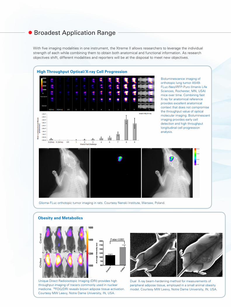
Broadest Application Range
With five imaging modalities in one instrument, the Xtreme II allows researchers to leverage the individual strength of each while combining them to obtain both anatomical and functional information. As research objectives shift, different modalities and reporters will be at the disposal to meet new objectives.
Glioma-FLuc orthotopic tumor imaging in rats. Courtesy Nenski Institute, Warsaw, Poland.
Bioluminescence imaging of orthotopic lung tumor A549-FLuc-Neo/iRFP-Puro (Imanis Life Sciences, Rochester, MN, USA) mice over time. Combining fast X-ray for anatomical reference provides excellent anatomical context that does not compromise the throughput value of optical molecular imaging. Bioluminescent imaging provides early cell detection and high throughput longitudinal cell progression analysis.
High Throughput Optical/X-ray Cell Progression
Unique Direct Radioisotopic Imaging (DRI) provides high throughput imaging of tracers commonly used in nuclear medicine. 18FDG/DRI reveals brown adipose tissue activation. Courtesy MW Leevy, Notre Dame University, IN, USA.
Dual X-ray beam-hardening method for measurements of peripheral adipose tissue, employed in a small animal obesity model. Courtesy MW Leevy, Notre Dame University, IN, USA.
Obesity and Metabolics

Fluorescence biomarker imaging using NIR molecular probes combined with high resolution X-ray reference provides insights of inflammatory responses in a preclinical model of PSA. Inflammation and new bone formation were assessed using in vivo non-invasive molecular imaging, using ProSense and Osteosense probes.
Bioluminescent cell detection combined with molecular fluorescent detections reveal intersecting cellular-molecular events in vivo. Combined bioluminescence and fluorescence imaging of subcutaneous PC3M-FLuc (Targeting Systems, Inc., El Cajon, CA, USA) tumor mouse using MMPSense 750 Fast.
Simultaneous angiogenesis and apoptosis biomarker imaging in a subcutaneous MDA-MB-231 tumor mouse.
Disease Biomarkers and Inflammation
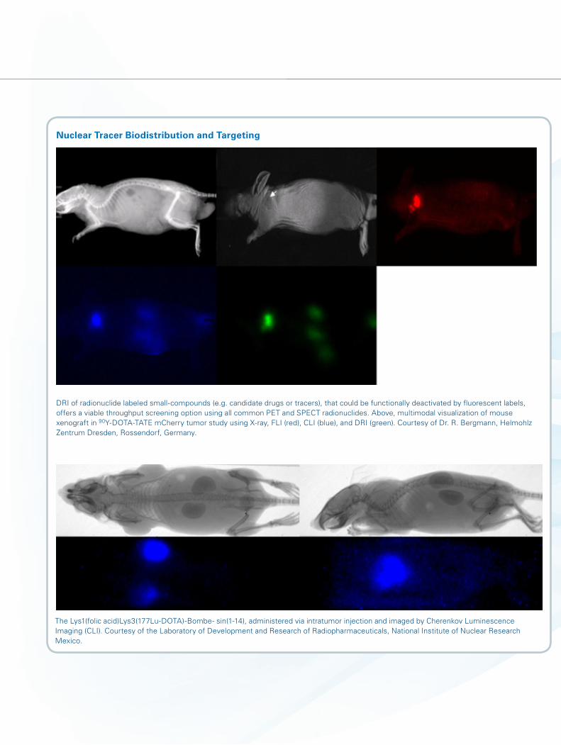
DRI of radionuclide labeled small-compounds (e.g. candidate drugs or tracers), that could be functionally deactivated by fluorescent labels, offers a viable throughput screening option using all common PET and SPECT radionuclides. Above, multimodal visualization of mouse xenograft in 90Y-DOTA-TATE mCherry tumor study using X-ray, FLI (red), CLI (blue), and DRI (green). Courtesy of Dr. R. Bergmann, Helmohlz Zentrum Dresden, Rossendorf, Germany.
The Lys1(folic acid)Lys3(177Lu-DOTA)-Bombe- sin(1-14), administered via intratumor injection and imaged by Cherenkov Luminescence Imaging (CLI). Courtesy of the Laboratory of Development and Research of Radiopharmaceuticals, National Institute of Nuclear Research Mexico.
Nuclear Tracer Biodistribution and Targeting
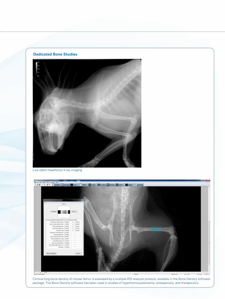
Live rabbit head/torso X-ray imaging.
Cortical long bone density of mouse femur is evaluated by a multiple ROI analysis protocol, available in the Bone Density software package. The Bone Density software has been used in studies of hyperhomocysteinemia, osteoporosis, and therapeutics.
Dedicated Bone Studies

Bruker BioSpin
www.bruker.com/XtremeII
© B
ruke
r 01
/17
T159
341
Optical
Camera Sensor Back-thinned, back-illuminated, 16-bit, Cooled, Grade 1 CCD 2.7 x 2.7 cm
CCD Operating Temperature - 90 °C absolute
Imaging Pixels 2048 x 2048, 13.5 microns
Field of View (FOV) cm2 7.2 x 7.2, 10 x 10, 12 x 12, 15 x 15, 18 x 18, 19 x 19
Lens f/1.1 - f/16, 58 mm fixed
Binning 1 x 1, 2 x 2, 4 x 4, 8 x 8, 16 x 16, 32 x 32
Illumination Source 400 W Xenon Lamp; 350 - 1100 nm spectrum
Excitation Fluorescence Filters Range 28 filters; 400 - 780 nm (additional filters available)
Dark Current (Typical) < 100 electrons/s
Emission Fluorescence Filters Range 6 filters; 518 - 850 nm; Patented Wide Angle Filters for Increased Uniformity (additional filters available)
Quantification Units Yes, absolute; Counts, ph/s/cm², ph/s/cm²/sr, pWatt/cm2
X-ray
Nominal Resolution (pixel size at maximum magnification)
< 25 μm
Radiation Safety 0.2 mR/hr max exposure at 5 cm from anywhere outside cabinet, FDA and TUV approved
X-ray Source 20 - 45 kV, 500 μA < 60 μm spot size
Beam Hardening, Geometric Magnifica-tion, and Energy Calibration
0, 0.1, 0.2, 0.4, 0.8 mm Al
Other
Physiological Monitoring Heated chamber, gas anesthesia compatible
AutoEvac Active waste gas removal from animal cabinet
Weight 140 kg
Imaging Chamber Interior Size (cm) 20 x 20 x 5 (L x W x H)
Imaging System Space Requirement (cm) 73 x 85 x 186 (W x D X H)
Power Requirements 100–240 V AC, 50/60 Hz, 15 A (System, heater, illuminator) 2.9 A (X-ray)
Software Yes, full featured analysis; unlimited site license
Computer (minimum specifications) Quad Core 2.8 GHz, 32 GB, 2133 MHz DDR4, SD RAM, 500 GB HD 24 inch UltraSharp Monitor
Optional
360° Projection Yes; MARS Unit, applicable for all imaging modalities
Nose Cone Manifold 3, 5, 8 mice and single rat nose cone EquaFlow ™ hyperbranch manifold
SafeAir ™ Anesthesia System Yes
Bone Mineral Density Analysis Yes, g/cm3, bone radius, asymmetry, peripheral X-ray density
SPF Chamber Yes, HEPA filtered and with quick connectors for easy access and transport
Imaging System Components: Specifications:


![In Vitro and In Vivo Uncaging and Bioluminescence Imaging ......molecular imaging in vitro and in vivo.[8] In our design, Tm/ Yb co-doped NaYF 4 core-shell nanoparticles [9] that have](https://static.fdocuments.net/doc/165x107/6120f332dd980c25b847715a/in-vitro-and-in-vivo-uncaging-and-bioluminescence-imaging-molecular-imaging.jpg)