2 wound dressing for the treatment of diabetic wounds: an ...

Original Article
In Vivo Performance of Chitosan/Soy-Based Membranesas Wound-Dressing Devices for Acute Skin Wounds
Tırcia C. Santos, PhD,1,2 Bernhard Horing,3 Kathrin Reise, MD,3 Alexandra P. Marques PhD,1,2
Simone S. Silva, PhD,1,2 Joaquim M. Oliveira, PhD,1,2 Joao F. Mano, PhD,1,2 Antonio G. Castro, PhD,2,4
Rui L. Reis, PhD,1,2 and Martijn van Griensven, PhD3,5
Wound management represents a major clinical challenge on what concerns healing enhancement and paincontrol. The selection of an appropriate dressing plays an important role in both recovery and esthetic ap-pearance of the regenerated tissue. Despite the wide range of available dressings, the progress in the wound caremarket relies on the increasing interest in using natural-based biomedical products. Herein, a rat wound-dressing model of partial-thickness skin wounds was used to study newly developed chitosan/soy (cht/soy)-based membranes as wound-dressing materials. Healing and repair of nondressed, cht/soy membrane-dressed,and Epigard�-dressed wounds were followed macroscopically and histologically for 1 and 2 weeks. cht/soymembranes performed better than the controls, promoting a faster wound repair. Re-epithelialization, ob-served 1 week after wounding, was followed by cornification of the outermost epidermal layer at the secondweek of dressing, indicating repair of the wounded tissue. The use of this rodent model, although in impairedhealing conditions, may enclose some drawbacks regarding the inevitable wound contraction. Moreover,being the main purpose the evaluation of cht/soy-based membranes’ performance in the absence of growthfactors, the choice of a clinically relevant positive control was limited to a polymeric mesh, without any growthfactor influencing skin healing/repair, Epigard. These new cht/soy membranes possess the desired featuresregarding healing/repair stimulation, ease of handling, and final esthetic appearance—thus, valuable prop-erties for wound dressings.
Introduction
Tissue repair and scar formation are main concerns ofwound care. The appropriate dressing selection plays an
important role both in the complete regeneration of the in-jured tissue and in the esthetic appearance.1 A wide range ofdressings and bandages, adaptable to various types ofwounds, are nowadays available in the wound care market,and are currently used in the clinics. The traditional wounddressing that simply covered and protected the wound2,3 hasbeen replaced by alternative dressings that allow control ofwound moisture4–7 and, more recently, by dressings with an(bio-) active role in the healing environment.8,9 However,most of the existing products need to be changed every fewdays after their application; in some cases, there is the needto replace the material to maintain/accelerate the ongoinghealing process.10 Therefore, further developments that
facilitate the healing process or address issues such as thecontrol of the chemical environment and bacterial infectionare required and desired.
Indiscriminately, synthetic,11–14 natural,8,15,16 or biologicalmaterials10,17,18 have been presented along the years, as thekey elements for controlling/modulating the healing mech-anisms and the outcomes of wound repair upon dressing.10
Among the recently proposed natural-origin materials forwound dressing, collagen2,9,19 chitosan,20–24 and silk25,26 oc-cupy a central position due to their nature, which mightpresent improved performance.
The positive impact of chitosan, a deacetylated derivativefrom chitin, concerning the healing mechanisms that in-clude the inflammatory process, is well documented. Thedemonstration of the chitosan anti-inflammatory activityand potential in promoting wound healing was reported byMori et al.27 Although chitosan activates macrophages, it
13B’s Research Group—Biomaterials, Biodegradables and Biomimetics, University of Minho, Headquarters of the European Instituteof Excellence on Tissue Engineering and Regenerative Medicine, AvePark, Guimaraes, Portugal.
2ICVS-3B’s–Portuguese Government Associate Laboratory, Braga/Guimaraes, Portugal.3Research Centre AUVA, Ludwig Boltzmann Institute for Experimental and Clinical Traumatology, Austrian Cluster for Tissue
Regeneration, Vienna, Austria.4Life and Health Sciences Research Institute, School of Health Sciences, University of Minho, Braga, Portugal.5Department of Trauma Surgery, Institute for Experimental Trauma Surgery, Klinikum rechts der Isar, Technical University Munich,
Munich, Germany.
TISSUE ENGINEERING: Part AVolume 19, Numbers 7 and 8, 2013ª Mary Ann Liebert, Inc.DOI: 10.1089/ten.tea.2011.0651
1
also induces gradual macrophage apoptosis in vitro (about50% programmed cell death in 6 h),27 thus preventing thepossible occurrence of septic shock and death.28 Macro-phage activation induced by chitosan resulted in increasedmetabolic activity, secretion of cytokines, growth factorsand inflammatory mediators, and enhanced phagocyticactivity.27 The production of macrophage inflammatoryprotein-2 (MIP-2) by chitosan-activated macrophagesstimulates epithelial cell proliferation29 and thus skin heal-ing. Additionally, a reduction on the influx of activatedtissue macrophages, which in turn decreases angiogenesis,fibroplasia, and connective tissue deposition, was also at-tributed to chitosan.30 These features may substantiate thehemostatic properties preventing bleeding,30 as well as theantibacterial activity when applied in skin wounds22,24,31
that chitosan also displays. In a clinical trial, Azad et al.32
have shown a positive effect of chitosan both on the re-epithelialization and regeneration of the granular layer ofthe skin where chitosan-dressed wounds healed fastercompared to controls. These data are in agreement withother studies demonstrating that chitosan induce the mi-gration of polymorphonuclear neutrophils (PMNs) at theearly stage of wound healing, when treating open skinwounds in dogs, enhancing the formation of granulationtissue and production of collagen by fibroblasts.33 In addi-tion, we have previously demonstrated that PMNs are notactivated by chitosan and, therefore, an acute inflammatoryresponse will not persist at the wound site in the presence ofchitosan.34
Soybean-rich foods have been recognized to decrease therisks of chronic diseases and protective effects against per-sistent inflammation.35 These features are mainly attributedto isoflavones.35 Among the isoflavones, genistein, a phy-toestrogen, showed to have favorable effects on cutaneouswound healing36,37 through an estrogen receptor-indepen-dent mechanism.36 Additionally, it was demonstrated thatgenistein is able to signal, via mitogen-activated protein ki-nase (MAPK) activation in macrophages, reducing woundpro-inflammatory cytokines.36
Chitosan/soy (cht/soy) membranes were previouslyproposed for biomedical applications based on the observa-tions of their in vitro capacity to enhance the proliferation offibroblasts38 and their impaired ability to activate PMNs.34
Additionally, a normal host reaction was observed aftersubcutaneous implantation in rats.39 Other important fea-tures of these cht/soy membranes, critical for wounddressings, are their simple manipulation and their transpar-ency. In this context and on the worldwide-recognized po-tential of chitosan and soy-based components for skinwounds regeneration, the aim of this study was to evaluatethe suitability of newly developed cht/soy-based mem-branes as dressings for partial thickness skin wounds.Among the many different animal models to study woundhealing and skin regeneration, excision of rat skin portions isa model widely used.8,17,22,40–45 Therefore, a wound-dressingrat model of partial-thickness skin wound, under impairedhealing conditions, was used to assess the suitability of thecht/soy membranes to promote wound healing. Compara-tively, Epigard�, a clinically accepted wound dresser, wasused as control. Epigard� is also only a dressing without anyfurther active substances.12,46 Thus, it is a clinically widelyused dressing similar to the one studied here.
Materials and Methods
Materials
Cht with a deacetylation degree of about 85% was pur-chased from Sigma, and the soy protein isolate (SI) was pro-vided by Loders Crocklaan. All other reagents were analyticalgrade and used as received. Cht/soy protein-blended mem-branes (average thickness of 84mm and 17 mm of diameter)were prepared by solvent casting according to a proceduredescribed elsewhere.38 Briefly, chitosan was dissolved in anaqueous acetic acid 2% (v/v) solution at a concentration of4 wt%. A soy suspension (1 wt%) was prepared by slowlydispersing the soy protein powder, under constant stirring, indistilled water with glycerol. After adjusting the pH to 8.0 – 0.3with 1 M sodium hydroxide, the dispersion was heated in awater bath at 50�C for 30 min. The cht and the SI solutions weremixed at a weight ratio of 75/25% cht/soy (CS75). Afterhomogenization, the CS75 solution was casted into Petridishes and dried at room temperature for 6 days. The neu-tralization of the membranes was obtained by immersion in0.1 M sodium hydroxide for about 10 min. Membranes werewashed with distilled water to remove all traces of alkali andthen dried at room temperature. The materials were sterilizedunder standard conditions with ethylene oxide.47
Animals
Twenty male Sprague-Dawley rats weighting between 230and 280 g were used for the study. Three groups were in-vestigated: membranes (wound directly covered with thecht/soy-based membranes); positive control (wound directlycovered with Epigard�—Biovision GmbH); and negativecontrol (no direct coverage of the wound). Epigard� is com-posed of a nontextile outer layer of polytetrafluorethyleneand an inner layer of soft elastic polyurethane that forms anopen matrix to which adsorbs the exudate from the woundbed. This dressing was chosen as positive control because itis extensively used in the clinical practice as a short-termwound dressing without growth factors.12,46
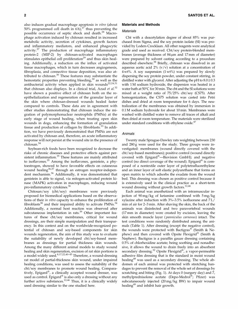
Each animal was anesthetized with an intramuscular in-jection of 90 mg/kg of ketamine combined with 5 mg/kgxylazine after induction with 3%–3.5% isofluorane and 7 L/min of air for 2–3 min. After shaving the skin, the back of theanimals was disinfected and two paravertebral wounds(17 mm in diameter) were created by excision, leaving theskin smooth muscle layer ( panniculus carnosus) intact. Thetest conditions were randomly distributed among the ani-mals (Table 1). After dressing (except the negative control),the wounds were protected with Bactigras� (Smith & Ne-phew) and then covered with Opsite Flexigrid� (Smith &Nephew). Bactigras is a paraffin gauze dressing containing0.5% of chlorhexidine acetate; being soothing and nonadhe-sive, it allows the wound to drain freely into an absorbentsecondary dressing.32 Opsite Flexigrid�, a vapor-permeableadhesive film dressing that is the standard in moist woundhealing48 was used as a secondary dressing. The whole ab-domen of each animal was protected with stretching ban-dages to prevent the removal of the whole set of dressings byscratching and biting (Fig. 1). At days 0 (surgery day) and 7,methylprednisolone acetate (Depo-Medrol�; Pfizer) wassubcutaneously injected (20 mg/kg BW) to impair woundhealing49 and inhibit hair growth.
2 SANTOS ET AL.
The animals were kept separately and received daily an-algesia with metamizole sodium (200 mg/g BW) and sedationwith diazepam (2.5 mg/125 mL water) in drinking water.
The bandages were changed every 3–4 days. Macroscopicanalysis of the wounds was carried out at days 3, 7, and 14and the images taken used for the planimetric evaluation ofthe healing process. The evaluation was performed with theLUCIA G� version 4.8 (Laboratory Imaging Ltd.) softwareby two independent researchers blinded to the experimentalcondition.
After 1 and 2 weeks, the animals were anesthetized withisoflurane and then euthanized by and intracardial overdoseof ketamin/xylazine. The wound area and the surroundinghealthy skin were explanted. Central wound cross sectionswere performed and samples were fixed for histological
analysis, in 3.7% formalin, and then paraffin embedded,sectioned, and stained according to a routine hematoxylinand eosin protocol.
The histological samples were analyzed using an Ax-ioplan Imager Z1 microscope (Zeiss) and included themeasurement of the length of the wound, allowing the es-tablishment of a correlation with the planimetric assessmentof the wound areas. The planimetric assessment comprisesthe wound area. This area is diminished during healing bycontraction and epithelialization. These entities cannot beseparated, but are present in all the groups. The fullwounded area and the healthy margins images were thenobtained after the composition of multiple standardizedhistological pictures.
Statistical analysis
Data from the planimetry and from the wound lengthmeasurements50 were analyzed by a single-factor ANOVAtest and the significance value was set at p < 0.05.
Results
Macroscopic analysis
The macroscopic characterization of the wounds duringthe observation period was based on the planimetric analysisof the wounded area. A qualitative evaluation of the healingarea and interface regions between healthy and newlyformed tissue was also performed.
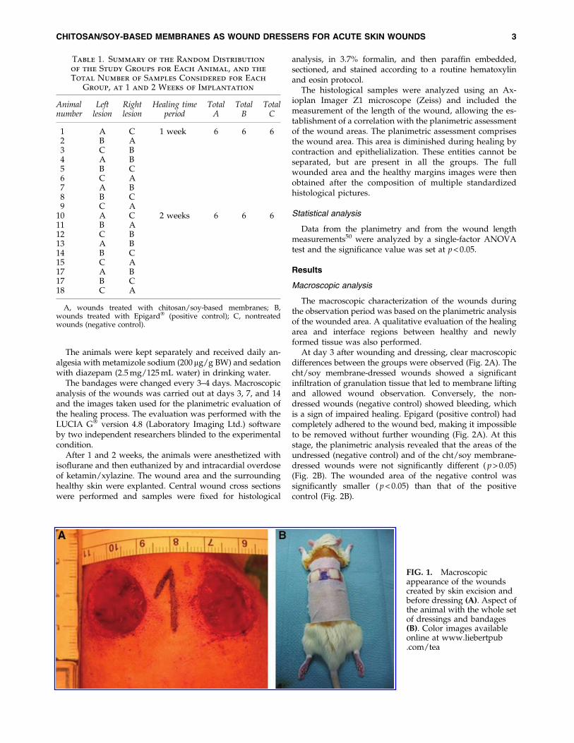
At day 3 after wounding and dressing, clear macroscopicdifferences between the groups were observed (Fig. 2A). Thecht/soy membrane-dressed wounds showed a significantinfiltration of granulation tissue that led to membrane liftingand allowed wound observation. Conversely, the non-dressed wounds (negative control) showed bleeding, whichis a sign of impaired healing. Epigard (positive control) hadcompletely adhered to the wound bed, making it impossibleto be removed without further wounding (Fig. 2A). At thisstage, the planimetric analysis revealed that the areas of theundressed (negative control) and of the cht/soy membrane-dressed wounds were not significantly different ( p > 0.05)(Fig. 2B). The wounded area of the negative control wassignificantly smaller ( p < 0.05) than that of the positivecontrol (Fig. 2B).
FIG. 1. Macroscopicappearance of the woundscreated by skin excision andbefore dressing (A). Aspect ofthe animal with the whole setof dressings and bandages(B). Color images availableonline at www.liebertpub.com/tea
Table 1. Summary of the Random Distribution
of the Study Groups for Each Animal, and the
Total Number of Samples Considered for Each
Group, at 1 and 2 Weeks of Implantation
Animalnumber
Leftlesion
Rightlesion
Healing timeperiod
TotalA
TotalB
TotalC
1 A C 1 week 6 6 62 B A3 C B4 A B5 B C6 C A7 A B8 B C9 C A
10 A C 2 weeks 6 6 611 B A12 C B13 A B14 B C15 C A17 A B17 B C18 C A
A, wounds treated with chitosan/soy-based membranes; B,wounds treated with Epigard� (positive control); C, nontreatedwounds (negative control).
CHITOSAN/SOY-BASED MEMBRANES AS WOUND DRESSERS FOR ACUTE SKIN WOUNDS 3
Despite some scratching of the secondary bandages, after7 days of dressing no signs of infection were detected in anyof the test groups (Fig. 2A). After this time period, the newepithelial tissue started to replace the granulation tissue inthe cht/soy membrane-dressed wounds, and the membraneswere easily lifted from the wound bed without bleeding. Animproved healing was observed for the cht/soy membrane-covered wounds versus the negative control, where somegranulation tissue was still present (Fig. 2A). In contrastEpigard� completely adhered to the wound bed, avoidingnew epidermis formation, and an attempt to remove it led tobleeding (Fig. 2A). Compared to both controls, the woundsdressed with the cht/soy membranes showed thinner mar-gins with an almost complete healing and new tissue for-mation replacing the excised epidermis (Fig. 2A). The woundof the negative control revealed a significant degree of con-traction. Despite the smaller wound area of the cht/soymembrane group, at day 7 the planimetric evaluation did notreveal significant differences in comparison to both controls( p > 0.05) (Fig. 2B).
The macroscopic analysis showed that the wound areasignificantly decreased from the operation day to the finalexcision time point (14 days) in all the tested conditions,which is an indication of neoepithelialization and replace-ment of the wounded tissue (Figs. 2A, B) by newly formedtissue. A significant and consecutive reduction of the woundarea was gradually observed in both negative and positivecontrols, although with a significantly delayed wound clo-sure (Figs. 2A, B). While comparing the contour and di-
mensions of the wounds at day 14 (Figs. 2A, B), it wasevident that the re-epithelialization and healing was alsomore efficient in the wounds dressed with the cht/soy-basedmembranes.
Histological analysis
After the first and second week of dressing, the woundsand surrounding healthy skin were excised, together withthe attached dressing in the case of the positive controls, andhistological analysis was performed. As during the processof sectioning and staining, the adherent Epigard� did notdetach from the wound bed, the analysis of the explantsincluded its integration with the growing tissue.
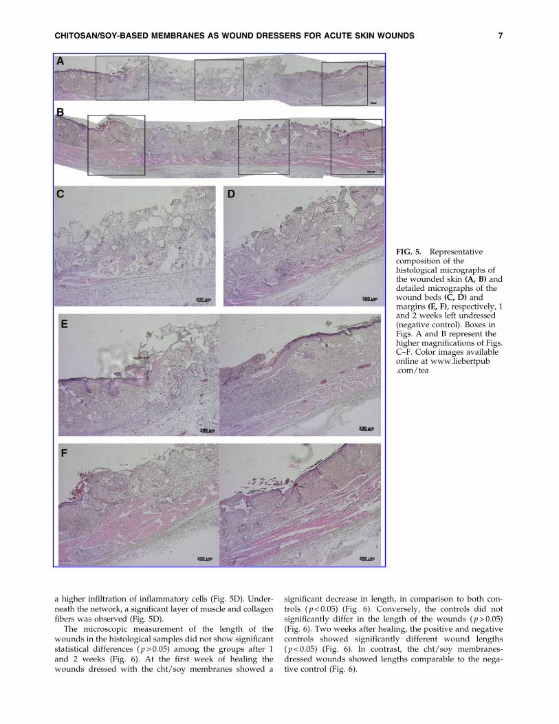
One week after dressing, the cht/soy-based membranes(Fig. 3A, C, E) seemed to enhance the formation of granu-lation tissue, in comparison to the negative control (un-dressed wounds) (Fig. 4A, C, E). Furthermore, the margins ofthe cht/soy membrane-dressed wounds presented a conti-nuity of the repairing tissue (Fig. 3E) that was not observedin the wound margins of the negative (Fig. 4E) and positive(Fig. 5E) controls. At this stage of healing, it was alreadypossible to identify some stratification of the tissue in themembrane-dressed wounds (Fig. 3C). These features werenot detectable either in the negative (Fig. 4C) or in the pos-itive controls (Fig. 5C). A disorganized mesh of cells, in-cluding a large amount of inflammatory cells, somefibroblasts, and collagen fibers, were observed in bothgroups. Furthermore, necrosis was detected in the
FIG. 2. Representative images of themacroscopic aspect of the excisionalwounds at the operation day (OpD)and subsequent healing at days 3, 7,and 14 after dressing with thechitosan/soy-based membranes, andin comparison with the negative andpositive controls (A). Follow-up of thewound area determined by planimetricanalysis (B). *Differences statisticallysignificant; A, �, and �p < 0.05 versusOpD; Cp < 0.05 versus negative controlat day 3 of dressing; Op < 0.05 versuspositive control at day 7 of dressing;fp < 0.05 versus membrane dressing atday 7. Color images available online atwww.liebertpub.com/tea
4 SANTOS ET AL.

nondressed wounds (Fig. 4C) but not in the Epigard�-dressed wounds (Fig. 5C). In fact, the material adhered to thewound bed and formed an intimate network composed bythe polymer matrix and the inflammatory cells. This networkstarted to vascularize, indicating a good integration of the
material into the wound bed (Fig. 5C). During the first weekof healing the presence of foreign body giant cells was notdetected in all test groups.
At the second week of dressing, the healing of the woundscovered by the cht/soy membranes (Fig. 3B, D, F) was
FIG. 3. Representativecomposition of thehistological micrographs ofthe wounded skin (A, B) anddetailed micrographs of thewound beds (C, D) andmargins (E, F), respectively, 1and 2 weeks after dressingwith the chitosan/soy (cht/soy) membrane. Boxes inFigs. A and B represent thehigher magnifications of Figs.C–F. Color images availableonline at www.liebertpub.com/tea
CHITOSAN/SOY-BASED MEMBRANES AS WOUND DRESSERS FOR ACUTE SKIN WOUNDS 5
enhanced in comparison to the negative (Fig. 4B, D, F) andpositive (Fig. 5B, D, F) controls. The wounds decreased insize (Fig. 3B), the margins were thinner (Fig. 3F) and thepreviously observed granulation tissue was replaced by amore stratified regenerated epidermis (Fig. 3D). Cornifica-tion of the outermost epidermal layer, although still near the
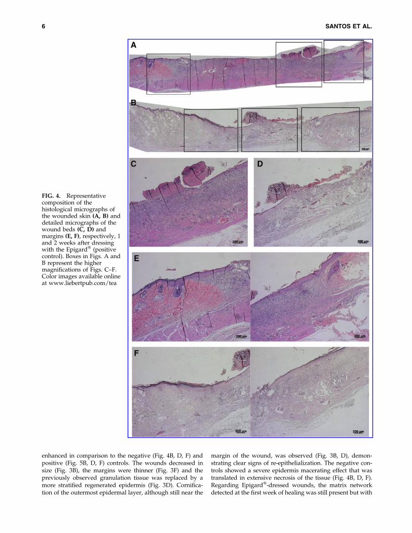
margin of the wound, was observed (Fig. 3B, D), demon-strating clear signs of re-epithelialization. The negative con-trols showed a severe epidermis macerating effect that wastranslated in extensive necrosis of the tissue (Fig. 4B, D, F).Regarding Epigard�-dressed wounds, the matrix networkdetected at the first week of healing was still present but with
FIG. 4. Representativecomposition of thehistological micrographs ofthe wounded skin (A, B) anddetailed micrographs of thewound beds (C, D) andmargins (E, F), respectively, 1and 2 weeks after dressingwith the Epigard� (positivecontrol). Boxes in Figs. A andB represent the highermagnifications of Figs. C–F.Color images available onlineat www.liebertpub.com/tea
6 SANTOS ET AL.
a higher infiltration of inflammatory cells (Fig. 5D). Under-neath the network, a significant layer of muscle and collagenfibers was observed (Fig. 5D).
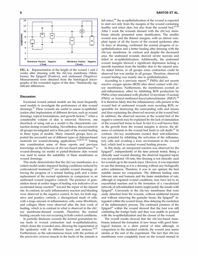
The microscopic measurement of the length of thewounds in the histological samples did not show significantstatistical differences ( p > 0.05) among the groups after 1and 2 weeks (Fig. 6). At the first week of healing thewounds dressed with the cht/soy membranes showed a
significant decrease in length, in comparison to both con-trols ( p < 0.05) (Fig. 6). Conversely, the controls did notsignificantly differ in the length of the wounds ( p > 0.05)(Fig. 6). Two weeks after healing, the positive and negativecontrols showed significantly different wound lengths( p < 0.05) (Fig. 6). In contrast, the cht/soy membranes-dressed wounds showed lengths comparable to the nega-tive control (Fig. 6).
FIG. 5. Representativecomposition of thehistological micrographs ofthe wounded skin (A, B) anddetailed micrographs of thewound beds (C, D) andmargins (E, F), respectively, 1and 2 weeks left undressed(negative control). Boxes inFigs. A and B represent thehigher magnifications of Figs.C–F. Color images availableonline at www.liebertpub.com/tea
CHITOSAN/SOY-BASED MEMBRANES AS WOUND DRESSERS FOR ACUTE SKIN WOUNDS 7
Discussion
Excisional wound animal models are the most frequentlyused models to investigate the performance of skin wounddressings.51 These wounds are useful to assess re-epithelial-ization after implantation of different devices, such as wounddressings, topical formulations, and growth factors,51 when aconsiderable volume of skin is removed. However, onedrawback of using rats as a model is the characteristic con-traction during wound healing. Nevertheless, this occurred inall groups investigated and is thus part of the wound healingin these types of models. Many research groups have re-ported the successful use of different types of chitosan-basedmaterials for wound-dressing applications.32,48,52–54 Takinginto consideration some of those reports and previousknowledge on the behavior of cht/soy-based membranes,34 awound-dressing rat model of partial-thickness skin woundwas used to assess the suitability of these membranes aswound dressings.
This study demonstrates that the cht/soy membranes, in arodent model under impaired healing conditions induced bycorticosteroid treatment,49 are suitable wound dressings, al-lowing the progress of a normal healing path and a fasterreplacement of the excised epidermis in comparison to anundressed wound (negative control). The presence of gran-ulation tissue at earlier stages of healing was indicative of anaccelerated tissue reaction55 toward the repair of the injuredsite. In contrast, an early inflammatory reaction and bleedingwere observed in the negative control. In fact, for both neg-ative and positive controls, a disorganized mesh of cells,with a large amount of inflammatory cells, some fibroblasts,and collagen fibers were observed after the first week ofhealing, which is in contrast to what is observed in the cht/soy membrane-dressed wounds. Therefore, the normalhealing cascade was not occurring in both control conditions.
In partially thickness wounds the formed granulation tis-sue leads to wound contraction and re-epithelialization,which closes the wounded area, allowing the regeneration ofthe epidermis with its different layers and annexes.43,51
Furthermore, as the subcutaneous tissue with the portion ofthe panniculus carnosus muscle in the backs of the animals is
left intact,56 the re-epithelialization of the wound is expectedto start not only from the margins of the wound containinghealthy and intact skin, but also from the wound bed.43,56
After 1 week the wounds dressed with the cht/soy mem-brane already presented some stratification. The smallerwound area and the thinner margins, with an almost com-plete repair of all the layers of the excised epidermis after14 days of dressing, confirmed the normal progress of re-epithelialization and a better healing after dressing with thecht/soy membrane. In contrast and despite the decreasedarea, the undressed wounds showed severe necrosis andfailed in re-epithelialization. Additionally, the undressedwound margins showed a significant depression lacking asmooth transition from the healthy skin to the injured area.As stated before, in all groups some contraction could beobserved but was similar in all groups. Therefore, observedwound healing was mainly due to epithelialization.
According to a previous report,34 PMNs did not secretereactive oxygen species (ROS) after direct contact with cht/soy membranes. Furthermore, the membranes exerted ananti-inflammatory effect by inhibiting ROS production byPMNs when stimulated with phorbol 12-myristate 13-acetate(PMA) or formyl-methionyl-leucyl-phenylalanine (fMLP).34
It is therefore likely that the inflammatory cells present at thewound bed of undressed wounds were secreting ROS, re-sponsible for destroying the surrounding cells and tissuesand thus explaining the observed necrosis at the wound bed.In addition, the observed necrosis at the wound bed of thenegative controls may be explained by the lack of stimulationof the wound bed tissue to heal. In fact it is well known that,as the growth from the wound margins is slower, the ab-sence of nutrients in the wound bed leads to cell death.43 Incontrast, cht/soy membranes exerted their anti-inflamma-tory potential by inhibiting the activation of the inflamma-tory cells and avoiding a toxic environment in the woundbed, which lead to normal wound healing process.
In this study, an unexpected reaction was observed for theEpigard�, independently of the time periods tested. Being aclinically used wound dressing, the observed impaired repairwas not predicted. Of note, this dressing is not clinically usedfor wounds up to the muscle layer. However, it was importantto use this dressing as it is a dressing without any biologicallyactive substances. Therefore, it was in our opinion the bestsuitable means for comparison. The different healing ratesbetween rats and humans and the faster metabolism of rats,although in impaired wound conditions, may have led to anexacerbated reaction and to the formation of a vascularizednetwork of cell-embedded matrix might justify the results withEpigard�. Conversely to the cht/soy membranes that wereeasily detached from the wounds, without additional traumaand without removing the granular tissue, the Epigard� in-tegrated within the wound tissue, thus delaying the resolutionof the inflammatory process. The continued presence of theEpigard� within the wound showed that the host was stillcombating the foreign body and thus was unable to proceedwith the re-epithelialization and the closure of the wound.
The overall results showed that the cht/soy-based mem-branes induced the formation of new tissue with good histo-logical features in a short period of time although, incomparison to the standard controls, the wound area seemssimilar at the end of the experiment. The fact that cht/soymembranes were completely loose on the wound seemed to
FIG. 6. Representation of the length of the wound 1 and 2weeks after dressing with the cht/soy membrane (Mem-brane), the Epigard (Positive), and undressed (Negative).Measurements were obtained from the histological micro-graphs of the wounded region of the skin. *Statistically sig-nificant differences.
8 SANTOS ET AL.
facilitate the spatial and increased progression of the newlyformed epithelium. It is also critical to emphasize that, besideswound closure, wound dressing must promote the formationof quality tissue, ideally without contraction. The cht/soy-based membranes appear not only to be able to regulate thewound moisture, which at extreme levels would impair epi-dermis repair, but also to provide the adequate coverage thatdoes not physically constrain the formation of new tissue.
Despite the relevance of the observations included in thisstudy, the use of a rodent model in wound-dressing evalu-ation, although in impaired healing conditions, may enclosesome drawbacks regarding the inevitable wound contrac-tion. Furthermore, since the main purpose was the evalua-tion of cht/soy-based membranes’ performance withoutgrowth factors, the choice of a clinically relevant positivecontrol was limited to a polymeric mesh, without anygrowth factor (Epigard�), which had a detrimental influencein wound healing and cutaneous repair.
Nevertheless, the present work strongly suggests that thenewly developed cht/soy-based membranes produced bysolvent casting methodology that had proven to promotelow in vitro activation of human PMNs isolated from circu-lating blood decrease the healing time period of partial-thickness skin wounds in impaired healing rats. Moreover,these cht/soy-based membranes showed an enhanced per-formance as compared to the negative and positive controls,inducing fast re-epithelialization and wound closure.
Acknowledgments
The author Tırcia C. Santos acknowledges the PortugueseFoundation for Science and Technology (FCT) for her PhDgrant (SFRH/BD/40861/2007). This work was developedunder the scope of the European Network of ExcellenceEXPERTISSUES (NMP3-CT-2004-5000283).
Disclosure Statement
The authors state no conflicts of interest.
References
1. White, R., and Morris, C. Mepitel: a non-adherent wounddressing with Safetac technology. Br J Nurs 18, 58, 2009.
2. Alvarez, O.M., Mertz, P.M., and Eaglstein, W.H. The ef-fect of occlusive dressings on collagen synthesis and re-epithelialization in superficial wounds. J Surg Res 35, 142,1983.
3. Nathan, P., MacMillan, B.G., and Holder, I.A. In situ pro-duction of a synthetic barrier dressing for burn wounds inrats. Infect Immun 12, 257, 1975.
4. Geronemus, R.G., and Robins, P. The effect of two newdressings on epidermal wound healing. J Dermatol SurgOncol 8, 850, 1982.
5. James, J.H., and Watson, A.C. The use of Opsite, a vapourpermeable dressing, on skin graft donor sites. Br J Plast Surg28, 107, 1975.
6. Wood, R.A., and Hughes, L.E. Silicone foam sponge for pi-lonidal sinus: a new technique for dressing open granulatingwounds. Br Med J 4, 131, 1975.
7. Tavis, M.J., Thornton, J.W., Harney, J.H., Woodroof, E.A.,and Bartlett, R.H. Graft adherence to de-epithelialized sur-faces: a comparative study. Ann Surg 184, 594, 1976.
8. Damour, O., Gueugniaud, P.Y., Berthin-Maghit, M., Rous-selle, P., Berthod, F., Sahuc, F., et al. A dermal substratemade of collagen—GAG—chitosan for deep burn coverage:first clinical uses. Clin Mater 15, 273, 1994.
9. Burget, A., Nathan, P., Holder, I.A., and Macmillan, B.G.The effect of a collagen dressing on contaminated surgicalwounds in rats. Langenbecks Arch Chir 343, 69, 1976.
10. Ovington, L.G. Advances in wound dressings. Clin Der-matol 25, 33, 2007.
11. Nathan, P., Macmillan, B.G., and Holder, I.A. Effect of a syn-thetic dressing formed on a burn wound in rats: a comparisonof allografts, collagen sheets, and polyhydroxyethyl-methacrylate in the control of wound infection. Appl Microbiol28, 465, 1974.
12. Stone, H.A., Edelman, R.D., and McGarry, J.J. Epigard: asynthetic skin substitute with application to podiatricwound management. J Foot Ankle Surg 32, 232, 1993.
13. Szycher, M., and Lee, S.J. Modern wound dressings: a sys-tematic approach to wound healing. J Biomater Appl 7, 142,1992.
14. Nathan, P., Law, E.J., MacMillan, B.G., Murphy, D.F., Ronel,S.H., D’Andrea, M.J., et al. A new biomaterial for the controlof infection in the burn wound. Trans Am Soc Artif InternOrgans 22, 30, 1976.
15. Fulton, J.E., Jr. The stimulation of postdermabrasion woundhealing with stabilized aloe vera gel-polyethylene oxidedressing. J Dermatol Surg Oncol 16, 460, 1990.
16. Okamoto, Y., Shibazaki, K., Minami, S., Matsuhashi, A.,Tanioka, S., and Shigemasa, Y. Evaluation of chitin andchitosan on open would healing in dogs. J Vet Med Sci 57,
851, 1995.17. King, W.W., Lam, P.K., Liew, C.T., Ho, W.S., and Li, A.K.
Evaluation of artificial skin (Integra) in a rodent model.Burns 23 Suppl 1, S30, 1997.
18. Leipziger, L.S., Glushko, V., DiBernardo, B., Shafaie, F.,Noble, J., Nichols, J., et al. Dermal wound repair: role ofcollagen matrix implants and synthetic polymer dressings.J Am Acad Dermatol 12, 409, 1985.
19. Norton, L., and Chvapil, M. Comparison of newer syn-thetic and biological wound dressings. J Trauma 21, 463,1981.
20. Keong, L.C., and Halim, A.S. In vitro models in biocompat-ibility assessment for biomedical-grade chitosan derivativesin wound management. Int J Mol Sci 10, 1300, 2009.
21. Wang, W., Lin, S.Q., Xiao, Y.C., Huang, Y.D., Tan, Y., Cai,L., et al. Acceleration of diabetic wound healing with chit-osan-crosslinked collagen sponge containing recombinanthuman acidic fibroblast growth factor in healing-impairedSTZ diabetic rats. Life Sci 82, 190, 2008.
22. Wang, C.C., Su, C.H., and Chen, C.C. Water absorbing andantibacterial properties of N-isopropyl acrylamide graftedand collagen/chitosan immobilized polypropylene nonwo-ven fabric and its application on wound healing enhance-ment. J Biomed Mater Res Part A 84A, 1006, 2008.
23. Qin, Y.M. The preparation and characterization of chitosanwound dressings with different degrees of acetylation. JAppl Polym Sci 107, 993, 2008.
24. Ong, S.Y., Wu, J., Moochhala, S.M., Tan, M.H., and Lu, J.Development of a chitosan-based wound dressing with im-proved hemostatic and antimicrobial properties. Biomater-ials 29, 4323, 2008.
25. Okabayashi, R., Nakamura, M., Okabayashi, T., Tanaka, Y.,Nagai, A., and Yamashita, K. Efficacy of polarized hy-droxyapatite and silk fibroin composite dressing gel on
CHITOSAN/SOY-BASED MEMBRANES AS WOUND DRESSERS FOR ACUTE SKIN WOUNDS 9

epidermal recovery from full-thickness skin wounds. JBiomed Mater Res B Appl Biomater 90, 641, 2009.
26. Schneider, A., Wang, X.Y., Kaplan, D.L., Garlick, J.A., andEgles, C. Biofunctionalized electrospun silk mats as a topicalbioactive dressing for accelerated wound healing. ActaBiomater 5, 2570, 2009.
27. Mori, T., Murakami, M., Okumura, M., Kadosawa, T., Uede,T., and Fujinaga, T. Mechanism of macrophage activation bychitin derivatives. J Vet Med Sci 67, 51, 2005.
28. Cerami A. Inflammatory cytokines. Clin Immunol Im-munopathol 62, S3, 1992.
29. Driscoll, K.E., Hassenbein, D.G., Howard, B.W., Isfort, R.J.,Cody, D., Tindal, M.H., et al. Cloning, expression, andfunctional characterization of rat MIP-2: a neutrophil che-moattractant and epithelial cell mitogen. J Leukoc Biol 58,
359, 1995.30. Diegelmann, R.F., Dunn, J.D., Lindblad, W.J., and Cohen,
I.K. Analysis of the effects of chitosan on inflammation,angiogenesis, fibroplasia, and collagen deposition in poly-vinyl alcohol sponge implants in rat wounds. Wound RepairRegen 4, 48, 1996.
31. Deng, C.M., He, L.Z., Zhao, M., Yang, D., and Liu, Y. Bio-logical properties of the chitosan-gelatin sponge wounddressing. Carbohydr Polym 69, 583, 2007.
32. Azad, A.K., Sermsintham, N., Chandrkrachang, S., andStevens, W.F. Chitosan membrane as a wound-healingdressing: characterization and clinical application. J BiomedMater Res B Appl Biomater 69, 216, 2004.
33. Ueno, H., Yamada, H., Tanaka, I., Kaba, N., Matsuura, M.,Okumura, M., et al. Accelerating effects of chitosan forhealing at early phase of experimental open wound in dogs.Biomaterials 20, 1407, 1999.
34. Santos, T.C., Marques, A.P., Silva, S.S., Oliveira, J.M., Mano,J.F., Castro, A.G., et al. In vitro evaluation of the behaviour ofhuman polymorphonuclear neutrophils in direct contactwith chitosan-based membranes. J Biotechnol 132, 218, 2007.
35. Coward, L., Barnes, N.C., Setchel, K.D.R., and Barnes, S.Genistein, Daidzein, and their &glycoside conjugates: anti-tumor isoflavones in soybean foods from American andAsian Diets. J Agric Food Chem 41, 1961, 1993.
36. Emmerson, E., Campbell, L., Ashcroft, G.S., and Hardman,M.J. The phytoestrogen genistein promotes wound healingby multiple independent mechanisms. Mol Cell Endocrinol321, 184, 2010.
37. Marini, H., Polito, P., Altavilla, D., Irrera, N., Minutoli, L.,Calo, M., et al. Genistein aglycone improves skin repair in anincisional model of wound healing: a comparison with ra-loxifene and oestradiol in ovariectomized rats. Br J Phar-macol 160, 1185, 2010.
38. Silva, S.S., Santos, M.I., Coutinho, O.P., Mano, J.F., and Reis,R.L. Physical properties and biocompatibility of chitosan/soyblended membranes. J Mater Sci Mater Med 16, 575, 2005.
39. Santos, T.C., Marques, A.P., Silva, R.M., Silva, S.S., Mano,J.F., Castro, A.G., et al. Chitosan improves the biologicalperformance of soy-based biomaterials. Tissue Eng Part A16, 2883, 2010.
40. Hong, H.J., Jin, S.E., Park, J.S., Ahn, W.S., and Kim, C.K.Accelerated wound healing by smad3 antisense oligonucleo-tides-impregnated chitosan/alginate polyelectrolyte complex.Biomaterials 29, 4831, 2008.
41. Burkatovskaya, M., Castano, A.P., Demidova-Rice, T.N.,Tegos, G.P., and Hamblin, M.R. Effect of chitosan acetatebandage on wound healing in infected and noninfectedwounds in mice. Wound Repair Regen 16, 425, 2008.
42. Noorjahan, S.E., and Sastry, T.P. An in vivo study of hy-drogels based on physiologically clotted fibrin-gelatin com-posites as wound-dressing materials. J Biomed Mater Res BAppl Biomater 71, 305, 2004.
43. Laplante, A.F., Germain, L., Auger, F.A., and Moulin, V.Mechanisms of wound reepithelialization: hints from a tissue-engineered reconstructed skin to long-standing questions.FASEB J 15, 2377, 2001.
44. Kweon, D.K., Song, S.B., and Park, Y.Y. Preparation of water-soluble chitosan/heparin complex and its application aswound healing accelerator. Biomaterials 24, 1595, 2003.
45. Burkatovskaya, M., Tegos, G.P., Swietlik, E., Demidova,T.N., P Castano, A., and Hamblin, M.R. Use of chitosanbandage to prevent fatal infections developing from highlycontaminated wounds in mice. Biomaterials 27, 4157, 2006.
46. Roth, R.R., and Winton, G.B. A synthetic skin substitute as atemporary dressing in Mohs surgery. J Dermatol Surg Oncol15, 670, 1989.
47. Reis, R.L., Mendes, S.C., Cunha, A.M., and Bevis, M.L.Processing and in vitro degradation of starch/EVOH ther-moplastic blends. Polym Int 43, 347. 1997.
48. Yusof, N.L., Wee, A., Lim, L.Y., and Khor, E. Flexible chitinfilms as potential wound-dressing materials: wound modelstudies. J Biomed Mater Res A 66, 224, 2003.
49. Wicke, C., Halliday, B., Allen, D., Roche, N.S., Scheuenstuhl,H., Spencer, M.M., et al. Effects of steroids and retinoids onwound healing. Arch Surg 135, 1265, 2000.
50. Kirkwood, B., and Sterne, J. Essential Medical Statistics, 2ndEdition. Hoboken, NJ: Wiley, 2003.
51. Davidson, J.M. Animal models for wound repair. ArchDermatol Res 290 Suppl, S1, 1998.
52. Ishihara, M., Nakanishi, K., Ono, K., Sato, M., Kikuchi, M.,Saito, Y., et al. Photocrosslinkable chitosan as a dressing forwound occlusion and accelerator in healing process. Bio-materials 23, 833, 2002.
53. Khan, T.A., and Peh, K.K. A preliminary investigation ofchitosan film as dressing for punch biopsy wounds in rats. JPharm Pharm Sci 6, 20, 2003.
54. Ueno, H., Mori, T., and Fujinaga, T. Topical formulationsand wound healing applications of chitosan. Adv DrugDeliv Rev 52, 105, 2001.
55. Atala, A., Lanza, R., Thomson, J., and Nerem, R. Principlesof Regenerative Medicine. London, UK: Academic Press, 2008.
56. Saulis, A., and Mustoe, T.A. Models of wound healing in growthfactor studies. In: Souba, W.W., and Wilmore, D.W., eds. Sur-gery Research. San Diego, CA: Academic Press, 2001, pp. 857.
Address correspondence to:Rui L. Reis, PhD
3B’s Research Group—Biomaterials, Biodegradablesand Biomimetics
University of MinhoHeadquarters of the European Institute of Excellence on Tissue
Engineering and Regenerative MedicineAvePark
4806-909 TaipasGuimaraes
Portugal
E-mail: [email protected]
Received: November 20, 2011Accepted: October17, 2012
Online Publication Date: February 19, 2013
10 SANTOS ET AL.