
In vivo dynamic process imaging using real-time optical-resolution
6
In vivo dynamic process imaging using real-time optical-resolution photoacoustic microscopy Wei Shi Peng Shao Parsin Hajireza Alexander Forbrich Roger J. Zemp Downloaded From: https://www.spiedigitallibrary.org/journals/Journal-of-Biomedical-Optics on 24 Nov 2021 Terms of Use: https://www.spiedigitallibrary.org/terms-of-use
Transcript of In vivo dynamic process imaging using real-time optical-resolution

In vivo dynamic process imaging usingreal-time optical-resolution photoacousticmicroscopy
Wei ShiPeng ShaoParsin HajirezaAlexander ForbrichRoger J. Zemp
Downloaded From: https://www.spiedigitallibrary.org/journals/Journal-of-Biomedical-Optics on 24 Nov 2021Terms of Use: https://www.spiedigitallibrary.org/terms-of-use

In vivo dynamic process imaging using real-timeoptical-resolution photoacoustic microscopy
Wei Shi, Peng Shao, Parsin Hajireza, Alexander Forbrich, and Roger J. ZempUniversity of Alberta, Department of Electrical and Computer Engineering, Edmonton, Alberta, Canada, T6G 2V4
Abstract. The authors demonstrate in vivo dynamic process imaging using a label-free real-time optical-resolutionphotoacoustic microscope (OR-PAM). This reflection-mode system takes advantage of a 532-nm fiber laser sourcewith a high pulse repetition rate of up to 600 kHz combined with a fast-scanning mirror system. Microvasculature inSCID mouse ears is imaged at near real-time (0.5 fps) for a 1 × 1 mm2 field of view (FOV) with micron-scale lateralresolution. We also demonstrate imaging of cardiac-induced microhemodynamics in murine microvasculature atreal-time frame-rates (30 fps) over a 250 × 250 μm2 FOV using real-time C-scan OR-PAM with ability to providesustained imaging with near real-time feedback for focusing and positioning. © The Authors. Published by SPIE under a Creative
Commons Attribution 3.0 Unported License. Distribution or reproduction of this work in whole or in part requires full attribution of the original publication,
including its DOI. [DOI: 10.1117/1.JBO.18.2.026001]
Keywords: photoacoustic microscopy; fiber laser.
Paper 12546P received Aug. 21, 2012; revised manuscript received Dec. 20, 2012; accepted for publication Jan. 2, 2013; publishedonline Feb. 1, 2013.
1 IntroductionOptical-resolution photoacoustic microscopy (OR-PAM) is anemerging imaging technology with high lateral resolution dueto tightly focused micron-scale laser spot size, and high contrastin tissue based on optical absorption. Maslov et al.,1 first dem-onstrated in vivo imaging of the microvasculature including sin-gle capillaries in mice using OR-PAM with a lateral resolutionof 5 μm at imaging depths >0.7 mm. Later, more studies onusing novel OR-PAM techniques for structural2 and func-tional2–6 imaging were reported. OR-PAM has demonstratedits potential applications for neuro-functional imaging, oxygensaturation imaging,3,4 blood velocity imaging,5 and transcranialimaging of whole brain murine cortical capillary networks.6 Inaddition, OR-PAM was reported to realize in vivo imaging ofamyloid plaques in a transgenic mouse model of Alzheimer’sdisease,7 as well as longitudinal monitoring of angiogenesisin a transgenic mouse model,8,9 demonstrating the potentialto monitor the efficacy of anti-angiogenic therapies.
In order to extend the applications of OR-PAM to clinicalapplications, ease of use and real-time operation will be key fac-tors to be implemented. Both laser pulse repetition rate (PRR)and scanning speed are important factors affecting the imagingspeed. Recently, Hu et al.10 developed a second-generationOR-PAM system based on mechanical translation of an imaginghead. They reported a 70-min image acquisition time for a 7.8 ×10 mm2 FOV with a pixel size of 2.5 × 2.5 μm2. Translating theimaging head instead of the living object accelerated the scan-ning speed by a factor of five.10 However, despite high imagequality, imaging speed was far below real-time rates. Xie etal.11 reported a laser-scanning OR-PAM with only laser lightbeing raster scanned by an x − y galvanometer mirror systemwhile keeping the ultrasonic transducer stationary. The systemenabled fast scanning speed but with imaging speed limited
mainly by their kHz PRR laser. In 2010, our group demonstratedlaser-scanningOR-PAM imaging using passively Q-switched microchip laserswith PRR exceeding 10 kHz and fiber lasers with 100 kHzPRR.12 Later, a 50 kHz fiber-laser operating at 1064 nm forOR-PAM is reported by Wang et al.13 In 2011, our group dem-onstrated an OR-PAM system using a fiber laser source withhigh repetition rate of up to 600 kHz capable of C-scan imagingat four frames per second.14 However, the system was limited toacquisition of only two to three volumetric datasets due tomemory limitations, and sustained real-time imaging was notpossible. Rao et al.15 reported a high-speed OR-PAM systemin an inverted microscope configuration with Au nanopar-ticle-assisted sub-diffraction-limit resolution. With a 100 kHzpulsed laser, a stationary ultrasonic transducer and a two-dimen-sional (2-D)-Galvo system scanning the collimated laser beamthrough the pupil of objective lens, their system demonstrated itsability to achieve in vivo imaging of microcirculation in mouseskin at 18 three-dimensional volumes per second with repeated2-D raster scans of 100 by 50 points for 100 × 50 μm2 imagesize with 0.23 μm point size and 256 A-line measurements ateach points. However, their setup worked only in transmissionmode, which limits its applications for thick soft tissue. Also,imaging microcirculation in their system required the assistanceof nanoparticle agents. In Ref. 16, a second generation OR-PAMsystem was used to acquire ECG-gated measurements of bloodpulse-waves in small vessels. This novel approach offered out-standing image quality and provided for the first time estimatesof pulse-wave velocities using photoacoustic imaging; however,real-time C-scan visualization was not possible. Yao et al.17
demonstrated an immersible MEMS-mirror scanning systemcapable of high volumetric frame rates and imaging of carbonparticles and red blood cells; however, they did not correlatemicrovascular hemodynamics with cardiac pulsations, whichcould be important in pulse-wave velocity studies. In our pre-vious studies,14 we were not able to image dynamic processesdue to previous data acquisition limitations of our system. In thispaper, we demonstrate an improved fast C-scan OR-PAM
Address all correspondence to: Roger J. Zemp, University of Alberta, Departmentof Electrical and Computer Engineering, Edmonton, Alberta, Canada, T6G 2V4.Tel: 1-780-492-1825; Fax: 1-780-492-1811; E-mail: [email protected]
Journal of Biomedical Optics 026001-1 February 2013 • Vol. 18(2)
Journal of Biomedical Optics 18(2), 026001 (February 2013)
Downloaded From: https://www.spiedigitallibrary.org/journals/Journal-of-Biomedical-Optics on 24 Nov 2021Terms of Use: https://www.spiedigitallibrary.org/terms-of-use
system, which enables both sustained imaging and real-timeimaging. We report in vivo label-free reflection-mode real-time OR-PAM imaging of micro-hemodynamics correlatedwith cardiac pulsations and anticipate this system can play animportant role in future functional imaging studies of neuro-nal-hemodynamic coupling.
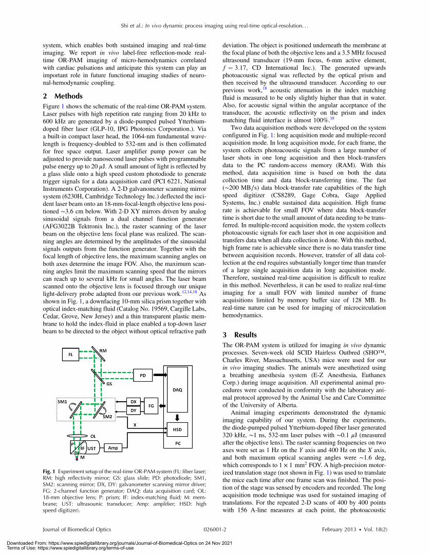
2 MethodsFigure 1 shows the schematic of the real-time OR-PAM system.Laser pulses with high repetition rate ranging from 20 kHz to600 kHz are generated by a diode-pumped pulsed Ytterbium-doped fiber laser (GLP-10, IPG Photonics Corporation.). Viaa built-in compact laser head, the 1064-nm fundamental wave-length is frequency-doubled to 532-nm and is then collimatedfor free space output. Laser amplifier pump power can beadjusted to provide nanosecond laser pulses with programmablepulse energy up to 20 μJ. A small amount of light is reflected bya glass slide onto a high speed custom photodiode to generatetrigger signals for a data acquisition card (PCI 6221, NationalInstruments Corporation). A 2-D galvanometer scanning mirrorsystem (6230H, Cambridge Technology Inc.) deflected the inci-dent laser beam onto an 18-mm-focal-length objective lens posi-tioned ∼3.6 cm below. With 2-D XY mirrors driven by analogsinusoidal signals from a dual channel function generator(AFG3022B Tektronix Inc.), the raster scanning of the laserbeam on the objective lens focal plane was realized. The scan-ning angles are determined by the amplitudes of the sinusoidalsignals outputs from the function generator. Together with thefocal length of objective lens, the maximum scanning angles onboth axes determine the image FOV. Also, the maximum scan-ning angles limit the maximum scanning speed that the mirrorscan reach up to several kHz for small angles. The laser beamscanned onto the objective lens is focused through our uniquelight-delivery probe adapted from our previous work.12,14,18 Asshown in Fig. 1, a downfacing 10-mm silica prism together withoptical index-matching fluid (Catalog No. 19569, Cargille Labs,Cedar, Grove, New Jersey) and a thin transparent plastic mem-brane to hold the index-fluid in place enabled a top-down laserbeam to be directed to the object without optical refractive path
deviation. The object is positioned underneath the membrane atthe focal plane of both the objective lens and a 3.5 MHz focusedultrasound transducer (19-mm focus, 6-mm active element,f ¼ 3.17, CD International Inc.). The generated upwardsphotoacoustic signal was reflected by the optical prism andthen received by the ultrasound transducer. According to ourprevious work,18 acoustic attenuation in the index matchingfluid is measured to be only slightly higher than that in water.Also, for acoustic signal within the angular acceptance of thetransducer, the acoustic reflectivity on the prism and indexmatching fluid interface is almost 100%.18
Two data acquisition methods were developed on the systemconfigured in Fig. 1: long acquisition mode and multiple-recordacquisition mode. In long acquisition mode, for each frame, thesystem collects photoacoustic signals from a large number oflaser shots in one long acquisition and then block-transfersdata to the PC random-access memory (RAM). With thismethod, data acquisition time is based on both the datacollection time and data block-transferring time. The fast(∼200 MB∕s) data block-transfer rate capabilities of the highspeed digitizer (CS8289, Gage Cobra, Gage AppliedSystems, Inc.) enable sustained data acquisition. High framerate is achievable for small FOV where data block-transfertime is short due to the small amount of data needing to be trans-ferred. In multiple-record acquisition mode, the system collectsphotoacoustic signals for each laser shot in one acquisition andtransfers data when all data collection is done. With this method,high frame rate is achievable since there is no data transfer timebetween acquisition records. However, transfer of all data col-lection at the end requires substantially longer time than transferof a large single acquisition data in long acquisition mode.Therefore, sustained real-time acquisition is difficult to realizein this method. Nevertheless, it can be used to realize real-timeimaging for a small FOV with limited number of frameacquisitions limited by memory buffer size of 128 MB. Itsreal-time nature can be used for imaging of microcirculationhemodynamics.
3 ResultsThe OR-PAM system is utilized for imaging in vivo dynamicprocesses. Seven-week old SCID Hairless Outbred (SHO™,Charles River, Massachusetts, USA) mice were used for ourin vivo imaging studies. The animals were anesthetized usinga breathing anesthesia system (E-Z Anesthesia, EuthanexCorp.) during image acquisition. All experimental animal pro-cedures were conducted in conformity with the laboratory ani-mal protocol approved by the Animal Use and Care Committeeof the University of Alberta.
Animal imaging experiments demonstrated the dynamicimaging capability of our system. During the experiments,the diode-pumped pulsed Ytterbium-doped fiber laser generated320 kHz, ∼1 ns, 532-nm laser pulses with ∼0.1 μJ (measuredafter the objective lens). The raster scanning frequencies on twoaxes were set as 1 Hz on the Y axis and 400 Hz on the X axis,and both maximum optical scanning angles were ∼1.6 deg,which corresponds to 1 × 1 mm2 FOV. A high-precision motor-ized translation stage (not shown in Fig. 1) was used to translatethe mice each time after one frame scan was finished. The posi-tion of the stage was sensed by encoders and recorded. The longacquisition mode technique was used for sustained imaging oftranslations. For the repeated 2-D scans of 400 by 400 pointswith 156 A-line measures at each point, the photoacoustic
Fig. 1 Experiment setup of the real-time OR-PAM system (FL: fiber laser;RM: high reflectivity mirror; GS: glass slide; PD: photodiode; SM1,SM2: scanning mirror; DX, DY: galvanometer scanning mirror driver;FG: 2-channel function generator; DAQ: data acquisition card; OL:18-mm objective lens; P: prism; IF: index-matching fluid; M: mem-brane; UST: ultrasounic transducer; Amp: amplifier; HSD: highspeed digitizer).
Journal of Biomedical Optics 026001-2 February 2013 • Vol. 18(2)
Shi et al.: In vivo dynamic process imaging using real-time optical-resolution. . .
Downloaded From: https://www.spiedigitallibrary.org/journals/Journal-of-Biomedical-Optics on 24 Nov 2021Terms of Use: https://www.spiedigitallibrary.org/terms-of-use

data collection time was 0.5 s. Block-data transfer time was aslightly over 0.5 s, resulting in a frame rate of 0.5 fps, whichmeans 1D A-scan rate of 80,000 Hz. Figure 2(a) shows amaximum-amplitude-projection (MAP) snapshot of microvas-culature in an SCID mouse ear to demonstrate the imagingcapability of our system. It clearly depicts the vessels includingsmall capillaries. This is consistent with previous resolutionstudies using our OR-PAM system with ∼6-μm resolution.12,14
Figure 2(b) shows a snapshot taken from a movie of microvas-culature MAP images in an SCID mouse ear as well but with themouse under 6 μm per step vertical translation for focusing.Vessels including capillaries are visualized. The correspondingmovie is displayed at 2 fps in Video 1. It includes a total of41-frame 1 × 1 mm2 images acquired at 0.5 fps. Some smallcapillaries show up and disappear in the video as the mouseis translated.
In our studies on imaging dynamic processes of micro-hemodynamics, the diode-pumped pulsed Ytterbium-dopedfiber laser generated 300-kHz, ∼1-ns, 532-nm laser pulseswith ∼0.1 μJ. Raster scanning frequencies of the fast-scanningmirrors were set as 15 Hz for the Y axis and 1.5 kHz for the Xaxis. Therefore, the data collection rate can reach 30 fps (two
times the slow axis scan rate). Also, due to the limitation of thescanning galvanometer mirror system, the maximum opticalscanning angles of both mirrors are set as ∼0.4 deg. The result-ing imaging area was 250 × 250 μm2. The mice were kept sta-tionary during the experiment. Multirecord acquisition modewas adapted as the data acquisition method to reach a frame-rate of 30 fps over a limited number of frame acquisitionswith 156 A-line measures at each point for 100 by 100 repeated2-D scans. The corresponding 1D A-scan rate was 300,000 Hz.Figure 3(a) shows a single frame from a movie representingMAP images of microvasculature in an SCID mouse ear.The corresponding movie (Video 2) is composed of 42-frame250 × 250 μm2 images taken at a frame rate of 30 fps. A peri-odic variation in photoacoustic intensity was observed, whichwe hypothesize is the flow of blood cells through the vesselsdue to cardiac cycle-induced pulse waves propagating througharterioles. We examined the photoacoustic signal amplitude overtime within the dashed box region A indicated in Fig. 3(a) byaveraging the intensity as seen in Fig. 3(b). The intensity varia-tion is about 35% with ∼5 peaks clearly depicted in ∼1.4 s,which is consistent with the heart rate of an anaesthetizedmouse measured independently by a pulse-oximeter. For com-parison, we examined the photoacoustic signal amplitude overtime within the dashed box region B and C indicated in Fig. 3(a)respectively, while region C did not include visible blood ves-sels. The intensity variation for B is similar to that of A, whilethe intensity variation for C is within 5%, much less comparedwith studies on region A and B. In addition, the pulse energyinstability of the diode-pumped pulsed Ytterbium-doped fiberlaser used here is within 1%.
4 DiscussionIn vivo images of microvasculature in SCID mouse ears undertranslations, with an average pixel separation of 2.5 μm withinan FOVof 1 × 1 mm2, were obtained at a frame-rate of 0.5 fps.Higher frame rate with sustained imaging is achievable forsmaller FOV (∼7 fps for a FOV of 250 × 250 μm). The stepsize for the vertical translation in Fig. 2 is 6 μm. So for 41frames, it moved ∼240 μm in total. Some small vessels includ-ing capillaries are seen to come into and out of focus with ver-tical translation. However, this is less evident for larger vessels.One explanation of this is that for targets larger than the spotsize, with OR-PAM (which, like conventional microscopyuses one-way focusing) the photoacoustic axial depth of field
Fig. 2 (a) An in vivo image of micro-vasculatures obtained from a hair-less SCID mouse ear to demonstrate the imaging capability of ourimaging system; (b) A snapshot from the in vivo movie composed of41 frame 1 mm × 1 mm images of microvasculature in an SCID hairlessmouse ear when the mouse was in a vertical movement; (Video 1,MPEG, 4.0 MB) [URL: http://dx.doi.org/10.1117/1.JBO.18.2.026001.1]displays at 2 fps while the actual system frame rate is about 0.5 fps;some small capillaries come out and disappear during the verticaltranslation.
Fig. 3 (a) A snapshot from the in vivomovie composed of 42 frame 250 × 250 μm2 images of microvasculature in an SCID hairless mouse ear acquiredat 30 fps; (Video 2, MPEG, 412 KB) [URL: http://dx.doi.org/10.1117/1.JBO.18.2.026001.2] displays at 15 fps, while microvasculature motions andblood intensity changes are clear to see. (b) Mean photoacoustic signal amplitude inside the dash box A indicated area in (a) versus time. (c) Meanphotoacoustic signal amplitude inside the dash box B and C indicated area in (a) respectively versus time.
Journal of Biomedical Optics 026001-3 February 2013 • Vol. 18(2)
Shi et al.: In vivo dynamic process imaging using real-time optical-resolution. . .
Downloaded From: https://www.spiedigitallibrary.org/journals/Journal-of-Biomedical-Optics on 24 Nov 2021Terms of Use: https://www.spiedigitallibrary.org/terms-of-use

is effectively much larger than the optical depth of field of thefocused beam (estimated as ∼35 μm).19 This, coupled with pooraxial resolution (∼500 μm) leads to lack of clear axial focusing/defocusing for large targets. The sustained high resolution fastimaging demonstrated the capability of our system to be used forreal-time focusing and positioning. Near real-time positioningwill permit panning over large areas with mm FOV windowsand then locating regions of interest for further studies.
Video 2 is the label-free reflection-mode OR-PAM demon-stration of imaging micro-hemodynamics. One observation,already noted, is the intensity variations that correlate with car-diac cycle. This may in part be explained by surges of bloodperiodically surging through arterioles due to cardiac-inducedpulse-waves. Another observation is that some vascular branchesseem to have negligible flow then seemingly random transientsurges. These effects deserve additional study and could bedue to microhemodynamic regulatory mechanisms. The effectsseen can be discounted as artifacts, however, because laser pulse-to-pulse intensity variation is less than 1% and scanning of sta-tionary phantom structures shows no motion. It is noteworthy topoint out that optical coherence tomography can provide excel-lent structural images of microvasculature.20,21 However, to date,these systems have not been able to provide video-rate imagingof comparable tissue volumes due to the necessity of requiringmultiple A-scan lines to extract motion of scatterers. Intravitalmicroscopy is a powerful technique that has been capable ofstudying microhemodynamics.22 However, this technique re-quires surgical exposure of thin transparent membranes and isnot suitable for noninvasive reflection-mode imaging.
The unique capabilities of our system may prove importantfor imaging cortical hemodynamics in functional brain mappingstudies. Real-time focusing and positioning should facilitatetranslation of the technique to clinical settings. While FOV islimited in our real-time scanning, mosaicing of small patchesshould provide larger-FOV images, as described by our recentwork.23
For 2-D laser scanning OR-PAM systems using a focusedtransducer, the FOV is limited by the focal width of the ultra-sonic transducer. Therefore, we used a low frequency (3.5 MHz)ultrasonic transducer to achieve a large FOV. The axial resolu-tion of the 3.5 MHz ultrasonic transducer can be calculated asnear 500 μm, sacrifice of axial resolution is tolerable for MAPimages. A future design could match the receiver bandwidth tothe bandwidth of the high-frequency photoacoustic signals toimprove signal-noise-ratio (SNR) at the expense of FOV.
In our in vivo studies, given that the optical focus is ∼150 μmbeneath the tissue surface, with an objective lense NA of 0.15,the surface spot size is ∼45 μm in diameter, and the calculatedsurface laser fluence is ∼5 mJ∕cm2, below the single pulse limitof 20 mJ∕cm2 set by the American National Standards Institute(ANSI).24 The spatial peak optical fluence at the focus in wateris ∼500 mJ∕cm2, which is still less than the damage thresholdobserved in small animals.25 In our work, light delivery is con-fined to a localized area, and no tissue damage is visible afterimaging.
In addition, for an average pixel separation of 2.5 μm, thereare on average N ¼ 45 μm∕2.5 μm ∼18 adjacent laser pulsesoverlapping on the skin surface. For 320 kHz laser PRR, theexposure time is t ∼ 56 μs, so the MPE for a pulse train isMPETrain ¼ 1.1CAt0.25 ¼ 95 mJ∕cm2, where CA is a wave-length-correction factor equal to unity for 400 to 700 nm wave-lengths. The average power limit set by ANSI is calculated as
MPEAverage ¼ MPETrain∕N ∼ 5 mJ∕cm2, which is our estimatedfluence per pulse at the skin surface. This means that we areessentially at our theoretical upper limit for pulse-repetitionrate; however, the repetition rate could be increased if pulse-energy can be lowered and SNR improved. In future studies,careful selection of focusing, repetition rate, and pulse energyparameters must be considered to avoid exceeding ANSI limits.On the other hand, for some preclinical applications, exceedingthese limits may be acceptable.
5 ConclusionWe have demonstrated an OR-PAM system capable of near real-time sustained imaging to aid focusing and positioning, and areal-time frame-limited mode capable of imaging micro-circu-lation pulsatile hemodynamics. This OR-PAM system enabledlabel-free reflection-mode imaging of micro-hemodynamics atreal-time rates. The fast acquisition capabilities of the systemmay pave the way for clinical adaptation and preclinical studiessuch as functional brain imaging.
AcknowledgmentsWe gratefully acknowledge funding from NSERC (355544-2008, 375340-2009, STPGP 396444), Terry-Fox Foundationand the Canadian Cancer Society (TFF 019237, TFF 019240,CCS 2011-700718), the Alberta Cancer Research Institute(ACB 23728), the Canada Foundation for Innovation,Leaders Opportunity Fund (18472), Alberta AdvancedEducation & Technology, Small Equipment Grants Program(URSI09007SEG), Microsystems Technology ResearchInitiative (MSTRI RES0003166), University of AlbertaStartup Funds, Alberta Ingenuity/Alberta Innovates scholarshipsfor graduate and undergraduate students, and CanadianFederation of University Women Edmonton Margaret BrineGraduate Scholarships for Women.
References1. K. Maslov et al., “Optical-resolution photoacoustic microscopy for in
vivo imaging of single capillaries,” Opt. Lett. 33(9), 929–931 (2008).2. S. Hu, K. Maslov, and L. V. Wang, “Noninvasive label-free imaging of
microhemodynamics by optical-resolution photoacoustic microscopy,”Opt. Express 17(9), 7688–7693 (2009).
3. S. Hu, K. Maslov, and L. V. Wang, “In vivo functional chronic imagingof a small animal model using optical-resolution photoacoustic micros-copy,” Med. Phys. 36(6), 2320–2323 (2009).
4. L. Song, K. Maslov, and L. V. Wang, “Multifocal optical-resolutionphotoacoustic microscopy in vivo,” Opt. Lett. 36(7), 1236–1237 (2011).
5. J. Yao et al., “In vivo photoacoustic imaging of transverse blood flowusing Doppler broadening of bandwidth,” Opt. Lett. 35(9), 1419–1421(2010).
6. S. Hu et al., “Functional transcranial brain imaging by optical-resolutionphotoacoustic microscopy,” J. Biomed. Opt. 14(4), 040503 (2009).
7. S. Hu et al., “Intravital imaging of amyloid plaques in a transgenicmouse model using optical-resolution photoacoustic microscopy,”Opt. Lett. 34(24), 3899–3901 (2009)
8. S. Oladipupo et al., “Conditional HIF-1 induction produces multistageneovascularization with stage-specific sensitivity to VEGFR inhibitorsand myeloid cell independence,” Blood 117(15), 4142–4153 (2011).
9. S. Oladipupo et al., “VEGF is essential for hypoxia-inducible factormediated neovascularization but dispensable for endothelial sprouting,”PNAS 108(32), 13264–13269 (2011).
10. S. Hu, K. Maslov, and L. V. Wang, “Second-generation optical-resolu-tion photoacoustic microscopy with improved sensitivity and speed,”Opt. Lett. 36(7), 1134–1136 (2011).
11. Z. X. Xie et al., “Laser-scanning optical-resolution photoacousticmicroscopy,” Opt. Lett. 34(12), 1771–1773 (2009).
Journal of Biomedical Optics 026001-4 February 2013 • Vol. 18(2)
Shi et al.: In vivo dynamic process imaging using real-time optical-resolution. . .
Downloaded From: https://www.spiedigitallibrary.org/journals/Journal-of-Biomedical-Optics on 24 Nov 2021Terms of Use: https://www.spiedigitallibrary.org/terms-of-use

12. W. Shi et al., “Optical resolution photoacoustic microscopy using novelhigh-repetition-rate passively Q-switched microchip and fiber lasers,”J. Biomed. Opt. 15(5), 056017 (2010).
13. Y. Wang et al., “Fiber-laser-based photoacoustic microscopy andmelanoma cell detection,” J. Biomed. Opt. 16(1), 011014 (2011).
14. W. Shi et al., “In vivo near-realtime volumetric optical-resolution photo-acoustic microscopy using a high-repetition-rate nanosecond fiber-laser,” Opt. Express 19(18), 17143–17150 (2011).
15. B. Rao et al., “Real-time four-dimensional optical-resolution photo-acoustic microscopy with Au nanoparticle-assisted subdiffraction-limit resolution,” Opt. Lett. 36(7), 1137–1139 (2011).
16. C. Yeh et al., “Photoacoustic microscopy of blood pulse wave,”J. Biomed. Opt. 17(7), 070504 (2012)
17. J. Yao et al., “Wide-field fast-scanning photoacoustic microscopy basedon a water-immersible MEMS scanning mirror,” J. Biomed. Opt. 17(8),080505 (2012)
18. J. C. Ranasinghesagara et al., “Photoacoustic technique for assessingoptical scattering properties of turbid media,” J. Biomed. Opt. 14(4),040504 (2009).
19. L. V. Wang and H. Wu, Biomedical Optics: Principles and Imaging,John Wiley & Sons Inc., Hoboken, New Jersey (2007).
20. A. Mariampillai et al., “ Optimized speckle variance OCT imaging ofmicrovasculature,” Opt. Lett. 35(8), 1257–1259 (2010).
21. G. Liu et al., “High-resolution imaging of microvasculature in humanskin in-vivo with optical coherence tomography,” Opt. Express 20(7),7694–7705 (2012).
22. A. M. Iga et al., “Quantitating therapeutic disruption of tumorblood flow with intravital video microscopy,” Cancer Res. 66(24),11517–11519 (2006).
23. P. Shao et al., “Mosaicing acquisition and processing for opticalresolution photoacoustic microscopy,” J. Biomed. Opt. 17(8), 080503(2012).
24. Laser Institute of America, American National Standard for Safe Use ofLasers ANSI Z136.1-2007, American National Standards Institute, Inc.(2007).
25. V. P. Zharov et al., “In vivo photoacoustic flow cytometry formonitoringof circulating single cancer cells and contrast agents,” Opt. Lett. 31(24),3623–3625 (2006).
Journal of Biomedical Optics 026001-5 February 2013 • Vol. 18(2)
Shi et al.: In vivo dynamic process imaging using real-time optical-resolution. . .
Downloaded From: https://www.spiedigitallibrary.org/journals/Journal-of-Biomedical-Optics on 24 Nov 2021Terms of Use: https://www.spiedigitallibrary.org/terms-of-use


![In vivo imaging of swimming micromotors using hybrid high … · 2020. 6. 15. · optical-ultrasound imaging technique, also called photoacoustic imaging (PAI).[28–32] PAI is ...](https://static.fdocuments.net/doc/165x107/6092d60f6674c8570e70cd4e/in-vivo-imaging-of-swimming-micromotors-using-hybrid-high-2020-6-15-optical-ultrasound.jpg)


