IN VIVO ANTIPLASMODIAL AND EFFECTS OF SUBCHRONIC ... · Sulaiman S. Rukayyah 1 ⃰, Ali A. Jigam 2...
15
International Research Journal of Natural Sciences Vol.3, No.2, pp.1-15, June 2015 Published by European Centre for Research Training and Development UK (www.eajournals.org) 1 ISSN 2053-4108(Print), ISSN 2053-4116(Online) IN VIVO ANTIPLASMODIAL AND EFFECTS OF SUBCHRONIC ADMINISTRATION OF TRICHILIA EMETICA LEAVES EXTRACTS Sulaiman S. Rukayyah 1 ⃰ , Ali A. Jigam 2 and Mohammed T. Aisha 1 1 Department of Biochemistry, Ibrahim Badamasi Babangida University, Lapai, Nigeria. 2 Department of Biochemistry, Federal University of Technology, Minna, Nigeria ABSRACT: Objective: The leave extracts of Trichilia emetica were investigated for antiplasmodial activity against Plasmodium berghei infections in mice and chronic dose effects of the methanolic extract were also studied. Methods: The plant leaves were successively extracted into three (3) extract forms (Hexane, ethylacetate and methanolic extracts). Plasmodium berghei (NK 65 Chloroquine sensitive strain) was inoculated in to twenty mice assigned for 5 groups of 4 mice each. Group I, II and III were treated with 300mg/Kg bw hexane, ethylacetate and methanol extracts respectively. Group IV with 5mg/kg bw chloroquine phosphate (standard) and group V with 20ml/kg bw normal saline (control). Another set of 40 mice were divided into two groups of twenty each (test and control) and some serum parameters were studies. The test animals were gavaged with 300mg/kg bw extract while controls were given normal saline over a period of 5weeks on alternate days. Histology of the liver, and kidney were carried out. Results: The presence of alkaloids, saponins, pholobatannins, flavonoids, cardiac glycosides and phenolic compounds have been detected and quantified. T. emetica extracts of hexane and methanol suppressed parasitemia in mice by 79.19 % and 95.83%respectively while ethylacetate extract has no activity. The weight of the test group was on a continuous decrease compare to the control while the reverse was the case in terms of the PCV. Glucose, total proteins, triacylglycerides, ALT and ALP levels all decrease significantly compared to the control group. AST level of the test group was significantly higher compare to the control. Histology revealed no damage to the kidney and liver. Conclusion: Hexane and methanolic extracts of Trichilia emetica have strong efficacy against malaria and a possible mechanism for this efficacy is its ability to lyses erythrocytes. KEYWORDS: Plasmodium berghei, curative, phytochemicals, spectrophotometer, serum. INTRODUCTION Malaria is the most important eukaryotic parasitic disease, threatening the livelihood of over 3.3billion people [1]. Efforts to disrupt the life cycle of the parasite by controlling the vector have had only limited success, while the usefulness of antimalaria drug is hampered by their lack of availability to those most in need and rapid evolution of drug resistant parasites. An effective, safe vaccine remains the most promising approach to controlling the disease [2]. Immense benefits have been derived by man from using medicinal herbs in disease management because they are relatively safer, more affordable and sometimes offer better therapeutic value than synthetic drugs [3]. Therapeutic properties ascribed to most of these herbs are linked to the phytochemical compounds contained in them. Phytochemicals such as alkaloids, glycosides, phenols, saponins,
Transcript of IN VIVO ANTIPLASMODIAL AND EFFECTS OF SUBCHRONIC ... · Sulaiman S. Rukayyah 1 ⃰, Ali A. Jigam 2...
International Research Journal of Natural Sciences
Vol.3, No.2, pp.1-15, June 2015
Published by European Centre for Research Training and Development UK (www.eajournals.org)
1 ISSN 2053-4108(Print), ISSN 2053-4116(Online)
IN VIVO ANTIPLASMODIAL AND EFFECTS OF SUBCHRONIC ADMINISTRATION
OF TRICHILIA EMETICA LEAVES EXTRACTS
Sulaiman S. Rukayyah 1 ,⃰ Ali A. Jigam 2 and Mohammed T. Aisha 1
1Department of Biochemistry, Ibrahim Badamasi Babangida University, Lapai, Nigeria. 2Department of Biochemistry, Federal University of Technology, Minna, Nigeria
ABSRACT: Objective: The leave extracts of Trichilia emetica were investigated for
antiplasmodial activity against Plasmodium berghei infections in mice and chronic dose effects of
the methanolic extract were also studied. Methods: The plant leaves were successively extracted
into three (3) extract forms (Hexane, ethylacetate and methanolic extracts). Plasmodium berghei
(NK 65 Chloroquine sensitive strain) was inoculated in to twenty mice assigned for 5 groups of 4
mice each. Group I, II and III were treated with 300mg/Kg bw hexane, ethylacetate and methanol
extracts respectively. Group IV with 5mg/kg bw chloroquine phosphate (standard) and group V
with 20ml/kg bw normal saline (control). Another set of 40 mice were divided into two groups of
twenty each (test and control) and some serum parameters were studies. The test animals were
gavaged with 300mg/kg bw extract while controls were given normal saline over a period of
5weeks on alternate days. Histology of the liver, and kidney were carried out. Results: The
presence of alkaloids, saponins, pholobatannins, flavonoids, cardiac glycosides and phenolic
compounds have been detected and quantified. T. emetica extracts of hexane and methanol
suppressed parasitemia in mice by 79.19 % and 95.83%respectively while ethylacetate extract has
no activity. The weight of the test group was on a continuous decrease compare to the control
while the reverse was the case in terms of the PCV. Glucose, total proteins, triacylglycerides, ALT
and ALP levels all decrease significantly compared to the control group. AST level of the test
group was significantly higher compare to the control. Histology revealed no damage to the
kidney and liver. Conclusion: Hexane and methanolic extracts of Trichilia emetica have strong
efficacy against malaria and a possible mechanism for this efficacy is its ability to lyses
erythrocytes.
KEYWORDS: Plasmodium berghei, curative, phytochemicals, spectrophotometer, serum.
INTRODUCTION
Malaria is the most important eukaryotic parasitic disease, threatening the livelihood of over
3.3billion people [1]. Efforts to disrupt the life cycle of the parasite by controlling the vector have
had only limited success, while the usefulness of antimalaria drug is hampered by their lack of
availability to those most in need and rapid evolution of drug resistant parasites. An effective, safe
vaccine remains the most promising approach to controlling the disease [2]. Immense benefits
have been derived by man from using medicinal herbs in disease management because they are
relatively safer, more affordable and sometimes offer better therapeutic value than synthetic drugs
[3]. Therapeutic properties ascribed to most of these herbs are linked to the phytochemical
compounds contained in them. Phytochemicals such as alkaloids, glycosides, phenols, saponins,

International Research Journal of Natural Sciences
Vol.3, No.2, pp.1-15, June 2015
Published by European Centre for Research Training and Development UK (www.eajournals.org)
2 ISSN 2053-4108(Print), ISSN 2053-4116(Online)
titerpenoids, flavonoids, etc have been suggested to possess antimalaria properties [4,5]. The
increase in discovery of more medicinal plants has demanded for increased scientific scrutiny of
their bioactivity so as to provide data that will help physician and patients make wise decision
before using them [3].
Trichilia emetica is a tree native to Savannah belt and open woodland of Africa [6]. It has many
different traditional uses [7] including treatment of convulsion, fever, jaundice, cold, epilepsies,
scabies, pneumonia and also as purgative, diuretic agents [6]. Some of these claims have been
buttress by scientific proves [8, 9, 10, 11, 12]. One major and overriding criterion in the selection
of herbal medicines for use in health services is safety. Plant extracts should not only be efficacious
but safe for consumption. Therefore, closely associated with screening of plant extracts for their
activities against microorganism or disease conditions is the need to know their toxic potentials
[13]. Although there has been earlier reports of its in vitro antiplasmodial activity but in vivo
antiplasmodial has not been found in literature. A plant with high in vitro activity many have no
in vivo activity and vice versa [14]. Therefore considering its wide ethnomedicine uses and broad
spectrum activity against microbes there is the need also to determine the effect of Trichilia
emetica on some vital body organs and key enzyme markers.
MATERIALS AND METHOD
Plant Materials
Fresh leaves of Trichilia emetica were collected in Bida Local Government Area of Niger State of
Nigeria between July and September, after identification by herbal practitioner and authentitication
at the Biological Sciences Department Federal University of Technology, Minna. These leaves
were air dried at room temperature and pounded into powdered form using pestle and mortar. The
powder was packaged in an air tight container, labelled and stored until analysed.
Preparation of Crude Extracts
Eighty grams of air dried sample Trichilia emetica was extracted exhaustively (48hours) in the
cold sequentially with two liters each of n-hexane, ethylacetate and methanol (Sigma-Aldrich
Europe) in that order separately. The marc was filtered with muslin cloth and solvent removed
under reduced pressure in a rotary evaporator. Each of the paste was poured into beaker and placed
on a water bath for complete evaporation of the organic solvent. The green pastes were weighed
and labelled prior to further analysis
Animals
Healthy Swiss albino mice of either sex of about 7weeks old weighing between 20-30g were
obtained from Department of Pharmacology, Faculty of Pharmaceutical Sciences, Ahmadu Bello
University, Zaria, Nigeria were used for the experiments. The mice were conveniently housed
under standard environmental conditions, temperature 27 + 20C, and 70% relative humidity, free
access to commercial food pellets, water and natural 12hours day light/night cycles. The
experiments were conducted in strict compliance with internationally accepted principles for
laboratory animals’ use and care as contained in the Canadian Council on Animal Care Guidelines
and Protocol Review [15].

International Research Journal of Natural Sciences
Vol.3, No.2, pp.1-15, June 2015
Published by European Centre for Research Training and Development UK (www.eajournals.org)
3 ISSN 2053-4108(Print), ISSN 2053-4116(Online)
Parasites
Plasmodium berghei Nk65 chloroquine sensitive strain was obtained from Nigerian Institute of
Medical Research (NIMR) Lagos, Nigeria and maintained in the laboratory by serial passage in
mice.
Phytochemical Screening
Phytochemical screening was carried out with aqueous extract and pulverized sample using
standard procedures as described by Edeoga et al. (2005), Akinyemi et al. (2005) and Kwada and
Tella (2009)[16,17,18]. Tannins, phlobatannins, saponins, and flavonoids were screened according
to the methods described by Edeoga et al. (2005) [16]. Cardiac glycosides (Keller-killani Test)
and alkaloids were screened according to the methods described by Akinyemi et al. ( 2005)[17].
Harbone’s (1973) Method of alkaloid determination was also used. Five grams of the samples were
weighed into 250ml beaker and 200ml of 10% acetic acid in ethanol was added and covered and
allowed to stand for 4 hours. This was filtered and the extract was concentrated on a water bath at
900C to one-quarter of the original volume. Concentrated ammonium hydroxide was added drop
wise to the extract until the precipitation was complete. The whole solution was allowed to settle
and the precipitate was collected and washed with dilute ammonium hydroxide and then filtered.
The residue is the alkaloid, which was dried and weighed [16]
Quantitative determination of flavonoids. Ten grams of the pulverized stem bark was extracted
repeatedly with 100ml of 80% aqueous methanol at room temperature. The whole solution was
filtered through whatman filter paper No 42 (125mm). The filtrate was transferred into evaporating
dish and placed on a water bath at 400C until a constant weight was maintained [18].The Obadani
and Ochuka (2001) method was used in saponin determination. Twenty grams of each plant sample
was weighed into a conical flask and 100cm3 of 20% aqueous ethanol were added. The samples
were heated over a hot water bath for 4 hour with continuous stirring at 550C. The mixture was
filtered and the residue re-extracted with another 200ml 20% ethanol. The combined extracts were
reduced to 40ml over water bath at about 900C. The concentrate was transferred into a 250ml
seperatory funnel and 20ml of diethyl ether was added and shaken vigorously. The aqueous layer
recovered while the ether layer was discarded. The purification process was repeated. 50ml of n-
butanol extracts were washed twice with 10ml of 5% aqueous sodium chloride. The remaining
solution was heated on a water bath at 400C. After evaporation the samples were dried in the oven
to a constant weight, the saponin content was calculated as a percentage of the sample used [16]
Total phenol was determined using Edeoga et al (2005) method. Two grams of the plant sample
was defatted with 100ml of diethyl ether using a soxhlet apparatus for 2 hours. The fat free sample
was boiled with 50ml of ether for 15 minutes for the extraction of the phenolic component. Five
millilitre of the extract was pipetted into a 50ml flask, then 10ml of distilled water was added. Two
millilitre of ammonium hydroxide solution and 5ml of concentrated amyl alcohol were also added.
The samples were made up to mark and left to react for 30 minutes for colour development. This
was measured at 505nm [16].
Quantitative determination of Cardiac glycosides: The quantity of glycosides in the pulverized
sample was evaluated using Baljet’s reagent (95ml 1% picric acid and 5ml 10% NaOH) as
Oluwaniyi et al. (2007). Digitalis cardiac glycosides develop an orange-red colour with Baljet’s
reagent. The intensity (absorbance) of the colour produced is proportional to the concentration of
International Research Journal of Natural Sciences
Vol.3, No.2, pp.1-15, June 2015
Published by European Centre for Research Training and Development UK (www.eajournals.org)
4 ISSN 2053-4108(Print), ISSN 2053-4116(Online)
cardiac glycosides. This colour formation is made use of for the quantitative estimation of cardiac
glycosides present in B. aegyptiaca. One gram of sample was extracted by soaking overnight with
10ml of 70% alcohol and filtered. The extracts were then purified using lead acetate and Na2HPO4
solutions before the addition of freshly prepared Baljet’s reagent. The intensity (absorbance) of
the colour produced was measured using a spectrophotometer at 495nm. A blank was carried out
at the same time using distilled water and Baljet’s reagent. The absorbance of the colour produced
is proportional to the concentration of the glycoside. A 0.02% solution of digitoxin solution was
prepared in chloroform-methanol (1:1 v/v) mixture (1ml = 0.2mg). Different volumes, viz. 1, 2, 3,
4 and 5ml (equivalent to 0.2, 0.4, 0.6, 0.8 and 1mg of digitoxin respectively) of the solution were
transferred, each into a dry Erlynmeyer flask. The solvent in each flask was evaporated on a water
bath and dissolved in 0.35ml 90% alcohol. Ten ml of distilled water and 10ml of freshly prepared
Baljet’s were added. Absorbances were read and standard curve was plotted. The concentration of
cardiac glycoside in B. aegyptiaca was then extrapolated from the standard curve [19].
Determination of condensed tannins (Proanthocyanidins): The method described by Iqbal et al.
(2001) was followed for the determination of condensed tannins in the extract. The samples were
extracted to quantitatively diffuse the phenolics present in the materials to liquid phase. For the
extraction process, aqueous acetone (70%) was used. Each of the dried (finely ground) sample
(200 mg) was taken in a glass beaker of approximately 25mL capacity. Ten mL of aqueous acetone
(70%) was added and the beaker was suspended in an ultrasonic water bath and subjected to
ultrasonic treatment for 20min at room temperature. The contents of the beaker was then
transferred to centrifuge tubes and subjected to centrifugation for 10 min at approximately 3000 g
at 4°C using a refrigerated centrifuge. The supernatant was collected and kept on ice. The pellet
left in the tube was transferred to the beaker using two portions of 5 mL each of 70% aqueous
acetone and again subjected the contents to ultrasonic treatment for 20 min. The supernatant was
again collected as described above. Butanol–HCl reagent (butanol–HCI 95:5 v/v) was prepared by
mixing 95 mL of n–butanol with 5 mL concentrated HCl (37%). Ferric reagent (2% ferric
ammonium sulfate in 2N HCl) was prepared by dissolving 2.0 g of ferric ammonium sulfate in 2N
HCl (16.6 mL of concentrated HCl was made up to 100 mL with distilled water to make 2N HCl).
The reagents were stored in dark bottles. In a 100 mm x 12 mm glass test tube, 0.5 mL of the tannin
extract diluted with 70% acetone was pipetted. The quantity of acetone was large enough to prevent
the absorbance (550 nm) in the assay from exceeding 0.6. Three mL of the butanol–HCl reagent
and 0.1 mL of the ferric reagent was added to the tubes. The tubes capped with a glass marble were
shaken using a Vortex and then placed on a heating block adjusted at 97 to 100°C for 60 min. After
cooling the tubes, absorbance was recorded at 550 nm. Absorbance of the unheated mixture
(considered as a suitable blank) was subtracted from the absorbance of heated mixture, which was
actual reading at 550 nm to be used for calculation of condensed tannins. Condensed tannins (%
in dry matter) as leucocyanidin equivalent were calculated by the formula:
(A 550 nm x 78.26 x Dilution factor)/(% dry matter)
This formula assumes that the effective E l%, 1 cm, 550 nm of leucocyanidin is 460 [20].
Safe dose and acute toxicity (LD50) Five groups of four mice were used and the animals were given extracts intraperitoneally (i.p) at
doses of 200, 400, 800, 1600, and 3000mg/kg body weight (b/w) respectively. Extracts were
dissolved with 2ml of dimenthylsulphoxide (DMSO) (Sigma Chemical St Louis, MO, USA) and

International Research Journal of Natural Sciences
Vol.3, No.2, pp.1-15, June 2015
Published by European Centre for Research Training and Development UK (www.eajournals.org)
5 ISSN 2053-4108(Print), ISSN 2053-4116(Online)
the volume was made up to 10ml with distilled water. A control group was given normal saline
(0.9% w/v NaCl) at 20ml/Kg bw. Mice were observed over 72 hours for clinical signs and mortality
was recorded. LD50 was obtained as the intercept of % mortality (y-axis) and dosages (x-axis) [21].
Antiplasmodal Screening Mice were pre-screened by microscopy of thin and thick tail tip blood smears. This was necessary
to exclude the possibility of test animals harbouring rodent Plasmodium species.
Curative Test This is a procedure whereby mice are infected and left for 72 hours before treatment with test and
standard drug as in the Rane test or established infection [22]. Twenty albino mice were selected
and divided into five groups. One group served as control, another group as standard and the others
as the test groups for hexane, ethylacetate and methanolic extracts respectively. The mice were
inoculated with 0.2ml of diluted donor blood (Plasmodium berghei approximately 1 x 10⁷ infected
red cells) by the intraperitoneal route. The animals were left for 72hours for the infection to be
established. The control group was given 0.2ml of 0.9w/v of normal saline. 300 mgkg-1 bw day-1
dose was selected for the plant extract, because it was the safe dose. On D3 i.e, after 72 hours of
infection, the plant extracts were administered subcutaneously once daily for 4 days from D3 to
D7. Thick and thin blood smears from the tail blood were examined for parasite every morning.
Chloroquine (5mg/kg bw) was used as standard drug because the parasites are sensitive to this
compound (Chloroquine sensitive strain of Plasmodium berghei) and was hence ran for
comparison.
Evaluation of the medium term effect dosage of crude extract in mice
Forty mice were kept in two groups (A and B) of the twenty each. Group A was used as test and
gavaged with 300mg/Kg bw of the extracts and group B given 20ml/Kg bw normal saline on
alternating days. All animals were monitored for different biochemical parameters at weekly
intervals for five weeks.
Weight of mice were taken with Avery Balance (W and T) Avery Ltd, Birmingham, UK. Packed
Cell Volume (PCV) was determined using the microhaematocrit method [23]. Serum glucose was
determined using Randox Glucose Kit (Cat GL364) based on the Glucose oxidase reaction. The
estimation serum total protein was carryout with Total Protein Randox kit (Cat No. TP 245) on
cupric ions in an alkaline medium, interact with protein peptides bonds resulting in the formation
of a coloured complex compound which is proportional to the amount of protein present in the
sample. Triglyceride was evaluated using Randox Triglycerides kit (Cat No. TR 210). The
triglycerides are determined after enzymatic hydrolysis with lipases. The indicator is a
quinoneimine formed from hydrogen peroxide, 4-aminophonazone and 4-chlorophenol under the
catalytic influence of peroxidase [24]. Estimation of AST and ALT was done using AST Randox
kit (Cat No AS 101 and Cat No. AL 100 respectively) based on method of Marghoob et al (2013)
[25]. The estimation ALP activity was done using Randox ALP kit (Cat. No 542) ALP hydrolyses
colourfulness p-nitrophenyl phosphate (pNPP) producing phosphate and coloured p-nitropenol at
alkaline pH [26].

International Research Journal of Natural Sciences
Vol.3, No.2, pp.1-15, June 2015
Published by European Centre for Research Training and Development UK (www.eajournals.org)
6 ISSN 2053-4108(Print), ISSN 2053-4116(Online)
Histopathological screening of tissues At the end of the treatment period, mice were sacrificed and liver and kidney collected in sterile
saline. Freshly dissected organs from each animal were cut rapidly and fixed in buffered neutral
formalin (10%). The tissues were dehydrate in ascending grades ethanol (70%, 80%, 90%, 95%
and 100%), cleared in 2 changes of Xylene, impregnated with 2 changes of molten paraffin and
finally embedded in wax. Tissue sections of 4-5µm in thickness were cut with a microtome and
stained with hematoxylin and eosin [27].
Statistical Analysis Results are expressed as means ± standard error of the mean. While Analysis of variance
(ANOVA) coupled with Duncan Multiple Range Test (DMRT) was used to test for the significant
differences between groups for Antiplasmodial bioassay, paired sample t-test was used to test for
significant differences for the biochemical parameters, PCV, mean weight change and enzyme
assay data using Statistical Package for Social Sciences (SPSS) version 16.0. A value of P<0.05
was accepted as significant.
RESULTS
The extract yields of Trichilia emetica obtained with different solvents are in the order: methanol
> hexane > ethyl acetate 10.48, 6.00 and 2.42% respectively.
Phytochemical Screening
Cardiac glycosides, pholobatannins, flavonoids, saponins and alkaloids were detected during
phytochemical screening with total phenols been most abundant (64.71%).
Table 1.0 Phytochemical contents of T. emetica and B. aegyptiaca extracts
Phytochemicals
Test T.emetica
Tannins
0.1% FeCl
%
Bromine H2O -
Pholobatannins Lead acetate -
1% HCl +
Saponins Frothing + 13.40
Emulsion +
Flavonoids Dilute NH3/Conc H2SO4 - 32.20
Lead Acetate +
Dilute NaOH -
Cardiac glycosides Keller-killeni +
Alkaloids Mayer’s test + 19.40
Total phenol Spectrophotometric test 64.71
+= present, - = absent

International Research Journal of Natural Sciences
Vol.3, No.2, pp.1-15, June 2015
Published by European Centre for Research Training and Development UK (www.eajournals.org)
7 ISSN 2053-4108(Print), ISSN 2053-4116(Online)
Table 2.0 Results of safe dose determination (Pre LD50) for Trichilia emetica
Dose Observations Mortality (mg/kg
bw. ip.)
200 Animal appears normal 0/4
400 No observable Changes 0/4
800 Initial restlessness but normal 0/4
1600 Slow activity, laboured breathing 0/4
3000 Somnolence with a single mortality 1/4
Selected dose = 300mg/kg bw
The suppression of parasitaemia in mice treated with different fractions T. emetica of is in figure
1.0. Ethylacetate fraction of T. emetica is not effective in the treatment of malaria, because the
mice in this particular group died before the control group. Hexane and methanolic fraction of T.
emetica, show promising effect on malaria parasite, with the methanolic fraction having higher
activity than the hexane extract with parasite suppression of 95.83% and 79.17% respectively.The
methanolic extract of Trichilia emetica completely clear B. berghei from circulation in the infected
mice after 23 days, the cured mice remained aparasitemic for over 60 days.
Figure 1.0: Suppression of parasitaemia in mice treated with different fractions of T. emetica
Weight Variations
The whole body weights of mice (Fig.2.0) administered T. emetica exhibited a drastic decrease in
week one (1) and two (2), with a minimal increase to week. However the weight decreased steadily
-50
0
50
100
150
200
250
3 4 6 8 10 12 14 16
Pa
rasi
tes
per
mic
roa
sco
pe
fiel
d
Period(Days)
CQ
NS
ee
he
me

International Research Journal of Natural Sciences
Vol.3, No.2, pp.1-15, June 2015
Published by European Centre for Research Training and Development UK (www.eajournals.org)
8 ISSN 2053-4108(Print), ISSN 2053-4116(Online)
after week three (3). The weight of control mice continue to increase from week one (1) to week
five (5). The results of fresh organ weights of mice expressed as percentage of whole body weights
are in Table 2.0. Only the stomach of the test showed some variations. The other organs of the test
mice were comparable in weight with those of the control.
Figure 2.0: Variations in weight (g) of mice dosed with T. emetica.
Table 3.0 Results of fresh organ weights of mice dosed with T. emetica extract
Organs Control Test
Liver 4.55 4.69
Kidney 1.27 1.25
Intestine 11.32 11.30
Spleen 0.39 0.45
Stomach 2.34 2.68*
* means the sample result is significantly different at 5% (p<0.05) across the row.
Packed Cell Volume
Variations in Packed Cell Volume in mice chronically dosed with T. emetica and the control is
represented in figure 3.0. The PCV of T. emetica were progressively decreasing from week one to
18
19
20
21
22
23
24
25
26
27
28
1 2 3 4 5
We
igh
t (g
)
Period (weeks)
Control
T.emetica

International Research Journal of Natural Sciences
Vol.3, No.2, pp.1-15, June 2015
Published by European Centre for Research Training and Development UK (www.eajournals.org)
9 ISSN 2053-4108(Print), ISSN 2053-4116(Online)
three, with a peak at week four and a decline afterward while the PCV for the control was
increasing in all the weeks
Figure 3.0: Variations in packed cell volume in mice dosed with T. emetica.
26.5
31.5
36.5
41.5
46.5
1 2 3 4 5
PC
V (
%)
Period (weeks)
Control
T.emetica
International Research Journal of Natural Sciences
Vol.3, No.2, pp.1-15, June 2015
Published by European Centre for Research Training and Development UK (www.eajournals.org)
10 ISSN 2053-4108(Print), ISSN 2053-4116(Online)
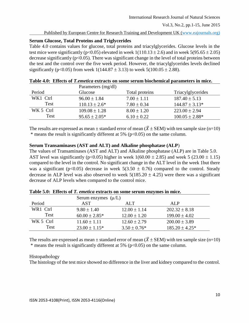
Serum Glucose, Total Proteins and Triglycerides
Table 4.0 contains values for glucose, total proteins and triacylglycerides. Glucose levels in the
test mice were significantly (p<0.05) elevated in week 1(110.13 2.6) and in week 5(95.65 2.05)
decrease significantly (p<0.05). There was significant change in the level of total proteins between
the test and the control over the five week period. However, the triacylglycerides levels declined
significantly (p<0.05) from week 1(144.87 3.13) to week 5(100.05 2.88).
Table 4.0: Effects of T.emetica extracts on some serum biochemical parameters in mice.
Parameters (mg/dl)
Period Glucose Total proteins Triacylglycerides
WK1 Ctrl
Test 96.00 1.84
110.13 2.6*
7.00 1.11
7.80 0.34
187.40 5.13
144.87 3.13*
WK 5 Ctrl
Test 109.08 1.28
95.65 2.05*
8.00 1.20
6.10 0.22
223.00 2.94
100.05 2.88*
The results are expressed as mean ± standard error of mean (�̅� SEM) with ten sample size (n=10)
* means the result is significantly different at 5% (p<0.05) on the same column.
Serum Transaminases (AST and ALT) and Alkaline phosphatase (ALP)
The values of Transaminases (AST and ALT) and Alkaline phosphatase (ALP) are in Table 5.0.
AST level was significantly (p<0.05) higher in week 1(60.00 2.85) and week 5 (23.00 1.15)
compared to the level in the control. No significant change in the ALT level in the week 1but there
was a significant (p<0.05) decrease in week 5(3.50 0.76) compared to the control. Steady
decrease in ALP level was also observed to week 5(185.20 4.25) were there was a significant
decrease of ALP levels when compared to the control mice.
Table 5.0: Effects of T. emetica extracts on some serum enzymes in mice.
Serum enzymes (/L)
Period AST ALT ALP
WR1 Ctrl
Test 9.80 1.40
60.00 2.85*
12.00 1.14
12.00 1.20
202.32 8.18
199.00 4.02
WK 5 Ctrl
Test 11.60 1.11
23.00 1.15*
12.60 2.79
3.50 0.76*
200.00 3.89
185.20 4.25*
The results are expressed as mean ± standard error of mean (�̅� SEM) with ten sample size (n=10)
* means the result is significantly different at 5% (p<0.05) on the same column.
Histopathology
The histology of the test mice showed no difference in the liver and kidney compared to the control.

International Research Journal of Natural Sciences
Vol.3, No.2, pp.1-15, June 2015
Published by European Centre for Research Training and Development UK (www.eajournals.org)
11 ISSN 2053-4108(Print), ISSN 2053-4116(Online)
DISCUSSIONS
Medicinal plants are considered to be the main sources of biologically active compounds that can
be used for the treatment of malaria [28].The presence of phytochemicals such as alkaloids,
saponins, flavonoids, pholobatannins and cardiac glycosides observed during qualitative screening
for secondary metabolites suggests that the hexane and methanolic extracts may excert some
mechanisms that counter the pathological processes of P. berghei infection. These metabolites
with profound antioxidant properties, among other mechanisms [29], may excert their
antiplasmodial effect by decreasing nitric oxide production in kupffer cells, resulting in killing the
parasites [30]. In addition, secondary metabolites such as alkaloids and glycosides have been
shown to posse direct antiplasmodial effects [31, 32]. It is imperative that hexane and methanolic
extracts contain more of these active phytochemicals, accounting for their antiplasmodial effects
in vivo. The result has clearly indicated that different solvent extracts of the same plant can exhibit
different antiplasmodial activities, just as extracts of different parts of the same plant [14]. Total
phenolic content showed positive correlation with the reducing power and lipid peroxidation
inhibition as earlier reported [29].
The continuous decrease in PCV value, for the first three weeks, could be attributed to either
chronic kidney or bone marrow disease [33] or ability of the extract to bind to essential minerals,
like iron required in the synthesis of red blood cells, causing a decrease in the concentration of
circulating red blood cells. After the third week, increase in PCV value indicates hemcentration
due to increase in red blood cells mass. The increase in red blood cells is suggestive of
polycythemia and a positive erythropoetic effect, thus enhancing the carrying capacity in the mice
[34]. This could be another possible mechanism (destruction of the infected red blood cells and
stimulation of its synthesis afterwards) with which this extract elucidate its antiplasmodial activity.
The fresh organ weight of kidney and spleen which are hematopoietic organs were not significantly
different (p>0.05) when compared to the control; this is the same with the liver and heart muscle.
The fresh organ weight of the stomach was significantly higher when compared to the control.
The decrease in glucose and triacylglycerides levels suggests that methanolic extract of T. emetica
contain active principle(s) that have antidiabetic property. This could be due to its ability to
stimulate the pancreas for insulin production [35]. Decrease in serum proteins could generally be
early indication of renal or liver or nutritional deficiency. Similar effects of some plant extract in
experimental animals have been documented [36].
In the assessment of organ damage by a xenobiotic, the determination of enzyme levels such as
ALT, AST and ALP are largely used [37]. In the present study, the activities of ALT and ALP
were decreased indicating that the extract has some hepatoprotective function and biliary
advantage [38]. The observed increase in AST level signifies damage to the liver, kidney, heart
muscle, erythrocyte or skeletal muscle, but there is no corresponding increase in the activity of
ALT (more specific and better parameter to detect liver injury) or ALP to suggest liver or kidney
damage. Histology of the liver, kidney and heart muscle revealed that no damage was done to these
organs.
International Research Journal of Natural Sciences
Vol.3, No.2, pp.1-15, June 2015
Published by European Centre for Research Training and Development UK (www.eajournals.org)
12 ISSN 2053-4108(Print), ISSN 2053-4116(Online)
The adverse effect of the extract reflected by highest AST activity (60u/l) in week one shows how
important these enzyme markers easily detect organ damage. Apart from the liver, kidney, heart
and skeletal muscle, AST is also found in erythrocytes. The decline in PCV value from the first
week with corresponding increase in AST activity in the first week suggest that destruction of
erythrocytes have contributed in the rise in AST activity. Also, when the extract stimulated
erythropoesis, the activity of AST was on a decrease with increase in PCV value. However, the
possibility skeletal muscle contributing to rise in AST level cannot be over rule
This report of in vivo antiplasmodial activity of T emetica extract is also a confirmation of the
earlier reports of its antiplasmodial activity in vitro by Kamanzi et al.,2004 and Bah et al., 2007
[39,40].Thus T. emetica has both in vitro and in vivo antiplasmodial activity[41,42].
IMPLICATION TO RESEARCH AND PRACTICE
The outcome of this research is of great importance to pharmaceutical industries for the
development of therapeutical active drug against malaria parasites. Isolation of the bioactive
phytochemical from these plant extracts will be of immense significance to drug manufacturers.
CONCLUSION
The present studies has confirmed that T. emetica does not only have an in vitro antiplasmodial
activity but also has in vivo antiplasmodial activity. It was also observed that long term
administration of the methanolic extract of T. emetica causes damage to erythrocytes and skeletal
muscle.
REFERENCES
1. World Health Organization (2012). Scaling up diagnostic testing, treatment and
Surveillance for malaria.
2. Leitner, W.W., Bergmam-leitner, E.S and Angov E(2010). Comparism of Plasmodium
berghei challenge models for the evalution of preerythrocytic malaria vaccines and their effect on
perceived vaccine efficacy. Malaria Journal, 9(1):14.
3. Oyewole, O.I. and Akingbala, P.F.(2011). Phytochemical analysis and hypolipidemic
properties of Jatropha tanjorensis leaf extract, European Journal of Medicinal Plants, 1(4):180-
185.
4. Omeregie, E.S. and Sisoda, B.S.(2012)In vitro antiplasmodial activity and cytotoxicity of
leaf extracts of Jatropha tanjorensis JL Ellis and Sojora, Bayero Journal of Pure and Applied
Sciences, 5(1):90-97.
5. Ravikuma,r S., Inbaneson, S.J. and Suganthi, P.(2012). In vitro antiplasmodial activity of
ethanolic extracts of south Indian medicinal plants against Plasmodium falciparum. Asian Pacific
Journal of Tropical Biomedicine. 1-9.
6. Sanogo, R.(2011). Medicinal plants traditionally used in Mali for Dysmenorrhea, Afri J
Tradi, Complementary and Alternate Medicine (AJTCAM), 8(5):90-96

International Research Journal of Natural Sciences
Vol.3, No.2, pp.1-15, June 2015
Published by European Centre for Research Training and Development UK (www.eajournals.org)
13 ISSN 2053-4108(Print), ISSN 2053-4116(Online)
7. Diallo, D,, Paulsen, B. S., Liljebac, Torun H. A. and Michaelsen Terje, E. (2003). The
Malian medicinal plant Trichilia emetica; studies on polysaccharides with complement fixing
ability. J Ethnopharmacol. 84(2–3):279–287
8. Champagne, D.E., Koul, O., Isman, M.B., Scudder, G.E. and Towerd, G.H.N.(1992).
Biological activity of limonoids from the Rutales. Phytochemistry. 31:377–394.
9. Sparg, S.G., Van Staden, J and Jager, A.K.(2000). Efficiency of traditionally used South
African plants against schistosomiasis. J Ethnopharmacol. 64:209–211
10. El Tahir, A., Satti, G.M.H and Khalid SA(1996). Antiplasmodial activity of selected
Sudanese medicinal plants with emphasis on Maytenus senegalensis (Lam.) Exell. J
Ethnopharmacol. 64:227–233.
11. Hoet, S., Opperdoes, F., Brun, R., Adjakidje, V. and Quetin-Leclercq J(2004). In vitro
antitrypanosomal activity of ethnopharmacologically selected Beninese plants. J Ethnopharmacol.
91(1):37–42.
12. Traore, M., Zhai, L and Chen, M. (2007). Cytotoxic kurubasch aldehyde from Trichilia
emetica. Nat Prod Res. 21(1):13–17.
13. Bulus, T., Atawodi, S.E. and Mamman, M.(2011). Acute toxicity effect of the aqueous
extract of Terminalia avicennioides on white albino rat. Science World Journal, 6 (2):1-4.
14. Atawodi, S.E., Bulus, T., Ibrahim, S., Ameh, D.A., Nok AJ, Mamman M et al. (2003). In
vitro trypanocidal effect of methanolic extract of some Nigeria savannah plants. African Journal
of Biotechnology, 2:317-321.
15. Ernest, D., Olfert, D.V., Brenda, M., Cross, V.M. and Mcwilliam, A.(1993)A. A Guide to
Care and Use of Experimental Animals. 2nd edition, Canadian Council on Animal Care.
16. Edeoga, H.O., Okwu, D.E. and Mbaebie, B.O.(2005). Phytochemical Constituents of some
medicinal plants, Afri J. Biotec 49: 685-687.
17. Akinyemi, O.K., Oladapo, O., Okwara, C.E., Ibe, C.C. and Fasure, K.A(2005). Screening
of crude extract of six medicinal plants used in south west Nigerian unorthodox medicine for anti-
methicillin resistance Staphyolococcus aureus activity, Biomedicial Central Complement
Alternate Medicine, 5: 6.
18. Kwada, A.D. and Tella, O. (2009)I.Determination of infochemicals and the phytochemical
screening of the foliage and stem bark of Senna siamea(lam.) in Yola, Adamawa State, J. Med
Plants Res, 3 (9):630.
19. Oluwaniyi, O.O., Ibiyemi, A.S., Usman, A.L.(2007). Effect of Detoxification on the
Nutrient content of Thevetia peruviana seed cake, Res. J. Applied Sci., 2(2)188-191.
20. Iqbal, Z., Sajid, M.S., Abbas, R.Z. and Shindu, Z.U.D (2001). Determination of condensed
tannin contents from different plants of kharimurat rangeland (Attock, Pakistan), J. Agric. Soc.
Sci, 7:114-116.
21. Ogbadoyi, E.O, Garba, M.H., Kabiru, A.Y., Mann, A and Okogun, J(2011)I. Therapeutic
evalution of Acacia nilotica (Linn) stem bark extract in experimental African typanosomiasis, Intl.
J. App. Res. Natrl. Pro. 4:11-18.
22. Jigam, A.A,. Akanya, H.O., Emmanuel, O.O., Dauda, B.E.N.(2010). In vivo
antiplasmodial, analgesic and anti-inflammatory effects of the root extracts of Acacia nilotica Del
(Leguminosae), Asian J. Exp Biol Sci,1:315-320.
23. Howlader, M.M.R., Begum. S., Islam, K, Hai, M.A. and Hossain M.G.(2004). Further
observations on the packed cell volume and haemoglobin concentration in cattles naturally
infected with Fasciola gigantica. 2:1
International Research Journal of Natural Sciences
Vol.3, No.2, pp.1-15, June 2015
Published by European Centre for Research Training and Development UK (www.eajournals.org)
14 ISSN 2053-4108(Print), ISSN 2053-4116(Online)
24. Rifai, N. and Warnick, C.K.(2006). Lipids, lipoproteins, Apolipoproteins and other
cardiovascular risk factors. In : Burtis AC, Ashwood ER, Burns DE, editors. Teitz textbook of
clinical chemistry and molecular diagnostics. 4th edition, St Lious, Missouri: Saunders(Elservier
Science USA), 944 945.
25. Marghoob, H., Mohd, A.H., Abdelmarouf, H.M.(2013). Comparative levels of ALT, AST,
ALP and GGT in liver associated diseases. European Journal of Experimental Biology, 3(2):280-
284.
26. Malhotra, R., Grover, V., Kapoor, A., Kapur, R.(2010). Alkaline phosphatase as a
periodontal disease marker. Indian J Dent Res, 21:531-536.
27. Pearse, A.E.(1985). Histochemistry: Theoretical and applied analytical technology,
Churchill-Livingstone, Edinburgh; 4th ed. Vol 2
28. George, V.C., Kumar, N., Rajkumar, V., Suresh, P.K.and Kumar, A.R.(2012 ).
Quantitative assessment of the relative antineoplastic potential of the n-butanolic leaf extract of
Annona muricata Linn in normal and immortalized Human cell lines, Asian Pacific J cancer Prev,
13699-704.
29. Germano, M.P., D'Angelo, V., Biasini, T., Sanogo, R., De Pasquale, R. and Catania, S.
(2006). Evaluation of the antioxidant properties and bioavailability of free and bound phenolic
acids from Trichilia emetica Vahl. J Ethnopharmacol. 105:368–373.
30. Dondorp, A.M., Fanello, C.I., Hendriksen, I.C. et al. (2010), Aquamat group. Artesunate
versus quinine in the treatment of severe falciparum malaria in Africa Childern(AQUAMAT): an
open label, randomized trial, Lancet, 376(9753)1647-1657.
31. Kayano, A.V., Lopes, S.C., Bueno, F.G., Cabral, E.C., Souza-Neiras, W.C., et al(2011). In
vitro and in vivo assessment of the antimalarial activity of Caesalpinia pluviosa. Malaria J,
10(1)112.
32. Boampong, J.N., Ameyaw, E.O., Kyei, S., Aboagye, B., Asare, K. et al.(2013). In vivo
antimalarial activity of stem bark extracts of Plumeria alba against Plasmodium berghei in printing
control region mice. Reports in partasitology, 3:24
33. Purves, W.K., David, S., Gordan, H.O. and Craig H(2004). The Science of biology,
Seventh edition, Sunderland mass: Sinauer Associates, pp 954.
34. Okpuzor, J., Ogbunugafor, H.A. and Kareem, G.K(2009). Hepatic and hematologic effects
of fractions of Globimetula braunii in normal albino rats, Excli Journal, 182-189.
35. Gad, S.C (2001). Principles and methods of Toxicology. Tayol and Francis, Philadephia P.
351-353.
36. Sodipo, O.A., Abdulrahman, F.I., Sandabe, U.K. and Akinniyi JA(2011). Biochemical
liver function with aqueous fruit extract of Solanum macrocarpum Linn. In albino rat acutely
administered triton-x to induce hyperlipidaemia. J App Pharm Sci, 01(08):89-93.
37. Ojo, O.O., Nadro, M.S. and Tella, I.O.(2006). Protection of rats by extracts of some
common Nigerian trees against acetaminophen-induced hepatotoxicity, Afri J 5 (9):755-760.
38. Luka, C.D., Saleh, B. and Mohammed A(2012). Effect of aqueos extract of Dioscorea
dementorum on some biochemical parameters in alloxan-induced diabetic rats, Asian J Exp Biol
Sci, 3(2):450-453.
39. Kamanzi, A.K., Schmid, C., Brun, R., Kone, M. W. and Traore, D.(2004).
Antitrypanosomal and antiplasmodial activity of medicinal plants from Cote d'Ivoire. J
Ethnopharmacol. 90(2–3):221–227.

International Research Journal of Natural Sciences
Vol.3, No.2, pp.1-15, June 2015
Published by European Centre for Research Training and Development UK (www.eajournals.org)
15 ISSN 2053-4108(Print), ISSN 2053-4116(Online)
40. Bah, S., Jäger, A.K., Adsersen, A., Diallo, D., Paulsen, B.S.(2007). Antiplasmodial and
GABA(A)-benzodiazepine receptor binding activities of five plants used in traditional medicine
in Mali, West Africa. J. Ethnopharmacol. 110(3):451–457.
41. Uchoa, V.T., de Paula, R..C., Krettli, L.G., Santana, A.E.G., Krettli, A.U.(2010).
Antimalaria activity of compounds and mixed fractions of Cecropia pachystachya, Drug Dev Res,
91: 82-91.
42. Andrade-Neto, V.E., Brandao, M.G., Oliveira, F.Q., Casali, V.W., Njaine, B., Zalis, M.G.,
Oliveira, L.A. and Kretti, A.U.(2004). Antimalaria activity of Biden pilosa L(Asteraceae) ethanol
extracts from wild plants collected in various localities or plants cultivated in human soil.
Phytother Res, 18:634-639.
FUTURE RESEARCH
Fractionation of plant extracts to determine the active compound(s)