In stent retenosis pathophysiology
49
DR NILESH TAWADE
-
Upload
nilesh-tawade -
Category
Health & Medicine
-
view
268 -
download
1
Transcript of In stent retenosis pathophysiology

DR NILESH TAWADE

Mr. Bachmann
1977
In the past decade, tremendous progress has been made in reducing the incidence of restenosis with the advent of the drug-eluting stent (DES).
With “POBA,” rates of acute and chronic vessel occlusion at 30% to 60%, secondary to acute and chronic recoil and constrictive remodeling.
The advent of bare-metal stents (BMS) appeared to eliminate the issue of acute and chronic recoil but introduced a new entity, neointimal hyperplasia (NIH),
Various studies has demonstrated a strong and linear relationship between NIH formation and late lumen loss (LLL)
The restenosis rates with BMS were reported to be between 16% and 44%, (with, in particular, long lesion length and small vessel caliber)

DES were thus conceived as the next step in tackling this iatrogenic entity of NIH.
large-scale reductions in restenosis rates reported at 0% in highly selective lesions and up to 16% in a broader range of patients and lesions with first-generation DES.
Despite the significant advances in the technology to reduce DES restenosis , incidence of in-stent restenosis (ISR) requiring target vessel revascularization (TVR), so-called DES failure, to be 5% to 10%,#
This leads to approximate around 2 lakhs repeat revascularization per year in united
states
# Garg S, Serruys PW. Coronary stents: current status. J Am Coll Cardiol. 2010;56:S1–S42

Classification of ISR ---Mehran et al
classification is prognostic ally important, and it may be used for appropriate and early patient triage
Recurrent ISR was more frequent with increasing grades of classification (9%, 20%, 34%, and 50% for classes I to IV,
as was diabetes (28%, 32%, 39%, and 48% in classes I to IV, respectively; P<0.01).
Angioplasty and stenting were used predominantly in classes I and II, whereas classes III and IV were treated with atheroablation
TLR increased with increasing ISR class; it was 19%, 35%, 50%, and 83% in classes I to IV, respectively (P<0.001)
Angiographic Patterns of In-Stent Restenosis Classification and Implications for Long-Term
OutcomeCirculation.1999; 100: 1872-1878

PATTERN OF RESTENOSIS
However, over one-fifth of ISR cases remain diffuse, and 10% to 20% are even occlusive
60% of ISR located at the proximal DES edge, as demonstrated in with either paclitaxel-eluting stent (PES) or sirolimus-eluting stent (SES) implantation
In DES it has been demonstrated to be usually focal .
BMS has been shown to be primarily diffuse.


BMS DES
neoatherosclerosis with formation of a necrotic core
calcification

ISR traditionally has been suggested as being potentially less benign with the recurrence of anginal symptoms alone.
However, emerging evidence now suggests that between 30% to 60% of ISR cases present with an acute coronary syndrome with unstable angina being the most common presentation.
and up to 5% of patients even reported to present with ST-elevation myocardial infarction (STEMI).
Rest present as stable angina profile

The Underlying Mechanisms of Restenosis With Drug-Eluting Stent
Biological Factors
Arterial Factors
Stent Factors
Implantation Factors

The Underlying Mechanisms of Restenosis With Drug-Eluting Stent

Stent FactorsPolymer drug release
kinetics
Type of DES? Type of drug?
Stent gap, non- uniform strut distribution, and drug
deposition
Polymer disruption,
peeling, and cracking
Stent fractures

Arterial factors
Wall shear stress
Progression of atherosclerosis within
a stented segment
“Thromborestenosis”
Vessel remodeling
Small vessels


Can be present in genetically predetermined individuals or be acquired following cytotoxic exposure to the drug.
Genes for resistance may operate in conventional pathways
• that hampers drug delivery , drug metabolism , apoptosis regulation and DNA repair
As examples, polymorphisms in the genes that encode mTOR or proteins involved in paclitaxel or sirolimus metabolism have been shown to confer drug resistance both in vitro and in vivo

The inflammatory reaction that occurs after arterial injury is a critical factor that influences the
extent of neointimal response, with the persistence of this inflammatory response beyond 90
days being strongly associated with delayed healing and implicated in an increased risk of LST
and restenosis long term.
SES triggers giant cell infiltrations and PES eosinophilic reactions around stent struts
The inflammatory responses associated with SES have been shown to persist beyond 180 days and up to 2 years
This is in contrast to BMS and the second-generation everolimus-eluting stent (EES-Xience V)
with a more biocompatible polymer, in which the inflammatory response have been
demonstrated to be limited to a period of 90 days and 12 months, respectively

Circulating MMPs recently have been demonstrated to be potentially useful in identifying patients at greater risk of developing ISR following DES implantation.
Significant eleations in MMP-9 levels at baseline and MMP-2 and MMP-9 levels 24 hours procedure have proven to be strongly associated with the development
of ISR following DES implantation.
Conversely low and near normal MMP-2 and MMP-9 levels were associated with a lack of a significant restenotic response.
Katsaros KM, Kastl SP, Zorn G, Maurer G, Wojta J, Huber K, Christ G, Speidl WS. Increased restenosis rate after implantation of drug-eluting stents in patients with elevated serum activity of matrix metalloproteinase-2
and -9. J Am Coll Cardiol Cardiovasc Interv. 2010; 3:90 –97.

In a recent, larger, well-designed trial, Papafaklis et al demonstrated the presence of significant numbers of “pockets” of low shear stress within stented segments, secondary to local geometric factors such as angulation or curvature, and that these pockets were significantly associated with
NIH formation at 6-month follow-up with BMS and PES.
Conversely, high shear stress can potentially directly inhibit SMC proliferation and therefore limit atherosclerosis or restenosis unless it progresses from a low-shear stress area.
Low shear stress consequently may lead to the accumulation of growth factors, mitogenic cytokines, and platelets, which may promote atherosclerosis or neointimal formation after vessel injury.
Wall shear stress refers to the principle that fluid dynamics and vessel geometry may play a potential role in the cause of focal plaque or neointimal formation.
Papafaklis MJ Am Coll Cardiol Intv. 2010;31189.

There is emerging evidence suggesting that chronic inflammation and/or incompetent endothelial
function induce late de novo neoatherosclerosis inside both BMS and DES, which may be an
important mechanism of the late phase ISR or thrombosis.
Interestingly, a significant difference in the timing of neoatherosclerosis development was noted in BMS and DES.
the earliest atherosclerotic change with foamy macrophage infiltration began at 4 months after SES implantation, whereas the same change in BMS lesions occurred beyond 2 years and remained a rare finding until 4 years1.
1.Nakazawa G, Vorpahl M, Finn AV, Narula J, Virmani R. One step forward and two steps back with drug-eluting stents. J Am Coll Cardiol Img 2009;2:623– 8.

The fibrous cap is infiltrate by
numerous foamy macrophages and is
markedly thinned

In addition, the earliest necrotic core formation began at 9 months in DES , whereas in BMS it occurred at 5 years
The incidence of neoatherosclerosis was greater in DES lesions (31%) than in BMS lesions (16%), and
the median stent duration with neoatherosclerosis was shorter in DES compared with BMS (420 days (13 MONTHS ) [361 to 683 days] vs. 2,160 days (5 YRS ) [1,800 to 2,880 days]).
Unstable lesions like the thin-cap fibroatheromas (TCFA) or intimal rupture had shorter implant duration for DES (1.5 years) compared with BMS (6.1 years).
These results represent that neoatherosclerosis in DES is more frequent and occurs earlier than in BMS, likely from different pathogenesis
1.Nakazawa G, Vorpahl M, Finn AV, Narula J, Virmani R. One step forward and two steps back with drug-eluting stents. J Am Coll Cardiol Img 2009;2:623– 8.
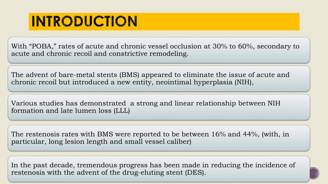
Further, there is a question about the difference in the induction of neoatherosclerosis among various DES.
Currently, the data are mostly available only in SES and PES, for which the trend toward more rapid neoatherosclerotic changes is observed in SES than in PES,
although the frequency of neoatherosclerosis in both DES is higher than BMS (cumulative
incidence up to 6 years: SES 38% vs. PES 24% vs. BMS 10%), indicating that differences of
drugs or polymers may influence on neointimal tissues
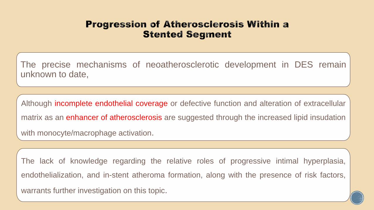
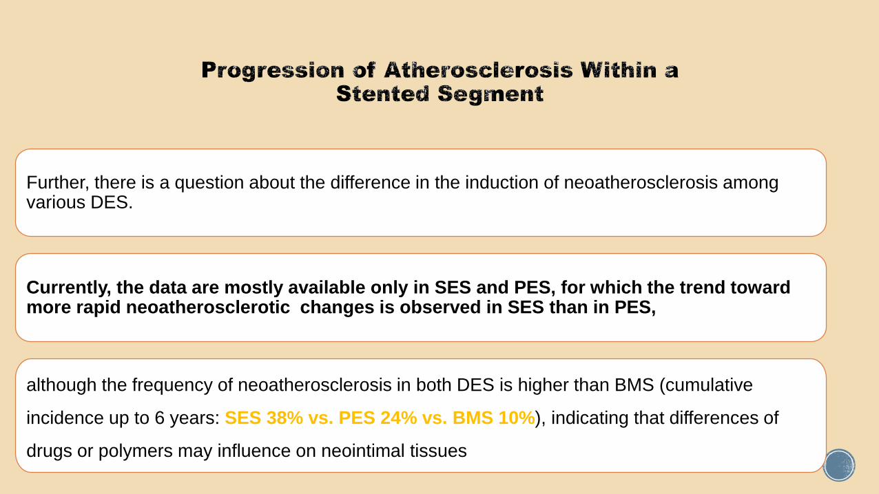
The precise mechanisms of neoatherosclerotic development in DES remainunknown to date,
Although incomplete endothelial coverage or defective function and alteration of extracellular
matrix as an enhancer of atherosclerosis are suggested through the increased lipid insudation
with monocyte/macrophage activation.
The lack of knowledge regarding the relative roles of progressive intimal hyperplasia,
endothelialization, and in-stent atheroma formation, along with the presence of risk factors,
warrants further investigation on this topic.
“Thromborestenosis” is a term first described by Oikawa et al to describe an intriguing theory in which
chronic thrombus formation may play an integral part in the development of ISR
Within their study, the incidences of thrombus and fibrin deposition were more frequently observed with
ISR lesions associated with SES implantation (12 of 13 cases) compared with BMS (2 of 8 cases).
Hypothetically the presence of older thrombi was speculated to be related to clinically silent
nonocclusive athero-thrombotic events in the preceding days to weeks before the clinical presentation of
occlusive thrombosis.
Whether this latter theory is also an explanation for the presence of thrombi seen with ISR is presently unclear
Intravascular ultrasound, angioscopic and histopathological characterisation of heterogeneous patterns of restenosis after sirolimus-eluting stent implantation: insights into potential “thromborestenosis” phenomenon. EuroIntervention.
2010;6: 380–387.

The phenomenon of the increased likelihood of restenosis occurring in
positively remodeled vessels. The amount of plaque present at the
lesion site can also influence the degree of IH following stenting

Stent FactorsPolymer drug release
kinetics
Type of DES? Type of drug?
Stent gap, non- uniform strut distribution, and drug
deposition
Polymer disruption,
peeling, and cracking
Stent fractures

Polymer Release Kinetics
Polymer release kinetics plays a key and fundamental role in the prevention of restenosis
The Paclitaxel In-Stent Controlled Elution Study (PISCES) trial showed that the duration of the drug release had a far greater impact on the inhibition of NIH than the dose of drug delivered
For example, 10 mcg of paclitaxel released over 10 days following DES implantation appeared to
have little effect on NIH formation, whereas the same dosage of drug released over a 30-day period
led to a profound reduction in NIH, with a more than halving (57% reduction) of the LLL.
Interestingly, 30 mcg of the same drug released over a 10-day period also was less effective.
Molecular biology studies have suggested that the genes responsible for the proliferative response potentially remain active for a period of up to 21 days after vessel injury.

These clinical findings therefore support the concept of a certain threshold of drug, delivered over a sustained prolonged period of time, being required to “dampen” down the inflammatory and subsequent NIH response

Schomig et al,1 in a meta-analysis of 16 trials comparing SES with PES, suggested the benefit of SES over PES over
a median 2-year follow-up, with a significant reduction in TVR (hazard ratio 0.74, 95% [confidence interval] CI 0.63 to
0.87, P<0.001) and stent thrombosis (hazard ratio 0.66, 95% CI 0.46 to 0.94, P0.02) without a mortality benefit.
The reasons for this apparent benefit have been suggested to be related to the slower polymer release kinetics of SES compared with PES.
But (SIRTAX) *trial have suggested a possible “catch-up” in LLL with SES over a 5-year follow-up, with no significant differences in LLL observed between the 2 groups
Schomig AJ Am Coll Cardiol. 2007;50:1373–1380
* Garg S, Serruys PW. Coronary stents: current status. J Am Coll
Cardiol. 2010;56:S1–S42.

and that important differences in the efficacy of differing types of DES existed
showed that the rates of restenosis with DES implantation were significantly higher in patients with diabetes mellitus
at 2-year follow-up
implanted with 4 different types of DES (Endeavor, SES, Taxus Express, and Liberty) in real-world practice
Data from the Swedish Coronary Angiography and Angioplasty Registry (SCAAR), registry involving >35 000 patients

Five-year unpublished follow-up data from registry continued to demonstrate differences in the
efficacy of the first- and second-generation DES in reducing the rate of restenosis, with a trend
toward better outcomes seen after nearly 2 years of EES use.
Higher restenosis rates were also evident in diabetic patients with Endeavor (relative risk
1.77, 95% CI 1.29 to 2.43) and SES (relative risk 1.25, 95% CI 1.04 to 1.51) when
compared with non diabetic patients.
In particular, the restenosis rates with Endeavor were twice as high in diabetic patient compared with other DES types
The EES releases 80% of the drug within 30 days and nearly all the drug within 4 months.
In the Spirit I, II, and III trials, a LLL of 0.10, 0.16, and 0.33 mm and TVR rates of 3.8%, 3.4%, and
4.6% were observed at 6, 12, and 24 months, respectively.
Conversely, the Endeavor (zotarolimus ) reported a LLL of 0.60 mm and 0.67 mm and TVR of
6.3% and 4.5%, respectively, in the Endeavor III and IV trials at 12 months.
The Endeavor, however, elutes 95% of its drug very rapidly (within 14 days); this is highly likely to
be the main reason for the poorer results.
However the next-generation Endeavor Resolute stent, consisting of the same cobalt chromium
metallic platform and the drug (zotarolimus) as the Endeavor stent, but with substantially longer
polymer drug release kinetics (180 days), reported an in-stent LLL equivalent to EES stents.

Stent Gap, Non-uniform Strut Distribution andDrug Deposition
Takebayashi et al classically described the number and distribution of DES struts, as
identified by IVUS, as being independent significant risk factors (fewer struts and
Nonuniform stent strut distribution) for NIH formation and the subsequent risk of restenosis.
Nonuniform DES strut distribution has been suggested to be attributable to features such
as stent design, stent gap, vessel curvature, coronary bifurcations, ostial lesions, stent
under or overexpansion, polymer peeling, and stent fracture
Small Vessels and Strut Thickness
Small-vessel disease is a recognized challenging lesion subset with
significant risks of restenosis.
Mechanism include
(1) a high degree of vessel stretch and injury,
(2) a smaller post procedural lumen area, and
(3) a higher metal density
Thicker stent struts have been linked to an increased risk of
restenosis with BMS and small vessels.
The underlying rationale is that a thinner stent strut would have less
of a “footprint” on the vessel wall with a consequential reduced
inflammatory response

Polymer disruption, peeling, and cracking, and subsequent exposure of bare-metal areas
have been demonstrated to occur in bench studies involving both first- and second
generation DES using light or scanning electron microscopy

Although there is no direct evidence to suggest that the integrity of the
polymer coating is a direct cause of restenosis, there are sufficient
theoretical concerns to warrant concern through nonuniform local drug
distribution or the disrupted polymer potentially acting as a nidus for an
ongoing inflammatory response.

Stent fracture related to DES implantation in coronary arteries was first reported in 2004.
Subsequent retrospective and prospective registries have quoted restenosis rates ranging from 15% to
100% in patients identified to have stent fractures.
In the only randomized controlled trial reporting the incidence of stent fracture and outcomes after DES
implantation is (LONG-DES-II study)*,in whch a 14% incidence of restenosis was observed.
The restenosis associated with DES fractures tends to occur fairly late and focally, reflecting the local
trauma sustained by the vessel at the fracture site
* Kim HS Int J Cardiol. 2009;133:354 –358

First, mechanical fatigue of the metallic stent can occur because of excessive movements
during cardiac contraction, especially at a hinge-point where the potential for 2 opposing
forces may occur at the same site.
In particular, this may occur in the right coronary artery or a saphenous vein graft, because of
their greater propensity for angulation and tortuosity
Second, a closed-cell design, is less likely to be able to withstand the pressures related to
excessive movements compared with the open-cell design. The incidence of stent fracture is
reported at less than 0.1% with the open cell design and approximately 2.3% with closed
cell design*

Long stents,
overlapping stents,
tight lesions that have been vigorously post dilated and expanded,
myocardial bridge sites, and
areas of significant curvature are all factors that may predispose patients to DES fracture.


A smaller post procedural minimal lumen diameter (MLD) and a greater residual stenosis
have been shown to be significant predictors of long-term patency and clinical outcomes.
The most plausible theory to explain the underlying mechanism relating stent under
expansion to restenosis is the so-called bigger-is-better paradigm
Effectively, if the minimum stent area is smaller at baseline, then the expected NIH
formation post-DES implantation would be more likely to be of more significance, whereas
if the minimum stent area was larger, then the same amount of NIH would be clinically
less relevant in causing binary restenosis

In a classical meta-analysis (n=2972 patients) investigating IVUS versus angiographic-
guided BMS implantation, Casella et al demonstrated at 6-month follow-up a reduction in
TVR (OR 0.62; 95% CI 0.49 to 0.78; P<0.00003), binary restenosis (OR 0.75; 95% CI
0.60 to 0.94; P<0.01), and major adverse cardiovascular events (OR 0.79; 95% CI 0.64 to
0.98; P<0.03)
Casella G, Klauss Catheter Cardiovasc Interv. 2003;59:314 –321.

Thrombocyte Activity Evaluation and Effects of Ultrasound Guidance in Long Intracoronary Stent
Placement (TULIP) STUDY AND The Angiography Versus Intravascular Ultrasound- Directed
(AVID) STUDY. Showed that there was definite benefit in lowering binary restenosis and TLR at
rate at angiographic follow-up in IVUS guided arm . [lower binary restenosis rate at 6-months
angiographic follow-up (23% vs. 46%, p=0.0082) and a subsequent decrease in TLR (4% vs.
14%, p=0.037)]
(TULIP Study) Circulation. 2003 Jan 7;107(1):62-7. (the AVID Trial) Circ Cardiovasc Interv. 2009 Apr;2(2):113-23..

But recently presented, multicenter trial Angiographic versus IVUS Optimization (AVIVO) trial in which.
IVUS versus angiographic-guided DES implantation in complex lesion was investigated, with 142 patients in each study arm.
At 30 days and 9 months, no significant differences were seen in the combined end point of MI, target lesion revascularization, TVR, or cardiac death (85.9% versus 83.1%, P<0.47)
Colombo A. AVIO: a prospective randomized trial Paper presented at: Transcatheter Cardiovascular
Therapeutics; September, 2010.

Geographical miss (GM) is essentially a failure to appropriately cover
an injured vessel, such as occurs after balloon associated vessel
barotrauma, or incomplete coverage of atherosclerotic plaque.
GM associated with SES implantation was investigated in the
prospective evaluation of the impact of Stent deployment Techniques
on cLinicaL outcomes of patients treated with the cypheR stent
(STLLR) study.
Longitudinal GM was defined as injured or diseased stenotic
segment not fully covered by DES, and axial GM was defined as
potentially under sizing or oversizing the balloon
multicenter prospective STLLR trial. Am J Cardiol. 2008; 101:1704 –1711

At 1-year follow-up, there was more than a 2-fold increase in TVR (5.1% versus 2.5%;
P<0.025) and a 3-fold increase in MI (2.4% versus 0.8%; P<0.04) in patients with GM.
These findings were almost exclusively related to longitudinal GM (6.1% versus 2.6%;
P<0.001) with two-thirds of cases being secondary to balloon injury outside the stent margins.
The lack of effect of axial GM (4.2% versus 4.3%; p non significant), in which it was shown that
the balloon-to-artery ratio or the occurrence of edge dissections (potentially associated with
axial GM) did not have a significant impact on the risk of restenosis and does perhaps argue
against the IVUS optimization of DES deployment.

The deployment of a DES in a clot-laden arterial segment has been shown in an ex vivo model
to lead to a 10-fold reduction in drug penetration into the vessel wall, which may potentially
affect clinical outcomes.
Despite these theoretical concerns, a recent meta-analysis of 13 trials (n>7244) has shown the
significant benefits of DES over BMS in primarily reducing TVR and recurrent MI.
The widespread use of glycoprotein-IIb/IIIa inhibitors and aspiration thrombectomy devices may
be the reasons why these concerns may have not materialized in clinical trials.
The concerns about reduced absorption of the drug from DES should be borne in mind in a
thrombus-laden vessel, especially when there has been inadequate resolution of thrombus and
DES implantation is to be considered.

Restenosis remains a common problem, independent of the type of angioplasty.
Both clinical and histologic studies of DES have demonstrated evidence of continuous neointimal
growth during long-term follow-up, which is designated as “late catch-up” phenomenon.
In-stent neoatherosclerosis is an important substrate for late stent failure for both BMS and DES,
especially in the extended phase.
early detection of neoatherosclerosis may be beneficial to improving long-term outcome of patients with
DES implants.
Apart from biological factors, there are potentially controllable factors are within arterial and stent
related factors
Ultimately, the implantation factors are the most important controllable factors from the perspective of
the interventional cardiologist