In situ Composite Ion-Triggered Gellan Gum Gel ...
51
Full Terms & Conditions of access and use can be found at https://www.tandfonline.com/action/journalInformation?journalCode=iphd20 Pharmaceutical Development and Technology ISSN: 1083-7450 (Print) 1097-9867 (Online) Journal homepage: https://www.tandfonline.com/loi/iphd20 In situ Composite Ion-Triggered Gellan Gum Gel Incorporating Amino Methacrylate Copolymer Microparticles: A Therapeutic Modality for Buccal Applicability Enas Elmowafy, Marco Cespi, Giulia Bonacucina & Mahmoud E. Soliman To cite this article: Enas Elmowafy, Marco Cespi, Giulia Bonacucina & Mahmoud E. Soliman (2019): In situ Composite Ion-Triggered Gellan Gum Gel Incorporating Amino Methacrylate Copolymer Microparticles: A Therapeutic Modality for Buccal Applicability, Pharmaceutical Development and Technology, DOI: 10.1080/10837450.2019.1659314 To link to this article: https://doi.org/10.1080/10837450.2019.1659314 Accepted author version posted online: 22 Aug 2019. Submit your article to this journal View related articles View Crossmark data
Transcript of In situ Composite Ion-Triggered Gellan Gum Gel ...

Full Terms & Conditions of access and use can be found athttps://www.tandfonline.com/action/journalInformation?journalCode=iphd20
Pharmaceutical Development and Technology
ISSN: 1083-7450 (Print) 1097-9867 (Online) Journal homepage: https://www.tandfonline.com/loi/iphd20
In situ Composite Ion-Triggered Gellan Gum GelIncorporating Amino Methacrylate CopolymerMicroparticles: A Therapeutic Modality for BuccalApplicability
Enas Elmowafy, Marco Cespi, Giulia Bonacucina & Mahmoud E. Soliman
To cite this article: Enas Elmowafy, Marco Cespi, Giulia Bonacucina & Mahmoud E. Soliman(2019): In situ Composite Ion-Triggered Gellan Gum Gel Incorporating Amino MethacrylateCopolymer Microparticles: A Therapeutic Modality for Buccal Applicability, PharmaceuticalDevelopment and Technology, DOI: 10.1080/10837450.2019.1659314
To link to this article: https://doi.org/10.1080/10837450.2019.1659314
Accepted author version posted online: 22Aug 2019.
Submit your article to this journal
View related articles
View Crossmark data

In situ Composite Ion-Triggered Gellan Gum Gel Incorporating Amino Methacrylate
Copolymer Microparticles: A Therapeutic Modality for Buccal Applicability
Enas Elmowafya, Marco Cespi
b , Giulia Bonacucina
b , and Mahmoud E. Soliman
a,
aDepartment of Pharmaceutics and Industrial Pharmacy, Faculty of Pharmacy, Ain
Shams University, Cairo, Egypt,Monazzamet Elwehda Elafrikeya Street, Abbaseyya, Cairo, Egypt, P.O. 11566
b School of Pharmacy, University of Camerino, Camerino, Italy Via S. Agsotino 1, 62032
* Corresponding author:
Enas Elmowafy
Department of Pharmaceutics and Industrial Pharmacy,
Faculty of Pharmacy, Ain Shams University, Cairo, 11566, Egypt
Tel.: +2 01005164052 Fax: +20 2 0224011507.
Email: [email protected] Acc
epte
d M
anus
cript

Abstract
The aim of the current investigation is to delineate the buccal applicability of an in situ
composite gel containing aceclofenac (AC) amino methacrylate copolymer microparticles (MPs),
surmounting limitations of oral existing conventional therapy. AC Eudragit RL100 MPs were
fabricated and statistically optimized using 224
1 factorial design. Better buccal applicability and
enhanced localization were achieved by combining the optimum MPs with in situ ion-activated
gellan gum gel. The crosslinking and gelation of in situ gel were investigated by morphological
and solid state characterizations. Suitability for buccal delivery and in vivo therapeutic efficacy
in inflammation model of rats were also assessed. Results showed that the best performing
formula displayed particle size (PS) of 51.00 µm and high entrapment efficiency (EE%) of
94.73%. MPs were successfully entrapped inside the gel network of the composite system.
Gelation tendency, pH, shear-thinning properties and mucoadhesivity of the prepared in situ
composite gel guaranteed its buccal suitability. Sustained AC release features and promising in
vitro anti-arthritic response were also demonstrated. Moreover, consistent and prolonged in vivo
anti-inflammatory effect was achieved, relative to standard AC. Taken together; this study
proves the potential of in situ composite gel as an appropriate therapeutic proposal for AC buccal
delivery.
Keywords
Eudragit RL100, Microparticles, Gellan gum, Ion-triggered gel, Aceclofenac, Buccal delivery
Accep
ted
Man
uscr
ipt

Introduction
Versatile polymers-based systems have remarkable aptitude for drug delivery via various
mucosal surfaces with reproducible qualities, offering meaningful therapeutic modulation
prospects (Momoh et al. 2014; Ferreira et al. 2015; Elmowafy E et al. 2017; Ferreira et al. 2018;
Elmowafy E, Abdal-hay A, et al. 2019; Elmowafy EM et al. 2019). Among different utilized
polymers in drug delivery systems (DDS) fabrication, poly(methacrylates) polymers (e.g.
eudragit RL, eudragit RS) have been widely employed for the last decades for bioencapsulation
and controlled drug release (Cui et al. 2009; Animesh et al. 2012; Soliman ME, Elmowafy, E.M.,
Almogerbi, A., Mansour, S.A., & Shamy, A.A 2016).
Considering these peculiarities, tailored poly(methacrylates) polymers based DDS have
been well-investigated in various routes including the buccal route (Reddy et al. 2011; Morales
et al. 2013; Mouftah et al. 2016). In particular, cationic aminomethacrylate copolymers (e.g.
eudragit RL100) could allow strong binding with negatively charged glycoproteins of mucus via
non-covalent bonds and hence, longer residence time on the buccal mucosal surface (Morales et
al. 2013; Garipova et al. 2018). Indeed, the buccal route offers great promises in drug delivery,
owing to high vasculature, and high permeability of the buccal mucosa increased patient
compliance and convenient application (Reddy et al. 2011; Khames 2019). Compared to the
dermal route, the buccal mucosa is 4-4000 times more permeable than the skin (Galey et al.
1976). Thus, accelerated drug absorption and augmented therapeutic effect could be realized;
issues of meaningful significance for either systemic effect or localized effect in buccal cavity
tissues for the treatment of mouth ulcers and periodontal disease (e.g. gingivitis) (Hassan M et al.
2011; Sanghai et al. 2016).
Accep
ted
Man
uscr
ipt

Depending on their outstanding merits, microparticles (MPs) have been widely employed
as buccal carriers, due to their capability of providing intimate contact with the large surface area
of buccal mucosa, sustained drug release and better patient acceptability (Patel VF et al. 2011).
Furthermore, MPs could avoid local irritation and uncomfortable mouthfeel of a foreign body
within the oral cavity (Sudhakar et al. 2006; Reddy et al. 2011). However, several issues should
be considered during their buccal administration, i.e. ease of applicability and prolonged buccal
retention. To fulfill such purposes, various delivery modes of MPs have been investigated,
including `tablets, aqueous suspensions, gels and aerosols (Patel VF et al. 2011; Reddy et al.
2011).
Undoubtedly, implementing gel systems; conventional preformed or in situ gels as MPs
carriers could have added merits over other alternatives. From delivery perspective, the first one;
conventional preformed gels have the following attributes: (i) providing thick gel barrier over
buccal mucosa with prolonged retention in the oral cavity and (ii) avoidance of quick and
premature removal by salivation and other mechanical activities like tongue movement and
swallowing (Cid et al. 2012; Suresh and Manhar 2014). Unfortunately, the application of an
accurate dose of these preformed gels might be inconvenient, because of the possible delivery of
variable drug amounts (Russo et al. 2016). In addition, frequent dosing is required to ensure the
intimate contact between the preformed gel and the absorbing site of application.
On the other hand, in situ gels are being adopted as a lucrative option by exhibiting a sol-
to-gel phase transition, combining the advantages of solutions and gels in the same formulation.
Thus, topical instillation of in situ gels in the form of sols could provide the following benefits:
(i) accurate dose application, (ii) enhanced spreadability, (iii) optimal coating of the absorbing
Accep
ted
Man
uscr
ipt

tissue, and (iv) prolonged residence time (Harish et al. 2009; Ibrahim et al. 2012; Irimia et al.
2018) .
In the current investigation, ion-triggered gellan gum gel has been chosen among other in
situ gels to facilitate the intimate and prolonged buccal application of mucoadhesive
aminomethacrylate copolymer, eudragit RL100, based MPs, owing to the following features: (i).
mucoadhesiveness of the anionic gellan gum (Elmowafy E et al. 2014), (ii) biodegradability, and
(iii) capability of formation of ionotropically cross-linked gels in the presence of multivalent
cations (Jana, Das, et al. 2013; Elmowafy E et al. 2014; Prezotti et al. 2014; Boni et al. 2016).
The in situ ion-triggered gel is designed by incorporating calcium chloride (chemical
source of calcium ions and cross-linker) and sodium citrate (complexing agent for free calcium)
into the gellan gum base. Sodium citrate sequesters free calcium ions, forming complexed
calcium form that is then liberated via complex dissociation in the physiological conditions in the
buccal cavity, in order to promote instantaneous gelation of gellan gum (Miyazaki et al. 1999;
Nagarwal and Pandit 2008). From a prolonged delivery perspective, blending gellan gum and
HPMC; a semisynthetic cellulose polymer is well-demonstrated in literature in order to
conceivably enhance gel structure and strength, augment viscosity and mucoadhesion and retard
drug release (Harish et al. 2009; Xie et al. 2019).
Therefore, in this work, the suitability of the aforementioned in situ composite system for
buccal delivery of a non-steroidal anti-inflammatory drug; aceclofenac (AC) has been exploited,
surmounting shortcomings of its oral formulae, viz. short elimination half-life (4h) and
considerable gastrointestinal disturbances (nausea, diarrhea and peptic ulceration) (Srinivas et al.
2010; Fathalla et al. 2014). Despite many previous works refer that AC topical application
presents a potential treatment of the inflammatory conditions reducing systemic side-effects
Accep
ted
Man
uscr
ipt

(Dasgupta et al. 2013; Raj et al. 2016; Ilić et al. 2018), sustained release in situ composite
systems intended for AC buccal delivery is still not available. To the best of our knowledge, this
study elucidates for the first time the critical parameters affecting in vitro physicochemical
attributes, sustained release and in vivo performance of the in situ composite systems;
crosslinking, gelation, and possible interaction between cationic eudragit RL100 and anionic
gellan gum in the composite system via morphological and solid-state characterization. The
mucoadhesiveness, in vitro anti-arthritic activity and in vivo anti-inflammatory activities of the
proposed system were also evaluated to give an indication about the efficiency of in situ ion-
triggered gel as a buccal drug delivery system.
Materials and Methods
Materials
AC (99.9% purity) was gifted from Bristol-Myers Squibb Company, Cairo, Egypt. Gellan gum,
polyvinyl alcohol (PVA), Tween 80 (T80), Poloxamer 188 (P188), Myrj 59 (M59) and
carrageenan were purchased from Sigma Chemical Co., St. Louis, USA. Eudragit RL100 was
supplied by Evonik Pharma, GmbH, Darmstadt, Germany. Dichloromethane (DCM), potassium
dihydrogen phosphate, disodium hydrogen phosphate, calcium chloride, sodium citrate, and
sodium chloride were supplied by ADWIC, El-Nasr chemical Co. Cairo, Egypt. HPMC
(Methocel E5, low viscosity) was kindly supplied by E.I.P.I.C.O. Company, Cairo, Egypt.
Spectra/Por dialysis membrane, 12.000–14.000 molecular weight cut off was purchased from
Spectrum Laboratories Inc., Rancho Dominguez, Canada.
Preparation and Statistical Optimization of Aceclofenac Eudragit RL100 Microparticles
AC Eudragit RL100 MPs were prepared using an emulsion solvent evaporation technique as
previously reported (Harsh et al. 2014). In brief, fixed amounts of Eudragit RL100 and AC,
Accep
ted
Man
uscr
ipt

dissolved in dichloromethane, were dropped in an aqueous solution containing O/W stabilizer.
The effect of different formulation variables on EE % and PS of MPs was optimized using 412
2
full factorial design. Three factors were estimated: two factors with two levels (A: Drug amount)
and (B: Surfactant concentration) while the third with four levels (C: type of surfactant) as
displayed in Table 1. The quantity of Eudragit RL100 (500 mg) and organic to aqueous volume
ratio (1:10) were kept constant in all experimental formulae.
[Table 1 near here]
Determination of Entrapment Efficiency % and Particle Size of Aceclofenac Eudragit RL100
Microparticles
An accurately weighed amount of AC Eudragit RL100 MPs was dissolved in methanol by
vortexing and assayed spectrophotometrically (UV-1601 PC, Shimadzu, Kyoto, Japan) at 274
nm after appropriate dilution. Entrapment efficiency was determined by dividing the actual
entrapped amount of AC to the total amount of AC. PS of MPs aqueous dispersions using
deionized water was measured by a laser diffraction particle size analyzer (Mastersizer X,
Malvern Instruments Ltd., UK) (Madhusudhan et al. 2010). The observed average particle size
was expressed as volume mean diameter (VMD) in µm (D[4,3]). SPAN as an indicator of
particle size distribution was also measured.
Preparation of Plain In Situ Gel
Preliminary trials were conducted for screening various concentrations of gellan gum (0.1 -
0.5%), sodium citrate (0.1-0.2%) and calcium chloride (0.05-0.1 %). Based on these trials, the in
situ gel in liquid form was prepared by placing equal concentrations of gellan gum and HPMC
(0.5%) in a well stirred and heated 0.2% w/v sodium citrate solution (at 90°C). After cooling to
below 40°C, 0.1% of calcium chloride was added into the sol (Harish et al. 2009). Freeze-drying
Accep
ted
Man
uscr
ipt

of the in situ gel was performed (freeze drying under vacuum for 48h using Chirst, alpha 1-2LD
plus, Germany), in order to conduct in vitro characterization tests, namely, morphologic analysis,
DSC and FT-IR (Kesavan et al. 2010; Xie et al. 2019) .
The Gelling Capacity, Gelation Time and pH of Plain In Situ Gel
The gelling capacity of in situ gel was determined visually using tube inversion method by
placing a glass vial containing 2 ml of freshly prepared simulated salivary fluid at pH 6.8 and the
produced gellan gum sol in a water bath and adjusting its temperature at 37 ± 0.5 °C. The gel
formation (speed “gelation time” and extent “gel stiffness”) and time interval for which
formulation remains as gel were assessed according to the following gelation capacity scoring
grades (Kesavan et al. 2010; Swain et al. 2019):
(+) Rapidly eroded in situ gel that forms after a few minutes
(++) Immediately formed in situ gel that remains for few hours
(+++)Immediately formed in situ gel that remains for an extended period of time
pH was determined using a pH meter (model 3510, Jenway, Burlington, NJ).
Preparation of In Situ Composite Gel (In Situ Gel Incorporating AC Eudragit RL100 MPs)
The optimized AC Eudragit RL100 MPs were incorporated into in situ gellan gum gel as
follows: precise weight of AC MPs was dispersed in the produced sol using a magnetic stirrer
and thoroughly mixed till even distribution of MPs throughout in situ gel can be visually
observed. The impact of MPs incorporation on pH and gelling capacity and time was
demonstrated, via measuring pH value and visual assessment of the formed in situ composite gel
respectively.
Accep
ted
Man
uscr
ipt

Viscosity Determination
The viscosity values of plain gel and in situ composite gel were measured using a DV-E
Viscometer (Brookfield Engineering Laboratories, Middleboro, MA, USA) using a 52 spindle at
speeds ranging from 0.05 to 100 rpm at 37°C over 10% torque. By plotting viscosity values
(Pa.s) versus angular velocity (rad/sec), the flow pattern was checked.
Mucoadhesivity Assessment
Based on the viscosity measurements, the mucoadhesive potential of the plain gel and in situ
composite gel was evaluated. The change in the viscosity of the mucin-gel dispersion from that
of each component alone was recorded (Hassan EE and Gallo 1990; Fini et al. 2011; Soliman
ME et al. 2018). The gel was allowed to be in contact with mucin (15% w/v) for an appropriate
contact time (3 min). The difference between ηmixture and the sum of (ηgel+ ηmucin)] is equal to the
contribution of the viscosity of the mucin-gel association (Δƞ; component of mucoadhesion)
using Eq. (1) (Fini et al. 2011):
∆𝜂 = [𝜂𝑚𝑖𝑥𝑡𝑢𝑟𝑒 − (𝜂𝑚𝑢𝑐𝑖𝑛 + 𝜂𝑔𝑒𝑙)] (1)
where: ηmucin, ƞgel, and ƞmixture are the viscosity coefficients of mucin, gel and mucin/gel mixture
respectively. The results were expressed in terms of the ratio between the component of
mucoadhesion and viscosity of gellan gum gel ( Δƞ/ ηgel ). Each recorded viscosity value was the
mean of three consecutive determinations.
Characterization of AC Eudragit RL100 MPs and In Situ Composite Gel
Morphologic Analysis of the Microstructure of MPs and In Situ Composite Gel
Morphological examination of MPs and freeze dried in situ composite gel (composite gel
powder) was conducted by means of scanning electron microscopy (SEM) (Zeiss, Germany). A
small amount of powder was spread on an aluminum stub and, after gold sputtering, was
Accep
ted
Man
uscr
ipt

visualized at 20 kV acceleration voltages under argon atmosphere and images were observed
under various magnifications.
In vitro Drug Release and Release Kinetics
In vitro drug release behavior, similar to the one attempted by (Elkomy et al. 2017), was done. In
details, an accurate volume of in situ composite gel equivalent to 2 mg AC was loaded in
cylinders of cellulose acetate membrane and placed in 100 ml simulated salivary fluid (pH 6.8)
maintained at 37 °C in shaking water bath at 50 rpm(Chan et al. 2019). The release of AC
powder, AC loaded in situ gel (control gel) and AC Eudragit RL100 MPs were also performed
for comparison. Control gel was prepared by dissolving AC in the minimum amount of ethanol
followed by gradual mixing to the preformed stirred gel. At preplanned time intervals, 1 mL was
sampled and replaced with an equal volume of fresh media. The quantity of AC released was
determined using UV spectrophotometry (UV-1601 PC, Shimadzu, Kyoto, Japan) at 274 nm.
In vitro release profiles of AC were fitted to different kinetic models (zero order, first order, and
Higuchi’s model), and the release mechanism was analyzed using the Korsmeyer-Peppas model.
Solid State Characterization
Differential Scanning Calorimetry (DSC):The thermal properties of AC, Eudragit RL100, 1:1
AC:Eudragit RL100 physical mixture , selected AC Eudragit RL100 MPs (PV3), gellan gum and
freeze dried in situ composite gel were estimated using DSC (Shimadzu-DSC 60, Kyoto, Japan)
at a heating rate of 10ºC/min over a temperature range of 25°C-300ºC.
Fourier Transform Infrared (FT-IR) Spectroscopy: FT-IR spectra of AC, Eudragit RL100, 1:1
AC: Eudragit RL100 physical mixture, selected AC Eudragit RL100 MPs (PV3), gellan gum and
freeze dried in situ composite gel were assessed by FT-IR spectrophotometer using KBr disc
method (Nicolet 6700 FT-IR; Thermal Scientific; Class 1 laser product; USA).
Accep
ted
Man
uscr
ipt

In vitro Anti-arthritic Activity
The anti-denaturation assay using bovine serum albumin was carried out on selected AC
Eudragit RL100 MPs (PV3), plain gellan gum gel and in situ composite gel as previously
described (Elisha et al. 2016). Diclofenac sodium was used as a positive control (final
concentrations of 7.81-1000 µg/mL). The percentage inhibition of protein denaturation was
computed for all studied concentrations by using Eq. (2):
% 𝑖𝑛ℎ𝑖𝑏𝑖𝑡𝑖𝑜𝑛 = 𝐴𝑏𝑠𝑐𝑜𝑛𝑡𝑟𝑜𝑙 −𝐴𝑏𝑠𝑡𝑒𝑠𝑡
𝐴𝑏𝑠𝑐𝑜𝑛𝑡𝑟𝑜𝑙 × 100 (2)
where: Abscontrol is the absorbance of albumin solution at λ=660 nm for 100% denatured protein
and Abstest is the absorbance of albumin mixed with tested samples.
The IC50 was calculated from a graph depicting protein inhibition against the different
concentrations. The values of concentrations (7.81-1000 µg/mL) were for the anti-inflammatory
drugs (diclofenac and aceclofenac). Plain gel was tested using the same concentration of in situ
composite gel without adding AC loaded MPs (PV3).
In vivo Anti-inflammatory Activity
Assessment of the anti-inflammatory activity of in situ composite gel was performed using the
carrageenan-induced rat-paw edema model as described previously (Patel B et al. 2011; Jana et
al. 2014). Male Sprague Dawley rats (weights, 150-200 g) were used in the study in accordance
with a protocol approved by the Ethical Committee of Faculty of Pharmacy, Ain-Shams
University on the use of animals. The fasted animals were divided into 4 groups (n = 6) and
treated as follows:
Group 1 (control group): received oral administration of 2.5 ml of 0.5% sodium carboxymethyl
cellulose (Na CMC) aqueous solution.
Accep
ted
Man
uscr
ipt

Group 2 (control group): received a buccal application of plain gellan gum gel (in situ gel in
liquid form).
Group 3 (standard group): received oral administration of pure aceclofenac (10 mg/kg body
weight) with 0.5% Na CMC aqueous solution.
Group 4 (test group): received a buccal application of in situ composite gel (equivalent to AC 10
mg/kg body weight) (in situ gel in liquid form).
Edema paw volumes were measured before and after carrageenan administration at different time
intervals with a plethysmometer (UGO-Basile, 7140, Comerio, Italy). % inhibition of paw edema
was calculated for all time intervals by using Eq. (3):
% 𝑖𝑛ℎ𝑖𝑏𝑖𝑡𝑖𝑜𝑛 = (1 − 𝐷𝑡
𝐷𝑐) × 100 (3)
Where: Dt is the difference in paw volume in drug-treated groups and Dc is the difference in paw
volume in control groups.
Statistical Analysis
All measured data are expressed as mean ± standard deviation (SD) of three or six independent
experiments. The factorial design was analyzed using Design-Expert® v.8.0.7.1 (Stat-Ease, Inc.,
Minneapolis, MN, USA). For statistical significance, either Student′s t-test or ANOVA was used
to compare the mean values using Graph Pad Instat software program.
Results and Discussion
In the current study, an in situ composite gel, incorporating AC Eudragit R100 MPs was
developed for optimal buccal applicability. As well-ascribed in literature, buccal administration
of anti-inflammatory drugs is a desirable strategy for both local and systemic effects (Perioli et
al. 2004; Perioli et al. 2007; Nazari et al. 2017). First, in preliminary work, many experiments
were done to check the ideal concentration ranges of eudragit RL100 and different surfactants
Accep
ted
Man
uscr
ipt

that fitted MPs formation and AC encapsulation (data not shown). The influence of inclusion of
different stabilizers and their concentrations as well as different drug amounts on Eudragit
RL100 MPs characteristics, viz. PS and EE% was assessed using the statistical design for the
conduct of experiments. Then, the best performing MPs were incorporated in the in situ gellan
gel, originating composite system. Specifically, gellan gum based carriers, attempted in this
work, have been considered lately for delivery of anti-inflammatory drugs through various
routes, presenting promising therapeutic outcomes (Mahdi et al. 2014; Mahdi et al. 2016;
Osmałek et al. 2017).
Preparation, Statistical Optimization, and Characterization of Aceclofenac Eudragit RL100
Microparticles
The emulsion solvent evaporation technique was selected for the fabrication of Eudragit
RL100 MPs as it is a simple and commonly documented method for the preparation of MPs with
high entrapment efficiency and satisfactory particle size for hydrophobic drugs. AC MPs were
successfully fabricated using different stabilizers (PVA, P188, T80, and M59).
PS is a critical parameter to consider for MPs buccal delivery and should be optimized as
it influences the mouthfeel and MPs palatability. Based on earlier literature, small particle size <
200 µm would be more appropriate in terms of reduced grittiness and rough mouthfeel (Patil et
al. 2016). In addition, the perception of oral grittiness was greatly reduced with increasing
viscosity of MPs dispersion medium (Lopez et al. 2016). Therefore, the formulation of MPs in
the gel system was attempted in our study to improve perceived grittiness besides ease of
applicability as mentioned earlier. Besides, high values of EE% are very crucial as it reflects the
capacity of the prepared system to accommodate drug and the amount of MPs to be delivered.
Thus, in this work, PS and EE % were statistically optimized.
Accep
ted
Man
uscr
ipt
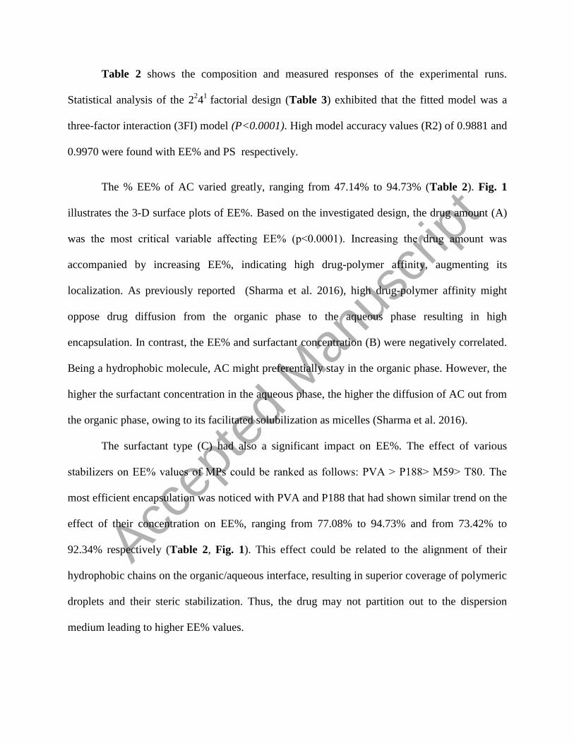
Table 2 shows the composition and measured responses of the experimental runs.
Statistical analysis of the 224
1 factorial design (Table 3) exhibited that the fitted model was a
three-factor interaction (3FI) model (P<0.0001). High model accuracy values (R2) of 0.9881 and
0.9970 were found with EE% and PS respectively.
The % EE% of AC varied greatly, ranging from 47.14% to 94.73% (Table 2). Fig. 1
illustrates the 3-D surface plots of EE%. Based on the investigated design, the drug amount (A)
was the most critical variable affecting EE% (p˂0.0001). Increasing the drug amount was
accompanied by increasing EE%, indicating high drug-polymer affinity, augmenting its
localization. As previously reported (Sharma et al. 2016), high drug-polymer affinity might
oppose drug diffusion from the organic phase to the aqueous phase resulting in high
encapsulation. In contrast, the EE% and surfactant concentration (B) were negatively correlated.
Being a hydrophobic molecule, AC might preferentially stay in the organic phase. However, the
higher the surfactant concentration in the aqueous phase, the higher the diffusion of AC out from
the organic phase, owing to its facilitated solubilization as micelles (Sharma et al. 2016).
The surfactant type (C) had also a significant impact on EE%. The effect of various
stabilizers on EE% values of MPs could be ranked as follows: PVA ˃ P188˃ M59˃ T80. The
most efficient encapsulation was noticed with PVA and P188 that had shown similar trend on the
effect of their concentration on EE%, ranging from 77.08% to 94.73% and from 73.42% to
92.34% respectively (Table 2, Fig. 1). This effect could be related to the alignment of their
hydrophobic chains on the organic/aqueous interface, resulting in superior coverage of polymeric
droplets and their steric stabilization. Thus, the drug may not partition out to the dispersion
medium leading to higher EE% values.
Accep
ted
Man
uscr
ipt

As for M59 and T80, decreased values of EE%, fitted between 56.42% and 89.60% and
between 47.14% and 73.75% respectively, were revealed. This could be explained on the basis of
the critical micelle concentration (CMC) values of both surfactants (0.1% for M59(Goodhart and
Martin 1962) versus 0.0014% for T80 (Wan and Lee 1974). Using concentrations higher than
their CMC values could contribute to micelles formation in the aqueous phase and hence, poor
AC encapsulation. The higher CMC value of M59 as compared to that of T80 might explain the
higher EE% values observed when using the former as a stabilizer.
Similarly, PS varied considerably (51.00 µm - 135.39 µm) as shown in Table 2 and Fig.
2. It is worthy to note that PS values were < 200 µm, thus, possessing a masking effect on oral
grittiness. Low span values ranged between 1.12 ± 0.03 and 4.83 ± 0.54, denoting narrow size
distribution (data not shown). Drug amount (A) did not significantly influence PS (p˃ 0.05),
while both factors B and C were found to had significant effects on PS (p˂ 0.001). Regarding
factor B, increasing concentration of either M59 or T80 yielded smaller particles and the reverse
was true for both PVA and P188. Typically, the formation of small-sized particles upon
increasing concentration of various surfactants could be explained by the alignment of surfactant
molecules on the organic/aqueous interface. The resulted coverage of oil droplets and reduction
of the interfacial tension between the organic and aqueous phase might have led to steric
stabilization with a subsequent decrease in PS. However, for PVA and P188, their chosen
concentrations might lead to deviation from the previously proposed effect and possible
agglomeration. This could be justified based on possible gelatinization of PVA, owing to the
possible formation of hydrogen bonds between intra- or inter- PVA molecules, and to the inter-
particle interaction of P188 chains (Blanco and Alonso 1997; Murakami et al. 1997).
Accep
ted
Man
uscr
ipt

Interestingly, factor C “surfactant type” was found to be the most influential factor on PS
(p˂0.0001). In contrast with the other stabilizers, the PS variation was the least in case of MPs
prepared with PVA, indicating its superior size-reducing effect. Furthermore, higher close PS
values were obtained with MPs prepared using P188 and T80, whereas the highest PS values
were obtained with MPs prepared using M59.
Due to such findings and considering suitable PS for buccal delivery and high EE%
(˃90%), MPs prepared using PVA (PV3 MPs) was selected for incorporation in the network of
in situ gellan gum gel and further investigations (PV3 loaded gel).
[Table 2 near here]
[Table 3 near here]
[Figure 1 near here]
[Figure 2 near here]
Preparation of Plain and Composite In Situ Ion Triggered Gellan Gum Gel, Gelling Capacity,
and pH
The in situ ion-triggered gel comprising of a combination of gellan gum and HPMC and
entrapping calcium ions was successfully prepared (El Maghraby et al. 2012). Herein, calcium
ions were entrapped inside the gellan gum-HPMC sol as citrate complex. The complexing agent,
sodium citrate reacted with calcium chloride, forming calcium citrate complex, ensuring sol
fluidity. Then, calcium ions were liberated in the presence of simulated salivary fluid, due to pH
change, resulting in ionic complexation and triggering instantaneous gelation (El Maghraby et al.
2012).
For in situ gelling systems, the gelling capacity is considered one of the prime
perquisites. The in situ gel is proposed to be in the sol state at room temperature before
Accep
ted
Man
uscr
ipt

administration, for ease of installation, and then undergoes rapid immediate gelation at the
application site at pH 6.8. Additionally, the formed in situ gel should resist dissolution and
maintain its gel consistency for an extended period of time. To achieve this target, optimum
concentrations of the gel base, as well as complexing agents should be obtained.
In the preliminary experiments, it was observed that gellan concentrations below 0.25%
were too low to form the gel network while those below 0.5% underwent gelation after few
minutes (˃5 min) “+ in gelling capacity scoring” or immediate gelation that remained for few
hours “++”. Thus, the optimum concentration of gellan gum was found to be 0.5%, forming a
stiff persistent gel within 15-20 seconds “+++”. Using a 1:1 ratio of gellan gum: HPMC yielded
immediately formed resistant gel structure upon contact with simulated salivary fluid pH 6.8 that
was retained for an extended period of time. Likewise, the concentrations of complexing agents
must ensure lack of free calcium ions in the vehicle, to remain in the fluid form prior to its
application, but following sol instillation, the complex must be dissociated to release sufficient
calcium ions to favor gelation. 0.1% calcium chloride and 0.2% sodium citrate concentrations
were found to be satisfactory to achieve such purpose. Fig. 3 (a) depicts that the prepared in situ
plain gel was homogenous and transparent showing no lumps. The gel homogeneity and absence
of lumps were also confirmed for the composite system (selected PV3 loaded gel), as displayed
in Fig. 3 (b), indicating that incorporating MPs inside in situ gel did not affect gelling capacity.
Considering the satisfactory range of buccal pH (5.5-7.5), the prepared in situ plain gel was
considered non-irritating and well tolerated with a pH value of 7.38±0.05 (Patel et al. 2006;
Dhiman et al. 2008). It is worth mentioning that the formation of a composite system by
combining MPs with the in situ gel did not affect the pH, showing similar value to that of plain
in situ gel (7.4±0.1; p˃0.05).
Accep
ted
Man
uscr
ipt

[Figure 3 near here]
Viscosity Determination
Fig. 4 highlights the viscosity values obtained for plain and in situ composite gel (PV3
loaded gel). The results imply that pseudoplastic behavior was demonstrated as previously
confirmed for gellan gum gels (Harish et al. 2009; Zhu et al. 2015). The shear-thinning behavior
allowed the ease of spreading on buccal mucosa. The high viscosity of the gel also has been
contributed to its mucoadhesive potential (Graciano et al. 2015). It is worthy to note that the
obtained viscosity values were similar for each angular velocity tested (p˃0.05), indicating that
incorporation of MPs in the in situ composite gel did not have an impact on its viscosity.
[Figure 4 near here]
Mucoadhesivity Assessment
Determination of mucoadhesive potential of formulations intended for mucosal delivery
is very essential to evaluate their in vivo performance, intensifying their contact with the
absorption surface (Elmowafy E and Soliman 2019). In particular, the attractive interaction
between DDS and the buccal membrane is highly warranted for better mucosal penetration
(Khutoryanskiy 2011) Utilizing the rheological characteristics of mucin and various
mucoadhesive polymers for the measurement of the mucoadhesivity has been reported
previously (Suvannasara et al. 2013; Soliman ME et al. 2018). Herein, a simple and reproducible
procedure for assessment of mucoadhesiveness of the in situ composite gel was performed
through monitoring viscometric changes of the mucin-gel mixture with a Brookfield viscometer
(Thirawong et al. 2008; Suvannasara et al. 2013). The viscosity enhancement that was noticed
after combining the gel with mucin in the mucin-gel mixture (ηmixture) could be taken as a direct
estimate of their interaction occurring in mucoadhesion “Δƞ; component of mucoadhesion”.
Accep
ted
Man
uscr
ipt

Our results confirmed such proposed mechanism as revealed from the rheological
synergism between mucin and either plain or PV3 loaded gel. The plain and PV3 loaded gels
showed component of mucoadhesion of 17382.33±4810.19 and 62726.67±2085.97 cp
respectively, suggesting greater synergism and hence, the higher mucoadhesive potential of the
in situ composite gel. In addition, PV3 loaded gel displayed a higher ratio of Δƞ/ƞgel (5.18),
almost 3.6 fold higher than that for plain gel (1.43). This could be attributed to its content of
cationic Eudragit RL100, with the possible formation of electrostatic interaction with anionic
gellan gum, allowing high retention of MPs inside the gel matrix.
Morphologic Analysis of the Microstructure of MPs and In Situ Composite Gel
SEM images of the prepared MPs, shown in Fig. 5 (a-d), confirmed the capability of the
selected stabilizers in attaining spherical non-aggregating particles with different surface
morphologies. Surface topography of the MPs prepared using P188 and M59 displayed smooth
surfaces. Contrastingly, the surfaces of MPs prepared using PVA and T80 were rough.
Interestingly, MPs prepared using either PVA or P188 exhibited some small pores.
The microstructure of in situ composite gel is depicted in Fig. 5 (e and f). The plain gel
showed irregular and relatively porous microstructure with a smooth surface, whereas PV3
loaded gel was rough indicating the incorporation of MPs in the porous structure of gellan gum
gel.
[Figure 5 near here]
In vitro Drug Release and Release Kinetics
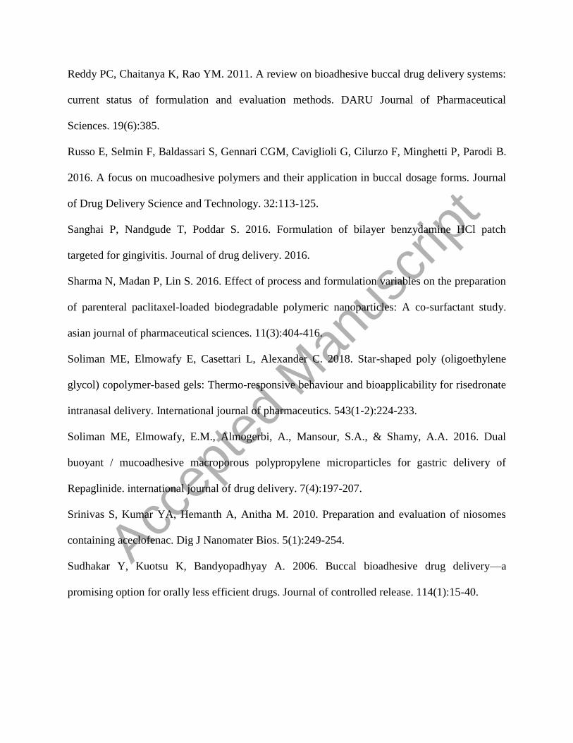
In vitro release profiles of AC from different formulae are illustrated in Fig. 6. AC
powder showed complete release in 3 h. AC is a weakly ionizable phenyl acetic acid derivative
with pH-dependent solubility, showing an experimentally reported high solubility in phosphate
Accep
ted
Man
uscr
ipt

buffer pH 6.8 (10.58 mg/mL, at 37°C) (Usha et al. 2008). Indeed, its weakly acidic nature could
guarantee its enhanced dissolution at neutral or slightly alkaline pH of the simulated salivary
fluid.
Expectedly, the release of AC from the control gel was relatively rapid, showing almost ˃
50% release in the first hour. However, a somewhat prolongation in the release of AC residual
amount was noticed, achieving 100% release within 8 h. This might be due to the hydration and
subsequent pH-dependent swelling behavior of gellan gum (Elmowafy E et al. 2014; Prezotti et
al. 2014; Osmałek et al. 2017), facilitating the penetration of dissolution medium and AC
diffusion.
In contrast, both selected AC Eudragit RL100 MPs (PV3 MPs) and in situ composite gel
(PV3 loaded gel) exhibited a more sustained release manner, reaching 50.61% and 55.33 %
release of AC respectively over 24 h. Slightly higher values of the release of AC were noticed
with MPs than its release in in situ composite gel within the first 4 h. The small size of MPs and
the presence of pores on their surface, as revealed in SEM image, yielded increased surface area,
facilitating the passage of dissolution medium inside the MPs matrix and hence, increasing AC
diffusion and release. This indicated that compact gel matrix of in situ composite gel caused
more retardation of AC release in the first 4 h as compared to MPs alone.
Afterward, a similar non-significant (p˃ 0.05) trend towards sustained AC fashion was obvious
for both formulae. Composite system porosity could guarantee drug leaching from MPs through
the pores and subsequent drug release through the gel network (Hoare and Kohane 2008). In
addition, the porous network of in situ composite gel could allow the penetration of dissolution
medium and might be at the base of lack of variation of AC release from both MPs and MPs
Accep
ted
Man
uscr
ipt
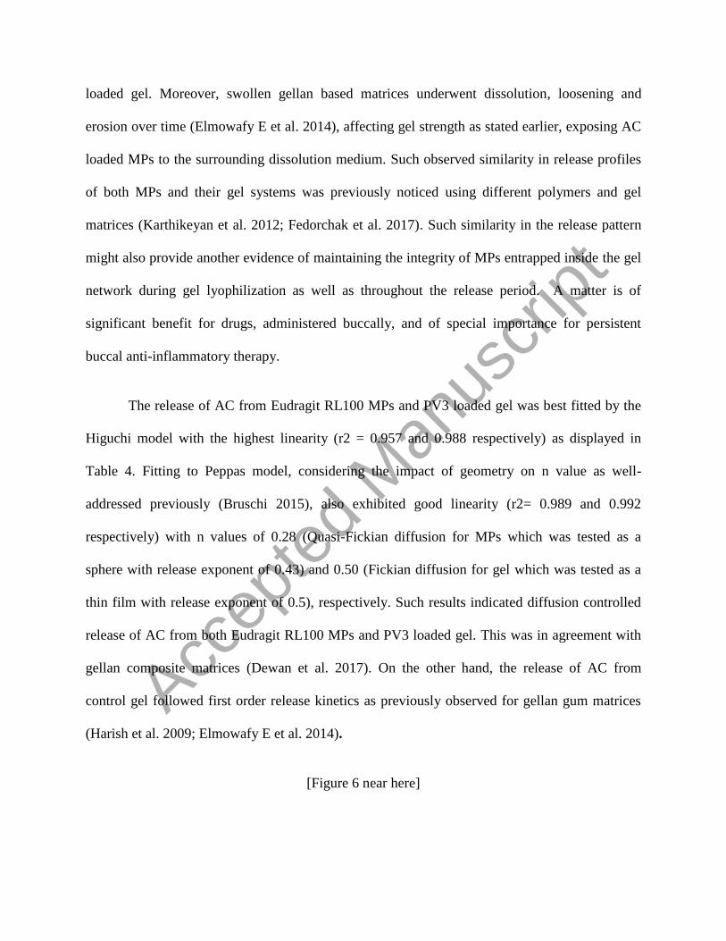
loaded gel. Moreover, swollen gellan based matrices underwent dissolution, loosening and
erosion over time (Elmowafy E et al. 2014), affecting gel strength as stated earlier, exposing AC
loaded MPs to the surrounding dissolution medium. Such observed similarity in release profiles
of both MPs and their gel systems was previously noticed using different polymers and gel
matrices (Karthikeyan et al. 2012; Fedorchak et al. 2017). Such similarity in the release pattern
might also provide another evidence of maintaining the integrity of MPs entrapped inside the gel
network during gel lyophilization as well as throughout the release period. A matter is of
significant benefit for drugs, administered buccally, and of special importance for persistent
buccal anti-inflammatory therapy.
The release of AC from Eudragit RL100 MPs and PV3 loaded gel was best fitted by the
Higuchi model with the highest linearity (r2 = 0.957 and 0.988 respectively) as displayed in
Table 4. Fitting to Peppas model, considering the impact of geometry on n value as well-
addressed previously (Bruschi 2015), also exhibited good linearity (r2= 0.989 and 0.992
respectively) with n values of 0.28 (Quasi-Fickian diffusion for MPs which was tested as a
sphere with release exponent of 0.43) and 0.50 (Fickian diffusion for gel which was tested as a
thin film with release exponent of 0.5), respectively. Such results indicated diffusion controlled
release of AC from both Eudragit RL100 MPs and PV3 loaded gel. This was in agreement with
gellan composite matrices (Dewan et al. 2017). On the other hand, the release of AC from
control gel followed first order release kinetics as previously observed for gellan gum matrices
(Harish et al. 2009; Elmowafy E et al. 2014).
[Figure 6 near here]
Accep
ted
Man
uscr
ipt

Solid State Characterization
Differential Scanning Calorimetry (DSC)
DSC thermogram of AC (Fig. 7) depicted its typical melting point (154.6ºC ) (Jana, Das,
et al. 2013; Patnaik et al. 2015). DSC curve of AC: Eudragit RL100 physical mixture exhibited
slight broadening of AC endothermic peak due to the dilution effect. The disappearance of AC
peak in PV3 confirmed its molecular dispersion and amorphization within MPs.
The thermogram of gellan gum exhibited an endothermic peak at 64.27°C corresponding
to the water liberation and an exothermic peak at 245.73°C resulting from polymer
decomposition. The plain gel was free from characteristic peaks of gellan gum, suggesting gellan
gum gelation and cross-linking. Loss of defined thermal events of gellan gum was noticed in the
thermograms of plain gel and in situ composite gel, denoting obvious retardation of its
degradation in the gel network and higher thermal stability.
[Figure 7 near here]
Fourier Transform Infrared (FT-IR) Spectroscopy
The infrared analysis is usually done to complement the thermal analysis data. It is a very
useful technique for evaluating the possible molecular interaction between drugs and particulate
matrices such as hydrogen bonding. It was previously postulated that the higher the value of the
OH and CO stretching peaks shift, the stronger the nature of the intermolecular hydrogen
bonding interaction. This could modulate drugs entrapment and release characteristics (Menjoge
and Kulkarni 2007; Elmowafy EM et al. 2008; Elmowafy E et al. 2014; Elmowafy E, Gad H, et
al. 2019).
The collected spectra of AC, Eudragit RL100, 1:1 physical mixture, selected MPs, gellan
gum, plain gel and in situ composite gel are illustrated in Fig. 8. AC spectrum showed
Accep
ted
Man
uscr
ipt
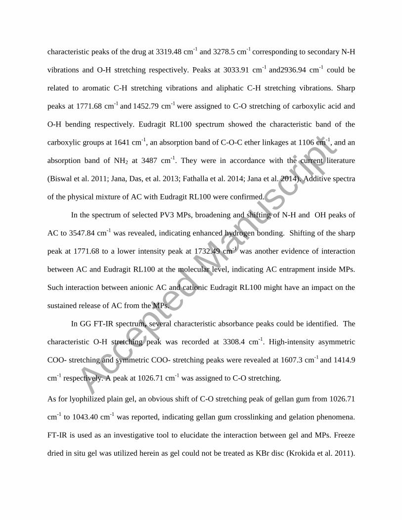
characteristic peaks of the drug at 3319.48 cm-1
and 3278.5 cm-1
corresponding to secondary N-H
vibrations and O-H stretching respectively. Peaks at 3033.91 cm-1
and2936.94 cm-1
could be
related to aromatic C-H stretching vibrations and aliphatic C-H stretching vibrations. Sharp
peaks at 1771.68 cm-1
and 1452.79 cm
-1 were assigned to C-O stretching of carboxylic acid and
O-H bending respectively. Eudragit RL100 spectrum showed the characteristic band of the
carboxylic groups at 1641 cm-1
, an absorption band of C-O-C ether linkages at 1106 cm-1
, and an
absorption band of NH2 at 3487 cm-1
. They were in accordance with the current literature
(Biswal et al. 2011; Jana, Das, et al. 2013; Fathalla et al. 2014; Jana et al. 2014). Additive spectra
of the physical mixture of AC with Eudragit RL100 were confirmed.
In the spectrum of selected PV3 MPs, broadening and shifting of N-H and OH peaks of
AC to 3547.84 cm-1
was revealed, indicating enhanced hydrogen bonding. Shifting of the sharp
peak at 1771.68 to a lower intensity peak at 1732.49 cm-1
was another evidence of interaction
between AC and Eudragit RL100 at the molecular level, indicating AC entrapment inside MPs.
Such interaction between anionic AC and cationic Eudragit RL100 might have an impact on the
sustained release of AC from the MPs.
In GG FT-IR spectrum, several characteristic absorbance peaks could be identified. The
characteristic O-H stretching peak was recorded at 3308.4 cm-1
. High-intensity asymmetric
COO- stretching and symmetric COO- stretching peaks were revealed at 1607.3 cm-1
and 1414.9
cm-1
respectively. A peak at 1026.71 cm-1
was assigned to C-O stretching.
As for lyophilized plain gel, an obvious shift of C-O stretching peak of gellan gum from 1026.71
cm-1
to 1043.40 cm-1
was reported, indicating gellan gum crosslinking and gelation phenomena.
FT-IR is used as an investigative tool to elucidate the interaction between gel and MPs. Freeze
dried in situ gel was utilized herein as gel could not be treated as KBr disc (Krokida et al. 2011).
Accep
ted
Man
uscr
ipt

As revealed in the spectrum of PV3 loaded gel, the O-H stretching peak of gellan gum was
shifted to a higher wavenumber from 3308.4 cm-1
to 3449.17 cm-1
. As a hydroxyl group-
containing polymer, gellan gum could form hydrogen bonding with MPs (NH2 of Eudragit
polymer) as confirmed by a shift in O-H stretching peak (Elmowafy E et al. 2014). The
disappearance of the characteristic band of the carboxylic groups of Eudragit RL100 at 1641 cm-
1 could provide evidence support of electrostatic interaction. Interestingly, characteristic peaks of
AC at 1771.68 and 1452.79 cm-1
were completely disappeared in MPs loaded gel. This could
predict the effective interaction between cationic MPs and anionic gellan gum gel at the
molecular level, profoundly sustained AC release. In addition, such interaction might have an
implication on the viscosity of the in situ gel which is highly warranted for buccal applicability.
[Figure 8 near here]
In vitro Anti-arthritic Activity
The albumin anti-denaturation method is used to investigate AC anti-arthritic effect. Non-
steroidal anti-inflammatory drugs have been proved to prevent protein denaturation as well as to
block COX enzyme, inhibiting the production of endogenous prostaglandins (Paul 1996).
Both Eudragit RL100 MPs and in situ composite gel possessed good anti-denaturation
activity with an obvious dose-dependent response (Fig. 9). Both formulae exhibited IC50 values
of 61.14 and 26.2 μg/mL respectively, evidencing effective results comparable to the positive
control, diclofenac sodium (IC50 value of 15.12 μg/mL). It is interesting to note that plain gel
showed a minimal response, starting from 125 μg/mL. The % inhibition of protein denaturation
for the plain gel was found to be 43.25% at the highest concentration used (1000 μg/mL),
compared to 81.94%, 84.63% and 89.35% for Eudragit RL100 MPs, in situ composite gel and
diclofenac sodium respectively. This might be at the base of higher IC50 value of in situ
Accep
ted
Man
uscr
ipt

composite gel than that of Eudragit RL100 MPs. Such promising activity of in situ composite gel
supports its use as a remedy for arthritis conditions.
[Figure 9 near here]
In vivo Anti-inflammatory Activity
Assessment of in vivo anti-inflammatory effect of AC from CMC solution and the in situ
composite gel was also done, using plain Na CMC solution and plain in situ gel as control
groups. Na CMC aqueous solution was extensively used in literature for in vivo anti-
inflammatory activity study as a control (Jana, Das, et al. 2013; Jana, Saha, et al. 2013). It was
reported to cause no inhibition of inflammatory response produced following the subplantar
injection of carrageenan. In this work, motivated by its minimal positive effect on in vitro anti-
arthritic activity study, the synergistic anti-inflammatory effect of the plain gel was also
investigated for comparison purposes. It was observed that in control groups receiving CMC
solution and plain gel, the subplantar injection of carrageenan produced local edema, denoting
lack of any significant influence on inhibition of inflammatory response (P˃0.05).
Both treated groups, that received standard AC in CMC solution and PV3 loaded gel,
displayed prominent inhibitory effect on edema swelling from the first 1 h and throughout the
whole study time as compared to the control group (p ˂ 0.05) (Fig. 10). A slower inhibitory
effect was demonstrated in the first 2 h for in situ composite gel, reaching 35.47% inhibition of
paw edema as compared to AC showing 63.08% inhibition then became similar in the next 2 h.
However, % inhibition of paw edema was declined after 5 h for AC in CMC solution, whereas,
in situ composite gel remained superior to AC over 8 h after buccal administration. The %
inhibition of PV3 loaded gel was increased by 2.84 fold than AC after 8 h (68.07% versus
23.90%), proving its sustained capability in suppressing paw swelling.
Accep
ted
Man
uscr
ipt

Such an improved and sustained inhibition of inflammatory response may be attributed to
the mucoadhesive potential of gellan gum gel reservoir. The proposed mucoadhesiveness of
gellan gum was achieved by exerting supramolecular interactions with mucin chains, favoring
MPs immobilization and residence in the buccal mucosa. This was profoundly coupled with a
continuous local concentration gradient of the entrapped drug over a prolonged period of time
(Meneguin et al. 2018; Prezotti et al. 2018).
[Figure 10 near here]
Conclusion
A buccal delivery system of an in situ composite gellan gum gel containing AC Eudragit
RL100 MPs was successfully developed in two steps. The first step aimed to prepare and
optimize AC Eudragit RL100 MPs by the emulsion solvent evaporation technique. The second
one explored the suitability of in situ ion triggered gellan gum gel to develop promising in situ
composite gel. The proposed in situ composite gel not only succeeded in sustaining AC release
but also ensured ease of buccal applicability of MPs as well as prolonged localization. A
sustained and prolonged drug-effects behavior of MPs loaded gel was demonstrated, relative to
standard drug.
Acknowledgments
The authors would like to acknowledge Evonik Pharma, GmbH, Darmstadt, Germany and
E.I.P.I.C.O. Company, Cairo, Egypt for their kind supply of Eudragit RL100 and HPMC
respectively. The authors would also like to thank Bristol-Myers Squibb Company, Cairo, Egypt
for their kind supply of aceclofenac.
Accep
ted
Man
uscr
ipt

Funding information
This manuscript received no sources of financial funding.
Disclosure of interest
The authors report no conflict of interest
References
Animesh K, Afrasim M, R Bommareddy R, Ayaz A, Shruthi R, G Shivakumar H. 2012.
Applicability and approaches of (Meth) acrylate copolymers (Eudragits) in novel drug delivery
systems. Current Drug Therapy. 7(4):219-234.
Biswal I, Dinda A, Mohanty S, Dhara M, Das D, Chowdary K, Si S. 2011. Influence of
Drug/Polymer Ratio on the Encapsulation Efficiency of Highly Hydrophilic Drug. Asian Journal
of Chemistry. 23(5):1973.
Blanco M, Alonso M. 1997. Development and characterization of protein-loaded poly (lactide-
co-glycolide) nanospheres. European Journal of Pharmaceutics and Biopharmaceutics.
43(3):287-294.
Boni FI, Prezotti FG, Cury BSF. 2016. Gellan gum microspheres crosslinked with trivalent ion:
effect of polymer and crosslinker concentrations on drug release and mucoadhesive properties.
Drug development and industrial pharmacy. 42(8):1283-1290.
Bruschi ML. 2015. Strategies to modify the drug release from pharmaceutical systems.
Woodhead Publishing.
Chan SY, Goh CF, Lau JY, Tiew YC, Balakrishnan T. 2019. Rice starch thin films as a potential
buccal delivery system: Effect of plasticiser and drug loading on drug release profile.
International Journal of Pharmaceutics. 562:203-211.
Accep
ted
Man
uscr
ipt

Cid YP, Pedrazzi V, de Sousa VP, Pierre MBR. 2012. In vitro characterization of chitosan gels
for buccal delivery of celecoxib: influence of a penetration enhancer. AAPS PharmSciTech.
13(1):101-111.
Cui F, Qian F, Zhao Z, Yin L, Tang C, Yin C. 2009. Preparation, characterization, and oral
delivery of insulin loaded carboxylated chitosan grafted poly (methyl methacrylate)
nanoparticles. Biomacromolecules. 10(5):1253-1258.
Dasgupta S, Dey S, Choudhury S, Mazumder B. 2013. Topical delivery of aceclofenac as
nanoemulsion comprising excipients having optimum emulsification capabilities: preparation,
characterization and in vivo evaluation. Expert opinion on drug delivery. 10(4):411-420.
Dewan M, Sarkar G, Bhowmik M, Das B, Chattoapadhyay AK, Rana D, Chattopadhyay D.
2017. Effect of gellan gum on the thermogelation property and drug release profile of Poloxamer
407 based ophthalmic formulation. International Journal of Biological Macromolecules.
102:258-265.
Dhiman M, Yedurkar P, Sawant KK. 2008. Formulation, characterization, and in vitro evaluation
of bioadhesive gels containing 5-fluorouracil. Pharmaceutical development and technology.
13(1):15-25.
El Maghraby GM, Elzayat EM, Alanazi FK. 2012. Development of modified in situ gelling oral
liquid sustained release formulation of dextromethorphan. Drug development and industrial
pharmacy. 38(8):971-978.
Elisha IL, Dzoyem J-P, McGaw LJ, Botha FS, Eloff JN. 2016. The anti-arthritic, anti-
inflammatory, antioxidant activity and relationships with total phenolics and total flavonoids of
nine South African plants used traditionally to treat arthritis. BMC complementary and
alternative medicine. 16(1):307.
Accep
ted
Man
uscr
ipt

Elkomy MH, El Menshawe SF, Abou-Taleb HA, Elkarmalawy MH. 2017. Loratadine
bioavailability via buccal transferosomal gel: formulation, statistical optimization, in vitro/in
vivo characterization, and pharmacokinetics in human volunteers. Drug Delivery. 24(1):781-791.
Elmowafy E, Abdal-hay A, Skouras A, Tiboni M, Casettari L, Guarino V. 2019.
Polyhydroxyalkanoate (PHA): applications in drug delivery and tissue engineering. Expert
review of medical devices.(just-accepted).
Elmowafy E, Gad H, Biondo F, Casettari L, Soliman ME. 2019. Exploring optimized methoxy
poly (ethylene glycol)-block-poly (ε-caprolactone) crystalline cored micelles in anti-glaucoma
pharmacotherapy. International journal of pharmaceutics.
Elmowafy E, Osman R, AH Ishak R. 2017. Polymer-based novel lung targeted delivery systems.
Current pharmaceutical design. 23(3):373-392.
Elmowafy E, Osman R, El-Shamy AE-HA, Awad GA. 2014. Nasal polysaccharides-glucose
regulator microparticles: Optimization, tolerability and antidiabetic activity in rats. Carbohydrate
polymers. 108:257-265.
Elmowafy E, Soliman ME. 2019. Losartan-chitosan/dextran sulfate microplex as a carrier to lung
therapeutics: Dry powder inhalation, aerodynamic profile and pulmonary tolerability.
International journal of biological macromolecules.
Elmowafy EM, Awad GA, Mansour S, El-Shamy AE-HA. 2008. Release mechanisms behind
polysaccharides-based famotidine controlled release matrix tablets. AAPS PharmSciTech.
9(4):1230-1239.
Elmowafy EM, Tiboni M, Soliman ME. 2019. Biocompatibility, biodegradation and biomedical
applications of poly (lactic acid)/poly (lactic-co-glycolic acid) micro and nanoparticles. Journal
of Pharmaceutical Investigation.1-34.
Accep
ted
Man
uscr
ipt

Fathalla D, Abdel-Mageed A, Abdel-Hamid F, Ahmed M. 2014. In-vitro and in-vivo evaluation
of niosomal gel containing aceclofenac for sustained drug delivery. International Journal of
Pharmaceutical Sciences Research. 2014.
Fedorchak MV, Conner IP, Schuman JS, Cugini A, Little SR. 2017. Long term glaucoma drug
delivery using a topically retained gel/microsphere eye drop. Scientific reports. 7(1):8639.
Ferreira IS, Bettencourt A, Bétrisey B, Gonçalves LM, Trampuz A, Almeida AJ. 2015.
Improvement of the antibacterial activity of daptomycin-loaded polymeric microparticles by
Eudragit RL 100: an assessment by isothermal microcalorimetry. International journal of
pharmaceutics. 485(1-2):171-182.
Ferreira IS, Kikhney J, Kursawe L, Kasper S, Gonçalves LM, Trampuz A, Moter A, Bettencourt
AF, Almeida AJ. 2018. Encapsulation in Polymeric Microparticles Improves Daptomycin
Activity Against Mature Staphylococci Biofilms—a Thermal and Imaging Study. AAPS
PharmSciTech. 19(4):1625-1636.
Fini A, Bergamante V, Ceschel GC. 2011. Mucoadhesive gels designed for the controlled release
of chlorhexidine in the oral cavity. Pharmaceutics. 3(4):665-679.
Galey WR, Lonsdale H, Nacht S. 1976. The in vitro permeability of skin and buccal mucosa to
selected drugs and tritiated water. Journal of Investigative Dermatology. 67(6):713-717.
Garipova V, Gennari C, Selmin F, Cilurzo F, Moustafine R. 2018. Mucoadhesive
interpolyelectrolyte complexes for the buccal delivery of clobetasol. Polymers. 10(1):85.
Goodhart FW, Martin AN. 1962. Solubilization of benzoic acid derivatives by polyoxyethylene
stearates. Journal of pharmaceutical sciences. 51(1):50-54.
Graciano TB, Coutinho TS, Cressoni CB, de Paula Freitas C, Pierre MBR, de Lima Pereira SA,
Shimano MM, da Cunha Frange RC, Garcia MTJ. 2015. Using chitosan gels as a toluidine blue
Accep
ted
Man
uscr
ipt

O delivery system for photodynamic therapy of buccal cancer: In vitro and in vivo studies.
Photodiagnosis and photodynamic therapy. 12(1):98-107.
Harish N, Prabhu P, Charyulu R, Gulzar M, Subrahmanyam E. 2009. Formulation and evaluation
of in situ gels containing clotrimazole for oral candidiasis. Indian journal of pharmaceutical
sciences. 71(4):421.
Harsh S, Patel K, Padhyay U. 2014. Formulation and evaluation of controlled release colon
targeted micro sponge of Aceclofenac. The Pharmacy Innovation Journal. 3(10):81-87.
Hassan EE, Gallo JM. 1990. A simple rheological method for the in vitro assessment of mucin-
polymer bioadhesive bond strength. Pharmaceutical research. 7(5):491-495.
Hassan M, Barakat N, El-Badry M, Shehata S. 2011. Formulation and in vitro/in vivo evaluation
of naproxen mucoadhesive buccal patches for local effect. Journal of drug delivery science and
technology. 21(5):423.
Hoare TR, Kohane DS. 2008. Hydrogels in drug delivery: Progress and challenges. Polymer.
49(8):1993-2007.
Ibrahim E-S, Ismail S, Fetih G, Shaaban O, Hassanein K, Abdellah N. 2012. Development and
characterization of thermosensitive pluronic-based metronidazole in situ gelling formulations for
vaginal application. Acta pharmaceutica. 62(1):59-70.
Ilić T, Savić S, Batinić B, Marković B, Schmidberger M, Lunter D, Savić M, Savić S. 2018.
Combined use of biocompatible nanoemulsions and solid microneedles to improve transport of a
model NSAID across the skin: In vitro and in vivo studies. European Journal of Pharmaceutical
Sciences. 125:110-119.
Accep
ted
Man
uscr
ipt

Irimia T, Dinu-Pîrvu C-E, Ghica M, Lupuleasa D, Muntean D-L, Udeanu D, Popa L. 2018.
Chitosan-based in situ gels for ocular delivery of therapeutics: A state-of-the-art Review. Marine
drugs. 16(10):373.
Jana S, Das A, Nayak AK, Sen KK, Basu SK. 2013. Aceclofenac-loaded unsaturated esterified
alginate/gellan gum microspheres: in vitro and in vivo assessment. International journal of
biological macromolecules. 57:129-137.
Jana S, Manna S, Nayak AK, Sen KK, Basu SK. 2014. Carbopol gel containing chitosan-egg
albumin nanoparticles for transdermal aceclofenac delivery. Colloids and surfaces B:
Biointerfaces. 114:36-44.
Jana S, Saha A, Nayak AK, Sen KK, Basu SK. 2013. Aceclofenac-loaded chitosan-tamarind
seed polysaccharide interpenetrating polymeric network microparticles. Colloids and Surfaces B:
Biointerfaces. 105:303-309.
Karthikeyan K, Durgadevi R, Saravanan K, Shivsankar K, Usha S, Saravanan M. 2012.
Formulation of bioadhesive carbomer gel incorporating drug-loaded gelatin microspheres for
periodontal therapy. Tropical Journal of Pharmaceutical Research. 11(3):335-343.
Kesavan K, Nath G, Pandit J. 2010. Preparation and in vitro antibacterial evaluation of
gatifloxacin mucoadhesive gellan system. Daru: journal of Faculty of Pharmacy, Tehran
University of Medical Sciences. 18(4):237.
Khames A. 2019. Hexyl alginate derivative, an amphiphilic innovative buccal film-forming
material of promising mechanical and release characteristics for the improvement of repaglinide
bioavailability. Drug design, development and therapy. 13:925-940. eng.
Khutoryanskiy VV. 2011. Advances in Mucoadhesion and Mucoadhesive Polymers.
Macromolecular Bioscience. 11(6):748-764.
Accep
ted
Man
uscr
ipt

Krokida M, Pappa A, Agalioti M. 2011. Effect of drying on Aloe's functional components.
Procedia Food Science. 1:1523-1527.
Lopez FL, Bowles A, Gul MO, Clapham D, Ernest TB, Tuleu C. 2016. Effect of formulation
variables on oral grittiness and preferences of multiparticulate formulations in adult volunteers.
European Journal of Pharmaceutical Sciences. 92:156-162.
Madhusudhan S, Panda AK, Parimalakrishnan S, Manavalan R, Manna P. 2010. Design, in vitro
and in vivo evaluation of glipizide Eudragit microparticles. Journal of microencapsulation.
27(4):281-291.
Mahdi MH, Conway BR, Mills T, Smith AM. 2016. Gellan gum fluid gels for topical
administration of diclofenac. International journal of pharmaceutics. 515(1):535-542.
Mahdi MH, Conway BR, Smith AM. 2014. Evaluation of gellan gum fluid gels as modified
release oral liquids. International journal of pharmaceutics. 475(1):335-343.
Meneguin AB, Beyssac E, Garrait G, Hsein H, Cury BS. 2018. Retrograded starch/pectin coated
gellan gum-microparticles for oral administration of insulin: A technological platform for
protection against enzymatic degradation and improvement of intestinal permeability. European
Journal of Pharmaceutics and Biopharmaceutics. 123:84-94.
Menjoge A, Kulkarni M. 2007. Mechanistic investigations of phase behavior in Eudragit® E
blends. International journal of pharmaceutics. 343(1-2):106-121.
Miyazaki S, Aoyama H, Kawasaki N, Kubo W, Attwood D. 1999. In situ-gelling gellan
formulations as vehicles for oral drug delivery. Journal of Controlled Release. 60(2):287-295.
Momoh M, Kenechukwu F, Adedokun M, Odo C, Attama A. 2014. Pharmacodynamics of
diclofenac from novel Eudragit entrapped microspheres. Drug delivery. 21(3):193-203.
Accep
ted
Man
uscr
ipt

Morales JO, Su R, McConville JT. 2013. The influence of recrystallized caffeine on water-
swellable polymethacrylate mucoadhesive buccal films. AAPS PharmSciTech. 14(2):475-484.
Mouftah S, Abdel-Mottaleb MM, Lamprecht A. 2016. Buccal delivery of low molecular weight
heparin by cationic polymethacrylate nanoparticles. International journal of pharmaceutics.
515(1-2):565-574.
Murakami H, Kawashima Y, Niwa T, Hino T, Takeuchi H, Kobayashi M. 1997. Influence of the
degrees of hydrolyzation and polymerization of poly (vinylalcohol) on the preparation and
properties of poly (DL-lactide-co-glycolide) nanoparticle. International journal of pharmaceutics.
149(1):43-49.
Nagarwal RC, Pandit J. 2008. Phase transition system: novel oral in-situ gel. Current drug
delivery. 5(4):282-289.
Nazari K, Kontogiannidou E, Ahmad RH, Gratsani A, Rasekh M, Arshad MS, Sunar BS,
Armitage D, Bouropoulos N, Chang M-W. 2017. Development and characterisation of cellulose
based electrospun mats for buccal delivery of non-steroidal anti-inflammatory drug (NSAID).
European Journal of Pharmaceutical Sciences. 102:147-155.
Osmałek T, Milanowski B, Froelich A, Szybowicz M, Białowąs W, Kapela M, Gadziński P,
Ancukiewicz K. 2017. Design and characteristics of gellan gum beads for modified release of
meloxicam. Drug Development and Industrial Pharmacy. 43(8):1314-1329.
Patel B, Banwait H, Parmar K, Patel M. 2011. Formulation and evaluation of topical aceclofenac
gel using different gelling agent. International Journal of drug development and research.
Patel V, Prajapat B, Patel J, Patel M. 2006. Physicochemical characterization and evaluation of
buccal adhesive patches containing propranolol hydrochloride. Current drug delivery. 3(3):325-
331.
Accep
ted
Man
uscr
ipt

Patel VF, Liu F, Brown MB. 2011. Advances in oral transmucosal drug delivery. Journal of
controlled release. 153(2):106-116.
Patil HG, Tiwari RV, Repka MA, Singh KK. 2016. Formulation and development of
orodispersible sustained release tablet of domperidone. Drug development and industrial
pharmacy. 42(6):906-915.
Patnaik S, Aditha SK, Rattan T, Kamisetti V. 2015. Aceclofenac-Soluplus® Nanocomposites for
Increased Bioavailability. Soft Nanoscience Letters. 5(02):13.
Paul A. 1996. Analgesic-antipyretics and antiinflammatory agents and drugs employed in the
treatment of gout. In The Pharmacological Basis of Therapeutics.617-657.
Perioli L, Ambrogi V, Angelici F, Ricci M, Giovagnoli S, Capuccella M, Rossi C. 2004.
Development of mucoadhesive patches for buccal administration of ibuprofen. Journal of
controlled release. 99(1):73-82.
Perioli L, Ambrogi V, Giovagnoli S, Ricci M, Blasi P, Rossi C. 2007. Mucoadhesive bilayered
tablets for buccal sustained release of flurbiprofen. AAPS PharmSciTech. 8(3):E20-E27.
Prezotti FG, Boni FI, Ferreira NN, Campana-Filho SP, Almeida A, Vasconcelos T, Gremião
MPD, Cury BSF, Sarmento B. 2018. Gellan Gum/Pectin Beads Are Safe and Efficient for the
Targeted Colonic Delivery of Resveratrol. Polymers. 10(1):50.
Prezotti FG, Cury BSF, Evangelista RC. 2014. Mucoadhesive beads of gellan gum/pectin
intended to controlled delivery of drugs. Carbohydrate polymers. 113:286-295.
Raj R, Mongia P, Ram A, Jain N. 2016. Enhanced skin delivery of aceclofenac via hydrogel-
based solid lipid nanoparticles. Artificial cells, nanomedicine, and biotechnology. 44(6):1434-
1439.
Accep
ted
Man
uscr
ipt
Reddy PC, Chaitanya K, Rao YM. 2011. A review on bioadhesive buccal drug delivery systems:
current status of formulation and evaluation methods. DARU Journal of Pharmaceutical
Sciences. 19(6):385.
Russo E, Selmin F, Baldassari S, Gennari CGM, Caviglioli G, Cilurzo F, Minghetti P, Parodi B.
2016. A focus on mucoadhesive polymers and their application in buccal dosage forms. Journal
of Drug Delivery Science and Technology. 32:113-125.
Sanghai P, Nandgude T, Poddar S. 2016. Formulation of bilayer benzydamine HCl patch
targeted for gingivitis. Journal of drug delivery. 2016.
Sharma N, Madan P, Lin S. 2016. Effect of process and formulation variables on the preparation
of parenteral paclitaxel-loaded biodegradable polymeric nanoparticles: A co-surfactant study.
asian journal of pharmaceutical sciences. 11(3):404-416.
Soliman ME, Elmowafy E, Casettari L, Alexander C. 2018. Star-shaped poly (oligoethylene
glycol) copolymer-based gels: Thermo-responsive behaviour and bioapplicability for risedronate
intranasal delivery. International journal of pharmaceutics. 543(1-2):224-233.
Soliman ME, Elmowafy, E.M., Almogerbi, A., Mansour, S.A., & Shamy, A.A. 2016. Dual
buoyant / mucoadhesive macroporous polypropylene microparticles for gastric delivery of
Repaglinide. international journal of drug delivery. 7(4):197-207.
Srinivas S, Kumar YA, Hemanth A, Anitha M. 2010. Preparation and evaluation of niosomes
containing aceclofenac. Dig J Nanomater Bios. 5(1):249-254.
Sudhakar Y, Kuotsu K, Bandyopadhyay A. 2006. Buccal bioadhesive drug delivery—a
promising option for orally less efficient drugs. Journal of controlled release. 114(1):15-40.
Accep
ted
Man
uscr
ipt

Suresh PK, Manhar S. 2014. Bioadhesive buccal gels impregnated with fluconazole:
formulation, in vitro and ex vivo characterization. Journal of Applied Pharmaceutical Science.
4(3):15.
Suvannasara P, Juntapram K, Praphairaksit N, Siralertmukul K, Muangsin N. 2013.
Mucoadhesive 4-carboxybenzenesulfonamide-chitosan with antibacterial properties.
Carbohydrate polymers. 94(1):244-252.
Swain GP, Patel S, Gandhi J, Shah P. 2019. Development of Moxifloxacin Hydrochloride loaded
in-situ gel for the treatment of periodontitis: In-vitro drug release study and antibacterial activity.
Journal of oral biology and craniofacial research. 9(3):190-200.
Thirawong N, Kennedy RA, Sriamornsak P. 2008. Viscometric study of pectin–mucin
interaction and its mucoadhesive bond strength. Carbohydrate Polymers. 71(2):170-179.
Usha AN, Mutalik S, Reddy MS, Ranjith AK, Kushtagi P, Udupa N. 2008. Preparation and, in
vitro, preclinical and clinical studies of aceclofenac spherical agglomerates. European journal of
pharmaceutics and biopharmaceutics. 70(2):674-683.
Wan LS, Lee PF. 1974. CMC of polysorbates. Journal of Pharmaceutical Sciences. 63(1):136-
137.
Xie H, Li L, Sun Y, Wang Y, Gao S, Tian Y, Ma X, Guo C, Bo F, Zhang L. 2019. An Available
Strategy for Nasal Brain Transport of Nanocomposite Based on PAMAM Dendrimers via In Situ
Gel. Nanomaterials. 9(2):147.
Zhu L, Ao J, Li P. 2015. A novel in situ gel base of deacetylase gellan gum for sustained
ophthalmic drug delivery of ketotifen: in vitro and in vivo evaluation. Drug design, development
and therapy. 9:3943.
Accep
ted
Man
uscr
ipt

Fig.1. 3D surface plots of EE% response for drug amounts (I) 50 mg and (II) 100 mg
Accep
ted
Man
uscr
ipt

Fig.2. 3D surface plots of PS response for drug amounts (I) 50 mg and (II) 100 mg
Accep
ted
Man
uscr
ipt

Fig.3. Sol-gel phase transition of in situ plain (a) and composite (b) gel at 37 °C upon contact with simulated
salivary fluid pH 6.8
Fig. 4. Viscosity measurements of the investigated plain gel and PV3 loaded gel. Experiments were performed
in triplicate; data are presented as average values±standard deviation.
0
2
4
6
8
10
12
14
0.0052 0.01 0.052 0.104 0.523 1.047 2.61 5.23 7.85 10.47
Vis
cosi
ty (
Pa.
s)
Angular velocity (rad/sec)
Plain gel PV3 loaded gel
Accep
ted
Man
uscr
ipt

Fig. 5. SEM of representative aceclofenac microparticles using different stabilizers (a) PVA, (b) P 188, (c) M
59 and (d) T 80, and in situ composite gel (e) plain gel and (f) PV3 loaded gel (magnification power X 1400,
1500, 4000, 1100, 50 and 50 for a-f respectively)
Accep
ted
Man
uscr
ipt
Fig. 6. In vitro release pattern of different aceclofenac formulations. Experiments were performed in
triplicate; data are presented as average values±standard deviation.
0
10
20
30
40
50
60
70
80
90
100
0 2 4 6 8 10 12 14 16 18 20 22 24
% A
cecl
ofe
nac
re
leas
e
Time (h)
AC drug
AC loaded gel
PV3 loaded gel
PV3 MPs
Accep
ted
Man
uscr
ipt

Fig. 7. DSC thermograms of aceclofenac, eudragit RL100, 1:1 physical mixture, PV3 MPs, gellan gum, plain
gel and PV3 loaded gel
0 50 100 150 200 250 300
Temprature °C
Aceclofenac Eudragit RL100 Physical Mixture PV3 MPs Gellan gum Plain gel PV3 loaded gelEn
do
ther
mic
Accep
ted
Man
uscr
ipt

Fig. 8. FT-IR spectra of aceclofenac, eudragit RL100, 1:1 physical mixture, PV3 MPs, gellan gum, plain gel
and PV3 loaded gel
5001000150020002500300035004000
wave number (cm-1)
Aceclofenac Eudrajit RL100 1:1 Physical mixture PV3 MPs
Gellan gum Plain gel PV3 loaded gel
Accep
ted
Man
uscr
ipt

Fig. 9. Anti-arthritic activity of PV3 loaded gel (compared to PV3 MPs and plain gel) and diclofenac sodium
as positive control. Experiments were performed in triplicate; data are presented as average values±standard
deviation. Concentration values were for diclofenac and aceclofenac drugs. The % inhibition for plain gel was
tested for gel formulation without adding PV3 MPs.
Fig. 10. The percentage inhibition of paw edema swelling of PV3 loaded gel (test) compared to aceclofenac
(standard). Experiments were performed on 6 rats; data are presented as average values±standard deviation.
0
10
20
30
40
50
60
70
80
90
100
7.81 15.63 31.25 62.5 125 250 500 1000
Inh
ibit
ion
of
pro
tein
den
atu
rati
on
%
Concentrtion (µg/mL)
Diclofenac sodium PV3 Plain gel PV3 loaded gel
0
10
20
30
40
50
60
70
80
1 2 3 4 5 6 7 8
Inh
ibit
ion
of
swe
llin
g %
Time (h)
Standard aceclofenac PV3 loaded gel
Accep
ted
Man
uscr
ipt

Figure Captions
Fig.1. 3D surface plots of EE% response for drug amounts (I) 50 mg and (II) 100 mg
Fig.2. 3D surface plots of PS response for drug amounts (I) 50 mg and (II) 100 mg
Fig.3. Fig.3. Sol-gel phase transition of in situ plain (a) and composite (b) gel at 37 °C upon contact with
simulated salivary fluid pH 6.8
Fig. 4. Viscosity measurements of the investigated plain gel and PV3 loaded gel. Experiments were performed
in triplicate; data are presented as average values±standard deviation.
Fig. 5. SEM of representative aceclofenac microparticles using different stabilizers (a) PVA, (b) P 188, (c) M
59 and (d) T 80, and in situ composite gel (e) plain gel and (f) PV3 loaded gel
Fig. 6. In vitro release pattern of different aceclofenac formulations. Experiments were performed in
triplicate; data are presented as average values±standard deviation.
Fig. 7. DSC thermograms of aceclofenac, eudragit RL100, 1:1 physical mixture, PV3 MPs, gellan gum, plain
gel and PV3 loaded gel
Fig. 8. FT-IR spectra of aceclofenac, eudragit RL100, 1:1 physical mixture, PV3 MPs, gellan gum, plain gel
and PV3 loaded gel
Fig. 9. Anti-arthritic activity of PV3 loaded gel (compared to PV3 MPs and plain gel) and diclofenac sodium
as positive control. Experiments were performed in triplicate; data are presented as average values±standard
deviation. Concentration values were for diclofenac and aceclofenac drugs. The % inhibition for plain gel was
tested for gel formulation without adding PV3 MPs.
Fig. 10. The percentage inhibition of paw edema swelling of PV3 loaded gel (test) compared to aceclofenac
(standard). Experiments were performed on 6 rats; data are presented as average values±standard deviation.
Accep
ted
Man
uscr
ipt

Table 1: 224
1 full factorial design
Factors Levels
A:Drug amount 50 mg 100 mg
B:Surfactant
concentration
0.25% 0.5%
C:Surfactant type PVA Poloxamer 188 Myrj 59 Tween 80
Accep
ted
Man
uscr
ipt

Table 2: composition of prepared aceclofenac microparticles according to the the 224
1 full
factorial design and their measured responses
MPs
code
Independent variables Responses
A:Drug
amount
(mg)
B:Surfactant
concentration
(%)
C:Surfactant
type
Y1: EE% Y2: PS (µm)
PV1 50 0.25 PVA 80.44 ± 0.32 58.75 ± 0.71
P1 50 0.25 Poloxamer 188 74.25 ± 1.78 114.53 ± 2.14
M1 50 0.25 Myrj 59 60.39 ± 0.57 116.83 ± 4.72
T1 50 0.25 Tween 80 73.75 ± 1.02 120.72 ± 0.37
PV2 50 0.5 PVA 77.08 ± 2.52 61.75 ± 0.87
P2 50 0.5 Poloxamer 188 73.42 ± 1.58 125.44 ± 0.40
M2 50 0.5 Myrj 59 56.42 ± 5.52 110.38 ± 2.08
T2 50 0.5 Tween 80 47.14 ± 1.47 77.90 ± 0.70
PV3 100 0.25 PVA 94.73 ± 0.43
51.00 ± 0.87
P3 100 0.25 Poloxamer 188 92.09 ± 0.66
80.74 ± 1.31
M3 100 0.25 Myrj 59 88.96 ± 0.22
135.39 ± 2.02
T3 100 0.25 Tween 80 73.37 ± 0.64
126.52 ± 3.04
PV4 100 0.5 PVA 86.32 ± 0.27 66.78 ± 0.16
P4 100 0.5 Poloxamer 188 92.34 ± 1.39 105.65 ± 1.69
M4 100 0.5 Myrj 59 89.60 ± 0.42 112.92 ± 1.13
T4 100 0.5 Tween 80 63.57 ± 0.97 106.72 ± 0.73
(Experiments were performed in triplicate; data are presented as average values±standard deviation “SD”)
Accep
ted
Man
uscr
ipt

Table 3: Sum of squares and F-value for the measured responses
Model term Sum of squares F-value
EE% PS EE% PS
Model 8421.35 34021.50 176.48 699.91
A 2871.21 8.164*10-3
901.28s
2.519*10-3
B 829.84 262.15 260.85s
80.89s
C 3461.33 25041.50 362.68s
2575.83s
AB 1.37 208.87 0.43 64.45s
AC 495.06 3358.33 51.87s
345.45s
BC 474.81 4508.09 49.75s
463.71s
ABC 291.73 642.56 30.57s
214.19s
ssignificant at P< 0.001
Table 4: Release kinetics of selected MPs and in situ composite gel
Formula First order Zero order Higuchi Peppas
R2
R2 R
2 R
2 (n value)
PV3 MPs 0.955 0.936 0.957 0.989(n=0.28)
PV3 loaded in
situ gel
0.821 0.843 0.988 0.992 (n=0.5)
Accep
ted
Man
uscr
ipt
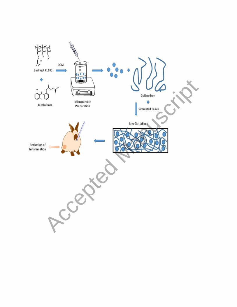
Accep
ted
Man
uscr
ipt