In Silico Analysis of β-Thalassemia Mutations in India and ... · Dibyajyoti Rabha1, Dipankar...
11
188 J Pharm Chem Biol Sci, September - November 2018; 6(3):188-198 Journal of Pharmaceutical, Chemical and Biological Sciences ISSN: 2348-7658 CODEN: JPCBBG September - November 2018; 6(3):188-198 Online available at https://www.jpcbs.info In Silico Analysis of β-Thalassemia Mutations in India and its Neighbouring South East Asian Countries Dibyajyoti Rabha 1 , Dipankar Baruah 2 , Paresh Kumar Sarma 2 , Jatin Sarmah 3 * 1 Dibyajyoti Rabha, DBT (Govt. of India) sponsored Bioinformatics Infrastructure Facility, Bodoland University, Kokrajhar, Assam, India 2 Dipankar Baruah, Associate Professor, Department of Pathology, Fakhruddin Ali Ahmed Medical College and Hospital, Barpeta, Assam, India 2 Paresh Kumar Sarma, Associate Professor, Department of Medicine, Fakhruddin Ali Ahmed Medical College and Hospital, Barpeta, Assam, India 3 Jatin Sarmah, Associate Professor, Department of Biotechnology & Co-ordinator, DBT (Govt. of India) sponsored Bioinformatics Infrastructure Facility, Bodoland University, Kokrajhar, Assam, India *CORRESPONDING AUTHOR Dr.Jatin Sarmah, Associate Professor, Department of Biotechnology & Co-ordinator, DBT (Govt. of India) sponsored Bioinformatics Infrastructure Facility, Bodoland University, Kokrajhar, Assam, India Email: [email protected] ARTICLE INFORMATION Received August 03, 2018 Revised September 12, 2018 Accepted September 14, 2018 Published October 03, 2018 INTRODUCTION Thalassemia is single gene disorder that occurs due to mutation in the Haemoglobin (Hb) gene and result in microcytic anemia [1]. HBA and HBB are the two sub-units of Haemoglobin with the α and β chain respectively. Thalassemia that arises due to mutation in the β -globin chain of Hb is called β-thalassemia consisting of more than 800 variants in the HBB gene described in the Beta Globin Gene Server [2-4]. Point mutations form the majority in HBB mutations. Mutations in promoter, introns, splice sites and exons affect normal functioning of the gene. Functions of the gene such as Research Article The work is licensed under ABSTRACT Thalassemia that arises due to mutation in the β-globin chain of Haemoglobin is called β-thalassemia. In silico analysis of the 4 common mutations- CD 26(G>A), IVS1-5(G>C), IVS1-1(G>T) and CD41/42(- CTTT) occurring frequently in India as well as in its South East Asian neighbouring countries were performed. Literature search on β-thalassemia mutation and their frequencies were performed on Pubmed database in the populations included in the study. The four common mutations found present in all the populations are analysed using bioinformatics tools for in silico analysis, genotype-phenotype correlation and interactome. One of the common mutations is used for predicting its secondary structure using online tool Protein Homology/analogy Recognition Engine v2.0 (PHYRE2). The pathogenicity of these mutations are assessed along with segregation and expression analysis. Structural and functional analysis of CD 26(G>A) was performed. The pathogenicity result for the mis- sense HBB variant CD 26(G>A) is predicted to have no pathogenicty expression but for the intronic mutations IVS1-5(G>C) and IVS1-1(G>T) and the frameshift mutation CD41/42(-CTTT) all are disease causing. Fatal phenotype can be identified by in silico analysis but with less accuracy in unknown or novel variants facilitating prenatal diagnosis, genetic counselling and preimplantation genetic diagnosis for the β-thalassemia patients and their families. KEYWORDS: β thalassemia; In silico; PHYRE2; I-TASSER; South East Asia
Transcript of In Silico Analysis of β-Thalassemia Mutations in India and ... · Dibyajyoti Rabha1, Dipankar...

188
J Pharm Chem Biol Sci, September - November 2018; 6(3):188-198
Journal of Pharmaceutical, Chemical and Biological Sciences
ISSN: 2348-7658
CODEN: JPCBBG
September - November 2018; 6(3):188-198 Online available at https:/ /www.jpcbs.info
In Silico Analysis of β-Thalassemia Mutations in India and its
Neighbouring South East Asian Countries
Dibyajyoti Rabha1, Dipankar Baruah2, Paresh Kumar Sarma2, Jatin Sarmah3*
1Dibyajyoti Rabha, DBT (Govt. of India) sponsored Bioinformatics Infrastructure Facility, Bodoland
University, Kokrajhar, Assam, India 2Dipankar Baruah, Associate Professor, Department of Pathology, Fakhruddin Ali Ahmed Medical College
and Hospital, Barpeta, Assam, India 2Paresh Kumar Sarma, Associate Professor, Department of Medicine, Fakhruddin Ali Ahmed Medical
College and Hospital, Barpeta, Assam, India 3Jatin Sarmah, Associate Professor, Department of Biotechnology & Co-ordinator, DBT (Govt. of India)
sponsored Bioinformatics Infrastructure Facility, Bodoland University, Kokrajhar, Assam, India
*CORRESPONDING AUTHOR
Dr.Jatin Sarmah, Associate Professor, Department of
Biotechnology & Co-ordinator, DBT (Govt. of India) sponsored
Bioinformatics Infrastructure Facility, Bodoland University,
Kokrajhar, Assam, India
Email: [email protected]
ARTICLE INFORMATION
Received August 03, 2018
Revised September 12, 2018
Accepted September 14, 2018
Published October 03, 2018
INTRODUCTION
Thalassemia is single gene disorder that occurs
due to mutation in the Haemoglobin (Hb) gene
and result in microcytic anemia [1]. HBA and
HBB are the two sub-units of Haemoglobin with
the α and β chain respectively. Thalassemia that
arises due to mutation in the β -globin chain of
Hb is called β-thalassemia consisting of more
than 800 variants in the HBB gene described in
the Beta Globin Gene Server [2-4].
Point mutations form the majority in HBB
mutations. Mutations in promoter, introns,
splice sites and exons affect normal functioning
of the gene. Functions of the gene such as
Research Article
The work is licensed under
ABSTRACT
Thalassemia that arises due to mutation in the β-globin chain of Haemoglobin is called β-thalassemia.
In silico analysis of the 4 common mutations- CD 26(G>A), IVS1-5(G>C), IVS1-1(G>T) and CD41/42(-
CTTT) occurring frequently in India as well as in its South East Asian neighbouring countries were
performed. Literature search on β-thalassemia mutation and their frequencies were performed on
Pubmed database in the populations included in the study. The four common mutations found present
in all the populations are analysed using bioinformatics tools for in silico analysis, genotype-phenotype
correlation and interactome. One of the common mutations is used for predicting its secondary
structure using online tool Protein Homology/analogy Recognition Engine v2.0 (PHYRE2). The
pathogenicity of these mutations are assessed along with segregation and expression analysis.
Structural and functional analysis of CD 26(G>A) was performed. The pathogenicity result for the mis-
sense HBB variant CD 26(G>A) is predicted to have no pathogenicty expression but for the intronic
mutations IVS1-5(G>C) and IVS1-1(G>T) and the frameshift mutation CD41/42(-CTTT) all are disease
causing. Fatal phenotype can be identified by in silico analysis but with less accuracy in unknown or
novel variants facilitating prenatal diagnosis, genetic counselling and preimplantation genetic
diagnosis for the β-thalassemia patients and their families.
KEYWORDS: β thalassemia; In silico; PHYRE2; I-TASSER; South East Asia

Rabha et al 189
J Pharm Chem Biol Sci, September - November 2018; 6(3):188-198
transcription, splicing and translation can be
affected.
Out of the total world thalassemics born every
year, 10% of them are born in India [5]. The first
case of thalassemia from India was described in
a non-Mediterranean person. Subsequently in
all parts of the country, cases of thalassemia
were documented [6]. β –thalassemia is the
most common single gene disorder in India [7].
β-thalassemia mutations have been found to be
relatively population specific [8-10]. According to
a WHO update in 2008, overall carrier frequency
of β-thalassemia in India is 3-4% [11, 12]. As
India is a country with a huge population, this
emphasized the need of prenatal diagnosis and
carrier status detection for containing the
disease. Nepal, a South Asian country shares
several similarities of mutation profiles with
that of India [13].
From different parts of the world over 150
different mutations causing β thalassemia have
been reported [14]. In South East Asian
countries including Thailand HbE/β-thalassemia
is a common thalassemia syndrome [15].
Our study is based on the surveys performed in
India, Nepal and other neighbouring South East
Asian countries like Myanmar, Indonesia,
Thailand and Malaysia to find out various
common mutations of HBB gene. This study
aimed for in silico analysis of common mutations
to study the pathogenicity of the variants and
their genotype-phenotype correlation. Such
comparative analysis of mutations of HBB gene
found in these countries will be useful for the β
thalassemia patients and their families for
conducting genetic counselling.
MATERIALS AND METHODS
Search strategies
For finding published reports of β thalassemia
mutations in India, Nepal and its neighbouring
countries, a PubMed database search was
conducted using the keywords: ―beta globin‖,
―gene‖, ―mutation‖, and/or ―beta thalassemia‖
and ―population's name such as India, Nepal,
Myanmar, Indonesia, Thailand and Malaysia‖.
India being the largest country with a huge
number of population, a thorough search for
papers on β thalassemia was performed on
PubMed database. The papers were screened
and included based on 3 criteria- (a) Patient’s or
carrier’s status, (b) The frequencies of the
mutations, and (c) molecular detection methods.
Type of mutations, geographical location,
ethnicity and the paper’s year of publication
were collected too.
Abstraction of mutation data
Using the keywords mentioned above, literature
search was conducted in PubMed database for
HBB mutations against the mentioned
populations and identified the reported
mutations available in the database.
Mutation selection
From the literatures collected mutation
frequencies of carriers and patients were
calculated found in the populations. For further
studies only the most frequent mutations were
collected. The frequently common mutations of
Indian population were then rechecked in the
population of the neighbouring countries. One
such frequently observed mis-sense mutation
was then selected for in silico and structural
analysis.
In silico analysis
The selected variant was analysed using
different software and servers as follows-
Structural and functional analysis
Using Phyre2 (V2.0), the structural and
functional analysis of the variants in protein
were determined and was compared to the HBB
protein sequence (UniProtKB/Swiss-Prot
P68871). For structure and function predictions
I-TASSER (Iterative Threading ASSEmbly
Refinement) server was used [16]. COACH
server performed the functional analysis [17].
PANNZER2 (Protein ANNotation with Z-scoRE)
tool was used for functional analysis.
Protein Interaction Network
The prediction for functional association of HBB
in network of proteins was performed by
STRING database version 10.0. STRING
database consists of known and predicted
protein-protein interactions. It includes direct
(physical) and indirect (functional) associations.
They begin from computational prediction, from
the knowledge that transfers between organisms
and from the interactions aggregated from other
databases (primary). The five main sources from
where interactions in STRING are derived are-
(a) Genomic context predictions, (b) High
throughput lab experiments, (c) co-expression,

Rabha et al 190
J Pharm Chem Biol Sci, September - November 2018; 6(3):188-198
(d) Automated text mining and (e) Previous
knowledge in databases.
SNP annotations
The pathogenic effect of the missense mutation
was assessed through application of several
available bioinformatics tools like Polymorphism
Phenotyping (PolyPhen-2) [18], nsSNPAnalyzer
[19]. They were used for predicting functional
effects of human nsSNPs. MutPred was used for
predicting the pathogenicity of amino acid
substitutions and their molecular mechanisms.
RESULTS
Selected studies
For analysis 59 articles were collected from
India. These articles included cases of minor,
major and intermedia thalassemia. These
articles contained 19,318 cases of β—
thalassemia patients.
From other populations and ethnic groups 25
articles were included from the South Asia and
South East Asian countries. The studies from
the articles on Indian population was
categorised into carriers and affected cases of
individuals.
From these 84 articles we found 4 common
mutations viz., IVS 1-5 (G→C), CD 41/42 (-
CTTT), IVS 1-1 (G→T) and Codon 26 (G>A).
In silico analysis
Among the common variants the four common
mutations i.e., IVS 1-5 (G→C), CD 41/42 (-
CTTT), IVS 1-1 (G→T) and Codon 26 (G>A) were
focused upon. The structural and functional
analysis of the missense mutation Codon 26
(G>A) or p.E27K was performed. Frameshift
mutations cause truncated protein and impaired
functional product. Various bioinformatics tools
were then used for analyzing the other three
mutations.
Protein sequence alignment
p.E27K was the subject of the protein sequence
alignment among the other species of
vertebrates. The degree of conservation was
investigated for each amino acid (Fig. 1). The
degree of conservation for Glutamic acid at
position 27 is highly conserved. Therefore, the
substitution of Glutamic acid (E) at position 27
by Lysine may cause pathogenicity.
Structure prediction
With the use of online tools, secondary structure
prediction was achieved for p.E27K. The
prediction of secondary structure of a protein is
based on threading model [16]. The structure
was predicted by I-TASSER. On I-TASSER top
five final models of predicted secondary
structures with 3-D predicted tertiary structures
were listed (Fig. 2A). To simulate the final
model based on pair-wise structure similarity of
the decoys (using SPICKER program), decoys
from a large structural conformation models
were generated. C-score measures the confidence
which for p.E27K is 1.24, estimated TM score =
0.88 ± 0.07 and RMSD = 2.3 ± 1.8Å. A higher
value of C-score signifies a model with a high
confidence and vice-versa [16, 20, 21]. Tm score
> 0.5 indicates a model of correct topology while
TM score < 0.17 means random similarity.
Fig. 1: Protein sequence alignment of HBB amino acid sequence among vertebrates using
Clustal Omega. The position and the residue at which the missense mutation p.E27K
occurred is indicated in the black box. The degree of conservation of residue is also
determined. E27 is a fully conserved residue among all the other organisms
Protein sequence alignment using Clustal Omega
Rabha et al 191
J Pharm Chem Biol Sci, September - November 2018; 6(3):188-198
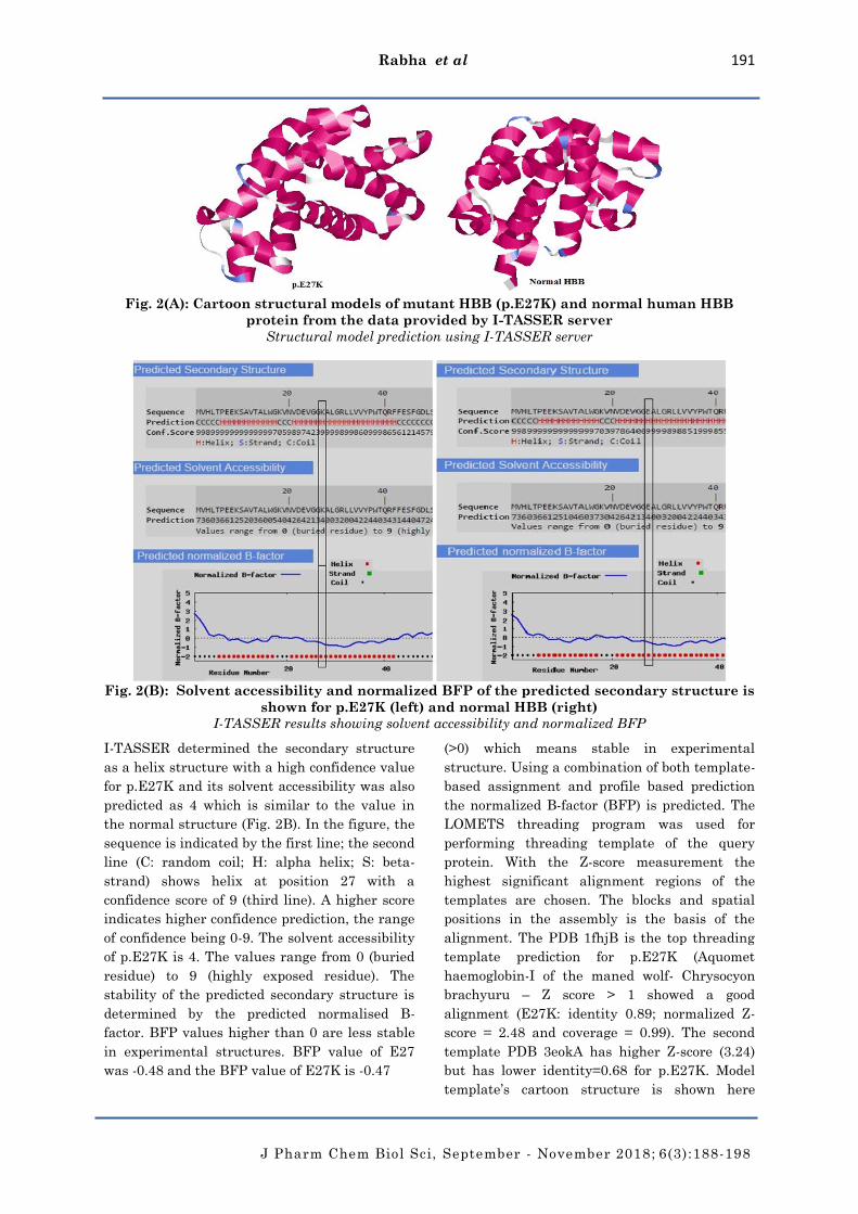
Fig. 2(A): Cartoon structural models of mutant HBB (p.E27K) and normal human HBB
protein from the data provided by I-TASSER server
Structural model prediction using I-TASSER server
Fig. 2(B): Solvent accessibility and normalized BFP of the predicted secondary structure is
shown for p.E27K (left) and normal HBB (right)
I-TASSER results showing solvent accessibility and normalized BFP
I-TASSER determined the secondary structure
as a helix structure with a high confidence value
for p.E27K and its solvent accessibility was also
predicted as 4 which is similar to the value in
the normal structure (Fig. 2B). In the figure, the
sequence is indicated by the first line; the second
line (C: random coil; H: alpha helix; S: beta-
strand) shows helix at position 27 with a
confidence score of 9 (third line). A higher score
indicates higher confidence prediction, the range
of confidence being 0-9. The solvent accessibility
of p.E27K is 4. The values range from 0 (buried
residue) to 9 (highly exposed residue). The
stability of the predicted secondary structure is
determined by the predicted normalised B-
factor. BFP values higher than 0 are less stable
in experimental structures. BFP value of E27
was -0.48 and the BFP value of E27K is -0.47
(>0) which means stable in experimental
structure. Using a combination of both template-
based assignment and profile based prediction
the normalized B-factor (BFP) is predicted. The
LOMETS threading program was used for
performing threading template of the query
protein. With the Z-score measurement the
highest significant alignment regions of the
templates are chosen. The blocks and spatial
positions in the assembly is the basis of the
alignment. The PDB 1fhjB is the top threading
template prediction for p.E27K (Aquomet
haemoglobin-I of the maned wolf- Chrysocyon
brachyuru – Z score > 1 showed a good
alignment (E27K: identity 0.89; normalized Z-
score = 2.48 and coverage = 0.99). The second
template PDB 3eokA has higher Z-score (3.24)
but has lower identity=0.68 for p.E27K. Model
template’s cartoon structure is shown here
Rabha et al 192
J Pharm Chem Biol Sci, September - November 2018; 6(3):188-198
(Fig.2A). C-score of the modelled template for
p.E27K = 1.24, estimated TM score=0.88 ± 0.07,
RMSD = 2.3 ± 1.8Å. C-score is typically in the
range (-5, 2). A model with a high confidence is
signified by a higher value of C-score and vice-
versa, a TM-score > 0.5 indicates a model of
correct topology and a TM-score < 0.17 means
random similarity. RMSD and TM-score values
indicate the similarity of the predicted
structures to the native structures.
The BFP or B-factor profile value of E27 and
E27K were -0.48 and -0.47 respectively. The
BFP value of p.E27K is slightly higher than that
of E27.
The LOMETS threading program performed the
threading template of p.E27K. The top threading
template prediction for it was based on PDB
1fhjB (Aquomet haemoglobin-I of the maned
wolf-Chrysocyon brachyuru – identity 0.89;
normalized Z-score of the threading alignments
= 2.48 and coverage = 0.99 [16]. A normal
sequence’s predicted threading template was
also based on 1fhjB – identity 0.90; normalized
Z-score = 2.49 and coverage = 0.99. There is just
a slight difference between the normal and
p.E27K.
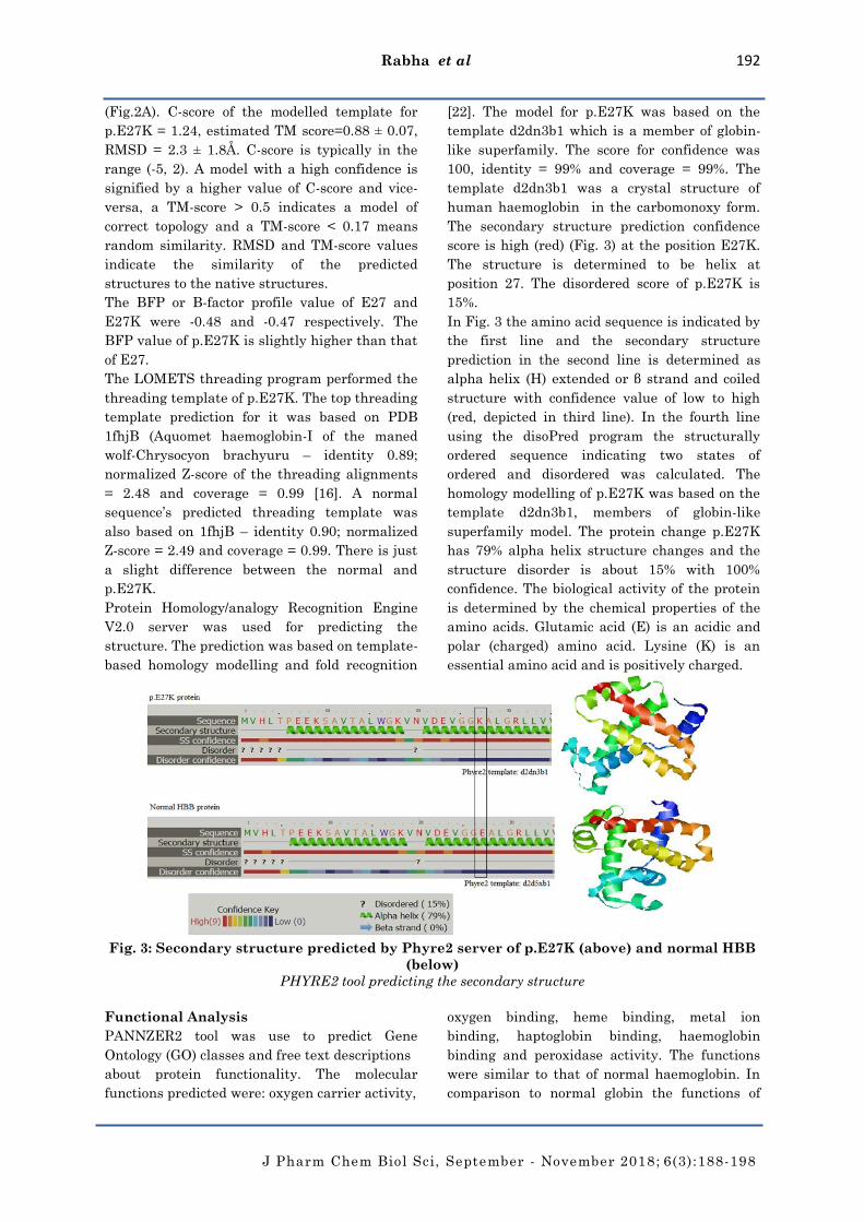
Protein Homology/analogy Recognition Engine
V2.0 server was used for predicting the
structure. The prediction was based on template-
based homology modelling and fold recognition
[22]. The model for p.E27K was based on the
template d2dn3b1 which is a member of globin-
like superfamily. The score for confidence was
100, identity = 99% and coverage = 99%. The
template d2dn3b1 was a crystal structure of
human haemoglobin in the carbomonoxy form.
The secondary structure prediction confidence
score is high (red) (Fig. 3) at the position E27K.
The structure is determined to be helix at
position 27. The disordered score of p.E27K is
15%.
In Fig. 3 the amino acid sequence is indicated by
the first line and the secondary structure
prediction in the second line is determined as
alpha helix (H) extended or β strand and coiled
structure with confidence value of low to high
(red, depicted in third line). In the fourth line
using the disoPred program the structurally
ordered sequence indicating two states of
ordered and disordered was calculated. The
homology modelling of p.E27K was based on the
template d2dn3b1, members of globin-like
superfamily model. The protein change p.E27K
has 79% alpha helix structure changes and the
structure disorder is about 15% with 100%
confidence. The biological activity of the protein
is determined by the chemical properties of the
amino acids. Glutamic acid (E) is an acidic and
polar (charged) amino acid. Lysine (K) is an
essential amino acid and is positively charged.
Fig. 3: Secondary structure predicted by Phyre2 server of p.E27K (above) and normal HBB
(below)
PHYRE2 tool predicting the secondary structure
Functional Analysis
PANNZER2 tool was use to predict Gene
Ontology (GO) classes and free text descriptions
about protein functionality. The molecular
functions predicted were: oxygen carrier activity,
oxygen binding, heme binding, metal ion
binding, haptoglobin binding, haemoglobin
binding and peroxidase activity. The functions
were similar to that of normal haemoglobin. In
comparison to normal globin the functions of

Rabha et al 193
J Pharm Chem Biol Sci, September - November 2018; 6(3):188-198
p.E27K predicted were similar. Oxygen
transport and blood coagulation were the top
listed biological functions predicted by
PANNZER2 while the functions predicted for
molecular were oxygen carrier activity, oxygen
binding, heme binding, metal ion binding and to
lesser extent haptoglobin binding, haemoglobin
binding and peroxidase activity.
Prediction of binding sites of p.E27K was
performed by the tool 3DLigandSite and no
effective change in binding sites were seen (Fig.
4). Though Glutamic acid residue at position 27
was substituted by Lysine residue there was no
change in the predicted binding sites.
Using COACH server I-TASSER performed the
functional analysis. Determination of ligand
binding sites, enzyme commission, and gene
ontology were performed by functional
homologous templates. On the basis of ligand
binding site analysis p.E27K matched to PDB
1fhjB (Crystal structure maned wolf-Chrysocyon
brachyurus-hemoglobin-I) with C-score = 1.24 as
hem for its binding factor.
Enzyme commission for p.E27K was determined
based on the PDB 1qvhA (C-score = 0.376 and
TM-score = 0.762, identity = 0.145) with
oxidoreductase activity. The PDB 1qvhA reveals
an unexpected geometry of the distal heme
pocket with two active sites i.e., 119 and 131. It
is an X-ray structure of ferric Escherichia coli
flavohemoglobin. Another comparison to PDB
1cqxA (crystal structure of the flavohemoglobin
from Alcaligenes eutrophus at 1.75 A resolution)
shows low enzyme commission (C-score = 0.360,
TM-score 0.713 and identity = 0.152) with no
active sites. From this we can say that this
protein has low enzymatic activity.
The figure (Fig. 4) consists all the predicted
binding sites with the number of ligands that
they contact, the average distance between the
residue and the residue conservation score. As
illustrated we do not see a change in the
residue’s contact in p.E27K (left) in comparison
with the normal HBB protein (right). For each
residue the average distance from conservation
score of each residue (range: 0-1.00) is defined.
The colour shows the binding site range. Low
distance indicates high accuracy and lower
coverage but increase in distance indicates less
accuracy and higher coverage.
Using gene ontology the functional analysis is
also investigated. The PDB 1dxtB matched to
p.E27K with coverage = 1.00, C-score = 0.91,
TM-score = 0.9699, identity = 0.99. The second
ranked template that matched p.E27K is 1g0bB
with coverage = 0.99, C-score = 0.77, TM-score =
0.9817 and identity = 0.83. This shows that the
protein functions were not changed due to the
amino acid change. Alterations are more
damaging to the quality of function of the
protein and not to the ontology.
Fig. 4: 3DLigandSite predicts potential binding sites (cluster) in comparison to normal
amino acid sequence through the data submitted by the Phyre2
Potential binding sites prediction using 3DLigandSite

Rabha et al 194
J Pharm Chem Biol Sci, September - November 2018; 6(3):188-198
Interactome analysis
The structure of a protein may change due to
mutation and with it the function also changes.
The interaction of various proteins with the
mutated protein may evolve phenotypic effect.
To investigate this, STRING v10.0 server was
used to inspect the interaction of various
proteins or genes with HBB in a network
system. The proteins that were predicted to
interact with HBB are as follows: NFE2
(Nuclear Factor Erythroid 2), HBG2
(Haemoglobin Gamma 2), AHSP (Alpha
Haemoglobin Stabilizing Protein), HBA2
(Haemoglobin sub-unit Alpha 2), HBZ
(Haemoglobin sub-unit Zeta), HBA1
(Haemoglobin sub-unit Alpha 1), HP
(Haptoglobin), HPR (Haptoglobin-Related-
Protein), HPX (Haemopexin) and CD163
(Cluster of Differentiation 163) (Fig. 5). The
function of a protein changes with the change in
its structure. Therefore, a change in B globin
structure may lead to interaction with other
proteins and may evolve the phenotype, hence,
altering the biological functions of haemoglobin.
Fig.5: Protein-protein interaction network generated by STRING (v10.0)
STRING (v10.0) generated interaction network
Pathogenicity Analysis
The pathogenicity of the variants were predicted
using several bioinformatics online tools
depending on the type of mutations. For
pathogenicity prediction of p.E27K, tools such as
MutPred2, PolyPhen2 and nsSNP Analyzer were
used. MutPred2 showed score = 0.428 for
p.E27K. A score threshold of 0.50 would suggest
pathogenicity. Polyphen2 with a score of 0.000
(sensitivity: 1.00; specificity: 0.00) predicted
p.E27K to be benign. nsSNP Analyzer predicted
the amino acid substitution in p.E27K to be
neutral with no pathogenic phenotypic effect
(Table 1).
The pathogenicity of the frameshift mutation CD
41/42(-CTTT) and two other intronic mutations
viz., IVS1-1(G>T) and IVS1-5(G>C) were also
predicted using Mutation Taster. For CD 41/42(-
CTTT), pathogenicity was predicted as disease
causing. Mutation Taster prediction of
pathogenicity for the two intronic mutations:
IVS1-5(G>C) and IVS1-1(G>T) were also disease
causing (Table 2).
Table 1: Pathogenicity prediction of the missense mutation CD 26G > A of the HBB gene
Mutation HGVS
Nomenclature
dbSNP Type PolyPhen2 nsSNP
Analyzer
MutPred
Codon
26G > A
c.79G > A rs33950507 Β+ Benign
(Score
0.000)
Neutral 0.428

Rabha et al 195
J Pharm Chem Biol Sci, September - November 2018; 6(3):188-198
Table 2: Bioinformatic analysis of pathogenicity of frameshift and intronic mutations of
HBB gene
Mutation HGVS Nomenclature dbSNP Type Mutation Taster
prediction
CD41/42(-CTTT) c.126_129delCTTT rs80356821 Β0 Disease causing
IVS1-5(G>C) c.92 + 5G > C rs33915217 Β+ Disease causing
IVS1-1(G>T) c.92 + 1G > T rs33971440 Β0 Disease causing
DISCUSSION
In this study; a total of 19,318 affected carriers
and their families of India were investigated.
From neighbouring country Nepal and countries
of South East Asia like Myanmar, Indonesia,
Thailand and Malaysia, total of 5,069 cases were
studied. Several studies have been performed on
β thalassemia patients over the past few decades
and all the reported mutations were reported in
this cohort.
Due to different functional mutations on the β
globin gene, HBB gene mutations occur. Among
those we have investigated 4 most frequently
reported mutations.
The mutation diversity in Indian cohorts of
border region is similar to neighbouring
countries of Nepal and other South East Asian
countries. This suggests historical immigration
and emigration of these populations.
The frequencies of different pathogenic HBB
variations were gathered for this study. For the
analysis of heterogenous and homogenous
populations and subpopulations and their
comparison such data is valuable. This data
would also be helpful in choosing the most cost-
effective strategy for screening of patients as
well as in premarital and carrier counselling.
Due to incomplete analysis of some reports there
maybe some bias. The reason for this may be due
to methodology used in each study. ARMS-PCR
and RFLPs were used in most studies for specific
analysis of the patients and other mutations
could have been missed though sequencing and
complete analysis of the genes in some studies
were needed for complete evaluation. For the
phenotype genotype analysis of patients
complete Sanger sequencing of the gene is
helpful.
The following 4 mutations have been reported in
almost all the populations taken into account in
this study: IVS1-5(G>C), CD 41/42(-CTTT),
IVS1-1(G>T) and CD 26(G>A).
CD 26(G>A) [HBB:c.79G>A] mutation lead to
E27 amino acid substitution with Lysine
(p.E27K). HbE is very common in South East
Asia as well as in the Indian sub-continent. The
HbE thalassemia is highly variable with some
patients being asymptomatic while others being
transfusion dependent. Through this study, the
goal is to improve management and counselling
of families.
In silico analysis
The biochemical analysis of mutations more
than 800 in the HBB gene calls for an intensive
effort and attention. However, all of those
mutations have not been characterized and their
clinical consequences remain unsolved. In silico
analysis makes this straightforward [23]. The
main functions of the Haemoglobin protein
include oxygen transport activity; heme binding,
haemoglobin binding, iron-iron binding and
mutations in the β globin subunit of the
haemoglobin protein may disrupt its functions.
The main molecular activities of the HBB
include oxygen carrier activity, oxygen binding,
heme binding, metal ion binding and to lesser
extent haptoglobin binding, haemoglobin
binding and peroxidase activity. The HBB
protein functions in tetramer and any variant in
its gene sequence may affect the amino acid
sequence, its expression as well as in the protein
function. The stability may also be changed due
to conformational and folding positions at mRNA
and consequently at the protein level. For mis-
sense mutation, I-TASSER and PHYRE2 were
used for investigating its structural study at the
DNA level. The structural data predicted by the
servers could assign phenotypes to novel
variants and as such would be helpful for
practitioners and geneticists.
For previously characterized and
uncharacterized mutations, structural and
pathogenic aberrations were investigated
computationally based on the changes brought to
the protein by each variant to evaluate the
confidence of the tools on novel mutations.
Change in protein stability by single point
Rabha et al 196
J Pharm Chem Biol Sci, September - November 2018; 6(3):188-198
mutation was predicted and investigated by
structural and functional analysis of p.E27K.
Frameshift mutation lead to truncated protein.
Prediction for such mutation was done by
Mutation Taster. To construct model, secondary
structures were determined using the native and
altered amino acid sequence. For investigating
protein stability, the solvent accessibility was
determined. Comparison of dynamic models
before and after mutation were done to evaluate
the altered and native protein models and its
consequences were explored in the protein’s
functions for biological functions (COACH),
enzyme activity, ligand binding sites, gene
ontology and binding sites to evaluate the
changes. For additional future studies, the
functional analysis in this study may be a good
model.
Because of more availability of software,
predictions of substitution mutations is easier
using in silico analysis than the prediction in
intronic and non-coding variants. Mutation
Taster was used for predicting pathogenicity of
the intronic variants. Pathogenicity analysis
showed the variants to be disease causing i.e., of
pathogenic character.
In silico evaluation of pathogenicity makes it
easier and faster to predict the effect of new and
novel mutations and could inform the β-
thalassemia families at risk. The data available
from this study indicates that the mutations in
the HBB gene’s non-coding regions maybe
responsible for some of the phenotypes which
may be scrutinized using several bioinformatics
servers (Table 2). Novel mutations not reported
in the SNP database should be investigated if
found in any thalassemic individuals. However,
segregation or expression analysis should
confirm this prediction. The advantage of
pathogenicity prediction of a mutation allows
confirmation of a defect in individuals and
facilitates genetic testing and counselling of
other high-risk family members.
Interactome network of proteins
From the interaction analysis, it can be seen
that the interaction is with ten proteins
including HBA1 and HBA2. They are mainly
involved in oxygen transport [24]. The other
proteins are NFE2, HBG2, AHSP, HBZ, HP,
HPR, CD163 and HPX. NFE2 possess DNA
binding transcription factor activity and
transcription co-activator activity, HBG2 possess
functions such as iron ion binding and oxygen
binding, acts as a chaperone. During normal
erythroid cell development it prevents the
aggregation (damaging) of alpha haemoglobin.
HBZ also functions in iron ion binding and
oxygen binding, HP possess activities like
serine-type endopeptidase activity and
haemoglobin activity. HPR also possess the
same functions such as the HP gene. CD163
possess the activity of scavenger receptor
activity and HPX is involved in heme
transporter activity. A change due to mutation
in the HBB gene would have an effect on the
interaction of proteins and factors, i.e.,
transcription, translation, development and
function of the Hb gene and consequently the
phenotype.
CONCLUSION
Mutations of high frequencies reported in this
region of the world was analyzed. Such study is
valuable for regions with no access to advanced
genetic technologies for analysis of panel based
studies. For performing prenatal diagnosis of
beta thalassemia this article will be of interest.
In silico analysis can be used to identify the fatal
phenotype but not with great accuracy for
unknown variants. For assessing pathogenicity
in novel mutations, computational analysis can
be used alongwith segregation and expression
analysis. In case of prenatal diagnosis of at-risk
patients, computational analysis could be used
to predict phenotype but careful analysis should
also be done. Through such studies functional
consequences of various mutations are known
which would give a high confidence for at-risk
families to undergo genetic counselling to know
the probability of the affected child. It gives the
families various options such as termination,
treatment of patients or PGD (preimplantation
genetic diagnosis).
ACKNOWLEDGEMENT
We gratefully acknowledge the Department of
Biotechnology (DBT), Government of India
sponsored Bioinformatics Infrastructure Facility
for providing the infrastructure to pursue the
work and Mr. Dibyojyoti Rabha was offered 6-
months studentship by the DBT.
CONFLICT OF INTEREST STATEMENT
The authors declare no conflict of interest in this
research article.

Rabha et al 197
J Pharm Chem Biol Sci, September - November 2018; 6(3):188-198
REFERENCES
1. Sarmah J, Baruah D, Sarma PK, Das D.
Dietary Habit, Anthropometric
measurements and haematological
parameters in correlation with prevalence
of iron-deficiency anaemia among never
married tribal female postgraduates of
Assam, India. Res Rev J Health Prof 2018;
8(2): 19-26.
2. Giardine B, Borg J, Viennas E, Pavlidis C,
Moradkhani K, Joly P, et al. Updates of the
HbVar database of human hemoglobin
variants and thalassemia mutations.
Nucleic Acids Res 2014; 42: D1063-D1069.
3. Huisman THJ, Carver MFH, Baysal E,
Efremov GD. Available from
http://globin.bx.psu.edu/hbvar/menu. html.
[(A database of human hemoglobin variants
and thalassemias)]; 2014.
4. Patrinos GP, Giardine B, Riemer C,
MillerW, Chui DH, Anagnou NP, et al.
Improvements in the HbVar database of
human hemoglobin variants and
thalassemia mutations for population and
sequence variation studies. Nucleic Acids
Res 2004; 32: D537- D541.
5. Bashyam MD, Bashyam L, Savithri GR,
Gopikrishna M, Sangal V, Devi ARR.
Molecular genetic analyses of beta
thalassemia in South India reveal rare
mutations in the beta globin gene. J Hum
Genet 2004; 49: 408-413.
6. Verma IC, Saxena R, Kohli S. Past, present
& future scenario of thalassaemic care &
control in India. Indian J Med Res 2011;
134: 507-521.
7. Verma IC. The challenge of genetic
disorders in India. Molecular genetics and
gene therapy- the new frontier. Amsterdam:
Scientific Communications; 1994; p11.
8. Setianingsih I, Williamson R, Daud D,
Harahap A, Marzuki S, Forrest S.
Phenotypic variability of Filipino beta (0)-
thalassemia/HbE patients in Indonesia. Am
J Hematol 1999; 62:7-12.
9. Colah R, Gorakshakar A, Nadkarni A,
Phanasgaonkar S, Surve R, Sawant P,
Mohanty D, Ghosh K. Regional
heterogeneity of beta-thalassemia
mutations in the multi ethnic Indian
population. Blood Cells Mol Dis 2009; 42:
241-246.
10. Colah R et al. Epidemiology of beta
thalassemia in western India: mapping the
frequencies & mutations in subversion of
Maharashtra and Gujrat. Br J Haematol
2010; 149: 739-747.
11. Population Reference Bureau (PRB). World
population data sheet. Washington DC:
Population Reference Bureau; 2010.
12. World Health Organization (WHO). Joint
WHO-TIF meeting on management of
hemoglobin disorders (2nd: 2008: Nicosia,
Cyprus). Geneva: World Health
Organization; 2008.
13. Mishra A et al. Distribution and ethnic
variation of α-thalassemia mutations in
Nepal. Nepal Med Coll J 2012; 14(1): 49-52.
14. Baysal E, Carver MFH. The beta and delta
thalassemia repository (eight edition).
Haemoglobin 1995; 19(3-4):213-236.
15. Wasi P, Pootrakul S, Pootrakul P.
Thalassemia in Thailand. Ann N Y Acad Sci
1980; 344: 352-363.
16. Roy A, Kucukural A, Zhang Y. I-TASSER: A
unified platform for automated protein
structure and function prediction. Nat
Protoc 2010; 5: 725-738.
17. Yang J, Roy A, Zhang Y. Protein-ligand
binding site recognition using
complementary binding-specific
substructure comparison and sequence
profile alignment. Bioinformatics 2013; 29:
2588-2595.
18. Adzhubei IA, Schmidt S, Peshkin L,
Ramensky VE, Gerasimova A, Bork P. A
method and server for predicting damaging
missense mutations. Nat Methods 2010; 7:
248-249.
19. Saunders CT, Baker D. Evaluation of
structural and evolutionary contributions to
deleterious mutation prediction. J Mol Bio
2012; 322: 891-901.
20. Roy A, Yang J, Zhang. Cofactor: an accurate
comparative algorithm for structure-based
protein function annotation. Nucleic Acids
Res 2012; 40: W471–W477.
21. Zhang Y. I-TASSER server for protein 3D
structure prediction. BMC Bioinformat
2008; 9: 40.
22. Kelley LA, Sternberg MJ. Protein structure
prediction on the web: a case study using
the PHYRE server. Nature Protoc 2009; 4:
363-371.

Rabha et al 198
J Pharm Chem Biol Sci, September - November 2018; 6(3):188-198
23. Alanazi M, Abduljaleel Z, Khan W, Warsy
AS, Elrobh M, Khan Z, Amri AA, Bazzi,
MD. In silico analysis of single nucleotide
polymorphism (SNPs) in human beta-globin
gene. PLoS One 2011; 6(10), e25876.
24. Franceschini A et al. STRING v9.1:
protein–protein interaction networks, with
increased coverage and integration. Nucleic
Acids Res 2013; 41: D808-D815.
Cite this article as:
Dibyajyoti Rabha, Dipankar Baruah, Paresh Kumar Sarma, Jatin Sarmah. In Silico Analysis of β-
Thalassemia Mutations in India and its Neighbouring South East Asian Countries. J Pharm
Chem Biol Sci 2018; 6(3): 188-198