Improving Pediatric Cardiology Consultation Methods by ...
60
University of South Florida Scholar Commons Graduate eses and Dissertations Graduate School 7-16-2007 Improving Pediatric Cardiology Consultation Methods by Introducing Digital Interactive 3-D Heart Models: A Proof of Concept Study Adam Verigan University of South Florida Follow this and additional works at: hp://scholarcommons.usf.edu/etd Part of the American Studies Commons , and the Biomedical Engineering and Bioengineering Commons is esis is brought to you for free and open access by the Graduate School at Scholar Commons. It has been accepted for inclusion in Graduate eses and Dissertations by an authorized administrator of Scholar Commons. For more information, please contact [email protected]. Scholar Commons Citation Verigan, Adam, "Improving Pediatric Cardiology Consultation Methods by Introducing Digital Interactive 3-D Heart Models: A Proof of Concept Study" (2007). Graduate eses and Dissertations. hp://scholarcommons.usf.edu/etd/3847
Transcript of Improving Pediatric Cardiology Consultation Methods by ...

University of South FloridaScholar Commons
Graduate Theses and Dissertations Graduate School
7-16-2007
Improving Pediatric Cardiology ConsultationMethods by Introducing Digital Interactive 3-DHeart Models: A Proof of Concept StudyAdam VeriganUniversity of South Florida
Follow this and additional works at: http://scholarcommons.usf.edu/etd
Part of the American Studies Commons, and the Biomedical Engineering and BioengineeringCommons
This Thesis is brought to you for free and open access by the Graduate School at Scholar Commons. It has been accepted for inclusion in GraduateTheses and Dissertations by an authorized administrator of Scholar Commons. For more information, please contact [email protected].
Scholar Commons CitationVerigan, Adam, "Improving Pediatric Cardiology Consultation Methods by Introducing Digital Interactive 3-D Heart Models: A Proofof Concept Study" (2007). Graduate Theses and Dissertations.http://scholarcommons.usf.edu/etd/3847

Improving Pediatric Cardiology Consultation Methods by Introducing
Digital Interactive 3-D Heart Models: A Proof of Concept Study
by
Adam Verigan
A thesis submitted in partial fulfillment of the requirements for the degree of
Master of Science in Biomedical Engineering Department of Chemical Engineering
College of Engineering University of South Florida
Major Professor: Don Hilbelink, Ph.D. William Lee III, Ph.D., P.E.
Karl Muffly, Ph.D.
Michael VanAuker, Ph.D.
Date of Approval: July 16, 2007
Keywords: Medical, Imaging, Segmentation, Education, Software
© Copyright 2007, Adam Verigan

i
Table of Contents
List of Tables iii
List of Figures iv
List of Abbreviations v
Glossary of Terms vi
Abstract vii
Chapter One: Introduction 1
Problem Statement 1 Solution Statement 2
Contribution 3
Chapter Two: Literature Review 4
Chapter Three: Materials and Methods 7 MR Data Acquisition 7
MR 3D Reconstruction 8 Software Development 9
User Interface 9 Heart Conditions List Panel 10
Diseased 3D Panel 11
Normal 3D Panel 12 Defect Description Panel 14
Diseased 2D Panel 15 Normal 2D Panel 16
System Intelligence 16 Survey Development 17
Chapter Four: Results 20
Segmented Heart Models 20 Software Components 22
User Interface 22 Heart Conditions List Panel 22

ii
3D Panels 24
Defect Description Panel 26 2D Panels 27
Survey Analysis 28 Parents 28
Pediatric Cardiologists 29
Chapter Five: Conclusion 31 Quantitative Analysis 31
Qualitative Analysis 31 Final Thoughts 32
References 34
Bibliography 36
Appendices 37 Appendix A: Parent Survey 38
Appendix B: Cardiologist Survey 41 Appendix C: Sample Java Code 43
Appendix D: Software Requirements Specification 44 Appendix E: Sample Heart Diagram 50

iii
List of Tables
Table 3.1 Heart conditions list processes 11
Table 3.2 Diseased 3D heart panel processes 12
Table 3.3 Normal 3D heart panel processes 13
Table 3.4 Defect description panel processes 14
Table 3.5 Diseased 2D panel processes 15
Table 3.6 Normal 2D panel processes 16

iv
List of Figures
Figure 4.1 Segmented repaired Tetralogy of Fallot model 20
Figure 4.2 Segmented normal heart model 21
Figure 4.3 EduView application 23
Figure 4.4 Heart conditions list panel 23
Figure 4.5 Diseased 3D heart surface model 25
Figure 4.6 Normal 3D heart surface model 25
Figure 4.7 Heart description panel 26
Figure 4.8 Tetralogy of Fallot 2D graphic 27
Figure 4.9 Normal heart 2D graphic 28
Figure D.1 Graphical user interface design 47
Figure D.2 Architectural model of the system 48
Figure D.3 Data-flow diagram of the system 49
Figure E.1 Sample heart diagram for Ventricular Septal Defect 50

v
List of Abbreviations
GUI Graphical User Interface
2-D Two-Dimensional
3-D Three-Dimensional
SRS Software Requirements Specification
ROI Region Of Interest
DT Dynamic Thresholding
STL Stereo Lithography
JDK Java Development Toolkit
IDE Integrated Development Environment
vi
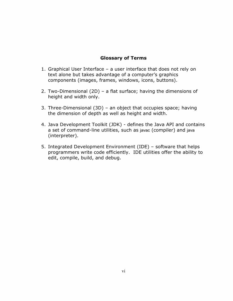
Glossary of Terms
1. Graphical User Interface – a user interface that does not rely on text alone but takes advantage of a computer’s graphics
components (images, frames, windows, icons, buttons).
2. Two-Dimensional (2D) – a flat surface; having the dimensions of height and width only.
3. Three-Dimensional (3D) – an object that occupies space; having
the dimension of depth as well as height and width.
4. Java Development Toolkit (JDK) - defines the Java API and contains
a set of command-line utilities, such as javac (compiler) and java (interpreter).
5. Integrated Development Environment (IDE) – software that helps
programmers write code efficiently. IDE utilities offer the ability to edit, compile, build, and debug.

vii
Improving Pediatric Cardiology Consultation Methods by Introducing Digital Interactive 3-D Heart Models:
A Proof of Concept Study
Adam Verigan
ABSTRACT
The purpose of a pediatric cardiology consultation is to inform, or
educate, the patient and family of all aspects surrounding a certain
congenital heart defect. Consultation education methods and
materials may include verbal descriptions, two-dimensional (2-D)
heart diagrams, and take-home pamphlets. Because the human heart
is a complex three-dimensional (3-D) object, the problem lies within
the clarity to which these methods are performed by the doctors and
understood by the patients and families. Therefore, during a
consultation the cardiologist must a) possess the ability to describe a
defect visually as well as verbally and b) ensure that the patient and
family have a clear understanding of the situation.
In this work a method to improve patient consultation is
outlined. Heart model segmentation methods from Cardiac MRA
images are discussed by using the Materialise Mimics 10.11 software.
EduView, the proposed software application solution, provides the user

viii
with traditional verbal descriptions and 2-D heart diagrams along with
the ability to interact with a digital 3-D human heart model. By
including a 3-D approach, the purpose is to assist the cardiologist in
explaining a defect while further educating the patient and family. Sun
Microsystems Java technology was utilized in order to program the
application. Implementation of the software solution is outlined and
the results from two surveys involving parents of children with
congenital heart defects and pediatric cardiologists are presented.
This study outlines a proof of concept. There is significant potential for
extending and marketing this tool for future clinical use.

1
Chapter One:
Introduction
Problem Statement
Patient consultations in any branch of medicine inherently
possess a two-fold challenge – communication and comprehension of
information between doctor and patient. Along with these challenges,
pediatric cardiology consultations must provide a means to relay
information in such a way that children (18 yrs and under) will
understand. The challenge is intensified within cardiology because of
the complexity of the human heart. In dealing with children patients
the role and importance of visual based learning becomes primary.
The problem with the current consultation approach lies within
the attempt to use flat, non-interactive two-dimensional (2D)
representations to explain the fully three-dimensional (3D) human
heart. Although anatomical heart models are a tangible 3D heart
representation they are fragile and inanimate. The education issue
can be addressed by utilizing current imaging techniques along with
2
automatic or semiautomatic segmentation and analysis of diagnostic
image scans.
Imaging technology methods, specifically magnetic resonance
imaging (MRI), computer tomography (CT) and ultrasound (US),
within cardiology practices continue to improve. Due to the
advancement in technology these techniques are becoming more
important in clinical diagnosis. Their non-invasive capability makes
them an ideal tool in cardiac treatment. There are benefits tied into
the improving quality and increasing quantity of the image scans. One
such benefit is found in the segmenting process. Segmented
anatomical models can be created more rapidly while improving in
quality. This pre and post processing of the captured data is designed
to further assist doctors in diagnosis and treatment.
This study focuses on the development of a more visually
engaging and interactive method to educate during a patient
consultation. The resulting application would not only assist the
patients and families but also assist the doctors.
Solution Statement
EduView is designed to be an alternate and interactive solution
for the education of congenital heart diseases. The solution is an
interactive program that allows a user to choose a congenital heart

3
defect to view. Once chosen the user has the ability to interact with
both a 3D ‘normal’ heart reference model along with a 3D ‘diseased’
heart reference model. This allows a user to see the differences
between the two models while allowing them to understand the 3D
shape complexity of the heart. Also, for each chosen heart disease
there will be a textual description of the malformation. Lastly, the
user can view the more traditional 2D heart image depictions of the
‘normal’ and ‘diseased’ heart.
Contribution
EduView goes beyond the bindings of literature and 2D image
representations into the interactive 3D realm. Because EduView is
software, it can be loaded onto any computer and taken anywhere.
Adding representative 3D heart reference models for both the normal
and diseased cases adds a new approach toward patient education.
Users have more control in what they want to see and understand.
This is achieved through the various heart conditions to choose from,
the textual and 2D descriptions of the diseased hearts, as well as the
ability to interact with the 3D heart models.

4
Chapter Two:
Literature Review
Current visual methods utilized by pediatric cardiologists for
patient consultation are take-home pamphlets with illustrations, heart
diagrams (see Appendix E), anatomical heart models, two-dimensional
(2D) heart animations, and looking at patient specific image scans (MR
or CT) [1]. These methods present a challenge to the patient and
family in fully understanding a complex 3D object. Because the
current visual methods lack the ability to express the full dimensions
of the heart, more emphasis is given to verbal descriptions. As a
result, patients and families must rely on the physician’s verbal
descriptions to piece together the problem in their mind.
In medicine, a specialized node exists called medical imaging.
This field is dedicated to generating visualizations to aid doctors and
other medical professionals in better understanding and visualizing the
anatomy of their patients. Also it allows them to perform mock
surgeries to gain valuable experience and practice before an actual
procedure. These visualizations draw on one main ingredient –

5
existing 2D image sequences. These captured images, generally from
CT and MRI, provide the datasets that allow for the three-dimensional
segmentation and visualization of the anatomy [2, 8].
Within cardiology, the ability to evaluate a patient’s heart
function non-invasively lies almost exclusively with cardiac MRI
volumetric data. CT may provide higher quality scans than MRI but
because the scanning process relies on X-rays it is considered too
risky. Modern cardiac MRI scanners have the ability to capture the
complete heart beat sequence. The scanners manufacturing software
provides the toolsets to generate maximum intensity projections (MIP)
of volumetric data [4, 9]. This function along with other segmenting
algorithms found in the software provides the cardiologists with patient
specific 3D heart models to view. Other cardiac segmentation
methods require the creation of mathematical models to map out the
geometry [6].
The need to provide better educational material during a patient
consultation has not been overlooked. Scientific Software Solutions
creates not only educational software but also educational field guides
and prints [3]. They provide some of the most in-depth teaching
materials for congenital heart disease. Their PedHeart suite includes
instructional material not only for medical professionals but also for
parents and adult patients [7].

6
In order to develop this application, a programming language
needed to be chosen. Java (Sun Microsystems, Inc., Santa Clara, CA)
was chosen for its portability and robust attributes. It possesses the
ability to function across platforms. Also, like the C programming
languages, Java is an object-oriented language (OOL). As a result, the
creation and manipulation of objects make it a very powerful
language. Java is well documented too. The Java API and tutorials for
Swing components and features are very useful [5]. Java3D is a
powerful object-oriented 3D renderer [10]. By default, it relies on
OpenGL for graphics processing. However, it can be tailored to
function with DirectX. Its libraries can be imported into a Java project
for ease of use. Because its framework is based on Java technology, it
was the logical choice to create the scenes in order to render the heart
reference models.

7
Chapter Three:
Materials and Methods
MR Data Acquisition
Studies were performed on a High Definition (HD) 1.5T super
conducting magnet (GE Signa Excite, GE Healthcare, Waukesha,
Wisconsin). In order to obtain the coronal, saggital, and transverse
planes, a series of three-dimensional (3D) fast multiplanar spoil
gradient (SPGR) echo images were acquired. The pulmonary CE-MRA
volumetric scans were ECG-gated and acquired during suspended
respiration. Each image slice was obtained in sequence of multiple
intervals or cardiac cycles of 20 s or less suspended respiration. If the
quality of the image acquisition was poor for a particular interval, the
interval would be re-scanned. 60 slices were acquired in order to
window the entire region of interest. For one study, the slice thickness
was 4 mm with 2 mm spacing between slices along with a repetition
time of 3.976 ms and an echo time of 1.452 ms. The other study had
a 4.8 mm slick thickness with 2.4 mm spacing between slices along
with a repetition time of 3.956 ms and an echo time of 1.436 ms. For

8
both studies, the images were captured at a 512 x 512 pixel resolution
with a pixel spacing of 0.8594\0.8594 mm. Between 9 and 14 images
were acquired per cardiac cycle.
MR 3D Reconstruction
Volumetric data from each MR study was imported and analyzed
using Mimics (Materialise, Leuven, Belgium) software. Once imported,
window levels were adjusted to assign the pixels that made up the
heart tissue a lighter shade of gray. In each study, a specific heart
structure was independently chosen by its pixel value and assigned a
colored mask that spanned throughout the entire image sequence. In
this manner, the data was segmented using semi-automatic methods
(thresholding, region growing, morphological edits) and manual editing
(multiple slice edit, edit mask in 3D).
3D volumetric surfaces were constructed from the segmented
data. Once the surfaces were calculated, they were imported into
Mimics finite element analysis (FEA) module. The surfaces ranged
from 50,000 to 170,000 faces or triangles. In order to import and
render the surface model for visualization, the models needed to be
remeshed. Within the FEA module, several tools for triangle reduction
and surface smoothing were used including triangle reduction, filter
sharp triangles, remesh part, detect self intersections, and smoothing.

9
The remeshed surface model was exported as an ASCII .stl model.
The .stl model was imported into Maya 6.5 (Autodesk Inc., San Rafael,
CA) to cleanup merged vertices and to convert the model into an .obj
file. The overall process to create and refine the 3D heart model from
MR scans was approximately 3 hours.
Software Development
Java was chosen as the programming language to create
EduView. EduView was developed using the latest development kit,
Java SE Development Kit (JDK), Version 6, on a Windows XP Pro
platform workstation. Java3D version 1.5 was used in order to create
the three-dimensional virtual scenes to render the 3D heart models. A
Radeon 9250 (Advanced Micro Devices Inc., Sunnyvale, CA) 256 MB
PCI version with OpenGL support graphics card was used to display the
virtual scenes during development.
User Interface
The application’s user interface design was built using Java
Swing components. The interface contains two halves, a top portion
and a bottom portion (Appendix D). The top half houses the heart
conditions list panel as well as the 3D diseased heart panel and the 3D
normal heart panel. The bottom half contains a congenital heart

10
defect textual description panel and both the 2D diseased heart panel
and the 2D normal heart panel. The placement of both the 3D and the
2D panels allows the user to give a direct comparison between the
chosen diseased heart and the normal heart. The size of the frame is
1020 pixels wide by 720 pixels high. User screen resolution should be
no less than 1024 pixels wide by 768 pixels high.
Heart Conditions List Panel
The heart conditions list, named ConditionListPanel in the
EduView.java source file, can contain multiple congenital heart defects.
The list is populated through reading the contents of a local text file.
Reading the text file is one of the first executions to be made by
EduView. The user can choose a condition in the list. Once a choice is
made, the intelligence of the system, both the MainManager and
ViewManager classes, are notified. This relay calls an update to the
defect description panel, the 3D diseased heart panel, and the 2D
diseased heart panel. Table 3.1 outlines the various processes of the
heart conditions list panel.

11
Table 3.1 Heart conditions list processes
Diseased 3D Panel
This module, named Defect3DPanel in the .java source files,
serves as one of two three-dimensional scenes of EduView. The
module consists of a virtual Canvas3D scene where the diseased heart
geometry and lights are loaded and rendered. The heart geometry
consists of vertices, faces, and materials in the form of an .obj file.
The module reads the .obj file and renders it to the scene. The
geometry is assigned to a BranchGroup object so that the scene is
aware of its existence. In order to view the loaded heart geometry, a
directional light is created and given a color and coordinates. Based
on action listeners and event handlers, the mouse is given control to
move the heart object around in the scene. The geometry has
Transform3D objects attached to its TransformGroup object. This
allows a user to rotate, translate, and zoom the heart object to see all
angles in virtual space. A reset button is provided if the user wishes
the heart model to return to its original position. Each selection of a
- put items read from the text file into a String array
- add elements to the list object from contents of the String array
- create the list - call to system intelligence if user changes the current condition

12
heart condition from the heart conditions list will refresh the scene and
pass initial position coordinates to the heart model for display. Table
3.2 outlines the various processes of the diseased 3D heart panel.
Table 3.2 Diseased 3D heart panel processes
Normal 3D Panel
This module, named Normal3DPanel in the .java source files,
serves as the last of the two three-dimensional scenes of EduView.
The module consists of a virtual Canvas3D scene where the normal
heart geometry and lights are loaded and rendered. The heart
geometry consists of vertices, faces, and materials in the form of an
.obj file. The module reads the .obj file and renders it to the scene.
The geometry is assigned to a BranchGroup object so that the scene is
- Create the scene
- Load the .obj file, assign it to the Scene object - Load heart position coordinates
- Assign Transform3D matrices to the
TransformGroup object - Assign the Scene object containing the geometry
to the TransformGroup object - Assign the TransformGoup object to the
BranchGroup object - Grant mouse control and assign it to the
BranchGroup object - Create directional light and finalize the scene
- Reset geometry if user clicks the reset model button
- Refresh scene with a new heart model if user chooses another heart condition to view

13
aware of its existence. In order to view the loaded heart geometry, a
directional light is created and given a color and coordinates. Based
on action listeners and event handlers, the mouse is given control to
move the heart object around in the scene. The geometry has
Transform3D objects attached to its TransformGroup object. This
allows a user to rotate, translate, and zoom the heart object to see all
angles in virtual space. A reset button is provided if the user wishes
the heart model to return to its original position. This panel is
initialized and rendered once at startup. User interaction has no affect
on this panel. Table 3.3 outlines the various processes of the normal
3D heart panel.
Table 3.3 Normal 3D heart panel processes
- Create the scene
- Load the .obj file, assign it to the Scene object - Load heart position coordinates
- Assign Transform3D matrices to the TransformGroup object
- Assign the Scene object containing the geometry
to the TransformGroup object - Assign the TransformGoup object to the
BranchGroup object - Grant mouse control and assign it to the
BranchGroup object - Create directional light and finalize the scene
- Reset geometry if user clicks the reset model button

14
Defect Description Panel
The congenital heart defect textual description panel, named
DescriptionPanel in the .java source files, displays the text that
describes the chosen congenital heart defect. It receives the path to
the defect description text file from the system intelligence.
JEditorPane and JScrollPane are the swing components that create and
hold the text within the panel. The text area is non-editable and has
the ability to be scrolled. Also, as the borders are approached, the
text is set to wrap on white-space, not on characters. The text file is
loaded in as a URL object and displayed in the editor pane. Upon user
interaction, the editor pane document object is cleared and the editor
pane is set with the new text from the URL object. Table 3.4 outlines
the various processes of the defect description panel.
Table 3.4 Defect description panel processes
- Create the JEditorPane object
- Create the JScrollPane object and add the JEditorPane object to it
- Read and load the text into a URL object - Set the editor pane with the URL object to display
- Refresh text area and set the editor pane with the URL object upon user interaction

15
Diseased 2D Panel
The diseased 2D panel, named Defect2DPanel in the .java source
files, holds and displays the diseased heart graphic. It provides the
user with the standard method of viewing a congenital heart defect.
The graphic displays the malformations for a chosen defect. The
graphic is drawn and rendered with the Defect2DImagePanel class
within the Defect2DPanel class. This is achieved using an Image
object that holds the heart graphic and is displayed on the canvas.
Each time a user changes the heart condition, the Defect2DPanel class
receives the path to the graphic along with the condition name. These
parameters are passed to the Defect2DImagePanel class to refresh
and update the Image object with the proper graphic. Table 3.5
outlines the various processes of the diseased 2D panel.
Table 3.5 Diseased 2D panel processes
- Create a panel to hold the Defect2DImagePanel object
- Pass the image path and defect name to the Defect2DImagePanel object
- Create and initialize the Image object - Draw the graphic to the canvas for display
- Refresh Image object and repaint canvas upon user interaction

16
Normal 2D Panel
The normal 2D panel, named Normal2DPanel in the .java source
files, holds and displays the normal heart graphic. It provides the user
with the standard method of viewing a normal heart. The graphic
displays the anatomy of the normal heart. The graphic is drawn and
rendered with the Normal2DImagePanel class within the
Normal2DPanel class. This is achieved using an Image object that
holds the heart graphic and is displayed on the canvas. This panel is
initialized and rendered once at startup. User interaction has no affect
on this panel. Table 3.6 outlines the various processes of the normal
2D panel.
Table 3.6 Normal 2D panel processes
System Intelligence
The core of the intelligence can be found in the public class
MainManager. The MainManager is aware of the path of all graphic
information for the chosen congenital heart defect, all the data
obtained from parsing the text file, the Normal3DPanel, the
- Create a panel to hold the Normal2DImagePanel object
- Pass the image path and defect name to the Normal2DImagePanel object
- Create and initialize the Image object - Draw the graphic to the canvas for display

17
Defect3DPanel, the Normal2DPanel, the Defect2DPanel, and the
DescriptionPanel. Within the class lies another class, ViewManager.
Apart from MainManager, ViewManager contains the details of updates
for the 3D model, the 2D graphic, and the defect description text file.
However, ViewManager does nothing unless the MainManager gives it
information. When a user chooses another heart condition, the
MainManager tells ViewManager to update the Defect3DPanel, the
DescriptionPanel, and the Defect2DPanel. ViewManager contains the
defect name and path to the heart model, the heart graphic, and the
heart description for a chosen heart condition.
The Parser class parses the text file, hearts.txt, into a DataSet
array list. The data in the array is the following: the directory to find
the heart information, the heart condition, the heart model, the heart
graphic, and the heart description. The class also creates a
ConditionList array list composed of conditions only. A programmer
has access to the data parsed from the text file by calling any of the
get methods in the DataSet class.
Survey Development
Two survey populations were selected in order to gather
statistical data to validate the design, quality, and effectiveness of
EduView. The first group was composed of parent volunteers from a
18
cardiac support group. Its members were comprised of children with
congenital heart defects and their families. The second group was
composed of pediatric cardiologists. These cardiologists routinely
provide standard pediatric cardiology consultations as a means of
relaying information about congenital heart defects.
The parent group received two presentations. The first
presentation was a demonstration using standard two dimensional
visuals and verbal descriptions given by a pediatric cardiologist. The
second presentation was a demonstration of EduView presented by its
developer. The clinician group received only a demonstration of
EduView presented by its developer. After the presentations, both
groups received a survey.
In order to conduct the surveys, an appropriate Institutional
Review Board (IRB) application was completed, processed, and
approved. The survey was designed to maintain the confidentiality of
the volunteer. Informed consent was not required because the survey
contained no identifiable tags. The parent survey (Appendix A) was
developed into four sections. The first section contained four non-
Likert scale format and nine 5-point scale Likert (Strongly Agree,
Agree, No Opinion, Disagree, Strongly Disagree) demographic or
background questions. The second section contained three 5-point
scale Likert questions about the standard 2D presentation. The third

19
section contained three 5-point scale Likert questions about EduView.
Sections two and three gauged the satisfaction of the user as well as
the understanding level of the user of the presentations. The last
section contained two 5-point scale Likert questions and two questions
asking the user for his/her consultation method preference. In
addition, a space was provided for additional comments or questions
regarding the presentations.
The clinician survey (Appendix B) was developed into three
sections. The first section contained four non-Likert scale format and
two 5-point scale Likert demographic or background questions. The
second section contained four 5-point scale Likert questions about the
user’s satisfaction with conventional patient consultation methods.
The last section contained seven 5-point scale Likert questions about
the user’s satisfaction and understanding of the EduView presentation.
In addition, a space was provided for additional comments or
questions regarding the presentations.

20
Chapter Four:
Results
Segmented Heart Models
Figure 4.1 shows the segmented defect representative heart
model. The model shown is of a repaired Tetralogy of Fallot MR
Figure 4.1 Segmented repaired Tetralogy of Fallot model

21
dataset with a right branch aortic arch. After remesh and cleanup, the
final .obj model contained 22,196 vertices and 44,700 triangles.
Figure 4.2 shows the segmented normal representative heart model.
Figure 4.2 Segmented normal heart model
After remesh and cleanup, the final .obj model contained 73,386
vertices and 24,462 triangles. The same FEA remesh algorithms and
cleanup methods were applied to both models. The normal heart
model loads and renders quicker (~ 2 s faster) in EduView than the
22
defect heart model. This is due to the smaller number of surface
triangles found on the normal heart model.
Software Components
User Interface
Using Java Swing components to create the interface was very
useful due to its container hierarchy method and its variety of tools.
The interface is broken down into JComponents. EduViewFrame
extends Jframe, which is a high-level Jcomponent that houses all
Jpanel components of EduView. All other classes of EduView extend
Jpanel. Each Jcomponent can be aligned and enhanced using the
layout and border features of Swing. Refer to Appendix C for sample
Java Swing code. Figure 4.3 is a screenshot of EduView in its final
form.
Heart Conditions List Panel
The conditions list is a JList component, which is filled using an
addElement function call. The amount of conditions found in the
parsed text file determines the size of the list. Each item in the list is
given an index. A user can either double-click a condition or select a
condition using the “Select Condition” button provided. The chosen
condition index is given to the MainManager class for updating the

23
Defect3DPanel, Defect2DPanel, and DescriptionPanel. Only one
condition can be selected at a time. Figure 4.4 shows the condition list
panel.
Figure 4.3 EduView application
Figure 4.4 Heart conditions list panel
24
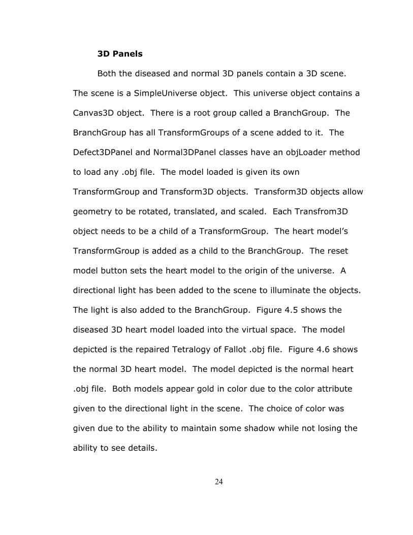
3D Panels
Both the diseased and normal 3D panels contain a 3D scene.
The scene is a SimpleUniverse object. This universe object contains a
Canvas3D object. There is a root group called a BranchGroup. The
BranchGroup has all TransformGroups of a scene added to it. The
Defect3DPanel and Normal3DPanel classes have an objLoader method
to load any .obj file. The model loaded is given its own
TransformGroup and Transform3D objects. Transform3D objects allow
geometry to be rotated, translated, and scaled. Each Transfrom3D
object needs to be a child of a TransformGroup. The heart model’s
TransformGroup is added as a child to the BranchGroup. The reset
model button sets the heart model to the origin of the universe. A
directional light has been added to the scene to illuminate the objects.
The light is also added to the BranchGroup. Figure 4.5 shows the
diseased 3D heart model loaded into the virtual space. The model
depicted is the repaired Tetralogy of Fallot .obj file. Figure 4.6 shows
the normal 3D heart model. The model depicted is the normal heart
.obj file. Both models appear gold in color due to the color attribute
given to the directional light in the scene. The choice of color was
given due to the ability to maintain some shadow while not losing the
ability to see details.

25
Figure 4.5 Diseased 3D heart surface model
Figure 4.6 Normal 3D heart surface model
26
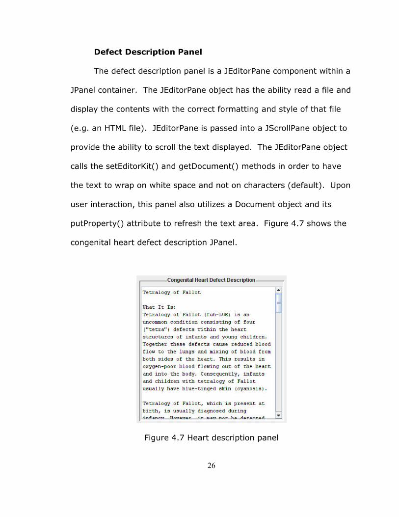
Defect Description Panel
The defect description panel is a JEditorPane component within a
JPanel container. The JEditorPane object has the ability read a file and
display the contents with the correct formatting and style of that file
(e.g. an HTML file). JEditorPane is passed into a JScrollPane object to
provide the ability to scroll the text displayed. The JEditorPane object
calls the setEditorKit() and getDocument() methods in order to have
the text to wrap on white space and not on characters (default). Upon
user interaction, this panel also utilizes a Document object and its
putProperty() attribute to refresh the text area. Figure 4.7 shows the
congenital heart defect description JPanel.
Figure 4.7 Heart description panel

27
2D Panels
Both the defect and normal 2D panels are comprised of two
classes. These classes extend JPanel. The defect 2D class,
Defect2DPanel, calls Defect2DImagePanel to update the defect graphic
at startup and for user interaction. The normal 2D class,
Normal2DPanel, calls Normal2DImagePanel to load the normal 2D
graphic at startup. The functionality of both the Defect2DPanel and
Normal2DPanel was split because of the need to add a border and use
the JLabel object. These classes setup the canvas and panel for
display. The Defect2DImagePanel and Normal2DImagePanel classes
are responsible for drawing the graphics. Figure 4.8 depicts the
Tetralogy of Fallot graphic while Figure 4.9 shows the normal heart.
Figure 4.8 Tetralogy of Fallot 2D graphic

28
Figure 4.9 Normal heart 2D graphic
Survey Analysis
As stated in chapter three, the participants in this study included
parents of children with congenital heart defects and pediatric
cardiologists.
Parents
In total, 11 parents volunteered to participate in the survey from
the cardiac support group. Of these, 7 or 64% were female and 4 or
36% were male. The age of the population group ranged from 25
years to 55 or more years. A variety of educational backgrounds
(highest achieved) were listed including the following: 1 with a

29
masters degree, 2 with a bachelors degree, 2 with an associates
degree, and 6 with high school diplomas. Of those 6 listed with high
school diplomas, 3 stated “some college.” One woman worked in a
health-related field as a pediatric cardiac sonographer. 100% stated
they were comfortable with using a computer. Lastly, every
participant or 100% stated that they preferred the interactive, digital
3D tool over the conventional consultation methods.
Pediatric Cardiologists
In total, 6 pediatric cardiologists volunteered to participate in
this study. Years of service as a clinician ranged from 1 to 20 or more
years. Of these, 100% stated that they frequently administer patient
consultations. The methods used by the clinicians include the
following: hand sketches, anatomical heart model, verbal description,
take home pamphlets, and 2D heart diagrams. 83% or 5 clinicians
stated they were comfortable with using a computer. 100% agreed
that current consultation methods require improvement and also
stated that the interactive, digital 3D heart models would assist in
patient and family education. Lastly, every participant or 100% stated
that if they had a choice, they would choose to utilize the interactive,
digital 3D tool and that the 3D approach is more useful than the 2D
methods.

30
Initial hypothesis was that the results would be dependent upon
demographic items. For instance, a well educated individual would not
find that the 3D solution is any more useful for educational purposes
than the standard methods. In fact, the data suggests that no
variable has an influence on the outcome. Preference of consultation
methods was not a function of any experience, background or
demographic item. Further analysis is not needed because in each
population group, regardless of any variable, the interactive, digital 3D
tool is preferred over standard consultation methods. There is no
correlation to make.

31
Chapter Five:
Conclusion
Quantitative Analysis
Due to the nature of the responses of the two population groups,
this portion of the analysis was rather straightforward. The parents (N
= 11) and the pediatric cardiologists (N = 6) provided us with useful
and encouraging data on the state of educational methods during a
patient consultation. Preliminary data analysis reveals that there is a
100% agreement between the parents and the cardiologists with
respect to preferring the 3D approach as a patient consultation
method. This suggests that there is a need for improving current
patient consultation methods. It also suggests that, for educational
purposes, the 3D approach would greatly assist patient and family
understanding of congenital heart defects.
Qualitative Analysis
It should be noted that 50% of the cardiologists and 100% of
the parents provided comments about EduView. These comments

32
brought to light the real-life requests and struggles of parents
concerning current and potential consultation methods. It exposed
their deep desire to grasp and understand fully the nature of their
child’s heart defect. It also shed light on the cardiologists desire to
improve their techniques. The possibility to utilize interactive 3D heart
models for future clinical use intrigued both the parents and the
cardiologists. To have the ability to explore the complexity of any
congenital heart defect in full 360 degrees, including within the
chambers, excited and prompted ideas for future additions from both
population groups.
Final Thoughts
EduView is an application that can be installed on any platform
(cross platform compatible). It is clean and simple to use. With
accurate human heart reference models and corresponding 2D heart
graphics, EduView can become a useful educational tool for patients
and patient families.
The future for EduView is bright. Because this is a proof of
concept study, EduView is a template or foundation for future
marketable revisions. The concept has now been validated with 100%
approval ratings from parents and cardiologists. In future versions,
EduView could display patient specific heart models in the place of the

33
representative diseased heart model. This would allow both the
patient and patient family the ability to study and understand their
child’s own diseased heart. Also, the idea of adding the fourth-
dimension (4D) appealed to every participant. Each parent expressed
desire to see the heart beating while simulating blood flow. Another
future task would be to implement treatment animations or 3D
visualizations to further aid in the understanding of various surgeries
or procedures. As a result of these possible revisions, EduView could
venture into the teaching tool market. Aspiring cardiologist residents
and medical students could utilize EduView for educational purposes.
There are a couple limitations to this concept that need to be
addressed. The process of segmenting the heart surface models is too
time heavy. EduView would rely on a quick turn around from MR data
into surface models to view and study. EduView also relies on MR data
that contains the entire heart. Because cardiac MR is primarily used as
a diagnostic tool, there is not a great need to capture the entire heart
structure. A slight limitation would be that EduView requires the user
to have both the JDK and Java3D installed to work properly. In order
for EduView to execute and display, system environment variables of
the operating system must be properly set. However, following the
Readme.txt file will assist and expedite the install process.

34
References
1. American Heart Association. The Consumer and Patient Education Materials page. Available at:
http://www.americanheart.org/presenter.jhtml?identifier=1200021. Accessed February 28, 2007.
2. Amira. The Overview page. Available at:
http://www.amiravis.com/overview.html. Accessed September 8, 2006.
3. Everett AD, Lim DS, Buck M, et al. Illustrated Field Guide to Congenital Heart Disease and Repair, 2nd ed. Charlottesville, VA:
Scientific Software Solutions Inc.; 2005.
4. GE Healthcare. The MR Clinical Applications page. Available at: http://www.gehealthcare.com/usen/mr/products/mrecho_cardiacimg.h
tml. Accessed June 11, 2007.
5. Java Sun Microsystems Inc. The Using Swing Components: Examples page. Available at:
http://java.sun.com/docs/books/tutorial/uiswing/examples/components/index.html. Accessed June 25, 2007.
6. Miquel ME, Hill DLG, Baker EJ, et al. Three-and four-dimensional
reconstruction of intra-cardiac anatomy from two-dimensional
magnetic resonance images. International Journal of Cardiovascular Imaging. 2003; 19: 239-254.
7. PedHeart Congenital Heart Software. The PedHeart Suite page.
Available at: http://www.pedheart.com/suite.php. Accessed September 2, 2006.
8. Schroeder W, Martin K, Lorensen B. The Visualization Toolkit, 3rd
ed. United States of America: Pearson Education Inc; 2002.

35
9. Toombs BD, Jing JM. Current concepts in the evaluation of vascular
disease – magnetic resonance and computed tomographic angiography. Texas Heart Institute Journal. 2000; 27(2): 170–192.
10. Walsh, AE, Gehringer, D. Java 3D API Jump-Start. Upper Saddle
River, NJ: Prentice Hall; 2002.

36
Bibliography
1. Bird, GL. An Update on Information Technology In Cardiac Medicine. Pediatric Cardiology Today. 2005; 3(3): 3-5.
2. Clason DL, Dormody TJ. Analyzing Data Measured by Individual
Likert-Type Items. Journal of Agricultrual Education. 35(4): 31-35.
3. Deitel HM, Deitel PJ. Java How to Program, 3rd ed. Upper Saddle River, NJ: Prentice Hall; 1999.
4. Huber ME, Paetsch I, Schnackenburg B, et al. Performance of a New Gadolinium-Based Intravascular Contrast Agent in Free-Breathing
Inversion-Recovery 3D Coronary MRA. Magnetic Resonance in Medicine. 2003; 49: 115-121.
5. Liang, YD. Introduction to Java Programming, 3rd ed. Upper Saddle
River, NJ: Prentice Hall; 2001.

37
Appendices

38
Appendix A: Parent Survey
The purpose of this survey is to determine the potential benefits (if any) of various
presentation methods that your physician could use in a pediatric cardiology
consultation. This survey is an effort to improve the current consultation methods to
better educate the patient/family of various congenital heart diseases. Your
experiences and opinions are very important to us, and we would greatly appreciate
your participation. Thank you very much for your cooperation.
Two-Dimensional (2D) – a flat surface; having the dimensions of height and width
only.
Three-Dimensional (3D) – an object that occupies space; having the dimension of
depth as well as height and width.
SECTION I: BACKGROUND INFORMATION
1) Are you:
A. Male
B. Female
2) Your age:
A. 18 – 24 years
B. 25 – 34 years
C. 35 – 44 years
D. 45 – 54 years
E. 55 years or more
3) The highest level of education you have achieved:
A. Completed high school or equivalent
B. A specialized certification
C. Associates degree
D. Bachelors degree
E. Masters degree
F. Doctorate degree
4) Do you work in a health-related field?
A. Yes
B. No
If yes, what do you do? _____________________________________________
Please read each statement and indicate the extent to which you agree or disagree.
5) You are very comfortable using a computer.
A. Strongly Agree B. Agree C. No Opinion D. Disagree E. Strongly Disagree
6) You consider yourself to be very knowledgeable regarding science as it relates to
human health.
A. Strongly Agree B. Agree C. No Opinion D. Disagree E. Strongly Disagree

39
Appendix A: (Continued)
7) You understand the difference between a 2D and a 3D object.
A. Strongly Agree B. Agree C. No Opinion D. Disagree E. Strongly Disagree
8) You have a clear understanding of your child’s heart defect.
A. Strongly Agree B. Agree C. No Opinion D. Disagree E. Strongly Disagree
9) You have a strong need or desire to understand your child’s heart defect.
A. Strongly Agree B. Agree C. No Opinion D. Disagree E. Strongly Disagree
10) You experience a high level of anxiety/stress as a result of your child’s heart
defect.
A. Strongly Agree B. Agree C. No Opinion D. Disagree E. Strongly Disagree
11) You experience a high level of anxiety/stress as a result of your lack of
understanding of your child’s heart defect.
A. Strongly Agree B. Agree C. No Opinion D. Disagree E. Strongly Disagree
12) Your cardiologist has done a satisfactory job of explaining the nature of your
child’s heart defect.
A. Strongly Agree B. Agree C. No Opinion D. Disagree E. Strongly Disagree
13) The level of usefulness of all diagrams, illustrations, etc. used by your
cardiologist to explain your child’s heart defect has been very good.
A. Strongly Agree B. Agree C. No Opinion D. Disagree E. Strongly Disagree
SECTION II
Please answer questions 14 - 16 after viewing the first presentation.
14) You understood the presentation regarding your child’s heart defect.
A. Strongly Agree B. Agree C. No Opinion D. Disagree E. Strongly Disagree
15) You experience a high level of anxiety/stress as a result of your lack of
understanding of your child’s heart defect.
A. Strongly Agree B. Agree C. No Opinion D. Disagree E. Strongly Disagree
16) The level of usefulness of all diagrams, illustrations, etc. used during this
presentation to explain your child’s heart defect has been very good.
A. Strongly Agree B. Agree C. No Opinion D. Disagree E. Strongly Disagree

40
Appendix A: (Continued)
SECTION III
Please answer questions 17 – 19 after viewing the second presentation.
17) You understood the presentation regarding your child’s heart defect.
A. Strongly Agree B. Agree C. No Opinion D. Disagree E. Strongly Disagree
18) You experience a high level of anxiety/stress as a result of your lack of
understanding of your child’s heart defect.
A. Strongly Agree B. Agree C. No Opinion D. Disagree E. Strongly Disagree
19) The level of usefulness of all diagrams, illustrations, etc. used during this
presentation to explain your child’s heart defect has been very good.
A. Strongly Agree B. Agree C. No Opinion D. Disagree E. Strongly Disagree
SECTION IV
20) Which presentation method did you prefer?
A) The first one
B) The second one
C) They were both about the same
21) Please briefly describe the reasoning behind your answer to question 19.
22) You understand the difference between a 2D and a 3D object.
A. Strongly Agree B. Agree C. No Opinion D. Disagree E. Strongly Disagree
23) The second method would be more effective if the computer illustration used was
an actual image of my child’s heart.
A. Strongly Agree B. Agree C. No Opinion D. Disagree E. Strongly Disagree
Thank you for completing this questionnaire.
If you have any additional comments or questions regarding this survey, please use
the space below.

41
Appendix B: Cardiologist Survey
The purpose of this survey is to determine the potential benefits (if any) of various
presentation methods that your physician could use in a pediatric cardiology
consultation. This survey is an effort to improve the current consultation methods to
better educate the patient/family of various congenital heart diseases. Your
experiences and opinions are very important to us, and we would greatly appreciate
your participation. Thank you very much for your cooperation.
Two-Dimensional (2D) – a flat surface; having the dimensions of height and width
only.
Three-Dimensional (3D) – an object that occupies space; having the dimension of
depth as well as height and width.
SECTION I: BACKGROUND INFORMATION
1) Years you have been a cardiologist.
A) Less than one year
B) 1 – 4 years
C) 5 – 9 years
D) 10 – 14 years
E) 15 – 19 years
F) 20 years or more
2) How often do you administer patient consultations?
A) Not at all
B) Occasionally
C) Frequently
3) If you answered ‘Yes’ to question #2, please circle the methods you utilize of the
following: (circle all that apply)
A) Verbal description
B) Hand sketches
C) 2D heart diagrams
D) Take home pamphlets
E) 2D heart animations
F) Patient MRI or CT scans
G) Anatomical heart model
H) Other:_______________________________________________________
4) If you answered ‘No’ to question #2, are you familiar with current pediatric
consultation methods?
A) Yes
B) No
Please read each statement and indicate the extent to which you agree or disagree.
5) You are very comfortable with using a computer.
A. Strongly Agree B. Agree C. No Opinion D. Disagree E. Strongly Disagree

42
Appendix B: (Continued)
6) You understand the difference between a 2D and 3D object.
A. Strongly Agree B. Agree C. No Opinion D. Disagree E. Strongly Disagree
SECTION II
7) Current consultation methods adequately inform patients and families.
A. Strongly Agree B. Agree C. No Opinion D. Disagree E. Strongly Disagree
8) Current consultation methods do not require improvement.
A. Strongly Agree B. Agree C. No Opinion D. Disagree E. Strongly Disagree
9) Current consultation methods need to be refined.
A. Strongly Agree B. Agree C. No Opinion D. Disagree E. Strongly Disagree
10) Having interactive 3D heart models would assist in patient and family education.
A. Strongly Agree B. Agree C. No Opinion D. Disagree E. Strongly Disagree
SECTION III
11) You understood the interactive digital consultation method presentation.
A. Strongly Agree B. Agree C. No Opinion D. Disagree E. Strongly Disagree
13) Interactive 3D heart models are easy to understand.
A. Strongly Agree B. Agree C. No Opinion D. Disagree E. Strongly Disagree
12) Having interactive 3D heart models would assist in patient and family education.
A. Strongly Agree B. Agree C. No Opinion D. Disagree E. Strongly Disagree
14) You would rather utilize conventional consultation methods.
A. Strongly Agree B. Agree C. No Opinion D. Disagree E. Strongly Disagree
15) If you had a choice, you would choose to utilize the digital, interactive 3D
approach.
A. Strongly Agree B. Agree C. No Opinion D. Disagree E. Strongly Disagree
16) This approach would be more useful if the malformed 3D heart model was
patient specific.
A. Strongly Agree B. Agree C. No Opinion D. Disagree E. Strongly Disagree
17) The interactive 3D approach is more useful than the 2D methods.
A. Strongly Agree B. Agree C. No Opinion D. Disagree E. Strongly Disagree
Thank you for completing this questionnaire.
If you have any additional comments or questions regarding this survey, please use
the space below.

43
Appendix C: Sample Java Code
/** main class of program. creates an EduViewFrame object**/
public class EduView
{
public static void main(String[] args)
{
//create the JFrame, EduViewFrame
JFrame frame = new EduViewFrame();
frame.setDefaultCloseOperation(JFrame.EXIT_ON_CLOSE);
frame.setVisible(true);
}
} //end EduView class
/** Class that contains all JComponents of the program **/
class EduViewFrame extends JFrame
{
//file that is parsed
private static String fileName = "src/data/hearts.txt";
private DataSet[] data;
String path = "src/data/heartModels/Tetralogy of Fallot/";
//set dimensions of the JFrame
private int WIDTH = 1020;
private int HEIGHT = 720;
Dimension dimension = new Dimension();
/** Constructor **/
public EduViewFrame()
{
//parse hearts.txt
Parser parser = new Parser(fileName);
//setup the JFrame
setTitle("EduView");
setSize(WIDTH, HEIGHT);
setResizable(false);
dimension.setSize(WIDTH, HEIGHT);
setPreferredSize(dimension);
setLayout(new BorderLayout());
Container contentPane = getContentPane();
…
} //end EduViewFrame class

44
Appendix D: Software Requirements Specification
System Features
Controller Description
Controller is a class to observe and then act when a user makes a button change in the GUI.
Stimulus/Response Sequences A user clicks one of the conditions to change the heart
displayed. Controller will note which condition was selected and set the appropriate 3D, 2D, and condition view by calling a
function on the Manager object. Functional Requirements
Function: Controller Inputs: Button choice
Source: Buttons are chosen by the user Effect: Call to Manager class Pre-condition: Waits for user interaction
Post-condition: Waits for user interaction
Manager Description
Manager is a class that receives input from the Controller class
and makes appropriate calls to the 3DView, 2DView, and ConditionView classes.
Stimulus/Response Sequences
Manager will receive input from the Controller class. The input
it receives will allow it to know which functions to call. It will call the functions with the appropriate parameters required by
the function. Functional Requirements
Function: Manager Inputs: 3D model, 2D image and condition view
Source: 3D model, 2D image and condition view from the Controller class
Effect: Calls to 3DView, 2DView, and ConditionView
Pre-Condition: Waits for input from Controller Post-Condition: Waits for input from Controller
3DView
Description

45
Appendix D: (Continued) 3DView is a class that renders both the heart reference model and diseased heart model.
Stimulus/Response Sequences
3DView will receive input from the Manager class. With the input, it will update by rendering the new model. 3DView will receive user interaction via the mouse. It will render as the
user rotates the heart model.
Functional Requirements Function: 3DView Inputs: Position coordinates, mouse click
Source: Mouse click performed by the user, position coordinates from the Manager class.
Effect: Render to its render viewport Pre-Condition: 3DView is rendered and displayed on the user’s screen.
Post-Condition: Global coordinates of heart model and plane are changed if user rotated it.
2DView Description
2DView is a class that renders a static background images of a normal heart and diseased heart.
Stimulus/Response Sequences
2DView will receive input from the Manager class. With the
input, it will update by rendering the new heart images.
Functional Requirements Function: 2DView
Inputs: Position coordinates Source: Position coordinates from the Manager class Effect: Render to its render viewport
Pre-Condition: 2DView is rendered and displayed on the user’s screen.
Post-Condition: The diseased heart image is different. ConditionView
Description ConditionView is a class that updates diseased heart
descriptions. Stimulus/Response Sequences

46
Appendix D: (Continued) ConditionView will receive input from the Manager class. With the input, it will update by entering the appropriate text for the
selected diseased heart.
Functional Requirements Function: ConditionView Inputs: Text from a file
Source: Text file location from the Manager class Effect: Render to its render viewport
Pre-Condition: ConditionView is rendered and displayed on the user’s screen.
Post-Condition: The description text is different.

47
Appendix D: (Continued)
Figure D.1 Graphical user interface design
Select
Reset Reset
Heart Defect Normal Heart

48
Appendix D: (Continued)
Controller
Figure D.2 Architectural model of the system
Manager
View Manager
GUI
Description View Defect 2D
View
Defect 3D
Heart Display
Condition
List
Normal 2D
View
Normal 3D
Heart Display
49
Appendix D: (Continued)
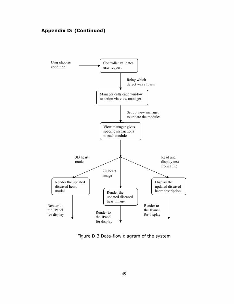
Figure D.3 Data-flow diagram of the system
User chooses
condition Controller validates
user request
Relay which
defect was chosen
Render the updated
diseased heart
model Render the
updated diseased
heart image
Display the
updated diseased
heart description
Render to
the JPanel
for display Render to
the JPanel
for display
Render to
the JPanel
for display
3D heart
model
2D heart
image
Read and
display text
from a file
View manager gives
specific instructions
to each module
Manager calls each window
to action via view manager
Set up view manager
to update the modules

50
Appendix E: Sample Heart Diagram
Figure E.1 Sample heart diagram for Ventricular Septal Defect


![Pediatric Cardiology Dysfunction for Students--2011[1]](https://static.fdocuments.net/doc/165x107/577d21651a28ab4e1e9523b1/pediatric-cardiology-dysfunction-for-students-20111.jpg)